Communication

Functionalizing NaGdF4:Yb,Er Upconverting Nanoparticles with Bone-Targeting Phosphonate Ligands: Imaging and In Vivo Biodistribution

1
CIC biomaGUNE, Paseo de Miramón 182, 20014 Donostia, Spain
2
Donostia International Physics Center (DIPC), Manuel Lardizabal Ibilbidea 4, 20018 Donostia, Spain
3
Ikerbasque, Basque Foundation for Science, 48011 Bilbao, Spain
*
Authors to whom correspondence should be addressed.
Current address: BASF SE, Department of Material Physics & Analytics, Carl-Bosch-Strasse 38, 67056 Ludwigshafen, Germany.
Submission received: 11 December 2018 / Revised: 12 April 2019 / Accepted: 19 April 2019 / Published: 30 April 2019

Abstract

:
Lanthanide-doped upconverting nanoparticles (UCNPs) transform near infrared light (NIR) into higher-energy UV and visible light by multiphotonic processes. Owing to such unique feature, UCNPs have found application in optical imaging and have been investigated for the NIR light activation of prodrugs, including transition metal complexes of interest in photochemotherapy. Besides, UCNPs also function as magnetic resonance imaging (MRI) contrast agents and positron emission tomography (PET) probes when labelled with radionuclides such as 18F. In this contribution, we report on a new series of phosphonate-functionalized NaGdF4:Yb,Er UCNPs that show affinity for hydroxyapatite (inorganic constituent of bones), and we discuss their potential as bone targeting multimodal (MRI/PET) imaging agents. In vivo biodistribution studies of 18F-labelled NaGdF4:Yb,Er UCNPs in rats indicate that surface functionalization with phosphonates favours the accumulation of nanoparticles in bones over time. PET results reveal leakage of 18F for phosphonate-functionalized NaGdF4:Yb,Er and control nanomaterials. However, Gd was detected in the femur for phosphonate-capped UCNPs by ex vivo analysis using ICP-MS, corresponding to 6–7% of the injected dose.

Graphical Abstract

1. Introduction

In recent years, our group developed a number of unconventional approaches for the photoactivation of metal-based anticancer prodrug complexes [1,2,3], including the use of lanthanide-doped upconverting nanoparticles (UCNPs) as near infrared (NIR) light triggers [4,5,6]. In this context, we and others reported on the capacity of UCNPs to prompt the photochemistry of several transition metal complexes such as Ru, Pt, Mn and Fe [7]. However, UCNPs have much more to offer besides their photophysics [8] since they can simultaneously act as multimodal imaging agents and be employed in theranostics. Gd-containing UCNPs are magnetic resonance imaging (MRI) contrast agents, while labelling with radionuclides such as 18F and 153Sm yields positron emission and single photon emission tomography (PET and SPECT) probes, respectively [9,10].
In this work, we explored the potential of phosphonate-functionalized NaGdF4:Yb,Er UCNPs to behave as MRI and PET imaging tools for bones, motivated by the long-term prospect of designing theranostic nanoconstructs for the photoactivated delivery of metal-based drugs. Actually, recent results highlight the possibility of adopting photodynamic therapy and related strategies for the treatment of spinal metastases [11] and to ablate tumors within vertebral bone [12]. Therefore, phosphonate ligands were selected for functionalization of UCNPs because of their capacity of accumulating in bones. The use of bisphosphonates for targeting bones has been recently explored in combination of superparamagnetic iron oxide nanoparticles (SPIONs) [13]. Several phosphonates, e.g., alendronate, etidronate, are used in the treatment of osteoporosis and investigated for their use against bone cancers [14]. Furthermore, a recent study by Li et al. [15] demonstrated that phosphonates have high affinity for the surface of UCNPs and show the ability of stabilizing colloidal dispersions of UCNPs in acidic conditions (e.g., in lysosomes), thus lowering their pro-inflammatory effects both in vitro and in vivo. Lastly, we recently demonstrated the functionalization and UCNP loading of photoactivatable Ru complexes modified with a phosphonate arm [4].
Herein, we investigated a set of 10 nm NaGdF4:Yb,Er NPs functionalized with three phosphonic acids, namely etidronic acid, alendronic acid and nitrilo(trimethylphosphonic acid) (3P), as MRI and PET agents and evaluated their biodistribution and in vivo accumulation capacity in bones.

2. Results and Discussion

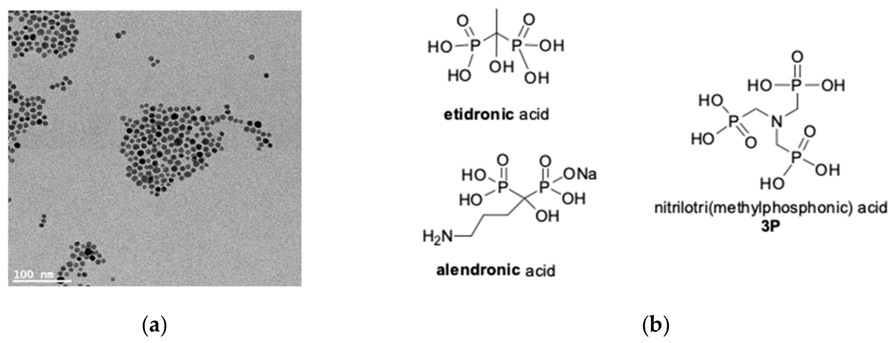
2.1. Synthesis and Surface Decoration of UCNPs NaGdF4:Yb,Er

Oleate-capped NaGdF4:Yb,Er nanoparticles were synthesized by thermal decomposition following a procedure previously reported by us and others [6,16]. The obtained UCNPs had homogeneous distribution in size and shape, displaying an average diameter of 9.5 ± 1.1 nm according to TEM measurements (Figure 1a and Figure S1). The relatively small size of these UCNPs is advantageous for MRI because of their high surface area, increasing the amount of Gd3+ ions accessible to water molecules and hence the relaxivity per Gd atom [17,18,19]. Conversely, small UCNPs such as the ones prepared in this work have modest upconversion properties due to solvent deactivation processes (Figure S2) [20].
Next, the surface of the oleate-capped nanoparticles was decorated with phosponates to achieve aqueous dispersibility and to assess the capacity of these ligands to drive accumulation of UCNPs into bones. Surface functionalization was performed by ligand exchange [21]. Accordingly to the different ligand solubility, oleic acid was exchanged for etidronic acid, alendronic acid or nitrilo(trimethylphosphonic acid) (3P) in a CHCl3:H2O mixture (Figure 1b), affording water dispersible nanoparticles where the negatively charged phosphonates are electrostatically tethered to the positive surface of the Gd-UCNPs.
All phosphonate-decorated UCNPs were characterized with FT-IR (Figure S3) and ICP-MS (Table S1) to confirm ligand functionalization and determine the quantity of Gd3+ per gram of functionalized nanoparticle.

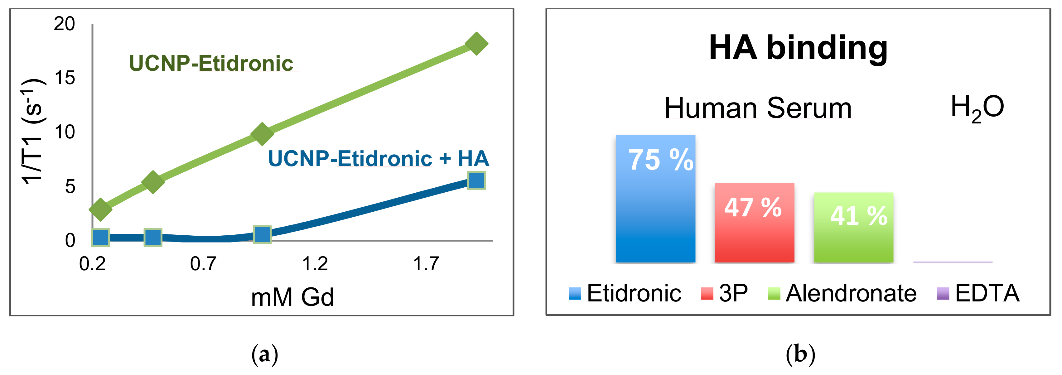
2.2. MRI Characterization and Interaction with Hydroxyapatite

UCNPs serve as contrast agents for T1-weighted MR imaging. T1 relaxation times in UCNPs depend on the accessibility of Gd3+ ions on the surface of the nanomaterials and can be strongly affected by different surface coatings [17]. For this reason, we investigated the longitudinal r1 relaxivity of the three phosphonate-functionalized NaGdF4:Yb,Er UCNPs and how this parameter responded to different media, including human serum as a mimic of blood components.
Relaxivity values for the three UCNPs were determined at 1.5 T (Figures S4–S9 and Table S2) and are summarized in Table 1. In aqueous solution, UCNPs coated with etidronate have the highest r1 (3.47 mM−1 s−1) compared to the alendronate and 3P analogues (r1 < 0.70 mM−1 s−1). In the case of etidronate UCNPs, a significant increase in r1 was observed upon changing the medium, while this was not the case for the other phosphonates.
In MES buffer, the r1 value of etidronate-capped UCNPs is five units higher than in water. In human serum r1 reached a value of 9.73 ± 0.02 mM−1 s−1, that is twice the relaxivity of Gadobutrol (gadovist) in water (3.30 mM−1 s−1) and in human blood plasma (4.70 mM−1 s−1) [22,23].
We also evaluated the capacity of the etidronate UCNPs to provide contrast by acquiring T1 weighted MRI images at 7 T in H2O (Figure S10) and in human serum (Figure 2) using a phantom. As shown in Figure 2, good contrast levels were obtained for etidronate UCNPs already at Gd concentrations of 0.25 mM.
The different behaviour of etidronate UCNPs in terms of relaxivity compared to the other phosphonate analogues can be rationalized in terms of colloidal stability. As shown by results obtained from DLS (dynamic light scattering, Figure S11), etidronate favours the dispersion of UCNPs, particularly in MES buffer, while nanoparticles capped with alendronate or 3P tend to aggregate and eventually precipitate in solution (Table S3), ultimately displaying a reduced number of Gd atoms exposed to solvent and hence lower r1 values.
Hydroxyapatite (HA) is a recognized analogue of bone mineral [14] and was employed as model to evaluate the affinity for bones of our phosphonate-functionalized UCNPs. To this aim, we performed relaxivity r1 measurements on solutions containing different concentrations of phosphonate-capped UCNPs that were incubated in the presence of 12.5 mg/mL HA (insoluble). In all cases, binding of UCNPs to HA resulted in lower r1 values compared to samples without HA. For example, etidronate-UCNPs showed remarkable affinity with HA already after 2 h of incubation. Figure 3a clearly demonstrated that UCNPs incubated with HA do not induce any T1 shortening effect in the medium until a concentration of 1 mM Gd is reached. At higher concentrations, the T1 shortening effects of etidronate-UCNPs are restored due to the saturation of HA surface. In order to quantify the affinity of UCNPs for HA, we compared relaxivity values for 2 mM Gd solutions of UCNPs in the presence and absence of the mineral. For etidronate nanoparticles a 75% affinity was found in human serum (Figure 3b). Similar behaviour was obtained for alendronate and 3P UCNPs upon 24 h of incubation with HA in human serum (Figure S12). However, the capacity of these nanomaterials to interact with HA was significantly lower (Figure 3b). For comparison, the same type of measurements was performed using UCNPs functionalized with EDTA (ethylenediaminetetraacetic acid) which decorates the nanoparticles through carboxylate groups instead of phosphonates. EDTA UCNPs have an r1 value in water of 3.9 mM−1 s−1 that is not modified in the presence of HA (Figure 3b), confirming that affinity of our UCNPs for this mineral is associated to phosphonate groups.

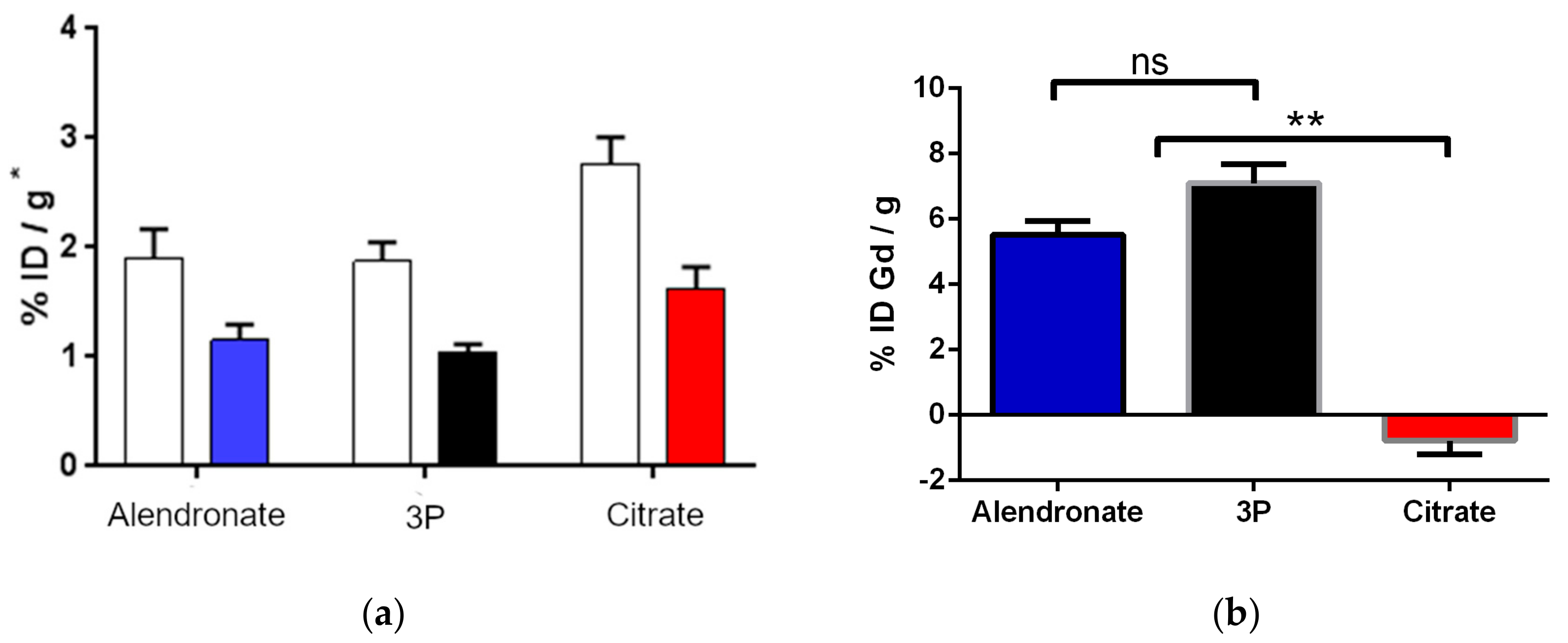
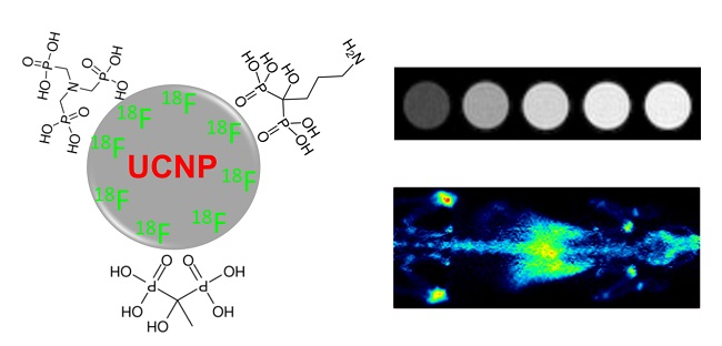
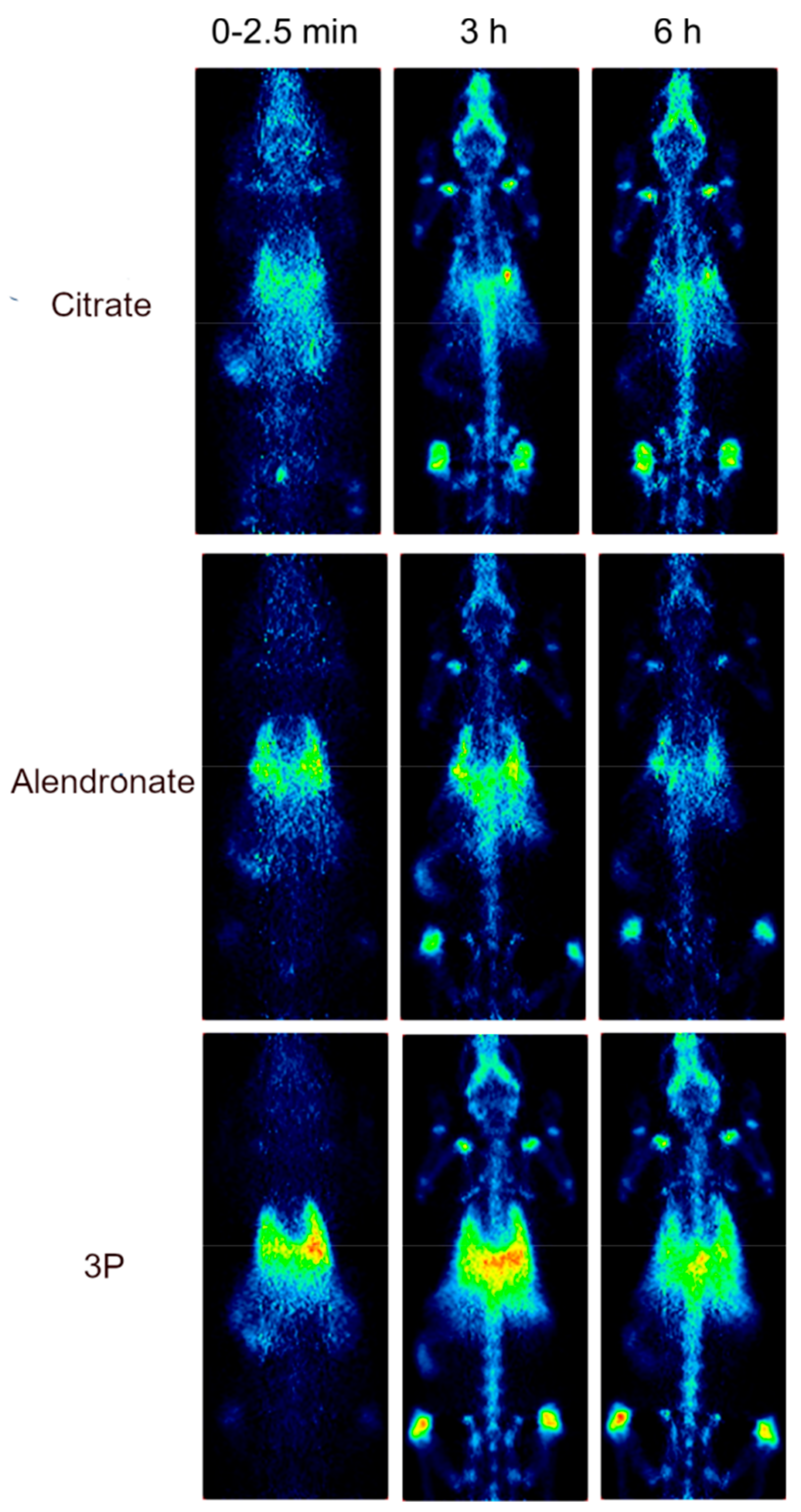
2.3. Biodistribution Study by Radiolabeling UCNPs

Nuclear imaging, and in particular PET, is routinely employed in the clinics to study the whole-body distribution of radiolabeled species with high sensitivity [24]. 18F-labelling of UCNPs has demonstrated to be an effective strategy to monitor the biodistribution of these nanomaterials in vivo [25,26,27], often in combination with other imaging modalities [28]. In this study, the observation of the UCNPs in vivo adhesion to the bones using MRI was discarded due to the intrinsic low sensitivity of the technique added to the specific characteristics of the bones: low water content, very short T2 relaxation times due to its rigid nature and susceptibility effects. Adopting the procedure reported by Li and coworkers [25], we radiolabelled phosphonate UCNPs by exposing the nanoparticles to [18F]F ions with the aim of visualizing via PET their biodistribution in healthy rodents and gain information about their eventual accumulation in bones. As previously described [25], this straightforward synthetic method afforded high incorporation ratios, with non-decay corrected radiochemical yields of 36%, 43% and 43% for citrate, 3P and alendronate-UCNPs, respectively, resulting in estimated specific activity values of 105 MBq/mg for citrate-UCNPs and of 126 MBq/mg for 3P and alendronate-UCNPs. Only etidronate functionalised UCNPs resulted in low radiochemical yields (<10%), and therefore were no further investigated in vivo. Nanoparticles decorated with citrate were used as control since this carboxylate ligand did not show any specific affinity to HA (as confirmed by MRI, data not shown). The stability (Figure S13) and radiochemical purity of alendronate, 3P and citrate UCNPs were confirmed by radio-TLC at different time points. The radiochemical purity of the labelled particles was >95% at t = 5 h of incubation in physiologic saline solution (see Figure S14 for representative example).
Biodistribution and clearance of the 18F-labeled UCNPs was obtained by PET imaging (Figure 4) after intravenous injection in the tail of 100 µL of a saline solution of the alendronate, 3P and citrate UCNPs (0.5 mg/mL). Direct inspection of the images (see Figure 4) clearly showed initial accumulation in the lungs and the liver, probably due to sequestration of the NPs by the mononuclear phagocyte system (MPS) which might be a consequence of aggregation of the NPs under physiologic conditions. At 3 h and 6 h after nanoparticle injection, 18F radioactivity was also clearly identified in the bones and joints, independently of the ligand coating. These initial results were confirmed by image quantification (Figure S15). All NPs, irrespective of the surface decoration, exhibited initial accumulation in the lungs, with values close to 4% of injected dose per cm3 of tissue (%ID/cc). Initial localization was also observed in the liver and kidneys, which was paralleled by high accumulation in the urine (results not shown) suggesting partial elimination of the NPs via urinary excretion or detachment of the label.
At early times, citrate nanoparticles (controls) displayed a pronounced accumulation in bladder and kidney compared to the phosphonate-functionalized UCNPs, suggesting that they were excreted more rapidly.
A progressive accumulation of the radioactivity in the bones was observed, which reached values close to 2%ID/cc in the case of citrate UCNPs. This result, confirmed by dissection/γ-counting (Figure 5a), was unexpected considering that the citrate had no affinity for HA, and suggested that 18F ions could leak partially from the nanoparticles, hence masking the real uptake of the labelled particles in bone tissue. For this reason, we further investigated the accumulation of NPs in bone by ex vivo analysis using ICP-MS. With that aim, the amount of Gd in the femurs was quantified (Figure 5b). The samples to be analysed by ICP-MS were obtained after digestion of the femurs harvested from the animals with HNO3. In the case of the citrate control (Figure 5b), no sign of Gd was detected by ICP-MS indicating that the totality of the [18F]F signal in the femur could be associated to free [18F]F. Conversely, Gd was detected by ICP-MS in the femur for phosphonate-capped UCNPs, corresponding to 6–7% (Figure 5b) of the injected dose per gram of bone. Although these results confirmed the leakage of 18F ions for alendronate, 3P and citrate UCNPs, they also indicated that phosphonate ligands were capable of promoting accumulation of UCNPs in bones to some extent, while the control nanomaterial did not. Although further studies are required, we speculate that phosphonate-capped UCNPs can escape the endothelium and reach the skeleton via a mechanism known as nanomaterial induced endothelial leakiness. In this process, nanomaterials enter and enlarge the nanosized gaps between microvascular capillary endothelial cells favouring their leakage to other tissue sites [29,30].

3. Materials and Methods

3.1. Materials

All chemical reagents, yttrium(III) acetate hydrate, ytterbium(III) acetate tetrahydrate, erbium(III) chloride hexahydrate, 1-octadecene (technical grade), oleic acid (technical grade), sodium hydroxide, ammonium fluoride, etidronic acid, alendronic acid, nitrilo(trimethylphosphonic acid) and solvents were purchased from Sigma-Aldrich (St. Louis, MO, USA).

3.2. Synthesis of UCNPs NaGdF4:Yb,Er

The NaGdF4:Yb,Er (rare earth element ratio 78/20/2 mol %) synthesis was carried out following a slightly modified literature procedure [6]. In a typical synthesis (total rare earth amount, 3 mmol), gadolinium (III) chloride hydrate (618 mg, 2.34 mmol), ytterbium(III) acetate tetrahydrate (253.8 mg, 0.60 mmol) and erbium (III) chloride hexahydrate (23 mg, 0.06 mmol) were dissolved in 1-octadecene (25 mL) and oleic acid (15 mL) in a 100 mL three-neck round-bottom flask with coil condenser. The suspension was heated up to 120 °C with a slow temperature ramp of 3.2 °C/min under stirring and vacuum. Once the reaction mixture reached such temperature, it was kept in these conditions for 30 min in order to form a clear solution and eliminate residual water and oxygen. The system was then allowed to cool to 50 °C under a flow of nitrogen gas.
A solution of sodium hydroxide (300 mg, 7.5 mmol) and ammonium fluoride (444 mg, 12.0 mmol) in methanol (8 mL) was added to the reaction flask dropwise in 10 min. The subsequent cloudy solution was stirred for 30 min at 50 °C and for 30 min at 70 °C under nitrogen to evaporate the entire amount of methanol from the solution. Successively, the system was heated up to 300 °C with a fast temperature ramp of 13.5 °C/min under stirring and nitrogen and maintained in such conditions for 1 h. The reaction mixture progressively changed from turbid to slightly yellow and transparent.
Next, the flask was left cooling to room temperature, and nanoparticles were purified by centrifugation (4500 rpm at 20 °C for 15 min) to remove any excess of reagents and solvents. The white pellet was washed once with of ethanol (30 mL) and once with THF/ethanol (5/30 mL) and recollected by centrifugation. Upconverting nanoparticles were dried at room temperature overnight. Typically, ca. 700 mg of NaGdF4:Yb,Er nanoparticles are obtained employing the reaction conditions described.

3.3. Functionalization of UCNPs/Ligand Exchange

Typically, 30 mg of UCNPs were dissolved in 2 mL of chloroform and 100 mg of organic ligand were dissolved in 10 mL of MES buffer (20 mM, pH 6). The solution mixture was left stirring overnight and the aqueous phase (supernatant) lyophilized. The solid obtained was then washed three times with 3 mL of ethanol (or a mixture of H2O:EtOH 1:3 in the case of etidronate) and left drying at room temperature for two days.

3.4. Relaxivity Measurements and HA Binding

All relaxivity results were obtained in triplicates by measuring at 37 °C 300 µL of aqueous solution containing different concentrations of UCNPs on a 1.5 T MiniSpec MQ60 (Bruker, Madrid, Spain. The affinity of UCNPs for HA was evaluated by adding a fixed quantity of HA (insoluble) in every sample and were directly measured on the 1.5 T MiniSpec at two time points (2 and 24 h).

3.5. Labeling of UCNPs with 18F

UCNPs with four different capping ligands (citrate, 3P, alendronate and etidronate) were labelled with fluorine-18 (positron emitter with T1/2 = 109.7 min). In brief, [18F]F (ca. 440 MBq) dissolved in 150 µL of 18O-enriched water (used for the production of 18F by proton irradiation) was added to the suspension of UCNPs (1.5 mg in 10 µL of distilled water). The reaction mixture was incubated for 10 min at room temperature and centrifuged to remove the unreacted [18F]F from [18F]UCNPs. The [18F]UCNPs were washed three times with distilled water by centrifugation (5 min, 10,000 rpm). Finally, the pellet was suspended in 150 µL of physiological saline solution (NaCl 9.0 g per liter). The amount of radioactivity in the final suspension was determined using a dose calibrator (CRC-25R PET dose calibrator, Capintec Inc., Ramsey, NJ, USA) and the non-decay corrected radiochemical yield was calculated as the ratio between the final and the starting amounts of radioactivity. Estimated specific activity values were calculated as the ratio between the final amount of radioactivity and the starting mass amount of UCNPs.
The stability of the NPs in physiologic saline solution was determined. With that aim, NPs were incubated at 37 °C and fractions were withdrawn at pre-selected time points, diluted with purified water (1:20) and further analysed by radio-Thin Layer Chromatography (radio-TLC, silica and 9:1 saline/ethanol as eluent). Under experimental conditions, 18F-labeled UCNPs remained at the seeding spot while free [18F]F eluted with the solvent front.

3.6. Gd3+ Determination from Ex Vivo Femur Bones

After the last imaging session (6.5 h post-administration, see Section 3.7.5) the animals were sacrificed and the bones harvested in order to conduct ex vivo analysis and determine the amount of Gd3+ by ICP-MS. We followed a protocol reported elsewhere [31]. Briefly, the femurs (n = 2 per UCNP type) treated with citrate, 3P, alendronate UCNPs were dissolved in 3 mL of HNO3 overnight. An aliquot of this solution was then diluted 50 times to use it as ICP sample. A non-treated femur was also digested in HNO3 overnight, to build a matrix for the calibration curve of Gd3+.

3.7. Instrumentation

3.7.1. Transmission Electron Microscopy (TEM)

TEM was performed on a JEOL JEM-1400 PLUS-HC microscope (Peabody, MA, USA) operating at 120 kV. The TEM samples were prepared by dropping 3 μL sample solutions (0.1–1 mg/mL in THF or in water) onto a 400-mesh carbon coated copper grid (3 mm in diameter) followed by the evaporation of the solvent under vacuum. The nanoparticle sizes were estimated from over 150 nanoparticles.

3.7.2. Fourier Transform Infrared (FTIR)

FTIR spectra of NaGdF4:Yb,Er UCNPs coated with oleic acid, citrate, EDTA, 3P, alendronate and etidronate were recorded on a Nicolet FTIR 6700 spectrometer (Thermo Fisher, Waltham, MA, USA) as KBr pellet.

3.7.3. Dynamic Light Scattering
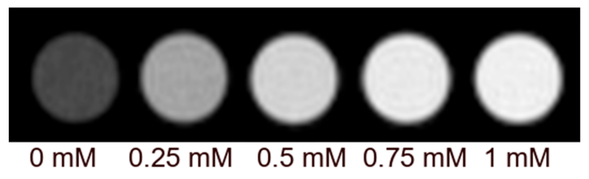
The hydrodynamic radius of the different functionalized NaGdF4:Yb,Er UCNPs was obtained measuring an aqueous solution containing the nanoparticles (0.5 mg/mL) with 173° scattering angle at 25 °C using a NanoSizer Malvern Nano-Zs (Malvern Panalytical, Malvern, UK).

3.7.4. MRI

T1 and T2 experiments were performed on a 1.5 T MiniSpec TD-NMR (Bruker) at 37 °C. Also, they were performed on a 7 Tesla Bruker Biospec 70/30 USR MRI system (Bruker Biospin GmbH, Ettlingen, Germany) at 37 °C, interfaced to an AVANCE III console. The BGA12-S imaging gradient (maximum gradient strength 400 mT/m switchable within 80 us and a 40 mm inner diameter quadrature volume resonator were used. R1 values were determined using a series of Spin echo measurements with increasing repetition time (TR) of 110.0, 130.0, 160.0, 190.0, 220.0, 280.0, 400.0, 600.0, 850.0, 1110.0, 1250.0, 1500.0, 1800.0, 2100.0, 2500.0, 3000.0, 4000.0 and 5000.0 ms, with an echo time (TE) of 10 ms. R2 values were measured using a multislice multiecho (MSME) spin echo sequence with a TR of 20,000 ms and TE ranging from 50 to 1600 ms, with an echo interval of 50 ms. All images were acquired with one average, 256 × 256 points and a resolution of 125 µm in plane, with a slice thickness of 2.0 mm. The images were fitted into Levenberg-Margardt method to calculate T1 and T2 values using Bruker’s Paravision 5.1 software.

3.7.5. PET

Healthy, 10–12 weeks aged Sprague-Dawley rats (Harlan, Udine, Italy) were used to investigate the biodistribution of the 18F-labelled NPs. Animals were maintained and handled in accordance with the Guidelines for Accommodation and Care of Animals (European Convention for the Protection of Vertebrate Animals Used for Experimental and Other Scientific Purposes) and internal guidelines, and experimental procedures were approved by the Ethical Committee of CIC biomaGUNE and local authorities (AE-biomaGUNE-0216, 26 June 2016). Rats were acclimated to the housing facility at 22–24 °C and 40–60% of humidity under light/dark conditions for at least five days prior to experiments.
For imaging sessions, animals were anesthetized with 5% isoflurane in pure oxygen. Anaesthesia was maintained afterwards with 2–3% isoflurane in pure oxygen. 18F-labelled NPs suspended in physiologic saline solution and prepared as previously described were appropriately diluted to a concentration of ca. 370 MBq/mL and 100 µL (containing ca. 37 MBq) were intravenously administered via one of the lateral tail veins.
After administration of the NPs, PET images were dynamically acquired on an eXplore Vista-CT camera (GE Healthcare, Las Rozas, Madrid, Spain) in four bed positions to cover the whole body of the animal (frames: 4 × 30 s, 4 × 1 min, 3 × 2 min, 4 × 4 min; total acquisition time = 28 min). Static imaging sessions (20 min each) were repeated at t = 3 and 6 h after administration of the labelled NPs. After each PET acquisition, a CT scan was performed for later attenuation correction during image reconstruction. Random and scatter corrections were also applied to the reconstructed images (2DOSEM iterative algorithm, four iterations). PET-CT images were co-registered and analysed using PMOD image processing tool. Volumes of interest (VOIs) were drawn in major organs on the CT images, translated to the PET images and the concentration of radioactivity in each organ was determined as a function of time. Injected dose and organ mass normalizations were finally applied to data to achieve percentage of injected dose per gram of tissue (%ID/g).

3.7.6. ICP-MS

After diluting the digested samples to a suitable concentration, Gd was determined by iCAP-Q ICPMS (Thermo Scientific, Bremen, Germany) equipped with an autosampler ASX-520 (Cetac Technologies Inc., Omaha, NE, USA). The instrumental operating conditions for the determination of the elements are summarized in Table 2. The quantification was based on at least five point external calibrations and Qtegra™ v2.6 (Thermo Scientific) software was used for the analysis.
Prior to analyses, instrument settings were optimized by infusion of TUNE B iCAP Q solution (Thermo Scientific).
The analyses were carried out in KED mode using He as collision gas in order to reduce possible polyatomic interferences. All the samples were measured in triplicate using Iridium 193 as internal standard. Blank samples containing 2% HNO3/0.5% HCl were infused before the calibration curve and wash samples (2% HNO3/0.5% HCl) were also infused and measured after calibration and between samples. Calibration samples were prepared at 25, 10, 5, 1, 0.1 µg/L in 2% HNO3/0.5% HCl from a certified stock solution from Inorganic Ventures (Lakewood, NJ, USA).

4. Conclusions

Our study shows that phosphonates are suitable ligands for functionalizing the surface of UCNPs in pursues of multimodal imaging agents. They confer promising MRI properties to these nanomaterials in term of r1 relaxivity and allow efficient labelling with 18F ions. These simple ligands favour the affinity of UCNPs for HA. The integration of in vivo PET imaging and ex vivo ICP-MS analysis demonstrated that indeed alendronate and 3P UCNPs have the capacity to accumulate in bones, although 18F-labeling is not an appropriate strategy to monitor in vivo such accumulation, as leakage of the radiolabel can mask the accumulation of UCNPs in bones.

Supplementary Materials

The following are available online at https://0-www-mdpi-com.brum.beds.ac.uk/2304-6740/7/5/60/s1. Table S1: Gd3+ determination per gram of functionalized UCNPs by ICP-MS; Table S2: Relaxivity r1 and r2 measurements and r1/r2 ratios for phosphonate-functionalized UCNPs obtained at 37 °C and 1.5 T; Table S3: Dynamic light scattering data for UCNPs in water and MES buffer (pH 6). Figure S1: TEM images (n = 150) of NaGdF4:Yb,Er capped with oleic acid and dissolved in THF; Figure S2: Emission spectra of NaYF4 Yb:Er UCNPs capped with oleic acid irradiated at 980 nm with 4.0 W; Figure S3: FT-IR spectra of NaGdF4:Yb,Er UCNPs capped with different ligand coatings and of their corresponding free ligand; Figures S4–S9: Relaxivity values for the three UCNPs in different solvents; Figure S10: T1 weighted MRI image of etidronate UCNPs (7 T, 37 °C) in H2O at different concentrations; Figure S11: Dymanic Light Scatering of UCNPs coated with etidronate, alendronate, 3P and citrate measured in H2O and MES buffer (pH 6); Figure S12: Relaxivity r1 values for 3P and alendronate UCNPs in the presence and in the absence of hydroxyapatite HA (12.5 mg/mL); Figure S13: TEM images of 3P and alendronate UCNPs in aqueous solution measured at different time points (t = 0 and 1 day); Figure S14: Diluted solution of 18F-labeled UCNPs (1:20) analysed by radio-TLC at t = 5 h of incubation in physiologic saline solution; Figure S15: Concentration of radioactivity in different organs at different time points for the different labeled NPs, as determined from PET imaging.

Author Contributions

Conceptualization, S.A.-d.C., E.R., J.L. and L.S.; methodology, S.A.-d.C., E.R., A.L.F., U.C., Z.B., D.O., V.G.-V., D.P.; formal analysis, S.A.-d.C., U.C., Z.B., D.O., V.G.-V., D.P.; investigation, S.A.-d.C., E.R., A.L.F., U.C., Z.B., D.O.; data curation, V.G.-V., D.P.; writing—original draft preparation, S.A.-d.C., J.L. and L.S.; writing—review and editing, S.A.-d.C., J.L. and L.S.; supervision, L.S. and J.L.; funding acquisition, L.S.

Funding

This research was funded by the Spanish Ministry of Economy and Competitiveness (grant CTQ2012-39315, CTQ2016-80844-R and BES-2013-065642) and by the MC CIG fellowship UCnanomat4iPACT (grant no. 321791).

Conflicts of Interest

The authors declare no conflict of interest.

References

  1. Alonso-de Castro, S.; Ruggiero, E.; Ruiz-de-Angulo, A.; Rezabal, E.; Mareque-Rivas, J.C.; Lopez, X.; López-Gallego, F.; Salassa, L. Riboflavin as a bioorthogonal photocatalyst for the activation of a PtIV prodrug. Chem. Sci. 2017, 8, 4619–4625. [Google Scholar] [CrossRef]
  2. Alonso-de Castro, S.; Cortajarena, A.L.; López-Gallego, F.; Salassa, L. Bioorthogonal Catalytic Activation of Platinum and Ruthenium Anticancer Complexes by FAD and Flavoproteins. Angew. Chem. Int. Ed. 2018, 57, 3143–3147. [Google Scholar] [CrossRef] [PubMed]
  3. Alonso-de Castro, S.; Terenzi, A.; Hager, S.; Englinger, B.; Faraone, A.; Martínez, J.C.; Galanski, M.; Keppler, B.K.; Berger, W.; Salassa, L. Biological activity of PtIV prodrugs triggered by riboflavin-mediated bioorthogonal photocatalysis. Sci. Rep. 2018, 8, 17198. [Google Scholar] [CrossRef] [PubMed]
  4. Ruggiero, E.; Garino, C.; Mareque-Rivas, J.C.; Habtemariam, A.; Salassa, L. Upconverting Nanoparticles Prompt Remote Near-Infrared Photoactivation of Ru(II)–Arene Complexes. Chem. Eur. J. 2016, 22, 2801–2811. [Google Scholar] [CrossRef]
  5. Ruggiero, E.; Hernández-Gil, J.; Mareque-Rivas, J.C.; Salassa, L. Near infrared activation of an anticancer PtIV complex by Tm-doped upconversion nanoparticles. Chem. Commun. 2015, 51, 2091–2094. [Google Scholar] [CrossRef]
  6. Ruggiero, E.; Habtemariam, A.; Yate, L.; Mareque-Rivas, J.C.; Salassa, L. Near infrared photolysis of a Ru polypyridyl complex by upconverting nanoparticles. Chem. Commun. 2014, 50, 1715–1718. [Google Scholar] [CrossRef]
  7. Ruggiero, E.; Alonso-De Castro, S.; Habtemariam, A.; Salassa, L. Upconverting nanoparticles for the near infrared photoactivation of transition metal complexes: New opportunities and challenges in medicinal inorganic photochemistry. Dalton Trans. 2016, 45, 13012–13020. [Google Scholar] [CrossRef]
  8. Hemmer, E.; Acosta-Mora, P.; Méndez-Ramos, J.; Fischer, S. Optical nanoprobes for biomedical applications: Shining a light on upconverting and near-infrared emitting nanoparticles for imaging, thermal sensing, and photodynamic therapy. J. Mater. Chem. B 2017, 5, 4365–4392. [Google Scholar] [CrossRef]
  9. Zhou, J.; Liu, Z.; Li, F. Upconversion nanophosphors for small-animal imaging. Chem. Soc. Rev. 2012, 41, 1323–1349. [Google Scholar] [CrossRef] [PubMed]
  10. Cao, T.; Yang, Y.; Sun, Y.; Wu, Y.; Gao, Y.; Feng, W.; Li, F. Biodistribution of sub-10nm PEG-modified radioactive/upconversion nanoparticles. Biomaterials 2013, 34, 7127–7134. [Google Scholar] [CrossRef] [PubMed]
  11. Wise-Milestone, L.; Akens, M.K.; Lo, V.C.K.; Yee, A.J.; Wilson, B.C.; Whyne, C.M. Local treatment of mixed osteolytic/osteoblastic spinal metastases: Is photodynamic therapy effective? Breast Cancer Res. Treat. 2012, 133, 899–908. [Google Scholar] [CrossRef]
  12. Lo, V.C.K.; Akens, M.K.; Wise-Milestone, L.; Yee, A.J.M.; Wilson, B.C.; Whyne, C.M. The benefits of photodynamic therapy on vertebral bone are maintained and enhanced by combination treatment with bisphosphonates and radiation therapy. J. Orthop. Res. 2013, 31, 1398–1405. [Google Scholar] [CrossRef]
  13. Panahifar, A.; Mahmoudi, M.; Doschak, M.R. Synthesis and in Vitro Evaluation of Bone-Seeking Superparamagnetic Iron Oxide Nanoparticles as Contrast Agents for Imaging Bone Metabolic Activity. ACS Appl. Mater. Interfaces 2013, 5, 5219–5226. [Google Scholar] [CrossRef] [PubMed]
  14. Zhang, S.; Gangal, G.; Uludağ, H. ‘Magic bullets’ for bone diseases: Progress in rational design of bone-seeking medicinal agents. Chem. Soc. Rev. 2007, 36, 507–531. [Google Scholar] [CrossRef]
  15. Li, R.; Ji, Z.; Dong, J.; Chang, C.H.; Wang, X.; Sun, B.; Wang, M.; Liao, Y.-P.; Zink, J.I.; Nel, A.E.; et al. Enhancing the Imaging and Biosafety of Upconversion Nanoparticles through Phosphonate Coating. ACS Nano 2015, 9, 3293–3306. [Google Scholar] [CrossRef] [PubMed]
  16. Boyer, J.-C.; Carling, C.-J.; Chua, S.Y.; Wilson, D.; Johnsen, B.; Baillie, D.; Branda, N.R. Photomodulation of Fluorescent Upconverting Nanoparticle Markers in Live Organisms by Using Molecular Switches. Chem. Eur. J. 2012, 18, 3122–3126. [Google Scholar] [CrossRef] [PubMed]
  17. Johnson, N.J.J.; Oakden, W.; Stanisz, G.J.; Scott Prosser, R.; van Veggel, F.C.J.M. Size-Tunable, Ultrasmall NaGdF4 Nanoparticles: Insights into Their T1 MRI Contrast Enhancement. Chem. Mater. 2011, 23, 3714–3722. [Google Scholar] [CrossRef]
  18. Wang, Z.; Carniato, F.; Xie, Y.; Huang, Y.; Li, Y.; He, S.; Zang, N.; Rinehart, J.D.; Botta, M.; Gianneschi, N.C. High Relaxivity Gadolinium-Polydopamine Nanoparticles. Small 2017, 13, 1701830. [Google Scholar] [CrossRef]
  19. Jacques, V.; Dumas, S.; Sun, W.-C.; Troughton, J.S.; Greenfield, M.T.; Caravan, P. High-relaxivity magnetic resonance imaging contrast agents. Part 2. Optimization of inner- and second-sphere relaxivity. Investig. Radiol. 2010, 45, 613–624. [Google Scholar] [CrossRef]
  20. Wen, S.; Zhou, J.; Zheng, K.; Bednarkiewicz, A.; Liu, X.; Jin, D. Advances in highly doped upconversion nanoparticles. Nat. Commun. 2018, 9, 2415. [Google Scholar] [CrossRef]
  21. Sedlmeier, A.; Gorris, H.H. Surface modification and characterization of photon-upconverting nanoparticles for bioanalytical applications. Chem. Soc. Rev. 2015, 44, 1526–1560. [Google Scholar] [CrossRef] [PubMed]
  22. Rohrer, M.; Bauer, H.; Mintorovitch, J. Comparison of Magnetic Properties of MRI Contrast Media Solutions at Different Magnetic Field Strengths. Investig. Radiol. 2005, 40, 715–724. [Google Scholar] [CrossRef]
  23. Pintaske, J.; Martirosian, P.; Graf, H.; Erb, G.; Lodemann, K.-P.; Claussen, C.D.; Schick, F. Relaxivity of Gadopentetate Dimeglumine (Magnevist), Gadobutrol (Gadovist), and Gadobenate Dimeglumine (MultiHance) in Human Blood Plasma at 0.2, 1.5, and 3 Tesla. Investig. Radiol. 2006, 41, 213–221. [Google Scholar] [CrossRef]
  24. Koziorowski, J.; Stanciu, A.; Gomez-Vallejo, V.; Llop, J. Radiolabeled Nanoparticles for Cancer Diagnosis and Therapy. Anticancer Agents Med. Chem. 2017, 17, 333–354. [Google Scholar] [CrossRef]
  25. Zhou, J.; Yu, M.; Sun, Y.; Zhang, X.; Zhu, X.; Wu, Z.; Wu, D.; Li, F. Fluorine-18-labeled Gd3+/Yb3+/Er3+ co-doped NaYF4 nanophosphors for multimodality PET/MR/UCL imaging. Biomaterials 2011, 32, 1148–1156. [Google Scholar] [CrossRef] [PubMed]
  26. Sun, Y.; Yu, M.; Liang, S.; Zhang, Y.; Li, C.; Mou, T.; Yang, W.; Zhang, X.; Li, B.; Huang, C.; et al. Fluorine-18 labeled rare-earth nanoparticles for positron emission tomography (PET) imaging of sentinel lymph node. Biomaterials 2011, 32, 2999–3007. [Google Scholar] [CrossRef]
  27. Liu, Q.; Sun, Y.; Li, C.; Zhou, J.; Li, C.; Yang, T.; Zhang, X.; Yi, T.; Wu, D.; Li, F. 18F-labeled magnetic-upconversion nanophosphors via rare-earth cation-assisted ligand assembly. ACS Nano 2011, 5, 3146–3157. [Google Scholar] [CrossRef]
  28. Li, Z.; Zhang, Y.; Han, G. Lanthanide-Doped Upconversion Nanoparticles for Imaging-Guided Drug Delivery and Therapy. In Advances in Nanotheranostics I: Design and Fabrication of Theranosic Nanoparticles; Dai, Z., Ed.; Springer Series in Biomaterials Science and Engineering; Springer: Berlin/Heidelberg, Germany, 2016; pp. 139–164. ISBN 978-3-662-48544-6. [Google Scholar]
  29. Wang, J.; Zhang, L.; Peng, F.; Shi, X.; Leong, D.T. Targeting Endothelial Cell Junctions with Negatively Charged Gold Nanoparticles. Chem. Mater. 2018, 30, 3759–3767. [Google Scholar] [CrossRef]
  30. Tan, Y.L.; Leong, D.T.; Ho, H.K. Inorganic Nanomaterials as Highly Efficient Inhibitors of Cellular Hepatic Fibrosis. ACS Appl. Mater. Interfaces 2018, 10, 31938–31946. [Google Scholar] [CrossRef]
  31. Darrah, T.H.; Prutsman-Pfeiffer, J.J.; Poreda, R.J.; Campbell, M.E.; Hauschka, P.V.; Hannigan, R.E. Incorporation of excess gadolinium into human bone from medical contrast agents. Metallomics 2009, 1, 479–488. [Google Scholar] [CrossRef] [PubMed]
Figure 1. (a) TEM image of Gd-UCNPs capped with oleic acid; (b) chemical structure of the phosphonate capping ligands.
Inorganics 07 00060 g001
Figure 2. 7T MRI phantom (37 °C) of etidronate-capped UCNPs in human serum.
Inorganics 07 00060 g002
Figure 3. (a) 1/T1 profile of etidronate UCNPs at 1.5 T (37 °C) before and after addition of 12.5 mg/mL hydroxyapatite (HA) in human serum; (b) HA binding of UCNPs capped with etidronate, alendronate, 3P and EDTA in human serum and H2O.
Inorganics 07 00060 g003
Figure 4. Biodistribution of 18F-labelled phosphonate and control UCNPs at different time points after intravenous injection (0.5 mg/mL).
Inorganics 07 00060 g004
Figure 5. (a) 18F detection in bones in vivo (color) and ex vivo (white), where in vivo units are % ID/mL (where ID is the injected dose), and corresponds to all skeleton, while ex vivo is only femur; (b) Gd detection in femur bones by ICP-MS. ICP-MS data is presented as mean ± SEM of two independent measurements. * = statistically significant difference (p < 0.05); ** = statistically significant difference (p < 0.01), ns = non-significant by one-way ANOVA followed by Tukey’s test.
Inorganics 07 00060 g005
Table 1. Longitudinal r1 relaxivity values determined at 1.5 T (37 °C) for the three phosphonate-functionalized UCNPs in different media.
r1 (mM−1 s−1)
H2OMES BufferHuman Serum
Gd-UCNP-etidronate3.47 ± 0.028.47 ± 0.029.73 ± 0.02
Gd-UCNP-alendronate0.59 ± 0.050.66 ± 0.030.27 ± 0.03
Gd-UCNP-3P0.70 ± 0.010.69 ± 0.070.76 ± 0.05
Table 2. ICP-MS operating conditions.
ParameterCondition
Nebulizer gas flow1.075 L min−1
Spray chamber2.70 °C
Extraction Lens−111.3 V
CCT focus lens0.6 V
Plasma power1550 w
Cooling gas flow14 L min−1
Auxiliary gas flow0.8 L min−1
Colision gas flow4.733 L min−1
Pole bias−18 V
CCT bias−21 V
Wash time30 s
Uptake time50 s

