Review

Iron Oxide Nanoparticles: An Alternative for Positive Contrast in Magnetic Resonance Imaging

by
Irene Fernández-Barahona
1,2,†,
María Muñoz-Hernando
1,3,†,
Jesus Ruiz-Cabello
2,4,5,6,
Fernando Herranz
1,4 and
Juan Pellico
4,7,*
1
NanoMedMol Group, Instituto de Química Medica (IQM), Consejo Superior de Investigaciones Científicas (CSIC), 28006 Madrid, Spain
2
Facultad de Farmacia, Universidad Complutense de Madrid, 28040 Madrid, Spain
3
Centro Nacional de Investigaciones Cardiovasculares Carlos III (CNIC), 28029 Madrid, Spain
4
Ciber de Enfermedades Respiratorias (CIBERES), 28029 Madrid, Spain
5
Center for Cooperative Research in Biomaterials (CIC biomaGUNE), Basque Research and Technology Alliance (BRTA), 20014 Donostia San Sebastián, Spain
6
IKERBASQUE, Basque Foundation for Science, 48013 Bilbao, Spain
7
School of Biomedical Engineering & Imaging Sciences, King’s College London, St. Thomas’ Hospital, London SE1 7EH, UK
*
Author to whom correspondence should be addressed.
These authors contributed equally to this work.
Submission received: 31 March 2020 / Revised: 7 April 2020 / Accepted: 8 April 2020 / Published: 10 April 2020

Abstract

:
Iron oxide nanoparticles have been extensively utilised as negative (T2) contrast agents in magnetic resonance imaging. In the past few years, researchers have also exploited their application as positive (T1) contrast agents to overcome the limitation of traditional Gd3+ contrast agents. To provide T1 contrast, these particles must present certain physicochemical properties with control over the size, morphology and surface of the particles. In this review, we summarise the reported T1 iron oxide nanoparticles and critically revise their properties, synthetic protocols and application, not only in MRI but also in multimodal imaging. In addition, we briefly summarise the most important nanoparticulate Gd and Mn agents to evaluate whether T1 iron oxide nanoparticles can reach Gd/Mn contrast capabilities.

Graphical Abstract

1. Introduction

Iron oxide nanoparticles (IONPs) are one of the most used nanomaterials in biomedicine. Among the reasons justifying this interest, their biocompatibility and magnetic properties are probably the most important. These properties have boosted their use in hyperthermia cancer treatment and, as imaging probes, in magnetic resonance imaging (MRI). When IONPs are prepared using “traditional” synthetic methods they show superparamagnetic properties. In other words, these nanoparticles show a very strong magnetic response when placed under the influence of a magnetic field, turning to zero when the magnetic field is off. Because of this, when placed inside MRI equipment, IONPs act as “small magnets”, suppressing the signal and, therefore, appearing as a dark spot, the so-called negative contrast. Due to the strong magnetic response, the concentration needed for an in vivo application is often low. However, based on this, IONPs have been, for a long time, the never fulfilled eternal promise to change the current clinical scenario in MRI. Currently, Gd-based compounds are the standard probes when an MRI scan is performed. It is well-known that, under certain circumstances, Gd compounds show important toxicity. This is particularly important for patients suffering from kidney problems. Besides the toxicity problems, Gd probes normally have a small molecular weight and are, after injection, rapidly extravasated, excluding them from many applications that require long circulating times. If Gd-based probes present these problems, why have IONPs not displaced them from clinical practice? Basically, because the signal provided by Gd compounds is much more useful for in vivo diagnosis than that provided by IONPs for many diseases. This is due to the dark, negative signal that traditional IONPs generate. Frequently, in many diseases, hypointense (dark) areas appear naturally in an MR image. If the image probe generates a dark signal over a dark background, diagnosis gets complicated. For this reason, in recent years, researchers have searched for an alternative that can join the good physicochemical properties of IONPs with the outstanding imaging properties of Gd compounds. This has led to numerous synthetic developments producing extremely small iron oxide nanoparticles that, being more paramagnetic than superpramagmetic, are capable of generating bright, positive contrast in MRI. Here, we will critically review these developments, highlighting achievements and considering what is left to accomplish to reach a point at which the use of IONPs in clinics is as frequent as the use of gadolinium compounds.

2. T1-BASED MR

2.1. Spin Density and Relaxation Times

Magnetic resonance imaging (MRI) uses differences in spin density or relaxation properties (T1, longitudinal or spin lattice relaxation time, T2, transversal or spin-spin relaxation time, T2*, apparent transversal relaxation time) to generate signal-to-noise ratio and contrast between different soft tissues in an image. Apart from these relaxation times that are interrogating different features of molecular dynamics and physical mechanisms, each soft tissue (liver, brain, heart, lung, etc.) will have a different chemical composition and different spin densities to encode an image using MRI. Water or fat are the usual predominant content of tissues; therefore, spin densities of tissues are often given by the concentration of water weighted by the relaxation properties and the acquisition parameters and type of sequence used. T1 is normally much longer than T2 and does not exhibit proportionality among them. Table 1 shows typical relaxation time values for different tissues at normal magnetic fields for clinical applications.

2.2. T1-Weighted or Positive Contrast Using Gradient and Spin Echo Sequences

For T1-contrast, imaging is performed by emphasising the differences in longitudinal components of magnetisation. This information can be coded with typical spin-echo or gradient-echo sequences. In the spin-echo sequences, two radiofrequency (RF) pulses (one at 90° followed by another at 180° or refocusing pulse) are used to acquire the signal. T1-weighted imaging in a spin-echo sequence is determined by the repetition time (i.e., the temporal distance between two consecutive 90-degree pulses, also called TR) whilst echo times (named TE hereinafter) are kept at the lowest possible value. The spin-echo sequences have the value of being immune to off-resonance artefacts caused by B0 inhomogeneities and to magnetic susceptibility shifts due to heterogenous tissues (such as multiple air-tissue interfaces in lung tissue or in some brain or abdominal regions) or to the presence of magnetic impurities (such as the presence of iron oxide nanoparticles).
In contrast to this alternative, a gradient-echo [1] image is formed with a single pulse (flip angle (θ) generally inferior to 90°), combined with the application of two lobes, also called dephasing and rephasing gradients, and the absence of a 180° refocusing pulse, per each RF excitation. Differently to the spin-echo methods, local susceptibilities are not refocused in this approach, so image quality is normally inferior and signal reduction is anticipated at gradient-echo sequences.
For our purpose here, to help to understand the contents of this review, for T1-contrast or positive contrast with nanoparticles, keeping a short TE in the pulse sequence should not be a problem for most MRI systems. Longer TE enables a T2* weighting contrast, which for certain nanoparticles can still be recommendable. However, for positive contrast or T1-contrast, TE should be kept as short as possible. The selection of the best θ to maximise the T1 contrast will then be critical for final optimisation. This flip angle finally enables T1 weighting and must be investigated to allow fast imaging (short TR) with this possibility. To do so, we need to take into consideration the expected T1 and T2 values of the different tissues for the magnetic field used in the study (see Table 1). As we can see from this table, T1 values generally will increase with B0, whereas T2 remain constant, so the properties as contrast agent are normally very different at low (<1.5 T) and high fields (>3 T).

3. Nanoparticles for Positive Contrast MRI

Despite the high resolution of anatomical features presented by MRI, sensitivity is one of the weak points. MRI relies on the differences in tissue proton density and therefore differences in tissues’ relaxation times to generate contrast. These differences allow discrimination between bone, air, and soft tissues in vivo. Nevertheless, discrimination of certain tissues and diseased areas gets complicated when these differences do not generate enough contrast. For this reason, contrast agents are regularly used to facilitate the diagnosis and characterisation of pathologies at cellular and molecular levels [2]. T1 or positive contrast agents shorten the longitudinal relaxation time in the areas or tissues where accumulation occurs, making brighter images. The majority of clinically available T1 contrast agents are paramagnetic metallo-chelates, composed of Gd, whose potential toxicity and short circulating times have driven the quest for improved positive contrast agents that overcome these limitations [3].
Nanostructured materials exhibit unique properties by virtue of their size. At this scale, quantum effects dominate material behaviour, conferring the size-dependent magnetic, electrical and optical behavior of nanomaterials. At the nanoscale, materials present an enhanced surface-to-volume ratio, extremely useful for bioconjugation purposes and targeted imaging. There is a broad variety of nanoparticulate T1 contrast agents mainly based on the incorporation of paramagnetic Gd or Mn (Table 2). Some of the most remarkable are summarised below.

3.1. Paramagnetic Gd2O3 Nanoparticles

Gadolinium-based contrast agents are the gold standard T1 contrast agents for MRI; however, their toxicity and reduced number of applications boosted the quest for alternatives. Most of Gd-based T1 contrast agents are based on organic molecules chelating Gd3+ ions to prevent toxicity. However, inorganic nanoparticles made up of Gd are increasingly common. Gadolinium oxide [4,5], gadolinium fluoride [6,7] and gadolinium phosphate [8] NPs have been synthesised and tried as T1 contrast agents. Most recent examples are focused on targeted T1 Gd2O3 NPs [9], dual T1T2 MRI probes [10], hybrid MRI/fluorescent probes [11,12] and theranostic (theraphy + diagnostic) probes [13] for tumour imaging. A Gd2O3-NP hybrid CT/MRI probe functionalised with bisphosphonate was used by Mastrogiacomo et al. to visualise calcium phosphate bone cement [14]. Dai et al. carried out a comparison between their PEGylated-Gd2O3 nanoparticles and the commercially available Magnevist, observing that their NPs presented a long half-life in blood and efficient MRI contrast, lower hepatic and renal toxicity and greater accumulation at the tumour site [15].

3.2. Paramagnetic MnO Nanoparticles

Manganese ions have emerged as a potential alternative to gadolinium as T1 enhancer; however, their toxicity, affecting the central nervous and the cardiovascular systems, has determined its clinical application [16,17]. Manganese oxide nanoparticles have emerged as the most suitable alternative to overcome these toxicity issues [18]. Size and shape control allow fine tuning of the relaxivity values of these nanoparticles. PEGylated MnO NPs have been demonstrated by several groups to be useful and non-toxic T1 contrast agents [19,20]. Their use for tumour detection is fairly popular, both in a non-specific and specific manner. Wang et al. achieved MnO NP accumulation in gliomas by elongating the circulation time of their MnO NPs, functionalising their surface with cysteine [21]. Chen et al. also managed to visualise mouse gliomas using MnO NPs, however using a glioma-specific moiety: folic acid [22]. Gallo et al. achieved M21 tumour visualisation in mice using RGD (arginine-glycine-aspartate peptide)-functionalised MnO nanoparticles [23]. Renal carcinoma T1 MR imaging was accomplished by Li et al., targeting MnO NPs using AS1411 aptamer [24]. Manganese oxide NPs have also been used as theranostic platforms for drug delivery and photothermal therapy at the tumour site [25,26,27]. MnO NPs have served as a platform to synthesise hybrid molecular imaging probes for both PET (Positron Emission Tomography)/MRI for tumour vasculature imaging using 64Cu [28] and fluorescence/MRI with Cy7.5 for lymph node mapping [29].

3.3. Organic Nanostructured Materials

Organic nanostructured materials have a long history as contrast agents for MRI. However, their use has been limited, as the relaxivity values of most of them, such as albumin- and dextran-based MR probes, are often insufficiently high.
Dendrimers are polymeric molecules with monomers branching out radially from a central core, forming a tree-like architecture [30]. As their synthesis is stepwise, resultant structures present narrow polydispersity, and terminal groups on the surface of the dendrimer can be included in a controlled manner [31]. The most commonly used monomers are polyamidoamine (PAMAM), polypropylimine (PPI), poly(ether imine) (PETIM) and poly-l-lysine (PLL) [32]. Due to plentiful anchoring sites for paramagnetic ions in their structure, they are appropriate nanoplatforms to integrate paramagnetic ions in their structure. Gadolinium (Gd3+) is one of the most repeatedly used ions in dendrimers for T1 MRI. The macromolecular size of these ions increases the rotational correlation times of integrated Gd3+, resulting in relaxivities larger than most of the clinically approved Gd-based contrast agents with low molecular weight. The use of these macromolecular probes has been demonstrated in lymphatic imaging [33], tumour detection [34], liver fibrosis staging [35], and colon cancer and brain theranostics [36]. Mn2+-based compounds have been also proposed as non-toxic alternatives to Gd chelates to image hepatocellular carcinoma and atherosclerosis [37,38]. Fan et al. have recently shown that Cu2+ can be integrated in dendrimeric structures to form a platform for tumour/metastasis imaging and chemotherapy [39].

3.4. Silica Based Nanoparticles

One of the main applications for SiO2 nanoparticles is drug delivery that, combined with the possibility of carrying paramagnetic ions in their pores or surface, yields excellent theranostic agents. Kim et al. used Mn-doped silica NPs to detect hepatocellular carcinoma [40]. Li et al. made use of a theranostic agent based on mesoporous manganese silica NPs loaded with doxorubicin to image and treat a breast cancer xenograft murine model [41]. Gd3+-containing silica NPs have also been used in vivo and/or in vitro as a T1 contrast agent [42,43,44]. Recently, Carniato et al. summarised the most remarkable examples of Gd-based mesoporous silica nanoparticles for MRI. In this work, the influence over the relaxometric properties of important factors such as the porosity, the localisation of the paramagnetic chelate and the surface properties of the mesoporous silica nanoparticles are discussed in detail [45]. For instance, Davis et al. reported mesoporous silica nanoparticles doped with Gd in the inner or outer part of the pore and in the surface of the particle showing great differences in the relaxivity values depending on the Gd localisation [46].

3.5. Liposomes

Liposomes are spherical structures formed by one or several concentric lipid bilayers with an aqueous phase inside [64]. Due to their amphiphilic composition, they can integrate hydrophobic and/or hydrophilic molecules. This, added to their outstanding biocompatibility, has boosted their use as nanocarriers in drug delivery and molecular imaging. There are numerous examples of liposome-based MRI probes carrying paramagnetic agents, mostly composed of Gd3+ and Mn2+ in the aqueous lumen [47,65,66,67,68]. Alternatively, some other liposomal contrast agents used in MRI carry the paramagnetic molecule in their lipid bilayer [48,69,70,71,72]. Recent applications of these probes include imaging of tumours [49,50,51,73], atherosclerotic plaque [52] and blood brain barrier permeability [74].

4. Iron Oxide Nanoparticles for MRI

Iron oxide nanoparticles have been mostly used as negative (T2) contrast agent. Their superparamagnetic behaviour, driven by the magnetic mono-domains at nanometric scale in the appropriate iron oxide phase, along with the great saturation magnetisation values, provide excellent T2 shortening in MRI images [75]. The limitations of T2-driven diagnosis has stimulated the development of iron oxide-based T1 agents [76]. IONPs act as T1 agent when certain physicochemical properties are fulfilled. These features, along with the synthetic method to achieve them, are summarised below.

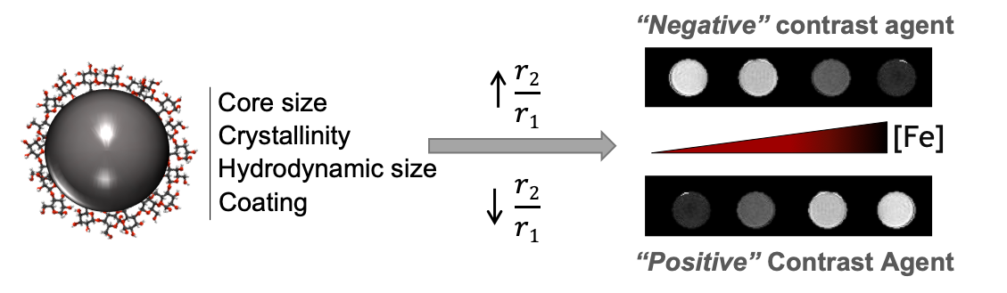
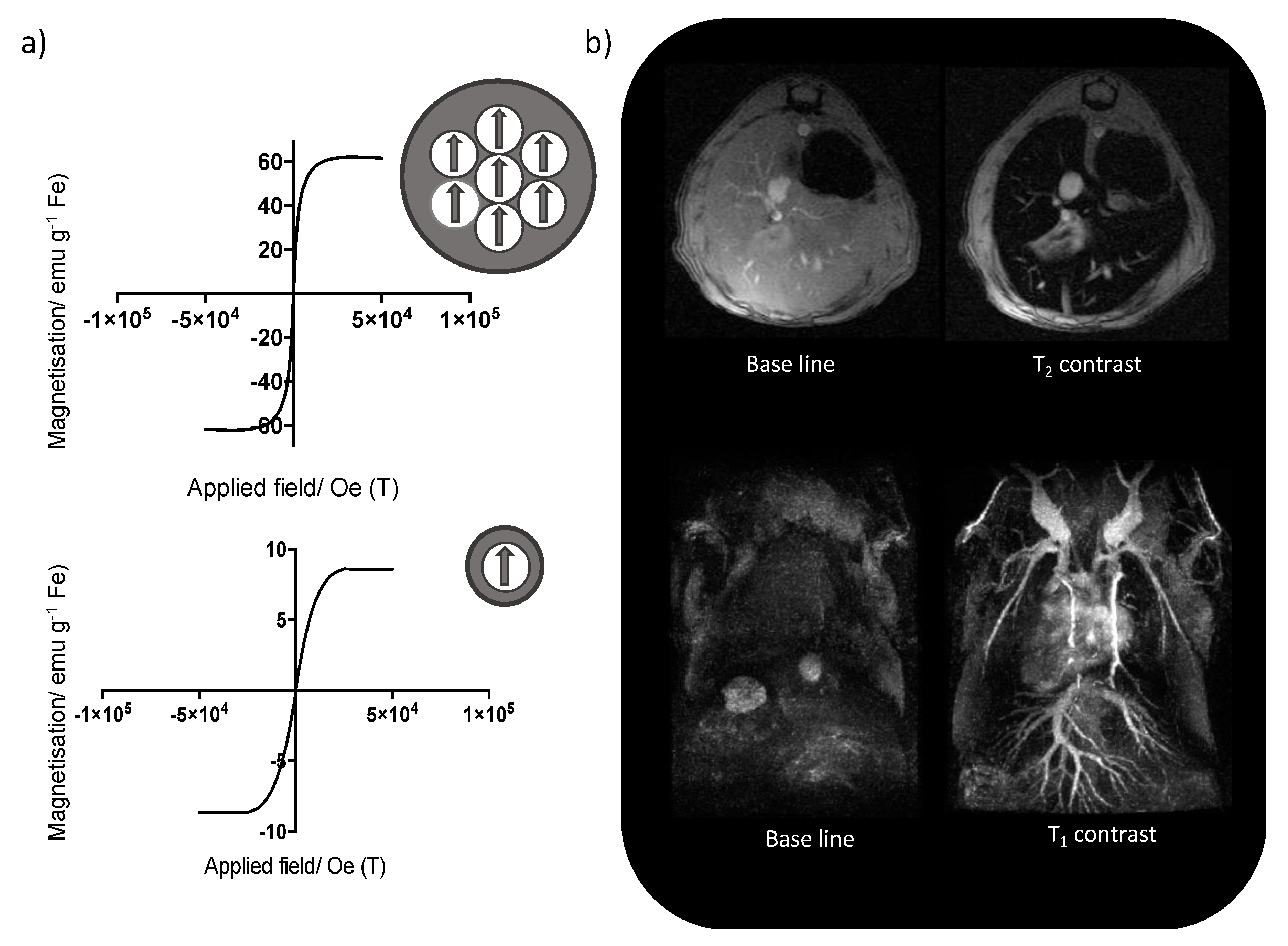
4.1. Physicochemical Properties

IONP components, the core and coating, play an important role in the contrast behaviour. The composition, size and shape of both components must be controlled since they determine the iron oxide phase, crystallinity, magnetic properties and the hydrodynamic size. All of these properties are key to develop an IONP-based T1 contrast agent.
In terms of size, we have to consider both the size of the core and the hydrodynamic size (core + coating). As a rule of thumb, in IONPs, the smaller the core the better the positive contrast. When the core size is decreased (<5 nm) the magnetic single domains decrease, leading to a spin canting effect, which provides five oriented and unpaired Fe3+/Fe2+ electrons, making the particle more paramagnetic than superparamagnetic (Figure 1). Smaller cores usually imply less crystallinity, with a subsequent decrease in the saturation magnetisation values (similar to paramagnetic materials). Therefore, maghemite (γ-Fe2O3) is often preferred for T1 contrast rather than magnetite (Fe3O4) where the crystallinity is usually higher.
Core size and composition are essential but so is an appropriate coating. The stability of the nanoparticles is crucial to avoid aggregation, which triggers multiple particles to act as a single magnetic domain and, hence, increases the T2 effect. In this regard, different coatings such as small molecules, macromolecules or proteins have served as stabilisers [77,78,79,80]. In addition, the size of the coating has been demonstrated to be a key factor in the T1 effect [81]. Large size coatings (i.e., large hydrodynamic size, >30–40 nm) restrict water access to the nanoparticle core increasing the outer-sphere contribution to the relaxation mechanism. Coatings providing ultrasmall hydrodynamic sizes (<7 nm) usually confer poor stability, with aggregation increasing the T2 effect. This is, however, an interesting challenge due to the renal clearance of ultrasmall IONPs, which increase their translational potential. Coatings rendering medium hydrodynamic sizes (10–20 nm) provide good colloidal stability with a thin coating that increases the water exchange rate, boosting the T1 effect.
Although these are general considerations, there is no general rule established to describe the best-case scenario for each T1-IONP. An optimal T1-weighted sequence, taking into account the physicochemical and relaxometric properties of the nanoparticles and selecting the experimental parameters to highlight them appropriately for each magnetic field, is mandatory. A great number of works have shown a wide variety of formulations, sizes and coatings with the magnetic field, pulse sequences, and acquisition parameters not always optimised for T1-weighted imaging. Table 3 summarises some of the reported T1-IONPs, depicting these differences and the influence over the relaxometric properties.

4.2. Synthesis

Co-precipitation and thermal decomposition are the most frequently used methods to produce iron oxide nanoparticles. In addition, the extensive work on iron oxide nanoparticles over the last decade is bringing new procedures every year for the synthesis of nanoparticles of different sizes, shapes and composition and, hence, magnetic behaviour. In this section, the most remarkable methods are briefly described pointing out those which render nanoparticles with positive contrast capabilities.

4.2.1. Co-Precipitation

The co-precipitation protocol is the most used throughout the literature. It is based on the precipitation of iron oxides throughout a mixture of aqueous solutions of ferrous and ferric salts at a 2:1 ratio under basic conditions. The success of the co-precipitation method lies in its simplicity, flexibility and the hydrophilicity of the nanoparticles. However, it also shows some associated drawbacks such as the lack of control over the uniformity of the nanoparticles [103]. Modifications in the co-precipitation protocol can render ultrasmall iron oxide nanoparticles (USPIO) for T1-MRI. A high increase in the temperature of the reaction, combined with different polymers, has been used to develop nanoparticles between 3 and 8 nm with associated longitudinal relaxivities of up to 9 mM−1 s−1 at 4.7 T and 31 mM−1 s−1 at 1 T [90,104].

4.2.2. Thermal Decomposition

This well-known method, based on the decomposition of organic precursors at very high temperatures, has been widely utilised because of the high uniformity, crystallinity and control over the size of the nanoparticles. These nanoparticles are hydrophobic and therefore only stable in hydrophobic solvents, which implies an extra reaction step to stabilise the nanoparticles in physiological media. Despite this inconvenience, the control over the hydrodynamic size allows the obtention of USPIO with potential in positive contrast [87]. Wei et al. described the thermal decomposition of Fe(oleate)3 in the presence of 1-tetradecene, 1-hexadecene and 1-octadecene, rendering nanoparticles from 2.5 to 7.0 nm of a maghemite core oxidised with trimethylamine N-oxide. The nanoparticles, stabilised in water using a ligand exchange reaction with a zwitterionic dopamine sulfonate (ZDS), showed a modest r1 = 5.2 mM−1 s−1 at 1.5 T and 1.5 mM−1 s−1 at 7 T [78]. Another remarkable example describes the synthesis of USPIO by decomposition of Fe(acac)3 at 300 °C in the presence of oleic acid, oleylamine, hexadecanediol and oleyl alcohol as solvent. Ligand exchange with different bisphosphonate-based ligands provided nanoparticles with a magnetic core of 3.6 nm and a maximum r1 = 11 mM−1 s−1 at 1.5 T [98].

4.2.3. Polyol Synthesis

Polyol synthesis has been gaining attention in the recent years for the synthesis of IONPs. This method allows for an easy scaling-up of the reaction in a single-step reaction producing hydrophilic nanoparticles, although aggregation often happens. The method essentially consists of the reduction of the organometallic precursor in the presence of different polyols such as trimethylene glycol, propylene glycol or ethylene glycol. The role of the polyol has been reported not only as stabiliser but also as reducing agent [105].
Concerning IONPs with T1 capabilities, the reduction of Fe(acac)3 in diethylene glycol at 200 °C, under an inert atmosphere provided nanoparticles with 8 nm of hydrodynamic size and 3 nm of core size. These small particles exhibited r1 = 12 mM−1 s−1 at 1.41 T with a low r2/r1 = 2.4 [106]. Another reported example depicts a one-pot reaction of 5.4 nm IONPs by reduction of Fe(acac)3 in the presence of triethylene glycol and HOOC-PEG-COOH at 260 °C. Under these conditions, the synthesised particles showed a r1 = 19.7 mM−1 s−1 at 1.5 T with a r2/r1 ratio of 2.0 [84].

4.2.4. Microwave Assisted Synthesis

Due to the simplicity, fast kinetics and reproducibility of the reactions, microwave-assisted synthesis of IONPs has grown lately. The use of microwaves ensures a very fast and homogeneous heating in the sample, which translates in a narrow size distribution of the nanoparticles [107]. Regarding the synthesis of IONPs for T1 contrast, our group has been deeply involved in the use of microwaves. Essentially, extremally small iron oxide cores (~2.5 nm) can be achieved with FeCl3 as iron source. The reaction is conducted at 100 °C for 10 min in the presence of hydrazine hydrate (reducing agent) and a surfactant, usually dextran or sodium citrate. In a first approach, a r1 = 5.97 mM−1 s−1 at 1.5 T was obtained using (FITC)-dextran or dextran 6KDa as surfactant [108,109]. After these first approaches, better longitudinal relaxivities were observed using sodium citrate as surfactant. In this case, an increase in the temperature of the reaction turns over the contrast capabilities from T2 to T1 with an r1 = 11.9 mM−1 s−1 at 1.5 T when the reaction is performed at 120 °C [81]. Very recently, Fernandez-Barahona et al. increased this value up to 15.7 mM−1 s−1, doping the iron oxide core with 4% mol of Cu [99].

5. In Vivo Applications

The use of iron oxide nanoparticles as contrast agents for MRI has usually been associated with T2-MRI. This is one of the main reasons why the use of iron oxide-based contrast agents for MRI is far from standard in clinical practice. The dark (T2) signal makes them difficult to distinguish from naturally occurring hypointense areas in many diseases—caused by calcium deposits, bleeding, other metals or any signal void present in the area of interest. Given this, the need for nontoxic positive contrast agents has led to intense research in the development of iron oxide nanoparticles that produce high T1-weighted MRI signals, becoming therefore, a very active topic.

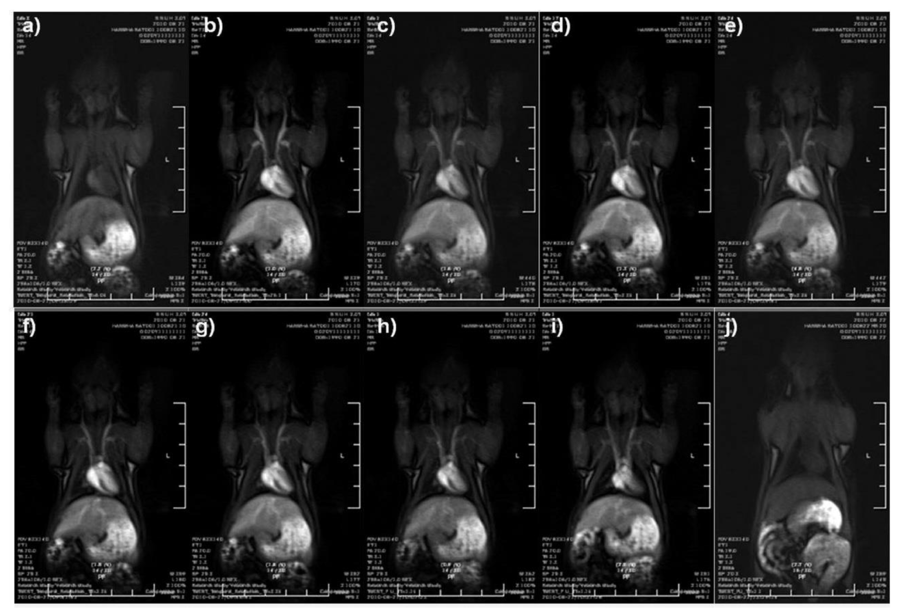
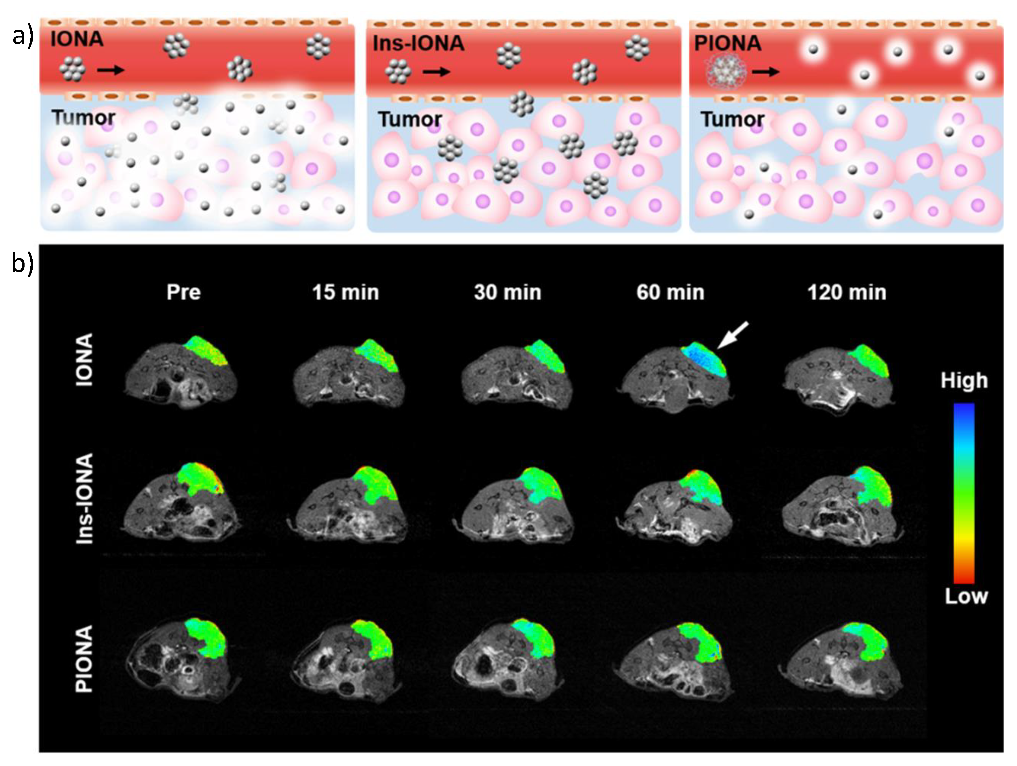
5.1. Iron-Based and Iron Oxide Nanoparticles

In recent years, several research groups have been developing new iron-based probes, mostly for T1-MRI but also for dual contrast MRI. Regarding T1-MRI, some researchers have been able to show, in vivo, a positive contrast effect. In 2013 Ju et al. [110] elaborated non-toxic synthetic melanin-like nanoparticles complexed with paramagnetic Fe3+ ions and stabilised with PEG (PEGylated Fe3+-MelNPs), which were inspired by the MRI signal-enhancing capability of natural melanin. These nanoparticles showed relaxivity values of r1 = 17 mM−1 s−1 and r2 =18 mM−1 s−1 at 3 T, which are higher than those of existing T1-MRI contrast agents based on gadolinium (Gd) or manganese (Mn). In vivo, PEGylated Fe3+-MelNPs showed a positive signal enhancement in the spleen and liver of healthy mice within 0.5 and 1.5 h, respectively, after intravenous injection. During the same year, Peng et al. [111] synthesised antiferromagnetic α-iron oxide-hydroxide (α-FeOOH) nanocolloids, with diameters of 2–3 nm, which were placed inside the mesopores of worm-like silica nanoparticles. These nanocomposites exhibited a low r2/r1 ratio of 1.9, making them suitable as T1-weighted contrast agents. The in vivo experiments carried out showed a positive enhancement in the brain, bladder and kidneys of healthy mice. Moreover, in 2015, Iqbal et al. [85] produced biocompatible silica-coated superparamagnetic IONPs with diameters of ~4 nm, which showed in vivo T1 contrast enhancement in the heart, liver, kidney and bladder of healthy mice. In addition, Macher et al. [93] developed an innovative T1 MRI contrast agent, ultrathin iron oxide nanowhiskers. These nanostructures, with dimensions of 2 × 20 nm, possessed a high surface-to-volume ratio, leading to a strong paramagnetic signal, a property suitable for T1 contrast. In vivo experiments showed a positive contrast enhancement in rat models after intraperitoneal (IP) and subcutaneous injection of the nanowhiskers. In 2019, Tao et al. [77] synthesised (poly(acrylic acid)-poly(methacrylic acid) iron oxide nanoparticles (PMAA-PTTM-IONPs) 4.34 nm in diameter with a low r2/r1 ratio of 2.78, adequate for T1-weighted contrast agents. In vivo experiments in mouse models showed biocompatibility and T1 contrast enhancement in liver and kidney.
Even though the previously described probes were shown to provide contrast enhancement during in vivo T1-MRI, no specific in vivo application was described for them; thus, only their biodistribution was tested. Nevertheless, different research groups have developed probes for particular applications such as magnetic resonance angiography (MRA) and tumour imaging.
MRA has been shown to be a very helpful technique in clinical imaging. With its use, several diseases could be detected including myocardial infarction, renal failure, atherosclerotic plaque, thrombosis and tumour angiogenesis. Regarding research carried out on MRA, in 2011 Kim et al. [87] synthesised ultrasmall iron oxide nanoparticles (ESION, size < 4 nm) capped using poly(ethylene glycol)-derivatised phosphine oxide (PO-PEG) ligands, which enabled clear observation of various blood vessels, with sizes down to 0.2 mm, during in vivo T1-MRI in rat models. They were able to maintain the bright signal of blood vessels for 1 h on dynamic time-resolved MR angiography, showing that this type of probe can be used for T1 enhanced blood pool MRI (Figure 2).
Furthermore, in 2014 Chan et al. [83] produced multidentate block-copolymer-stabilised ultrasmall superparamagnetic iron oxide nanoparticles (MDBC-USPIOs), with diameters <5 nm and a r2/r1 ratio of 4.74 to test them as a promising T1-positive contrast agent for in vivo MRI. Results from in vivo MRI showed a strong blood signal enhancement after their intravenous injection in mouse models. During the same year, Liu et al. [91] developed glutathione-coated iron oxide nanoparticles (GSH-IONPs) with sizes of around 3.75 nm and a r2/r1 ratio of 2.28 as a novel T1-MRI contrast agent. The in vivo results showed a strong vascular enhancement at the carotid artery and superior sagittal sinus of healthy mice models, making it a promising contrast agent for thrombus detection.
In 2015, Bhavesh et al. [108] proposed an extremely fast microwave synthesis of fluorescein-labelled dextran-coated extremely small IONPs for their use as a contrast agent for T1-weighted MRI. This method yielded very small NPs with hydrodynamic diameters of 21.5 nm and with a r2/r1 ratio of 4.7 suitable for T1-MRI. In vivo MRA of healthy mice showed a clear depiction of the main vascular architecture; furthermore, high quality visualisation of small vessels was maintained even 90 min post NP injection, highlighting the advantage of these NPs to be used as a contrast agent for blood pool imaging applications. In 2017, Pellico et al. [81] described the changes produced on the relaxometric properties of IONPs when the thickness of their organic coating is modified. For that purpose, this group synthesised all IONPs using a microwave-driven synthesis method; however, in order to change the coating layer thickness, different syntheses were carried out heating at different temperatures. Results showed that IONPs with a thinner coating yielded an excellent positive T1-MRI contrast, more specifically NPs synthesised at 120 °C. These NPs, with a hydrodynamic size of 7.9 nm and a r2/r1 ratio of 1.9 showed great contrast enhancement in both body and brain MRA of healthy mice, providing the possibility of better visualising their vasculature. In 2018, Vangijzegem et al. [98] synthesised very small PEGylated iron oxide nanoparticles (VSIONPs) with core sizes of 3.5 nm that showed positive contrast in in vivo MRA, enabling the heart chamber and the vena cava of healthy mice to be observed.
Regarding research on T1-weighted MR tumour imaging, in 2014 Wu et al. [112] produced mesoporous silica NPs with drug-labelled USPIONs confined within the mesoporous matrix (Fe-MSNs) as a pH-responsive theranostic platform. The tumour accumulation, driven by the enhanced permeability and retention effect (EPR), was confirmed by the T1 enhancement in the affected site. Moreover, the unique metal–ligand coordination bonding between Fe species and the anticancer drug molecules provided the carrier with a pH-responsive drug release feature, triggering a controlled drug release under the acidic microenvironment in the tumour area. Moreover, Shen et al. [94] in 2017 synthesised extremely small 3.6 nm magnetic iron oxide nanoparticles (ES-MIONs) functionalised with dimeric RGD peptide (RGD2) and PEG methyl ether (mPEG), which were afterwards loaded with the anticancer drug doxorubicin hydrochloride (DOX), as a tumour targeting theranostic platform. In this probe, the ES-MIONs serve as the contrast agent for T1-MRI, while RGD2 is used for tumour targeting and DOX for chemotherapy. In vivo experiments on tumour-bearing mice showed an enhanced T1-MRI signal when using this platform together with partial regression of tumours due to active targeting and chemotherapy. Furthermore, Li et al. [88] in 2019 developed dynamically reversible iron oxide nanoparticle assemblies (IONAs) consisting of ES-IONs cross-linked by small molecular aldehyde derivative ligands. The linkage of these ligands is cleaved in the presence of acidic environments, such as tumours, releasing ES-IONs that are capable of producing contrast in T1-weighted MRI (Figure 3a). To demonstrate the specificity of the tandem linkage-cleavage, pH-insensitive cross-linked iron oxide nanoparticles (Ins-IONAs) and micelle-like pH sensitive polymer-assisted iron oxide nanoparticle assemblies (PIONAs) of similar sizes were used as controls (Figure 3a). In vivo experiments on tumour-bearing mice clearly indicated that the IONAs could amplify the T1-MRI signal, whilst Ins-IONAs and PIONAs showed significantly lower intensities (Figure 3b).
In addition to the undertaken research to find new probes that serve as contrast agents for T1-weighted MRI, there have been some research groups that have focused on developing probes for dual contrast MRI, that is, probes that are capable of producing both negative (T2) and positive (T1) contrast. In MRI, T1-weighted images typically provide better spatial resolution, while T2-weighted images can provide enhanced detection of lesions. For this reason, dual contrast probes could potentially provide better imaging information leading to higher diagnostic accuracy. Regarding research in this field, Jung et al. [86] in 2014 synthesised 5 nm SPIONs that, as shown using in vivo MRA experiments in rat models, were capable of producing both positive and negative contrast in MRA by using different acquisition pulses. On the one hand, ultrashort echo (UTE) sequence, which differently to the sequences explained in Section 2 use a radial sampling of the k-space to minimise TE, positively enhanced vascular signals in MR angiography, providing highly resolved vessel structures. In addition, typical gradient sequences such as fast low angle shot (FLASH) acquisition yielded strong negative vessel contrast, resulting in a higher number of discernible vessel branches. Moreover, in 2018 Alipour et al. [82] developed 11 nm silica coated cubic SPIONs as a dual-mode contrast agent for T1 and T2 MRI. In vivo investigations on a 3 T MRI scanner demonstrated both positive and negative contrast enhancement 70 min post intravenous injection in healthy rat models.

5.2. Doped Iron Oxide Nanoparticles

Throughout the past years, some metals have been integrated into IONPs in order to get better T1 enhancement. Among different metals, focus remains on gadolinium (Gd), which are not included since our goal is focusing on alternatives, and manganese (Mn); however, there are some studies that use other metals such as copper (Cu) and europium (Eu).
Manganese-based contrast agents, similarly to Gd contrast agents, can significantly enhance the positive contrast in T1-weighted MRI. Furthermore, they can serve as contrast agents for visualising sub-anatomic structures and activities of brain and heart by taking advantage of Mn2+ as a biological calcium analogue. Nevertheless, their high toxicity limits their clinical applications, and for that reason, it is highly desirable to develop alternative non-toxic manganese contrast agents for clinical MRI. Several research groups have been recently developing manganese-containing iron oxide nanoparticles as alternative positive MRI contrast agents. In 2013, Li et al. [113] synthesised nontoxic ultrasmall manganese ferrite (MnFe2O4) nanoparticles of 2‒3 nm that were shown to enhance the T1 contrast in the liver, kidneys and brain of healthy mice during in vivo MRI. Similarly, in 2014 Huang et al. [114] produced Mn-doped IONPs of around 5 nm in diameter capable of functioning as a contrast agent for T1-weighted MRI having a low r2/r1 ratio of 2.6. Post injection, in vivo T1-MRI showed a brighter signal in the liver regions of healthy mice. Furthermore, in 2015 Zhang et al. [115] synthesised bovine serum albumin-coated manganese-doped IONPs (MnIO-BSA), 5 nm in size and with a low r2/r1 ratio of 2.18, as a theranostic platform for tumour targeting. The probe, once accumulated in the tumour, was able to provide contrast in T1-weighted MRI and be heated using an external NIR light source for photothermal therapy (PTT) purposes. The in vivo MRI experiments using a tumour-bearing mouse model exhibited significant signal enhancement (about two times) at the tumour site. Furthermore, it was demonstrated that hyperthermia caused by the photothermal effect of the MnIO-BSA nanoparticles under NIR laser irradiation resulted in significant death of the cancer cells.
Only a few studies use copper or europium as doping metals. In 2019, our group synthesised extremely small citrate-coated iron oxide nanoparticles doped with Cu as a T1-weighted contrast agent [99]. After studying several Cu doping amounts, IONPs doped with 4% mol of Cu (Cu4-NPs) were chosen as the best probe. Cu4-NPs presented a hydrodynamic diameter of ~15 nm and a core size of ~3.5 nm. Moreover, they showed a high r1 value of 15.7 mM−1 s−1 at 1.5 T and a low r2/r1 ratio of 2.1. Interestingly, the reason for this increase in the r1 values was due to the distribution of Cu atoms within the iron oxide structure. In vivo MRA showed that Cu4-NPs provided high-quality images with fine details of the vasculature up to 30 min post injection (Figure 4). In order to explore a different probe application, RGD molecules were conjugated to the surfaces of Cu4-NPs for active tumour targeting. In vivo T1-MRI experiments on tumour-bearing mice showed an increased positive signal on the tumour after intravenous injection of the RGD-Cu4-NP, further confirming their use as a positive contrast agent. Europium was used by Yang et al. [100] in 2015; this group produced citrate-coated europium-doped iron oxide nanocubes, 14 nm in size, with high r1 values of 36.8 mM−1 s−1 at 0.5 T and a low r2/r1 ratio of 2.65 for T1-weighted MRI. In vivo results showed an increase in signal brightness in the heart of healthy mice.

5.3. Multimodal T1-Iron Oxide Nanoparticles

The combination of MRI with complementary imaging techniques such as PET or SPECT (Single Photon Emission Computed Tomography), allows protocols to be developed that exploit their synergy. Although a great number of multimodal IONPs have been described for T2-weighted MRI, only a few examples have been reported for IONPs acting as positive contrast agents [116].
These examples are focused on the combination of biocompatible iron oxide nanoparticles with radionuclides, such as Galium-68 (68Ga) or Technetium-99m (99mTc) for PET and SPECT, respectively.
68Ga has been successfully used with IONPs in several multimodal experiments. In 2016, Pellico et al. developed a T1-weighted MRI/PET platform consisting of extremely small 68Ga-doped IONPs synthesised using a microwave-driven protocol, which were diversely functionalised for their use in several applications. For example, 68Ga core-doped IONPs functionalised with RGD were synthesised for tumour angiogenesis targeting. This probe presented a hydrodynamic size of 20.6 nm with core sizes of 2.5 nm. Furthermore, it showed a r1 value of 5.7 mM−1 s−1 at 1.5 T and a low r2/r1 ratio of 3.9, making it suitable for T1-weighted MRI. In vivo MRI experiments in tumour-bearing mouse models showed a brighter signal in the tumour 24 h post NP injection.114 In addition, in 2018 the group synthesised a biorthogonal nano-radiotracer for in vivo pretargeted molecular imaging of atherosclerosis. This probe was based on the in vivo tetrazine ligation of an imaging and a targeting moiety, which were functionalised with a tetrazine (Tz) and a trans-cyclooctene (TCO), respectively. The imaging part consisted of 68Ga-doped IONPs that provided simultaneous PET and T1-MRI signals, while the atherosclerosis targeting part consisted of an oxidised LDL (Low-Density Lipoprotein)-targeting IgM antibody (E-06). 68Ga-IONP-Tz presented a hydrodynamic diameter of 15.5 nm with a core size of 2.8 nm. Moreover, they showed a high r1 value of 7.1 mM−1 s−1 at 1.5 T and a low r2/r1 ratio of 2.5, making them useful as contrast agents for T1-weighted MRI. Ex vivo T1-MRI of the aorta of an atherosclerotic mouse model showed a brighter signal on the atherosclerotic plaque [117].
Regarding research carried out on 99mTc-doped IONPs, in 2013 Sandiford et al. [118] produced PEGylated bisphosphonate-coated USPIOs to use as a multimodal platform in T1-MRI and SPECT. This probe showed a high r1 value of 9.5 mM−1 s−1 at 3 T and a low r2/r1 ratio of 2.9, enabling their use as positive contrast agents. In vivo MRI experiments in healthy BALB/C mice showed a strong T1 effect post NP injection resulting in a substantial increase in the signal from blood, making vessels, the heart compartments and other highly vascularised organs such as the spleen visible (Figure 5a). This biodistribution was also observed using SPECT/CT with the nanoparticles circulating 40 min after i.v. injection (Figure 5b).
Besides the use of T1-IONPs for dual MRI(T1)/PET or SPECT imaging, G. Wang et al. reported an example for MRI(T1)/CT imaging. In this case, ultrasmall magnetite nanoparticles were combined with Au nanocages rendering F-AuNC@Fe3O4. The ultrasmall Fe3O4 particles (2.2 nm) showed a r1 = 6.3 mM−1 s−1, providing a T1-enhancement in MRI, whilst a strong X-ray attenuation was observed due to the AuNC [119].

6. Conclusions and Future Perspectives

In the last decade, significant effort has been made in the design of iron oxide formulations for T1 contrast due to the associated toxicity in the application of the traditional Gd3+ contrast agents. We have summarised the most important features to optimise IONPs as a T1 contrast agent, i.e., ultrasmall core size with moderate crystallinity (usually maghemite (γ-Fe2O3)) and high colloidal stability with hydrodynamic sizes ranging from 5 to 20 nm. With the focus on the physicochemical characteristics of T1-IONPs such as iron phase, core size, type of coating or the surface charge, there is still room for improvement using the described synthetic procedures or novel strategies. Although the optimisation of these properties brings associated higher T1 performance, the longitudinal relaxivities are still far from those obtained in some Gd3+-based nanoparticles such as gadofullerens, gadolinium oxide particles, Gd-nanocages or Gd3+ conjugated to mesoporous silica nanoparticles [120]. It is noteworthy that the application of Gd3+-based agents has been thoroughly studied for many years, whilst the potential of IONPs as T1 agents is an emerging field of research. Therefore, there is still a lack of consensus to determine the best scenario for T1-IONPs. To date, the variability of the particles showing different physicochemical properties and MRI responses is high. However, a broad number of applications have been reported including not only MRI but also multimodal imaging for molecular imaging applications or metal-doped particles for therapeutic purposes. In addition, other issues such as biocompatibility, pharmacokinetics or delivery pathways must be studied in advance to guarantee their clinical translation. In conclusion, there is great potential in the development of T1-IONPs with several concerns to be evaluated in detail. Further studies must bring a clear conclusion about the best properties and then adapt the formulations to clinical requirements.

Funding

F.H. was funded through the Spanish Ministry for Economy and Competitiveness (MEyC), grant number [SAF2016-79593-P] and the Ministry of Science, grant number [RED2018-102469-T]. J.R-C. was funded through the Spanish Ministry for Economy and Competitiveness (MEyC), grant number [SAF2017-84494-C2-R], and the Gobierno Vasco, Dpto. Industria, Innovación, Comercio y Turismo under the ELKARTEK Program (Grant No. KK-2019/bmG19). CIC biomaGUNE is supported by the Maria de Maeztu Units of Excellence Program from the Spanish State Research Agency—Grant No. MDM-2017-0720.

Conflicts of Interest

The authors declare no conflict of interest.

References

  1. Markl, M.; Leupold, J. Gradient echo imaging. J. Magn. Reson. Imaging 2012, 35, 1274–1289. [Google Scholar] [CrossRef] [PubMed]
  2. Lohrke, J.; Frenzel, T.; Endrikat, J.; Alves, F.C.; Grist, T.M.; Law, M.; Lee, J.M.; Leiner, T.; Li, K.-C.; Nikolaou, K.; et al. 25 Years of Contrast-Enhanced MRI: Developments, Current Challenges and Future Perspectives. Adv. Ther. 2016, 33, 1–28. [Google Scholar] [CrossRef] [PubMed] [Green Version]
  3. Na, H.B.; Hyeon, T. Nanostructured T1 MRI contrast agents. J. Mater. Chem. 2009, 19, 6267–6273. [Google Scholar] [CrossRef]
  4. Bridot, J.-L.; Faure, A.-C.; Laurent, S.; Riviere, C.; Billotey, C.; Hiba, B.; Janier, M.; Josserand, V.; Coll, J.-L.; Elst, L.V.; et al. Hybrid Gadolinium Oxide Nanoparticles: Multimodal Contrast Agents for in Vivo Imaging. J. Am. Chem. Soc. 2007, 129, 5076–5084. [Google Scholar] [CrossRef] [PubMed]
  5. Park, J.Y.; Baek, M.J.; Choi, E.S.; Woo, S.; Kim, J.H.; Kim, T.J.; Jung, J.C.; Chae, K.S.; Chang, Y.; Lee, G.H. Paramagnetic Ultrasmall Gadolinium Oxide Nanoparticles as Advanced T1 MRI Contrast Agent: Account for Large Longitudinal Relaxivity, Optimal Particle Diameter, and In Vivo T1 MR Images. ACS Nano 2009, 3, 3663–3669. [Google Scholar] [CrossRef]
  6. Evanics, F.; Diamente, P.R.; van Veggel, F.C.J.M.; Stanisz, G.J.; Prosser, R.S. Water-Soluble GdF3 and GdF3/LaF3 NanoparticlesPhysical Characterization and NMR Relaxation Properties. Chem. Mater. 2006, 18, 2499–2505. [Google Scholar] [CrossRef]
  7. Cheung, E.N.M.; Alvares, R.D.; Oakden, W.; Chaudhary, R.; Hill, M.L.; Pichaandi, J.; Mo, G.C.H.; Yip, C.; Macdonald, P.M.; Stanisz, G.J.; et al. Polymer-Stabilized Lanthanide Fluoride Nanoparticle Aggregates as Contrast Agents for Magnetic Resonance Imaging and Computed Tomography. Chem. Mater. 2010, 22, 4728–4739. [Google Scholar] [CrossRef]
  8. Hifumi, H.; Yamaoka, S.; Tanimoto, A.; Citterio, D.; Suzuki, K. Gadolinium-Based Hybrid Nanoparticles as a Positive MR Contrast Agent. J. Am. Chem. Soc. 2006, 128, 15090–15091. [Google Scholar] [CrossRef]
  9. Shen, Z.; Fan, W.; Yang, Z.; Liu, Y.; Bregadze, V.I.; Mandal, S.K.; Yung, B.C.; Lin, L.; Liu, T.; Tang, W.; et al. Exceedingly Small Gadolinium Oxide Nanoparticles with Remarkable Relaxivities for Magnetic Resonance Imaging of Tumors. Small 2019, 15, 1903422. [Google Scholar] [CrossRef]
  10. Mortezazadeh, T.; Gholibegloo, E.; Alam, N.R.; Dehghani, S.; Haghgoo, S.; Ghanaati, H.; Khoobi, M. Gadolinium(III) oxide nanoparticles coated with folic acid-functionalized poly(β-cyclodextrin-co-pentetic acid) as a biocompatible targeted nano-contrast agent for cancer diagnostic: In vitro and in vivo studies. Magn. Reson. Mater. Phys. Biol. Med. 2019, 32, 487–500. [Google Scholar] [CrossRef]
  11. Kumar, S.; Meena, V.K.; Hazari, P.P.; Sharma, S.K.; Sharma, R.K. Rose Bengal attached and dextran coated gadolinium oxide nanoparticles for potential diagnostic imaging applications. Eur. J. Pharm. Sci. 2018, 117, 362–370. [Google Scholar] [CrossRef] [PubMed]
  12. Qiu, Q.; Wen, Y.; Dong, H.; Shen, A.; Zheng, X.; Li, Y.; Feng, F. A highly sensitive living probe derived from nanoparticle-remodeled neutrophils for precision tumor imaging diagnosis. Biomater. Sci. 2019, 7, 5211–5220. [Google Scholar] [CrossRef] [PubMed]
  13. Cui, D.; Lu, X.; Yan, C.; Liu, X.; Hou, M.; Xia, Q.; Xu, Y.; Liu, R. Gastrin-releasing peptide receptor-targeted gadolinium oxide-based multifunctional nanoparticles for dual magnetic resonance/fluorescent molecular imaging of prostate cancer. Int. J. Nanomed. 2017, 12, 6787–6797. [Google Scholar] [CrossRef] [PubMed] [Green Version]
  14. Mastrogiacomo, S.; Kownacka, A.E.; Dou, W.; Burke, B.P.; de Rosales, R.T.; Heerschap, A.; Jansen, J.A.; Archibald, S.J.; Walboomers, X.F. Bisphosphonate Functionalized Gadolinium Oxide Nanoparticles Allow Long-Term MRI/CT Multimodal Imaging of Calcium Phosphate Bone Cement. Adv. Healthc. Mater. 2018, 7, 1800202. [Google Scholar] [CrossRef] [PubMed] [Green Version]
  15. Dai, Y.; Wu, C.; Wang, S.; Li, Q.; Zhang, M.; Li, J.; Xu, K. Comparative study on in vivo behavior of PEGylated gadolinium oxide nanoparticles and Magnevist as MRI contrast agent. Nanomed. Nanotechnol. Biol. Med. 2018, 14, 547–555. [Google Scholar] [CrossRef]
  16. Jiang, Y.; Zheng, W. Cardiovascular toxicities upon manganese exposure. Cardiovasc. Toxicol. 2005, 5, 345–354. [Google Scholar] [CrossRef]
  17. Sidoryk-Wegrzynowicz, M.; Aschner, M. Manganese toxicity in the central nervous system: The glutamine/glutamate-γ-aminobutyric acid cycle. J. Intern. Med. 2013, 273, 466–477. [Google Scholar] [CrossRef] [Green Version]
  18. Wolf, G.; Baum, L. Cardiovascular toxicity and tissue proton T1 response to manganese injection in the dog and rabbit. Am. J. Roentgenol. 1983, 141, 193–197. [Google Scholar] [CrossRef]
  19. Chevallier, P.; Walter, A.; Garofalo, A.; Veksler, I.; Lagueux, J.; Begin-Colin, S.; Felder-Fleschc, D.; Fortin, M.-A. Tailored biological retention and efficient clearance of pegylated ultra-small MnO nanoparticles as positive MRI contrast agents for molecular imaging. J. Mater. Chem. B 2014, 2, 1779–1790. [Google Scholar] [CrossRef] [Green Version]
  20. Huang, H.; Yue, T.; Xu, Y.; Xu, K.; Xu, H.; Liu, S.; Yu, J.; Huang, J. PEGylation of MnO nanoparticles via catechol-Mn chelation to improving T1-weighted magnetic resonance imaging application. J. Appl. Polym. Sci. 2015, 132, 2–9. [Google Scholar] [CrossRef]
  21. Wang, P.; Yang, J.; Zhou, B.; Hu, Y.; Xing, L.; Xu, F.; Shen, M.; Zhang, G.; Shi, X. Antifouling manganese oxide nanoparticles: Synthesis, characterization, and applications for enhanced MR imaging of tumors. ACS Appl. Mater. Interfaces 2017, 9, 47–53. [Google Scholar] [CrossRef] [PubMed]
  22. Chen, N.; Shao, C.; Qu, Y.; Li, S.; Gu, W.; Zheng, T.; Ye, L.; Yu, C. Folic Acid-Conjugated MnO Nanoparticles as a T1 Contrast Agent for Magnetic Resonance Imaging of Tiny Brain Gliomas. ACS Appl. Mater. Interfaces 2014, 6, 19850–19857. [Google Scholar] [CrossRef] [PubMed]
  23. Gallo, J.; Alam, I.S.; Lavdas, I.; Wylezinska-Arridge, M.; Aboagye, E.O.; Long, N.J. RGD-targeted MnO nanoparticles as T1 contrast agents for cancer imaging-the effect of PEG length in vivo. J. Mater. Chem. B 2014, 2, 868–876. [Google Scholar] [CrossRef]
  24. Li, J.; Wu, C.; Hou, P.; Zhang, M.; Xu, K. One-pot preparation of hydrophilic manganese oxide nanoparticles as T1 nano-contrast agent for molecular magnetic resonance imaging of renal carcinoma in vitro and in vivo. Biosens. Bioelectron. 2018, 102, 1–8. [Google Scholar] [CrossRef] [PubMed]
  25. Li, B.; Gu, Z.; Kurniawan, N.; Chen, W.; Xu, Z.P. Manganese-Based Layered Double Hydroxide Nanoparticles as a T1-MRI Contrast Agent with Ultrasensitive pH Response and High Relaxivity. Adv. Mater. 2017, 29, 1700373. [Google Scholar] [CrossRef] [PubMed]
  26. McDonagh, B.H.; Singh, G.; Hak, S.; Bandyopadhyay, S.; Augestad, I.L.; Peddis, D.; Sandvig, I.; Sandvig, A.; Glomm, W.R. L-DOPA-Coated Manganese Oxide Nanoparticles as Dual MRI Contrast Agents and Drug-Delivery Vehicles. Small 2016, 12, 301–306. [Google Scholar] [CrossRef] [PubMed] [Green Version]
  27. Cheng, Y.; Zhang, S.; Kang, N.; Huang, J.; Lv, X.; Wen, K.; Ye, S.; Chen, Z.; Zhou, X.; Ren, L. Polydopamine-Coated Manganese Carbonate Nanoparticles for Amplified Magnetic Resonance Imaging-Guided Photothermal Therapy. ACS Appl. Mater. Interfaces 2017, 9, 19296–19306. [Google Scholar] [CrossRef] [Green Version]
  28. Zhan, Y.; Shi, S.; Ehlerding, E.B.; Graves, S.A.; Goel, S.; Engle, J.W.; Liang, J.; Tian, J.; Cai, W. Radiolabeled, Antibody-Conjugated Manganese Oxide Nanoparticles for Tumor Vasculature Targeted Positron Emission Tomography and Magnetic Resonance Imaging. ACS Appl. Mater. Interfaces 2017, 9, 38304–38312. [Google Scholar] [CrossRef]
  29. Zhan, Y.; Zhan, W.; Li, H.; Xu, X.; Cao, X.; Zhu, S.; Liang, J.; Chen, X. In vivo dual-modality fluorescence and magnetic resonance imaging-guided lymph node mapping with good biocompatibility manganese oxide nanoparticles. Molecules 2017, 22, 2208. [Google Scholar] [CrossRef] [Green Version]
  30. McMahon, M.T.; Bulte, J.W.M. Two decades of dendrimers as versatile MRI agents: A tale with and without metals. Wiley Interdiscip. Rev. Nanomed. Nanobiotechnol. 2018, 10, e1496. [Google Scholar] [CrossRef]
  31. Dias, A.P.; da Silva Santos, S.; da Silva, J.V.; Parise Filho, R.; Ferreira, E.I.; El Seoud, O.; Giarolla, J. Dendrimers in the context of nanomedicine. Int. J. Pharm. 2020, 573, 118814. [Google Scholar] [CrossRef] [PubMed]
  32. Hu, J.; Hu, K.; Cheng, Y. Tailoring the dendrimer core for efficient gene delivery. Acta Biomater. 2016, 35, 1–11. [Google Scholar] [CrossRef] [PubMed]
  33. Opina, A.C.; Wong, K.J.; Griffiths, G.L.; Turkbey, B.I.; Bernardo, M.; Nakajima, T.; Kobayashi, H.; Choyke, P.L.; Vasalatiy, O. Preparation and long-term biodistribution studies of a PAMAM dendrimer G5–Gd-BnDOTA conjugate for lymphatic imaging. Nanomedicine 2015, 10, 1423–1437. [Google Scholar] [CrossRef] [PubMed] [Green Version]
  34. Swanson, S.D.; Kukowska-Latallo, J.F.; Patri, A.K.; Chen, C.; Ge, S.; Cao, Z.; Kotlyar, A.; East, A.T.; Baker, J.R. Targeted gadolinium-loaded dendrimer nanoparticles for tumor-specific magnetic renosance contrast enhancement. Int. J. Nanomedicine 2008, 3, 201–210. [Google Scholar]
  35. Li, F.; Yan, H.; Wang, J.; Li, C.; Wu, J.; Wu, S.; Rao, S.; Gao, X.; Jin, Q. Non-invasively differentiating extent of liver fibrosis by visualizing hepatic integrin αvβ3 expression with an MRI modality in mice. Biomaterials 2016, 102, 162–174. [Google Scholar] [CrossRef]
  36. Alamdari, N.H.; Alaei-Beirami, M.; Shandiz, S.A.S.; Hejazinia, H.; Rasouli, R.; Saffari, M.; Ebrahimi, S.E.S.; Assadi, A.; Ardestani, M.S. Gd3+-Asparagine-Anionic Linear Globular Dendrimer Second-Generation G2 Complexes: Novel Nanobiohybrid Theranostics. Contrast Media Mol. Imaging 2017, 2017, 3625729. [Google Scholar]
  37. Wang, R.; Luo, Y.; Yang, S.; Lin, J.; Gao, D.; Zhao, Y.; Liu, J.; Shi, X.; Wang, X. Hyaluronic acid-modified manganese-chelated dendrimer-entrapped gold nanoparticles for the targeted CT/MR dual-mode imaging of hepatocellular carcinoma. Sci. Rep. 2016, 6, 33844. [Google Scholar] [CrossRef]
  38. Nguyen, T.H.; Bryant, H.; Shapsa, A.; Street, H.; Mani, V.; Fayad, Z.A.; Frank, J.A.; Tsimikas, S.; Briley-Saebo, K.C. Manganese G8 dendrimers targeted to oxidation-specific epitopes: In vivo MR imaging of atherosclerosis. J. Magn. Reson. Imaging 2015, 41, 797–805. [Google Scholar] [CrossRef] [Green Version]
  39. Fan, Y.; Zhang, J.; Shi, M.; Li, D.; Lu, C.; Cao, X.; Peng, C.; Mignani, S.; Majoral, J.-P.; Shi, X. Poly(amidoamine) Dendrimer-Coordinated Copper(II) Complexes as a Theranostic Nanoplatform for the Radiotherapy-Enhanced Magnetic Resonance Imaging and Chemotherapy of Tumors and Tumor Metastasis. Nano Lett. 2019, 19, 1216–1226. [Google Scholar] [CrossRef]
  40. Kim, S.M.; Im, G.H.; Lee, D.G.; Lee, J.H.; Lee, W.J.; Lee, I.S. Mn2+-doped silica nanoparticles for hepatocyte-targeted detection of liver cancer in T1-weighted MRI. Biomaterials 2013, 34, 8941–8948. [Google Scholar] [CrossRef]
  41. Li, X.; Zhao, W.; Liu, X.; Chen, K.; Zhu, S.; Shi, P.; Chen, Y.; Shi, J. Mesoporous manganese silicate coated silica nanoparticles as multi-stimuli-responsive T1-MRI contrast agents and drug delivery carriers. Acta Biomater. 2016, 30, 378–387. [Google Scholar] [CrossRef] [PubMed]
  42. Pellico, J.; Ellis, C.M.; Miller, J.; Davis, J.J. Water gated contrast switching with polymer–silica hybrid nanoparticles. Chem. Commun. 2019, 55, 8540–8543. [Google Scholar] [CrossRef] [PubMed]
  43. Wartenberg, N.; Fries, P.; Raccurt, O.; Guillermo, A.; Imbert, D.; Mazzanti, M. A Gadolinium Complex Confined in Silica Nanoparticles as a Highly Efficient T1/T2 MRI Contrast Agent. Chem. Eur. J. 2013, 19, 6980–6983. [Google Scholar] [CrossRef] [PubMed]
  44. Taylor, K.M.L.; Kim, J.S.; Rieter, W.J.; An, H.; Lin, W.; Lin, W. Mesoporous Silica Nanospheres as Highly Efficient MRI Contrast Agents. J. Am. Chem. Soc. 2008, 130, 2154–2155. [Google Scholar] [CrossRef]
  45. Carniato, F.; Tei, L.; Botta, M. Gd-Based Mesoporous Silica Nanoparticles as MRI Probes. Eur. J. Inorg. Chem. 2018, 2018, 4936–4954. [Google Scholar] [CrossRef]
  46. Davis, J.J.; Huang, W.-Y.; Davies, G.-L. Location-tuned relaxivity in Gd-doped mesoporous silica nanoparticles. J. Mater. Chem. 2012, 22, 22848–22850. [Google Scholar] [CrossRef] [Green Version]
  47. Fossheim, S.L.; Fahlvik, A.K.; Klaveness, J.; Muller, R.N. Paramagnetic liposomes as MRI contrast agents: Influence of liposomal physicochemical properties on the in vitro relaxivity. Magn. Reson. Imaging 1999, 17, 83–89. [Google Scholar] [CrossRef]
  48. Leclercq, F.; Cohen-Ohana, M.; Mignet, N.; Sbarbati, A.; Herscovici, J.; Scherman, D.; Byk, G. Design, Synthesis, and Evaluation of Gadolinium Cationic Lipids As Tools for Biodistribution Studies of Gene Delivery Complexes. Bioconjug. Chem. 2003, 14, 112–119. [Google Scholar] [CrossRef]
  49. Qiu, L.H.; Zhang, J.W.; Li, S.P.; Xie, C.; Yao, Z.W.; Feng, X.Y. Molecular imaging of angiogenesis to delineate the tumor margins in glioma rat model with endoglin-targeted paramagnetic liposomes using 3T MRI. J. Magn. Reson. Imaging 2015, 41, 1056–1064. [Google Scholar] [CrossRef]
  50. Song, Y.; Li, W.; Meng, S.; Zhou, W.; Su, B.; Tang, L.; Zhao, Y.; Wu, X.; Yin, D.; Fan, M.; et al. Dual integrin αvβ 3 and NRP-1-Targeting Paramagnetic Liposome for Tumor Early Detection in Magnetic Resonance Imaging. Nanoscale Res. Lett. 2018, 13, 380. [Google Scholar] [CrossRef] [Green Version]
  51. Ren, L.; Chen, S.; Li, H.; Zhang, Z.; Zhong, J.; Liu, M.; Zhou, X. MRI-guided liposomes for targeted tandem chemotherapy and therapeutic response prediction. Acta Biomater. 2016, 35, 260–268. [Google Scholar] [CrossRef]
  52. Woodside, D.G.; Tanifum, E.A.; Ghaghada, K.B.; Biediger, R.J.; Caivano, A.R.; Starosolski, Z.A.; Khounlo, S.; Bhayana, S.; Abbasi, S.; Craft, J.W., Jr.; et al. Magnetic Resonance Imaging of Atherosclerotic Plaque at Clinically Relevant Field Strengths (1T) by Targeting the Integrin α4β1. Sci. Rep. 2018, 8, 3733. [Google Scholar] [CrossRef] [PubMed] [Green Version]
  53. Rieter, W.J.; Kim, J.S.; Taylor, K.M.; An, H.; Lin, W.; Tarrant, T.; Lin, W. Hybrid Silica Nanoparticles for Multimodal Imaging. Angew. Chem. Int. Ed. 2007, 46, 3680–3682. [Google Scholar] [CrossRef] [PubMed]
  54. Lechevallier, S.; Mauricot, R.; Gros-Dagnac, H.; Chevreux, S.; Lemercier, G.; Phonesouk, E.; Golzio, M.; Verelst, M. Silica-Based Nanoparticles as Bifunctional and Bimodal Imaging Contrast Agents. Chempluschem 2017, 82, 770–777. [Google Scholar] [CrossRef] [PubMed]
  55. Mignot, A.; Truillet, C.; Lux, F.; Sancey, L.; Louis, C.; Denat, F.; Boschetti, F.; Bocher, L.; Gloter, A.; Stéphan, O.; et al. A Top-Down Synthesis Route to Ultrasmall Multifunctional Gd-Based Silica Nanoparticles for Theranostic Applications. Chem. Eur. J. 2013, 19, 6122–6136. [Google Scholar] [CrossRef]
  56. Gizzatov, A.; Hernández-Rivera, M.; Keshishian, V.; Mackeyev, Y.; Law, J.J.; Guven, A.; Sethi, R.; Qu, F.F.; Muthupillai, R.; da Graça Cabreira-Hansen, M.; et al. Surfactant-free Gd3+-ion-containing carbon nanotube MRI contrast agents for stem cell labeling. Nanoscale 2015, 7, 12085–12091. [Google Scholar] [CrossRef] [Green Version]
  57. Servant, A.; Jacobs, I.; Bussy, C.; Fabbro, C.; Da Ros, T.; Pach, E.; Ballesteros, B.; Prato, M.; Nicolay, K.; Kostarelos, K. Gadolinium-functionalised multi-walled carbon nanotubes as a T1 contrast agent for MRI cell labelling and tracking. Carbon 2016, 97, 126–133. [Google Scholar] [CrossRef]
  58. Moghaddam, S.E.; Hernández-Rivera, M.; Zaibaq, N.G.; Ajala, A.; da Graça Cabreira-Hansen, M.; Mowlazadeh-Haghighi, S.; Willerson, J.T.; Perin, E.C.; Muthupillai, R.; Wilson, L.J. A New High-Performance Gadonanotube-Polymer Hybrid Material for Stem Cell Labeling and Tracking by MRI. Contrast Media Mol. Imaging 2018, 2018, 2853736. [Google Scholar] [CrossRef]
  59. Richard, C.; Doan, B.T.; Beloeil, J.C.; Bessodes, M.; Tóth, É.; Scherman, D. Noncovalent Functionalization of Carbon Nanotubes with Amphiphilic Gd3+ Chelates: Toward Powerful T1 and T2 MRI Contrast Agents. Nano Lett. 2008, 8, 232–236. [Google Scholar] [CrossRef]
  60. Rieter, W.J.; Taylor, K.M.L.; An, H.; Lin, W.; Lin, W. Nanoscale Metal−Organic Frameworks as Potential Multimodal Contrast Enhancing Agents. J. Am. Chem. Soc. 2006, 128, 9024–9025. [Google Scholar] [CrossRef] [Green Version]
  61. Wang, D.; Zhou, J.; Chen, R.; Shi, R.; Zhao, G.; Xia, G.; Li, R.; Liu, Z.; Tian, J.; Wang, H.; et al. Controllable synthesis of dual-MOFs nanostructures for pH-responsive artemisinin delivery, magnetic resonance and optical dual-model imaging-guided chemo/photothermal combinational cancer therapy. Biomaterials 2016, 100, 27–40. [Google Scholar] [CrossRef] [PubMed]
  62. Taylor, K.M.L.; Rieter, W.J.; Lin, W. Manganese-Based Nanoscale Metal−Organic Frameworks for Magnetic Resonance Imaging. J. Am. Chem. Soc. 2008, 130, 14358–14359. [Google Scholar] [CrossRef] [PubMed]
  63. He, M.; Chen, Y.; Tao, C.; Tian, Q.; An, L.; Lin, J.; Tian, Q.; Yang, H.; Yang, S. Mn–Porphyrin-Based Metal–Organic Framework with High Longitudinal Relaxivity for Magnetic Resonance Imaging Guidance and Oxygen Self-Supplementing Photodynamic Therapy. ACS Appl. Mater. Interfaces 2019, 11, 41946–41956. [Google Scholar] [CrossRef] [PubMed]
  64. Mulder, W.J.M.; Strijkers, G.J.; van Tilborg, G.A.F.; Griffioen, A.W.; Nicolay, K. Lipid-based nanoparticles for contrast-enhanced MRI and molecular imaging. NMR Biomed. 2006, 19, 142–164. [Google Scholar] [CrossRef]
  65. Magin, R.L.; Wright, S.M.; Niesman, M.R.; Chan, H.C.; Swartz, H.M. Liposome delivery of NMR contrast agents for improved tissue imaging. Magn. Reson. Med. 1986, 3, 440–447. [Google Scholar] [CrossRef]
  66. Koenig, S.H.; Brown, R.D.; Kurland, R.; Ohkit, S. Relaxivity and binding of Mn2+ ions in solutions of phosphatidylserine vesicles. Magn. Reson. Med. 1988, 7, 133–142. [Google Scholar] [CrossRef]
  67. Devoisselle, J.M.; Vion-Dury, J.; Galons, J.P.; Confort-Gouny, S.; Coustaut, D.; Canioni, P.; Cozzone, P.J. Entrapment of Gadolinium-DTPA in Liposomes: Characterization of Vesicles by P-31 NMR Spectroscopy. Investig. Radiol. 1988, 23, 719–724. [Google Scholar] [CrossRef]
  68. Tilcock, C.; Unger, E.; Cullis, P.; MacDougall, P. Liposomal Gd-DTPA: Preparation and characterization of relaxivity. Radiology 1989, 171, 77–80. [Google Scholar] [CrossRef]
  69. Kabalka, G.W.; Davis, M.A.; Moss, T.H.; Buonocore, E.; Hubner, K.; Holmberg, E.; Maruyama, K.; Huang, L. Gadolinium-labeled liposomes containing various amphiphilic Gd-DTPA derivatives: Targeted MRI contrast enhancement agents for the liver. Magn. Reson. Med. 1991, 19, 406–415. [Google Scholar] [CrossRef]
  70. Kabalka, G.W.; Buonocore, E.; Hubner, K.; Davis, M.; Huang, L. Gadolinium-labeled liposomes containing paramagnetic amphipathic agents: Targeted MRI contrast agents for the liver. Magn. Reson. Med. 1988, 8, 89–95. [Google Scholar] [CrossRef]
  71. Trubetskoy, V.S.; Cannillo, J.A.; Milshtein, A.; Wolf, G.L.; Torchilin, V.P. Controlled delivery of Gd-containing liposomes to lymph nodes: Surface modification may enhance MRI contrast properties. Magn. Reson. Imaging 1995, 13, 31–37. [Google Scholar] [CrossRef]
  72. Bertini, I.; Bianchini, F.; Calorini, L.; Colagrande, S.; Fragai, M.; Franchi, A.; Gallo, O.; Gavazzi, C.; Luchinat, C. Persistent contrast enhancement by sterically stabilized paramagnetic liposomes in murine melanoma. Magn. Reson. Med. 2004, 52, 669–672. [Google Scholar] [CrossRef] [PubMed]
  73. Sipkins, D.A.; Cheresh, D.A.; Kazemi, M.R.; Nevin, L.M.; Bednarski, M.D.; Li, K.C. Detection of tumor angiogenesis in vivo by αvβ3-targeted magnetic resonance imaging. Nat. Med. 1998, 4, 623–626. [Google Scholar] [CrossRef] [PubMed]
  74. Aryal, M.; Papademetriou, I.; Zhang, Y.Z.; Power, C.; McDannold, N.; Porter, T. MRI Monitoring and Quantification of Ultrasound-Mediated Delivery of Liposomes Dually Labeled with Gadolinium and Fluorophore through the Blood-Brain Barrier. Ultrasound Med. Biol. 2019, 45, 1733–1742. [Google Scholar] [CrossRef] [PubMed]
  75. Qin, J.; Laurent, S.; Jo, Y.S.; Roch, A.; Mikhaylova, M.; Bhujwalla, Z.M.; Muller, R.N.; Muhammed, M.A. High-Performance Magnetic Resonance ImagingT2 Contrast Agent. Adv. Mater. 2007, 19, 1874–1878. [Google Scholar] [CrossRef]
  76. Bao, Y.; Sherwood, J.A.; Sun, Z. Magnetic iron oxide nanoparticles as T1 contrast agents for magnetic resonance imaging. J. Mater. Chem. C 2018, 6, 1280–1290. [Google Scholar] [CrossRef]
  77. Tao, C.; Zheng, Q.; An, L.; He, M.; Lin, J.; Tian, Q.; Yang, S. T1-Weight Magnetic Resonance Imaging Performances of Iron Oxide Nanoparticles Modified with a Natural Protein Macromolecule and an Artificial Macromolecule. Nanomaterials 2019, 9, 170. [Google Scholar] [CrossRef] [Green Version]
  78. Wei, H.; Bruns, O.T.; Kaul, M.G.; Hansen, E.C.; Barch, M.; Wiśniowska, A.; Chen, O.; Chen, Y.; Li, N.; Okada, S.; et al. Exceedingly small iron oxide nanoparticles as positive MRI contrast agents. Proc. Natl. Acad. Sci. USA 2017, 114, 2325–2330. [Google Scholar] [CrossRef] [Green Version]
  79. Wang, G.; Zhang, X.; Skallberg, A.; Liu, Y.; Hu, Z.; Mei, X.; Uvdal, K. One-step synthesis of water-dispersible ultra-small Fe3O4 nanoparticles as contrast agents for T1 and T2 magnetic resonance imaging. Nanoscale 2014, 6, 2953. [Google Scholar] [CrossRef]
  80. Illés, E.; Szekeres, M.; Tóth, I.Y.; Farkas, K.; Földesi, I.; Szabó, Á.; Iván, B.; Tombácz, E. PEGylation of Superparamagnetic Iron Oxide Nanoparticles with Self-Organizing Polyacrylate-PEG Brushes for Contrast Enhancement in MRI Diagnosis. Nanomaterials 2018, 8, 776. [Google Scholar] [CrossRef] [Green Version]
  81. Pellico, J.; Ruiz-Cabello, J.; Fernández-Barahona, I.; Gutiérrez, L.; Lechuga-Vieco, A.V.; Enríquez, J.A.; Morales, M.P.; Herranz, F. One-Step Fast Synthesis of Nanoparticles for MRI: Coating Chemistry as the Key Variable Determining Positive or Negative Contrast. Langmuir 2017, 33, 10239–10247. [Google Scholar] [CrossRef] [PubMed] [Green Version]
  82. Alipour, A.; Soran-Erdem, Z.; Utkur, M.; Sharma, V.K.; Algin, O.; Saritas, E.U.; Demir, H.V. A new class of cubic SPIONs as a dual-mode T1 and T2 contrast agent for MRI. Magn. Reson. Imaging 2018, 49, 16–24. [Google Scholar] [CrossRef] [Green Version]
  83. Chan, N.; Laprise-Pelletier, M.; Chevallier, P.; Bianchi, A.; Fortin, M.A.; Oh, J.K. Multidentate block-copolymer-stabilized ultrasmall superparamagnetic iron oxide nanoparticles with enhanced colloidal stability for magnetic resonance imaging. Biomacromolecules 2014, 15, 2146–2156. [Google Scholar] [CrossRef] [PubMed]
  84. Hu, F.; Jia, Q.; Li, Y.; Gao, M. Facile synthesis of ultrasmall PEGylated iron oxide nanoparticles for dual-contrast T1- and T2-weighted magnetic resonance imaging. Nanotechnology 2011, 22, 245604. [Google Scholar] [CrossRef] [PubMed] [Green Version]
  85. Iqbal, M.Z.; Ma, X.; Chen, T.; Ren, W.; Xiang, L.; Wu, A. Silica-coated super-paramagnetic iron oxide nanoparticles (SPIONPs): A new type contrast agent of T1 magnetic resonance imaging (MRI). J. Mater. Chem. B 2015, 3, 5172–5181. [Google Scholar] [CrossRef]
  86. Jung, H.; Park, B.; Lee, C.; Cho, J.; Suh, J.; Park, J.; Kim, Y.; Kim, J.; Cho, G.; Cho, H. Dual MRI T1 and T2(*) contrast with size-controlled iron oxide nanoparticles. Nanomed. Nanotechnol. Biol. Med. 2014, 10, 1679–1689. [Google Scholar] [CrossRef] [PubMed]
  87. Kim, B.H.; Lee, N.; Kim, H.; An, K.; Park, Y.I.; Choi, Y.; Shin, K.; Lee, Y.; Kwon, S.G.; Na, H.B.; et al. Large-scale synthesis of uniform and extremely small-sized iron oxide nanoparticles for high-resolution T1 magnetic resonance imaging contrast agents. J. Am. Chem. Soc. 2011, 133, 12624–12631. [Google Scholar] [CrossRef]
  88. Li, F.; Liang, Z.; Liu, J.; Sun, J.; Hu, X.; Zhao, M.; Liu, J.; Bai, R.; Kim, D.; Sun, X.; et al. Dynamically Reversible Iron Oxide Nanoparticle Assemblies for Targeted Amplification of T1-Weighted Magnetic Resonance Imaging of Tumors. Nano Lett. 2019, 19, 4213–4220. [Google Scholar] [CrossRef]
  89. Li, P.; Chevallier, P.; Ramrup, P.; Biswas, D.; Vuckovich, D.; Fortin, M.A.; Oh, J.K. Mussel-Inspired Multidentate Block Copolymer to Stabilize Ultrasmall Superparamagnetic Fe3O4 for Magnetic Resonance Imaging Contrast Enhancement and Excellent Colloidal Stability. Chem. Mater. 2015, 27, 7100–7109. [Google Scholar] [CrossRef]
  90. Li, Z.; Yi, P.W.; Sun, Q.; Lei, H.; Li Zhao, H.; Zhu, Z.H.; Smith, S.C.; Lan, M.B.; Lu, G.Q. Ultrasmall Water-Soluble and Biocompatible Magnetic Iron Oxide Nanoparticles as Positive and Negative Dual Contrast Agents. Adv. Funct. Mater. 2012, 22, 2387–2393. [Google Scholar] [CrossRef]
  91. Liu, C.L.; Peng, Y.K.; Chou, S.W.; Tseng, W.H.; Tseng, Y.J.; Chen, H.C.; Hsiao, J.K.; Chou, P.T. One-Step, Room-Temperature Synthesis of Glutathione-Capped Iron-Oxide Nanoparticles and their Application in In Vivo T1-Weighted Magnetic Resonance Imaging. Small 2014, 10, 3962–3969. [Google Scholar] [CrossRef] [PubMed]
  92. Luo, Y.; Yang, J.; Yan, Y.; Li, J.; Shen, M.; Zhang, G.; Mignanic, S.; Shi, X. RGD-functionalized ultrasmall iron oxide nanoparticles for targeted T1-weighted MR imaging of gliomas. Nanoscale 2015, 7, 14538–14546. [Google Scholar] [CrossRef] [PubMed]
  93. Macher, T.; Totenhagen, J.; Sherwood, J.; Qin, Y.; Gurler, D.; Bolding, M.S.; Bao, Y. Ultrathin iron oxide nanowhiskers as positive contrast agents for magnetic resonance imaging. Adv. Funct. Mater. 2015, 25, 490–494. [Google Scholar] [CrossRef]
  94. Shen, Z.; Chen, T.; Ma, X.; Ren, W.; Zhou, Z.; Zhu, G.; Zhang, A.; Liu, Y.; Song, J.; Li, Z.; et al. Multifunctional Theranostic Nanoparticles Based on Exceedingly Small Magnetic Iron Oxide Nanoparticles for T1-Weighted Magnetic Resonance Imaging and Chemotherapy. ACS Nano 2017, 11, 10992–11004. [Google Scholar] [CrossRef] [PubMed]
  95. Shen, L.H.; Bao, J.F.; Wang, D.; Wang, Y.X.; Chen, Z.W.; Ren, L.; Zhou, X.; Ke, X.B.; Chen, M.; Yang, A.Q. One-step synthesis of monodisperse, water-soluble ultra-small Fe3O4 nanoparticles for potential bio-application. Nanoscale 2013, 5, 2133–2141. [Google Scholar] [CrossRef]
  96. Taboada, E.; Rodríguez, E.; Roig, A.; Oró, J.; Roch, A.; Muller, R.N. Relaxometric and Magnetic Characterization of Ultrasmall Iron Oxide Nanoparticles with High Magnetization. Evaluation as Potential T1 Magnetic Resonance Imaging Contrast Agents for Molecular Imaging. Langmuir 2007, 23, 4583–4588. [Google Scholar] [CrossRef]
  97. Tromsdorf, U.I.; Bruns, O.T.; Salmen, S.C.; Beisiegel, U.; Weller, H. A Highly Effective, Nontoxic T1 MR Contrast Agent Based on Ultrasmall PEGylated Iron Oxide Nanoparticles. Nano Lett. 2009, 9, 4434–4440. [Google Scholar] [CrossRef]
  98. Vangijzegem, T.; Stanicki, D.; Boutry, S.; Paternoster, Q.; Vander Elst, L.; Muller, R.N.; Laurent, S. VSION as high field MRI T1 contrast agent: Evidence of their potential as positive contrast agent for magnetic resonance angiography. Nanotechnology 2018, 29, 265103. [Google Scholar] [CrossRef]
  99. Fernández-Barahona, I.; Gutiérrez, L.; Veintemillas-Verdaguer, S.; Pellico, J.; Morales MD, P.; Catala, M.; del Pozo, M.A.; Ruiz-Cabello, J.; Herranz, F. Cu-Doped Extremely Small Iron Oxide Nanoparticles with Large Longitudinal Relaxivity: One-Pot Synthesis and in Vivo Targeted Molecular Imaging. ACS Omega 2019, 4, 2719–2727. [Google Scholar] [CrossRef] [Green Version]
  100. Yang, L.; Zhou, Z.; Liu, H.; Wu, C.; Zhang, H.; Huang, G.; Ai, H.; Gao, J. Europium-engineered iron oxide nanocubes with high T1 and T2 contrast abilities for MRI in living subjects. Nanoscale 2015, 7, 6843–6850. [Google Scholar] [CrossRef] [Green Version]
  101. Zeng, L.; Ren, W.; Zheng, J.; Cui, P.; Wu, A. Ultrasmall water-soluble metal-iron oxide nanoparticles as T1-weighted contrast agents for magnetic resonance imaging. Phys. Chem. Chem. Phys. 2012, 14, 2631–2636. [Google Scholar] [CrossRef] [PubMed]
  102. Yin, X.; Russek, S.E.; Zabow, G.; Sun, F.; Mohapatra, J.; Keenan, K.E.; Boss, M.A.; Zeng, H.; Liu, P.J.; Viert, A.; et al. Large T1 contrast enhancement using superparamagnetic nanoparticles in ultra-low field MRI. Sci. Rep. 2018, 8, 11863. [Google Scholar] [CrossRef] [PubMed] [Green Version]
  103. Ali, A.; Hira Zafar, M.Z.; ul Haq, I.; Phull, A.R.; Ali, J.S.; Hussain, A. Synthesis, characterization, applications, and challenges of iron oxide nanoparticles. Nanotechnol. Sci. Appl. 2016, 9, 49–67. [Google Scholar] [CrossRef] [PubMed] [Green Version]
  104. Tao, C.; Chen, Y.; Wang, D.; Cai, Y.; Zheng, Q.; An, L.; Lin, J.; Tian, Q.; Yang, S. Macromolecules with Different Charges, Lengths, and Coordination Groups for the Coprecipitation Synthesis of Magnetic Iron Oxide Nanoparticles as T1 MRI Contrast Agents. Nanomaterials 2019, 9, 699. [Google Scholar] [CrossRef] [PubMed] [Green Version]
  105. Joseyphus, R.J.; Kodama, D.; Matsumoto, T.; Sato, Y.; Jeyadevan, B.; Tohji, K. Role of polyol in the synthesis of Fe particles. J. Magn. Magn. Mater. 2007, 310, 2393–2395. [Google Scholar] [CrossRef]
  106. Hu, F.; MacRenaris, K.W.; Waters, E.A.; Liang, T.; Schultz-Sikma, E.A.; Eckermann, A.L.; Meade, T.J. Ultrasmall, Water-Soluble Magnetite Nanoparticles with High Relaxivity for Magnetic Resonance Imaging. J. Phys. Chem. C 2009, 113, 20855–20860. [Google Scholar] [CrossRef] [Green Version]
  107. Fernández-Barahona, I.; Muñoz-Hernando, M.; Herranz, F. Microwave-Driven Synthesis of Iron-Oxide Nanoparticles for Molecular Imaging. Molecules 2019, 24, 1224. [Google Scholar] [CrossRef] [Green Version]
  108. Bhavesh, R.; Lechuga-Vieco, A.V.; Ruiz-Cabello, J.; Herranz, F. T1-MRI Fluorescent Iron Oxide Nanoparticles by Microwave Assisted Synthesis. Nanomaterials 2015, 5, 1880–1890. [Google Scholar] [CrossRef] [Green Version]
  109. Pellico, J.; Ruiz-Cabello, J.; Saiz-Alía, M.; del Rosario, G.; Caja, S.; Montoya, M.; Fernández de Manuel, L.; Morales, M.P.; Gutiérre, L.; Galiana, B.; et al. Fast synthesis and bioconjugation of 68Ga core-doped extremely small iron oxide nanoparticles for PET/MR imaging. Contrast Media Mol. Imaging 2016, 11, 203–210. [Google Scholar] [CrossRef] [Green Version]
  110. Ju, K.Y.; Lee, J.W.; Im, G.H.; Lee, S.; Pyo, J.; Park, S.B.; Lee, J.H.; Lee, J.K. Bio-inspired, melanin-like nanoparticles as a highly efficient contrast agent for T1-weighted magnetic resonance imaging. Biomacromolecules 2013, 14, 3491–3497. [Google Scholar] [CrossRef]
  111. Peng, Y.K.; Liu, C.L.; Chen, H.C.; Chou, S.W.; Tseng, W.H.; Tseng, Y.J.; Kang, C.C.; Hsiao, J.K.; Chou, P.T. Antiferromagnetic iron nanocolloids: A new generation in vivo T1 mri contrast agent. J. Am. Chem. Soc. 2013, 135, 18621–18628. [Google Scholar] [CrossRef] [PubMed]
  112. Wu, M.; Meng, Q.; Chen, Y.; Xu, P.; Zhang, S.; Li, Y.; Zhang, L.; Wang, M.; Yao, H.; Shi, J. Ultrasmall confined Iron oxide nanoparticle MSNs as a pH-responsive theranostic platform. Adv. Funct. Mater. 2014, 24, 4273–4283. [Google Scholar] [CrossRef]
  113. Li, Z.; Wang, S.X.; Sun, Q.; Zhao, H.L.; Lei, H.; Lan, M.B. Ultrasmall Manganese Ferrite Nanoparticles as Positive Contrast Agent for Magnetic Resonance Imaging. Adv. Healthc. Mater. 2013, 2, 958–964. [Google Scholar] [CrossRef] [PubMed]
  114. Huang, G.; Li, H.; Chen, J.; Zhao, Z.; Yang, L.; Chi, X.; Chen, Z.; Wang, X.; Gao, J. Tunable T1 and T2 contrast abilities of manganese-engineered iron oxide nanoparticles through size control. Nanoscale 2014, 6, 10404–10412. [Google Scholar] [CrossRef] [PubMed]
  115. Zhang, M.; Cao, Y.; Wang, L.; Ma, Y.; Tu, X.; Zhang, Z. Manganese doped iron oxide theranostic nanoparticles for combined T1 magnetic resonance imaging and photothermal therapy. ACS Appl. Mater. Interfaces 2015, 7, 4650–4658. [Google Scholar] [CrossRef] [PubMed]
  116. Lee, N.; Yoo, D.; Ling, D.; Cho, M.H.; Hyeon, T.; Cheon, J. Iron Oxide Based Nanoparticles for Multimodal Imaging and Magnetoresponsive Therapy. Chem. Rev. 2015, 115, 10637–10689. [Google Scholar] [CrossRef] [PubMed]
  117. Pellico, J.; Fernández-Barahona, I.; Benito, M.; Gaitán-Simón, Á.; Gutiérrez, L.; Ruiz-Cabello, J.; Herranz, F. Unambiguous detection of atherosclerosis using bioorthogonal nanomaterials. Nanomed. Nanotechnol. Biol. Med. 2019, 17, 26–35. [Google Scholar] [CrossRef] [Green Version]
  118. Sandiford, L.; Phinikaridou, A.; Protti, A.; Meszaros, L.K.; Cui, X.; Yan, Y.; Frodsham, G.; Williamson, P.A.; Gaddum, N.; Botnar, R.M.; et al. Bisphosphonate-anchored pegylation and radiolabeling of superparamagnetic iron oxide: Long-circulating nanoparticles for in vivo multimodal (T1 MRI-SPECT) imaging. ACS Nano 2013, 7, 500–512. [Google Scholar] [CrossRef]
  119. Wang, G.; Gao, W.; Zhang, X.; Mei, X. Au Nanocage Functionalized with Ultra-small Fe3O4 Nanoparticles for Targeting T1T2Dual MRI and CT Imaging of Tumor. Sci. Rep. 2016, 6, 28258. [Google Scholar] [CrossRef] [Green Version]
  120. Pellico, J.; Ellis, C.M.; Davis, J.J. Nanoparticle-Based Paramagnetic Contrast Agents for Magnetic Resonance Imaging. Contrast Media Mol. Imaging 2019, 2019, 1845637. [Google Scholar] [CrossRef]
Figure 1. (a) Change in the magnetic behaviour of iron oxide nanoparticles with the decrease of the core size, from superparamagnetic (top) to paramagnetic (bottom), (b) Mouse liver T2-weighted MRI using iron oxide nanoparticles with bigger core size (top), T1-weighted MR angiography using iron oxide nanoparticles with smaller core sizes (bottom).
Inorganics 08 00028 g001
Figure 2. ESION-enhanced in vivo MR images with dynamic time-resolved MR sequence acquired at (a) 0 s (b) 30 s, (c) 1 min, (d) 2 min, (e) 3min, (f) 5 min, (g) 10 min, (h) 30 min, (i) 60 min, and (j) 1 day after the injection. Reproduced with permission from [87], published by ACS, 2011.
Inorganics 08 00028 g002
Figure 3. (a) Schematic illustration of the structural changes of iron oxide nanoparticle assemblies (IONAs), Ins−IONAs and polymer-assisted IONAs (PIONAs) in blood flow. The IONAs and Ins−IONAs are stable in blood circulation. After accumulation in tumour, IONAs further disassemble into dispersed ESIONs for non-linear amplification of MR imaging, while Ins−IONAs remain in the quenched T1MR state. PIONAs gradually dissociate during the blood circulation even before their accumulation in tumour. (b) T1-weighted MR images of tumour-bearing mice before and after i.v. injection of IONAs, Ins−IONAs and PIONAs. Modified and reproduced with permission from [88], published by ACS, 2019.
Inorganics 08 00028 g003
Figure 4. MRI (T1-weighted imaging) body angiography in healthy mice, before (a) and after the intravenous injection, 0.04 mmol Fe kg−1, of Cu4-NPs at (b) 15 min (c) 30 min (d) 45 min. Reproduced with permission from [99].
Inorganics 08 00028 g004
Figure 5. (a) In vivo MRI studies with PEG(5)-BP-USPIO: T1-weighted images showing the increase in signal from blood in the vessels and the heart (ii) at different time points (t = 0 min, pre-injection). (b) Maximum intensity projection SPECT-CT images after i.v. injection of radiolabelled (99mTc) PEG(5)-BP-USPIO at the first (i, 40 min) and last (ii, 200 min) time points. Labels: H = heart, S = spleen, K = kidney, A = aorta, M = myocardium, LV = left ventricle, J = jugular vein, AA = aortic arch, VC = vena cava, L = liver, B = bladder. Modified and reproduced with permission from [118].
Inorganics 08 00028 g005
Table 1. Typical relaxation times values of some tissues at clinical magnetic fields.
1.5 T3 T
TissueT1 (ms)T2 (ms)T1 (ms)T2 (ms)
BrainGrey matter1150100160070
White matter80080110060
CSF45002200
Skeletal muscle100035140030
Fat25060
Blood14002901900275
Liver5805581056
Cardiac muscle103042140047
Table 2. Composition, hydrodynamic diameter (DH, nm) and r1 (mM−1 s−1) at magnetic field B0 (T) of some reported T1 based nanoparticles.
NanomaterialCompositionDH Size (nm)r1 (mM−1 s−1)B0 (T)Ref.
Paramagnetic inorganic NPs-GadoliniumCore-shell Gd2O3@polisiloxane3.3 ± 0.88.87[4]
d-glucuronic acid-coated Gd2O319.91.5[5]
Citrate-coated GdF3, AEP-coated GdF3/LaF3129.38.8 ± 0.23[6]
PAA25-stabilized GdF3/CeF3 NPAs7040 ± 21.5[7]
PGP/dextran-K0123.2 ± 7.813.90.5[8]
ES-GON-PAA<270.2 ± 1.81.5[9]
Gd2O3@PCD-FA131 ± 4.63.953[10]
Gd2O3-FI-PEG-BBN52.34.233[13]
Bisphosphonate-functionalised Gd2O37015.413[14]
PEG-Gd2O336.35 ± 1.9293[15]
Paramagnetic inorganic NPs-ManganeseMnO@PDn24.8 ± 0.24.41.41[19]
mPEG-SA-dopamine-MnO12016.143[20]
l-cysteine-functionalised PEG-coated Mn3O4213.3 ± 2.43.660.5[21]
FA-TETT-MnO1224.837[22]
MnO@AUA@PEG5000@RGD56.7 ± 13.21.449.4[23]
PEG-MnO15.08 ± 2.712.943[24]
Mn-LDH489.48-[25]
MnCO3@polydopamine1738.37[27]
NOTA-Mn3O4@PEG-TRC10532.6 ± 4.50.544.7[28]
Mn3O4@PEG-Cy7.510 ± 2.30.537[29]
DendrimersPAMAM G5-BnDOTA-Gd6.512.983[33]
Folic acid-G5-DOTA-Gd-26 ± 0.062[34]
Den-cRGD-DOTA-Gd13.27.1 ± 0.34.7[35]
Gd3+-G2-Gd-Aspargine90 1.5[36]
(Au)100G5.NH2-FI-DOTA(Mn)-HA245.35.420.5[37]
PAMAM G8-DTPA-Mn13.3 ± 1.23.5 ± 0.11.5[38]
G5.NHAc-Pyr/Cu(II)153.2 ± 4.60.70240.5[39]
LiposomesDPPC/DPPG Gd-Liposomes72 ± 61.130.5[47]
MCO-I-68-Gd/DNA liposomes150 [48]
Mab-Gd-SLs129.9 ± 40.98.061.5[49]
RGD- and ATWLPPR- functionalised Gd-liposomes89.9~63[50]
RGD-CPGd-L1284.2411.7[51]
THI0567-targeted liposomal-Gd150–2502 × 105/particle1[52]
Silica NPsMn-SiO225 ± 26.73[40]
Doxorubicin-loaded SiO2@MnSiO31504.343[41]
Silyated Gd complex-coated [Ru(bpy)3]Cl23719.73[53]
Gd-Si-DTTA7528.83[44]
Gd-DOTA-MSNs66.3 ± 6.633.57 ± 1.297[46]
Gd-DTPA-33420 ± 218.70.5[54]
SRPs8.311.91.5[55]
Carbon nanotubes (CNTs)Gd ultrashort single-walled CNTs-901.5[56]
Gd-MWNT-6.617[57]
PAA-GNTs-1501.5[58]
MWNT/GdL-50.30.5[59]
Metal–organic frameworks (MOFs)Eu-, Gd-, Tb- doped MOFs100 × 3535.83[60]
Core-shell PB@MIL-100(Fe)1001.33[61]
C(RGDfK)-MnMOFs50–100 × 7504.09.4[62]
PCN-222(Mn)24135.31[63]
Table 3. Hydrodynamic diameter (DH, nm), core size (nm) and r1, r2 (mM−1 s−1) at magnetic field B0 (T) of reported T1-IONPs.
SampleDH (nm)Core Size (nm)r1
(mM−1 s−1)
r2
(mM−1 s−1)
B0 (T)(ref)
Cubic IONP18113.436.83[82]
MDBC-USPIO243.44.822.561.5[83]
Pegylated SPIONs10.15.419.739.51.5[84]
Fe3O4@SiO230 - 4041.27.83[85]
SPION20 ± 75–1013.3140.901.4[86]
ESIONs-34.7829.253[87]
IONAs1795.121.33[88]
Cat-MDBC/USNP203.4 ± 1.86.837.11.4[89]
UMIONs7.53.3 ± 0.58.335.14.7[90]
GSH-IO NPs4.19 ± 0.313.72 ± 0.123.638.284.7[91]
Fe3O4-PEG-RGD212.52.7 ± 0.21.4-0.5[92]
UTIO-nanowhiskers-2 × 206.1311.151.4[93]
C-ESION1207.94.211.922.91.5[81]
ES-MION3-3.68.822.71.5[94]
Ultrasmall Fe3O4-1.91.412.877[95]
Fe2O3-water8 ± 24.9 ± 0.617.635.81.5[96]
Fe2O3-Citrate18 ± 45 ± 114.566.91.5[96]
Fe3O4-PMAA-PTTM-4.34 ± 1.5424.267.20.5[77]
Fe3O4-PEG110010–1547.317.51.4[97]
PEG750-VSION19.83.5 ± 0.61.7440.69.4[98]
PEG2000-VSION22.23.5 ± 0.61.1231.19.4[98]
Ultrasmall Fe3O45.81.78.2016.671.4[79]
Ultrasmall Fe3O45.82.26.1528.621.4[79]
Metal-Doped IONPs
Cu4-NP16.13.515.732.81.5[99]
EuIO-14 nanocubes14.0 ± 1.914.0 ± 1.936.79 ± 1.1697.52 ± 2.160.5[100]
ZnFe2O4-47.9314.641.5[101]
NiFe2O4-56.8512.921.5[101]
Zn0.3Fe2.7O4@SiO2-1861516570.13 × 10−3[102]

