PEEK with Reinforced Materials and Modifications for Dental Implant Applications


Abstract
:1. Introduction
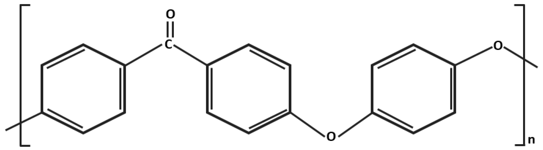
2. What Is PEEK?
3. PEEK Reinforcement
4. Surface Modification of PEEK for Osseointegration
5. Conclusions
Acknowledgments
Author Contributions
Conflicts of Interest
References
- Brånemark, P.I.; Adell, R.; Breine, U.; Hansson, B.O.; Lindstrom, J.; Ohlsson, A. Intraosseous anchorage of dental prostheses. I. Experimental studies. Scand. J. Plast. Reconstr. Surg. 1969, 3, 81–100. [Google Scholar] [CrossRef] [PubMed]
- Lautenschlager, E.P.; Monaghan, P. Titanium and titanium alloys as dental materials. Int. Dent. J. 1993, 43, 245–253. [Google Scholar] [PubMed]
- Renouard, F.; Nisand, D. Impact of implant length and diameter on survival rates. Clin. Oral Implants Res. 2006, 17, 35–51. [Google Scholar] [CrossRef] [PubMed]
- Lee, W.T.; Koak, J.Y.; Lim, Y.J.; Kim, S.K.; Kwon, H.B.; Kim, M.J. Stress shielding and fatigue limits of poly-ether-ether-ketone dental implants. J. Biomed. Mater. Res. B Appl. Biomater. 2012, 100, 1044–1052. [Google Scholar] [CrossRef] [PubMed]
- Huiskes, R.; Ruimerman, R.; Van Lenthe, G.H.; Janssen, J.D. Effects of mechanical forces on maintenance and adaptation of form in trabecular bone. Nature 2000, 405, 704–706. [Google Scholar] [CrossRef] [PubMed]
- Schalock, P.C.; Menné, T.; Johansen, J.D.; Taylor, J.S.; Maibach, H.I.; Lidén, C.; Bruze, M.; Thyssen, J.P. Hypersensitivity reactions to metallic implants-Diagnostic algorithm and suggested patch test series for clinical use. Contact Dermat. 2012, 66, 4–19. [Google Scholar] [CrossRef] [PubMed]
- Nakamura, K.; Kanno, T.; Milleding, P.; Ortengren, U. Zirconia as a dental implant abutment material: A systematic review. Int. J. Prosthodont. 2010, 23, 299–309. [Google Scholar] [PubMed]
- Özkurt, Z.; Kazazoğlu, E. Zirconia dental implants: A literature review. J. Oral Implantol. 2011, 37, 367–376. [Google Scholar] [CrossRef] [PubMed]
- Akagi, K.; Okamoto, Y.; Matsuura, T.; Horibe, T. Properties of test metal ceramic titanium alloys. J. Prosthet. Dent. 1992, 68, 462–467. [Google Scholar] [CrossRef]
- Kelly, J.R.; Denry, I. Stabilized zirconia as a structural ceramic: An overview. Dent. Mater. 2008, 24, 289–298. [Google Scholar] [CrossRef] [PubMed]
- Eschbach, L. Nonresorbable polymers in bone surgery. Injury 2000, 31, 22–27. [Google Scholar] [CrossRef]
- Fujihara, K.; Huang, Z.M.; Ramakrishna, S.; Satknanantham, K.; Hamada, H. Performance study of braided carbon/PEEK composite compression bone plates. Biomaterials 2003, 24, 2661–2667. [Google Scholar] [CrossRef]
- Fujihara, K.; Huang, Z.M.; Ramakrishna, S.; Satknanantham, K.; Hamada, H. Feasibility of knitted carbon/PEEK composites for orthopedic bone plates. Biomaterials 2004, 25, 3877–3885. [Google Scholar] [CrossRef] [PubMed]
- Kurtz, S.M.; Devine, J.N. PEEK biomaterials in trauma, orthopedic, and spinal implants. Biomaterials 2007, 28, 4845–4869. [Google Scholar] [CrossRef] [PubMed]
- Kurtz, S.M. PEEK Biomaterials Handbook; Elsevier Science: Waltham, MA, USA, 2012; pp. 30–31. [Google Scholar]
- Andreiotelli, M.; Wenz, H.J.; Kohal, R.J. Are ceramic implants a viable alternative to titanium implants? A systematic literature review. Clin. Oral Implants Res. 2009, 20, 32–47. [Google Scholar] [CrossRef] [PubMed]
- Skinner, H.B. Composite technology for total hip arthroplasty. Clin. Orthop. 1988, 235, 224–236. [Google Scholar] [CrossRef]
- Manish, G.; Chandu, G.; Sunil, K.M.; Siddharth, G. Titanium allergy: A literature review. Indian J. Dermatol. 2014, 59, 630. [Google Scholar]
- Wang, H.; Xu, M.; Zhang, W.; Kwok, D.T.; Jiang, J.; Wu, Z.; Chu, P.K. Mechanical and biological characteristics of diamond-like carbon coated poly aryl-ether-ether-ketone. Biomaterials 2010, 31, 8181–8187. [Google Scholar] [CrossRef] [PubMed]
- Becker, W.; Doerr, J.; Becker, B.E. A novel method for creating an optimal emergence profile adjacent to dental implants. J. Esthet. Restor. Dent. 2012, 24, 395–400. [Google Scholar] [CrossRef] [PubMed]
- Koutouziz, T.; Richardson, J.; Lundgren, T. Comparative soft and hard tissue responses to titanium and polymer healing abutments. J. Oral Implantol. 2011, 37, 174–182. [Google Scholar] [CrossRef] [PubMed]
- Val, J.E.M.S.D.; Gómez-Moreno, G.; Martínes, C.P.A.; Ramírez-Fernández, M.P.; Granero-Marín, J.M.; Gehrke, S.A.; Calvo-Guirado, J.L. Peri-implant tissue behavior around non-titanium material: Experimental study in dogs. Ann. Anat. 2016, 206, 104–109. [Google Scholar]
- Santing, H.J.; Meijer, H.J.A.; Raghoebar, G.M.; Özcan, M. Fracture strength and failure mode of maxillary implant-supported provisional single crowns: A comparison of composite resin crown fabricated directly over PEEK abutments and solid titanium abutments. Clin. Implant Dent. Relat. Res. 2012, 14, 882–889. [Google Scholar] [CrossRef] [PubMed]
- Sandler, J.; Werner, P.; Shaffer, M.S.; Demchuk, V.; Altstädt, V.; Windle, A.H. Carbon-nanofibre-reinforced poly(ether ether ketone) composites. Compos. Part A Appl. Sci. Manuf. 2002, 33, 1033–1039. [Google Scholar] [CrossRef]
- Schwitalla, A.D.; Emara, M.A.; Spintig, T.; Lackmann, J.; Müller, W.D. Finite element analysis of the biomechanical effects of PEEK dental implants on the peri-implant bone. J. Biomech. 2015, 48, 1–7. [Google Scholar] [CrossRef] [PubMed]
- Najeeb, S.; Zafar, M.S.; Khursid, Z.; Siddiqui, F. Applications of polyetheretherketone (PEEK) in oral implantology and prosthodontics. J. Prosthodont. Res. 2016, 60, 12–19. [Google Scholar] [CrossRef] [PubMed]
- Rust-Dawicki, A.M.; Cook, S.D. Preliminary evaluation of titanium-coated PEEK implants. J. Oral Implantol. 1995, 21, 75–77. [Google Scholar]
- Wiesli, M.G.; Med, M.D.; Özcan, M. High-performance polymers and their potential application as medical and oral implant materials: A review. Implant Dent. 2015, 24, 448–457. [Google Scholar] [CrossRef] [PubMed]
- Lewinstein, I.; Banks-Sills, L.; Eliasi, R. Finite element analysis of a new system (IL) for supporting an implant-retained cantilever prosthesis. Int. J. Oral Maxillofac. Implants 1995, 10, 355–366. [Google Scholar] [PubMed]
- Vallittu, P. Some aspects of the tensile strength of unidirectional glass fibre-polymethyl methacrylate composite used in dentures. J. Oral Rehabilit. 1998, 25, 100–105. [Google Scholar] [CrossRef]
- Zafar, M.S.; Ahmed, N. Nanoindentation and surface roughness profilometry of poly methyl methacrylate denture base materials. Technol. Health Care 2014, 22, 573–581. [Google Scholar] [PubMed]
- Martin, R.; Ishida, J. The relative effects of collagen fiber orientation, porosity, density, and mineralization on bone strength. J. Biomech. 1989, 22, 419–426. [Google Scholar] [CrossRef]
- Rho, J.Y.; Ashman, R.B.; Turner, C.H. Young’s modulus of trabecular and cortical bone material: Ultrasonic and microtensile measurements. J. Biomech. 1993, 26, 111–119. [Google Scholar] [CrossRef]
- Borchers, L.; Reichart, P. Three-dimensional stress distribution around a dental implant at different stages of interface development. J. Dent. Res. 1983, 62, 155–159. [Google Scholar] [CrossRef] [PubMed]
- Staines, M.; Robinson, W.; Hood, J. Spherical indentation of tooth enamel. J. Mater. Sci. 1981, 16, 2551–2556. [Google Scholar] [CrossRef]
- Rees, J.; Jacobsen, P. The elastic moduli of enamel and dentine. Clin. Mater. 1993, 14, 35–39. [Google Scholar] [CrossRef]
- Cavalli, V.; Giannini, M.; Carvalho, R.M. Effect of carbamide peroxide bleaching agents on tensile strength of human enamel. Dent. Mater. 2004, 20, 733–739. [Google Scholar] [CrossRef] [PubMed]
- Chun, K.J.; Choi, H.H.; Lee, J.Y. Comparison of mechanical property and role between enamel and dentin in the human teeth. J. Dent. Biomech. 2014, 5, 1–7. [Google Scholar] [CrossRef] [PubMed]
- Sheets, C.G.; Earthmann, J.C. Natural intrusion and reversal in implant assisted prosthesis: Evidence of and a hypothesis for the occurrence. J. Prosthet. Dent. 1993, 70, 513–520. [Google Scholar] [CrossRef]
- Sarot, J.R.; Contar, C.M.M.; Cruz, A.C.C.D.; Magini, R.S. Evaluation of the stress distribution in CFR-PEEK dental implants by the three-dimensional finite element method. J. Mater. Sci. Mater. Med. 2010, 21, 2079–2085. [Google Scholar] [CrossRef] [PubMed]
- Schwitalla, A.D.; Spintig, T.; Kallage, I.; Müller, W.D. Pressure behavior of different PEEK materials for dental implants. J. Mech. Behav. Biomed. Mater. 2016, 54, 295–304. [Google Scholar] [CrossRef] [PubMed]
- Rabiei, A.; Sandukas, S. Processing and evaluation of bioactive coatings on polymeric implants. J. Biomed. Mater. Res. Part A 2013, 101, 2621–2629. [Google Scholar] [CrossRef] [PubMed]
- Barkamo, S.; Wennerberg, A.; Hoffman, M.; Kjellin, P.; Breding, K.; Handa, P.; Stenport, V. Nano-hydroxyapatite-coated PEEK implants: A pilot study in rabbit bone. J. Biomed. Mater. Res. Part A 2013, 101, 465–471. [Google Scholar] [CrossRef] [PubMed]
- Suska, F.; Omar, O.; Emanuelsson, L.; Taylor, M.; Gruner, P.; Kinbrum, A.; Hunt, D.; Hunt, T.; Taylor, A.; Palmquist, A. Enhancement of CRF-PEEK osseointegration by plasma-sprayed hydroxyapatite: A rabbit model. J. Biomater. Appl. 2014, 29, 234–242. [Google Scholar] [CrossRef] [PubMed]
- Wu, X.; Liu, X.; Wei, J.; Ma, J.; Deng, F.; Wei, S. Nano-TiO2/PEEK bioactive composite as a bone substitute material: In vitro and in vivo studies. Int. J. Nanomed. 2012, 7, 1215–1225. [Google Scholar]
- Wang, N.; Li, H.; Lü, W.; Li, J.; Wang, J.; Zhang, Z.; Liu, Y. Effects of TiO2 nanotubes with different diameters on gene expression and ossointegration of implants in minipigs. Biomaterials 2011, 32, 6900–6911. [Google Scholar] [CrossRef] [PubMed]
- Wang, L.; He, S.; Wu, X.; Liang, S.; Mu, Z.; Wei, J.; Deng, F.; Deng, Y.; Wei, S. Polyetheretherketone/nano-fluorohydroxyapatite composite with antimicrobial activity and osseointegration properties. Biomaterials 2014, 35, 6758–6775. [Google Scholar] [CrossRef] [PubMed]
- Najeeb, S.; Khurshid, Z.; Matinlinna, J.P.; Siddiqui, F.; Nassani, M.Z.; Baroudi, K. Nanomodified Peek Dental Implants: Bioactive Composites and Surface Modification—A Review. Int. J. Dent. 2015. [Google Scholar] [CrossRef] [PubMed]
- Johansson, P.; Jimbo, R.; Kjellin, P.; Currie, F.; Chrcanovic, B.R.; Wennerberg, A. Biomechanical evaluation and surface characterization of a nano-modified surface on PEEK implants: A study in the rabbit tibia. Int. J. Nanomed. 2014, 9, 3903. [Google Scholar] [CrossRef] [PubMed]
- Waser-Althaus, J.; Salamon, A.; Waser, M.; Padeste, C.; Kreutzer, M.; Pieles, U.; Müller, B.; Peters, K. Differentiation of human mesenchymal stem cells on plasma-treated polyetheretherketone. J. Mater. Sci. Mater. Med. 2014, 25, 515–525. [Google Scholar] [CrossRef] [PubMed]
- Randolph, S.; Fowlkes, J.; Rack, P. Focused, nanoscale electron-beam-induced deposition and etching. Crit. Rev. Solid State Mater. Sci. 2006, 31, 55–89. [Google Scholar] [CrossRef]
- Mantese, J.V.; Brown, I.G.; Cheung, N.W.; Collins, G.A. Plasma-immersion ion implantation. MRS Bull. 1996, 21, 52–56. [Google Scholar] [CrossRef]
- Ha, S.; Mayer, J.; Koch, B.; Wintermantel, E. Plasma-sprayed hydroxylapatite coating on carbon fibre reinforced thermoplastic composite materials. J. Mater. Sci. Mater. Med. 1994, 5, 481–484. [Google Scholar] [CrossRef]
- Han, C.; Lee, E.; Kim, H.; Koh, Y.; Kim, K.N. The electron beam deposition of titanium on polyetheretherketone (PEEK) and the resulting enhanced biological properties. Biomaterials 2010, 31, 3465–3470. [Google Scholar] [CrossRef] [PubMed]
- Wen, J.; Lu, T.; Wang, X.; Xu, L.; Wu, Q.; Pan, H.; Wang, D.; Liu, X.; Jiang, X. In vitro an in vivo evaluation of silicate-coated polyetheretherketone fabricated by electron beam evaporation. ACS Appl. Mater. Interfaces 2016, 8, 13197–13206. [Google Scholar] [CrossRef] [PubMed]
- Lu, T.; Liu, X.; Qian, S.; Cao, H.; Qiao, Y.; Mei, Y.; Chu, P.K.; Ding, C. Multilevel surface engineering of nanostructured TiO2 on carbonfiber-reinforced polyetheretherketone. Biomaterials 2014, 35, 5731–5740. [Google Scholar] [CrossRef] [PubMed]
- Lu, T.; Qian, S.; Meng, F.; Ning, C.; Liu, X. Enhanced osteogenic activity of poly ether ether ketone using calcium plasma immersion ion implantation. Colloids Surf. B Biointerfaces 2016, 142, 192–198. [Google Scholar] [CrossRef] [PubMed]
- Chen, M.; Ouyang, L.; Lu, T.; Wang, H.; Meng, F.; Yang, Y.; Ning, C.; Ma, J.; Liu, X. Enhanced bioactivity and bacteriostatis of surface fluorinated polyetheretherketone. ACS Appl. Mater. Interfaces 2017, 9, 16824–16833. [Google Scholar] [CrossRef] [PubMed]
- Zhao, Y.; Wong, H.M.; Wang, W.; Li, P.; Xu, Z.; Chong, E.Y.W.; Yan, C.H.; Yeung, K.W.K.; Chu, P.K. Cytocompatibility, osseointegration, and bioactivity of three-dimensional porous and nanostructured network on polyetheretherketone. Biomaterials 2013, 34, 9264–9277. [Google Scholar] [CrossRef] [PubMed]
- Xu, A.; Liu, X.; Gao, X.; Deng, F.; Deng, Y.; Wei, S. Enhancement of osteogenesis on micro/nano-topographical carbon fiber-reinforced polyetheretherketone-nanohydroxyapatite biocomposite. Mater. Sci. Eng. C 2015, 48, 592–598. [Google Scholar] [CrossRef] [PubMed]
- Yee, R.S.L.; Zhang, K.; Ladewig, B.P. The effects of sulfonated poly(ether ether ketone) ion exchange preparation conditions on membrane properties. Membranes 2013, 3, 182–195. [Google Scholar] [CrossRef] [PubMed]
- Henneuse-Boxusa, C.; Boxusa, T.; Dulière, E.; Pringallea, C.; Tesolina, L.; Adriaensenb, Y.; Marchand-Brynaert, J. Surface amination of PEEK film by selective wet-chemistry. Polymer 1998, 39, 5359–5369. [Google Scholar] [CrossRef]
- Conceição, T.F.; Bertolino, J.R.; Barra, G.M.O.; Pires, A.T.N. Poly (ether ether ketone) derivatives: Synthetic route and characterization of nitrated and sulfonated polymers. Mater. Sci. Eng. C 2009, 29, 575–582. [Google Scholar] [CrossRef]
- Qahtani, M.S.A.A.; Wu, Y.; Spintzyk, S.; Krieg, P.; Killinger, A.; Schweizer, E.; Stephan, I.; Scheideler, L.; Geis-Gerstorfer, J.; Rupp, F. UV-A and UV-C light induced hydrophilization of dental implants. Dent. Mater. 2015, 31, e157–e167. [Google Scholar] [CrossRef] [PubMed]
- Poulsson, A.H.; Eglin, D.; Zeiter, S.; Camenisch, K.; Sprecher, C.; Agarwal, Y.; Nehrbass, D.; Willson, J.; Richards, R.G. Osseointegration of machined, injection moulded and oxygen plasma modified PEEK implants in a sheep model. Biomaterials 2014, 35, 3717–3728. [Google Scholar] [CrossRef] [PubMed]
- Schwitalla, A.; Müller, W.D. PEEK Dental Implants: A Review of the Literature. J. Oral Implantol. 2013, 39, 743–749. [Google Scholar] [CrossRef] [PubMed]
- Huang, R.; Shao, P.; Burns, C.; Feng, X. Sulfonation of poly(ether ether ketone) (PEEK): Kinetic study and characterization. J. Appl. Polym. Sci. 2001, 82, 2651–2660. [Google Scholar] [CrossRef]
- Nieminen, T.; Kallela, I.; Wuolijoki, E.; Kainulainen, H.; Hiidenheimo, I.; Rantala, I. Amorphous and crystalline polyetheretherketone: Mechanical properties and tissue reactions during a 3-year follow-up. J. Biomed. Mater. Res. Part A 2008, 84, 377–383. [Google Scholar] [CrossRef] [PubMed]
- Wenz, L.; Merritt, K.; Brown, S.; Moet, A.; Steffee, A. In vitro biocompatibility of polyetheretherketone and polysulfone composites. J. Biomed. Mater. Res. 1990, 24, 207–215. [Google Scholar] [CrossRef] [PubMed]
- Rupp, F.; Gittens, R.A.; Scheideler, L.; Marmur, A.; Boyan, B.D.; Schwartz, Z.; Geis-Gerstorfer, J. A review on the wettability of dental implant surfaces I: Theoretical and experimental aspects. Acta Biomater. 2014, 10, 2894–2906. [Google Scholar] [CrossRef] [PubMed]
- Gittens, R.A.; Scheideler, L.; Rupp, F.; Hyzy, S.I.; Geis-Gerstorfer, J.; Schwartz, Z.; Boyan, B.D. A review on the wettability of dental implant surfaces II: Biological and clinical aspects. Acta Biomater. 2014, 10, 2907–2918. [Google Scholar] [CrossRef] [PubMed]


| Material | Elastic Modulus (GPa) | References |
|---|---|---|
| Titanium | 110 | Lee, 2012 [4] |
| Cobalt-Chromium | 180–210 | Wiesli, 2015 [28] |
| Zirconia | 210 | Lee, 2012 [4] |
| Porcelain | 68.9 | Lewinstein, 1995 [29] |
| PMMA | 3–5 | Vallittu, 1998; Zafar, 2014 [30,31] |
| PEEK | 3–4 | Sandler, 2002 [24] |
| CFR-PEEK | 18 | Sandler, 2002 [24] |
| Continuous CFR-PEEK (Endolign®) | 150 | Schwitalla, 2015 [25] |
| GFR-PEEK | 12 | Lee, 2012 [4] |
| Cortical bone | 14 | Martin, 1989; Rho, 1993 [32,33] |
| Cancellous bone | 1.34 | Borchers and Reichart, 1983 [34] |
| Enamel | 40–83 | Staines, 1981; Rees, 1993; Cavalli, 2004 [35,36,37] |
| Dentin | 15–30 | Rees, 1993; Chun, 2014 [36,38] |
| Surface Modifications | Procedures | Material | References |
|---|---|---|---|
| Coating | Plasma spraying | Hydroxyapatite (HA), titanium (Ti) | Rust-Dawicki, 1995; Suska, 2014; Ha, 1994 [27,45,53] |
| Spin coating | Nanosized HA crystals containing surfactans, organic solvent, an aquous solution of Ca(NO3)2 and H3PO4 | Barkarmo, 2012; Johansson, 2014 [43,49] | |
| Electron-beam evaporation (EBE) | Ti; Silicate | Han, 2010; Wen, 2016 [54,55] | |
| Plasma immersion ion implantation (PIII) | Titanium dioxide (TiO2); calcium (Ca); water (H2O); Argon (Ar) | Wang, 2014; Lu, 2014; Lu, 2016; Chen, 2017 [47,56,57,58] | |
| Surface topographical modifications | Acid etching | Sulfuric acid | Zhao, 2013 [59] |
| Sandblasting | TiO2, alumina (Al2O3) | Suska, 2014; Xu, 2015 [44,60] | |
| Chemical modifications | Sulphonation | Sulfonate groups (–SO3–) | Yee, 2013 [61] |
| Amination | Amine functions | Henneuse-Boxus, 1998 [62] | |
| Nitration | Nitrate functions | Conceição, 2009 [63] | |
| Incorporating with bioactive properties | Bioactive inorganic materials | Nano-TiO2 (n-TiO2); nano-fluorohydroxyapatite (n-FHA) | Wu, 2012; Wang, 2014 [45,47] |
| Improving hydrophylicity | UV irradiation | UV-A light, UV-C light | Qahtani, 2015 [64] |
| Plasma gas treatment | Oxygen plasma | Waser-Althaus, 2014; Xu, 2015; Poulsson, 2014 [50,60,65] |
© 2017 by the authors. Licensee MDPI, Basel, Switzerland. This article is an open access article distributed under the terms and conditions of the Creative Commons Attribution (CC BY) license (http://creativecommons.org/licenses/by/4.0/).
Share and Cite
Rahmitasari, F.; Ishida, Y.; Kurahashi, K.; Matsuda, T.; Watanabe, M.; Ichikawa, T. PEEK with Reinforced Materials and Modifications for Dental Implant Applications. Dent. J. 2017, 5, 35. https://0-doi-org.brum.beds.ac.uk/10.3390/dj5040035
Rahmitasari F, Ishida Y, Kurahashi K, Matsuda T, Watanabe M, Ichikawa T. PEEK with Reinforced Materials and Modifications for Dental Implant Applications. Dentistry Journal. 2017; 5(4):35. https://0-doi-org.brum.beds.ac.uk/10.3390/dj5040035
Chicago/Turabian StyleRahmitasari, Fitria, Yuichi Ishida, Kosuke Kurahashi, Takashi Matsuda, Megumi Watanabe, and Tetsuo Ichikawa. 2017. "PEEK with Reinforced Materials and Modifications for Dental Implant Applications" Dentistry Journal 5, no. 4: 35. https://0-doi-org.brum.beds.ac.uk/10.3390/dj5040035