Evaluation of Profile Changes in Class II Individuals Treated by Means of Herbst Miniscope Appliance

 , ,
, , 
Abstract
:1. Introduction
2. Materials and Methods
2.1. Sample
- a full Class II molar relationship;
- ANB > 4°,
- Overjet > 6 mm,
- an age range of 9–15 years,
- good quality radiographs, and
- cervical vertebral maturation stage (CVMS)2 or >3 [22].
- hyperdivergent subjects (mandibular plane angle equal to or greater than the normal value plus a standard deviation 26° ± 4°) [23],
- tooth agenesis,
- previous orthodontic treatment, and
- periodontal diseases.
- a total of 22 patients (14 girls, 8 boys, mean age 11.9 ± 1.3 years, HBT group) were treated with Herbst MiniScope® appliance. Transversal discrepancies and/or crossbites, if present, were treated with orthopedic or orthodontic expansion of the palate before functional treatment [24]. The average age of therapy onset was 10 years, 8 months for females and 11 years, 11 months for male. After the orthopedic phase, the patients underwent orthodontic treatment with self-ligating brackets [25] and initial thermoelastic or superelastic archwires [26];
- the CTR group comprised 22 patients (14 boys, 8 girls; mean age 10.6 ± 1.3,). The CTR group followed the same inclusion and exclusion criteria as the treated group.
2.2. Cephalometric Analysis
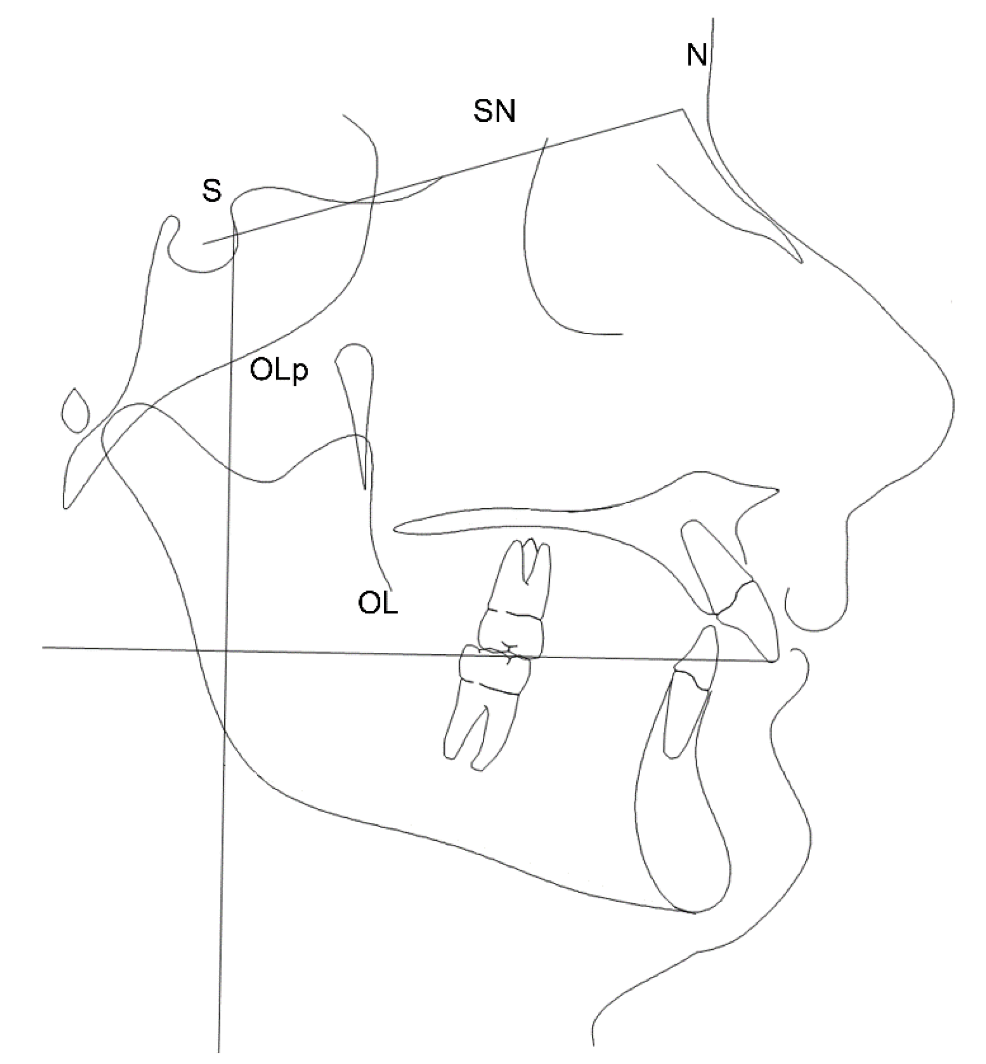
2.2.1. Reference Lines
- NSL: (nasion-sella line) S to N line used in cephalometric orientation
- OL: (occlusal line) line that goes from U1 to the distobuccal cusp of the first permanent upper molar. Used as a reference line in the detection of cephalometric measurements
- OLp: (perpendicular line to the occlusal plane) perpendicular to OL passing through T point. It represents the line from which the distances to the measuring points were evaluated.
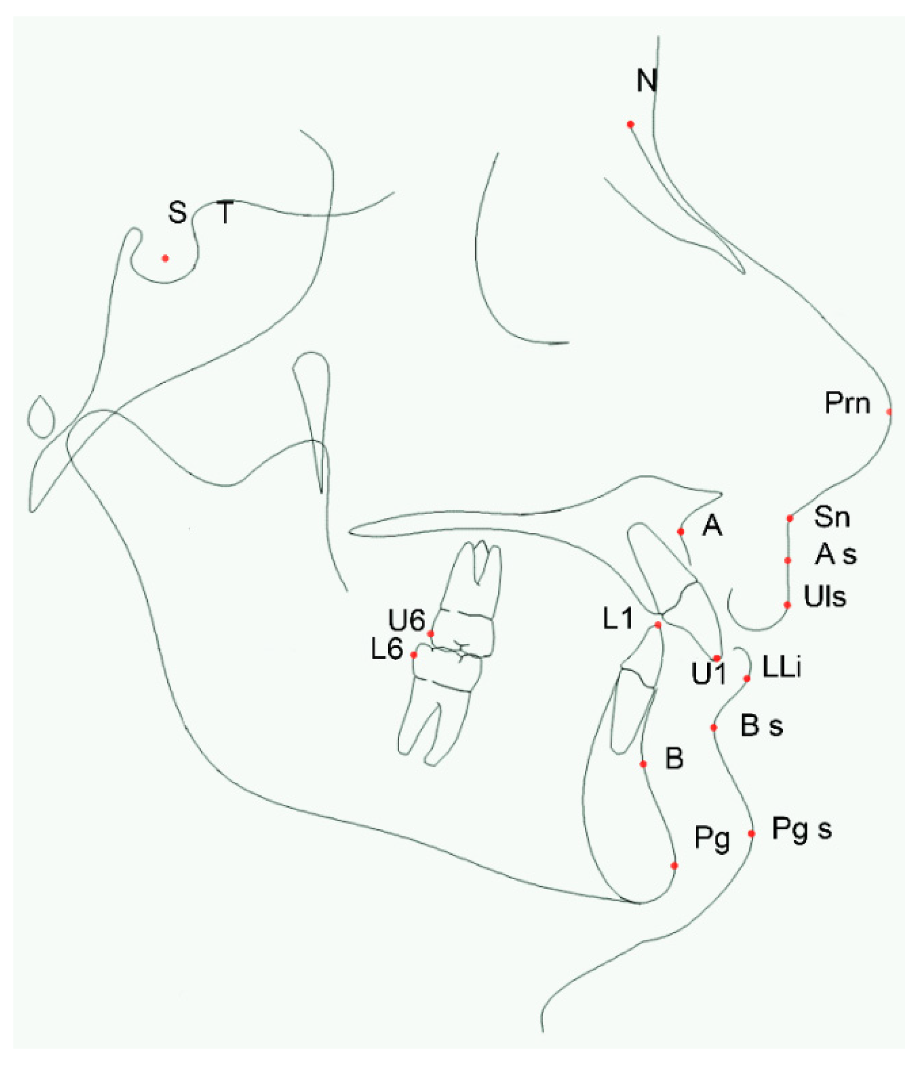
2.2.2. Measuring Points
- S: (sella) point located at the center of the sella turcica
- T point
- N: (nasion) earlier point of the nasofrontal suture
- U1: (superior incision) lower point of the upper central incisors
- Prn: (nasal point), the most anterior point of the nasal apex
- ANS: (anterior nasal spine), most anterior point of maxillary bone
- A: (point A), posterior point of the anterior convexity of the maxilla
- A soft: (A soft point), maximum concavity of anterior lip superior point
- Sn: (nasolabial point), the point of maximum concavity of the nasolabial curvature
- ULs: (upper lip), the most forward point of the upper lip
- U6: (upper first molar), distal point of upper first molar
- L1: (lower incision) upper point of the lower central incisors
- LLi: (lower lip), most anterior point of the lower lip
- L6: (lower first molar), distal point of inferior first molar
- Sub: (inferior labial sulcus), the posterior point of the inferior labial sulcus
- B: (B point), maximum concavity of anterior mandibular bone
- Po: (pogonion) anterior point of the mandibular symphysis
- Pos: (soft tissue pogonion), most anterior point of the soft tissues of the lower jaw
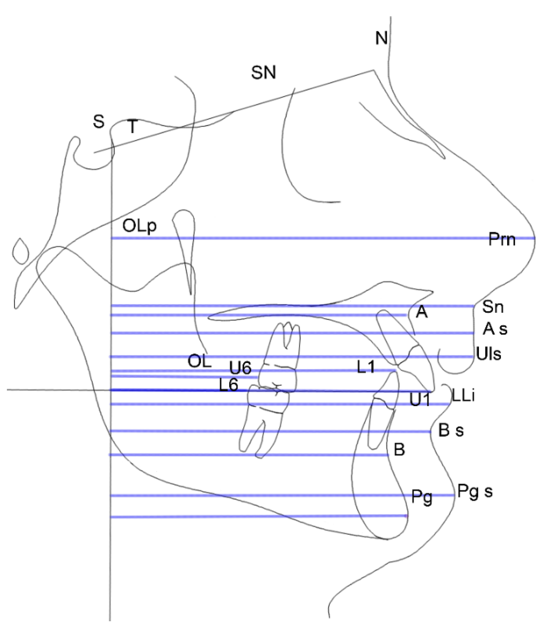
2.2.3. Linear Measuring Procedures
- SN: sella-nasion line
- OLp-U1: line that evaluates the position of superior incisor compared to the perpendicular line to the occlusal line
- OLp-Prn: line that evaluates the nasal prominence compared to the perpendicular line to the occlusal plane (nasal growth)
- OLp-ANS: line that evaluates the anterior nasal spine point compared to the perpendicular line to the occlusal plane
- OLp-A: line that evaluates the maxillary’s position compared to the perpendicular line to the occlusal plane
- OLp-As: line that evaluates the position of A point of the soft tissue compared to the perpendicular line to the occlusal plane
- OLp-Sn: line that evaluates the position of the infranasal point compared to the perpendicular line to the occlusal plane (filtrum)
- OLp-ULs: line that evaluates the position of the upper lip compared to the perpendicular line to the occlusal plane
- OLp-U6: line that evaluates the position of upper first molar compared to the perpendicular line to the occlusal plane
- OLp-L1: line that evaluates the position of lower incisor compared to the perpendicular line to the occlusal plane
- OLp-LLi: line that evaluates the position of the lower lip compared to the perpendicular line to the occlusal plane
- OLp-L6: line that evaluates the position of the lower first molar compared to the perpendicular line to the occlusal plane
- OLp-B: line that evaluates the mandible’s position compared to the perpendicular line to the occlusal plane
- OLp-Sub: line that evaluates the position of the mandibular sulcus compared to the perpendicular line to the occlusal plane (concavity of the mandibular sulcus)
- OLp-Po: line that evaluates the position of the pogonion compared to the perpendicular line to the occlusal plane
- OLp-Pos: a line that evaluates the position of the soft tissue pogonion compared to the perpendicular line to the occlusal plane.
2.3. Statistical Analysis
3. Results
- of 2 mm, on mean average, of the lower lip (OLp-LLI), p = 0.030
- of 4.3 mm, on mean average, of the sublabial sulcus (OLp-sublabial), p = 0.002.
4. Discussion
5. Conclusions
Author Contributions
Funding
Conflicts of Interest
References
- McLain, J.B.; Proffit, W.R. Oral health status in the United States: Prevalence of malocclusion. J. Dent. Educ. 1985, 49, 386–396. [Google Scholar]
- Ngan, P.W.; Byczek, E.; Scheick, J. Longitudinal evaluation of growth changes in class II division 1 subjects. Semin. Orthod. 1997, 3, 322–331. [Google Scholar] [CrossRef]
- Rongo, R.; Bucci, R.; Adaimo, R.; Amato, M.; Martina, S.; Valletta, R.; D’Antò, V. Two-dimensional versus three-dimensional Frӓnkel Manoeuvre: A reproducibility study. Eur. J. Orthod. 2019. [Google Scholar] [CrossRef]
- Dimberg, L.; Arnrup, K.; Bondemark, L. The impact of malocclusion on the quality of life among children and adolescents: A systematic review of quantitative studies. Eur. J. Orthod. 2015, 37, 238–247. [Google Scholar] [CrossRef]
- McComb, J.L.; Wright, J.L.; Fox, N.A.; O’Brien, K.D. Perceptions of the risks and benefits of orthodontic treatment. Community Dent. Health 1996, 13, 133–138. [Google Scholar]
- Bowman, S.J.; Johnston, L.E. Much ado about facial esthetics. In Treatment Timing: Orthodontics in Four Dimensions; Monograph No. 39; Craniofacial Growth Series; McNamara, J.A., Ed.; Center for Human Growth and Development, University of Michigan: Ann Arbor, MI, USA, 2001; pp. 199–217. [Google Scholar]
- Aelbers, C.M.; Dermaut, L.R. Orthopedics in orthodontics: Part I, Fiction or reality–a review of the literature. Am. J. Orthod. Dentofac. Orthop. 1996, 110, 513–519. [Google Scholar] [CrossRef]
- Dermaut, L.R.; Aelbers, C.M. Orthopedics in orthodontics: Fiction or reality. A review of the literature–Part II. Am. J. Orthod. Dentofac. Orthop. 1996, 110, 667–671. [Google Scholar] [CrossRef]
- D’Antò, V.; Bucci, R.; Franchi, L.; Rongo, R.; Michelotti, A.; Martina, R. Class II functional orthopaedic treatment: A systematic review of systematic reviews. J. Oral. Rehabil. 2015, 42, 624–642. [Google Scholar] [CrossRef] [PubMed] [Green Version]
- Martina, R.; Cioffi, I.; Galeotti, A.; Tagliaferri, R.; Cimino, R.; Michelotti, A.; Valletta, R.; Farella, M.; Paduano, S. Efficacy of the Sander bite-jumping appliance in growing patients with mandibular retrusion: A randomized controlled trial. Orthod. Craniofac. Res. 2013, 16, 116–126. [Google Scholar] [CrossRef]
- Lucchese, A.; Carinci, F.; Brunelli, G. Skeletal effects induced by twin block in therapy of class II malocclusion. Eur. J. Inflamm. 2012, 10, 83–87. [Google Scholar]
- DiBiase, A.T.; Lucchesi, L.; Qureshi, U.; Lee, R.T. Post-treatment cephalometric changes in adolescent patients with Class II malocclusion treated using two different functional appliance systems for an extended time period: A randomized clinical trial. Eur. J. Orthod. 2019. [Google Scholar] [CrossRef] [PubMed]
- Souki, B.Q.; Vilefort, P.L.C.; Oliveira, D.D.; Andrade, I., Jr.; Ruellas, A.C.; Yatabe, M.S.; Nguyen, T.; Franchi, L.; McNamara, J.A., Jr.; Cevidanes, L.H. Three-dimensional skeletal mandibular changes associated with Herbst appliance treatment. Orthod. Craniofac. Res. 2017, 20, 111–118. [Google Scholar] [CrossRef] [PubMed]
- Paduano, S.; Spagnuolo, G.; Biase, G.; Cioffi, I. Treatment of a Class II Division Patient with Severe Skeletal Discrepancy by Using a Custom Made TPA Proclination Spring. Open Dent. J. 2013, 7, 109–117. [Google Scholar] [CrossRef] [PubMed] [Green Version]
- Ruf, S.; Pancherz, H. Temporomandibular joint remodeling in adolescents and young adults during Herbst treatment: A prospective longitudinal magnetic resonance imaging and cephalometric radiographic investigation. Am. J. Orthod. Dentofac. Orthop. 1999, 115, 607–618. [Google Scholar] [CrossRef]
- Tomblyn, T.; Rogers, M.; Andrews, L., 2nd; Martin, C.; Tremont, T.; Gunel, E.; Ngan, P. Cephalometric study of Class II Division 1 patients treated with an extended-duration, reinforced, banded Herbst appliance followed by fixed appliances. Am. J. Orthod. Dentofac. Orthop. 2016, 150, 818–830. [Google Scholar] [CrossRef]
- Pancherz, H. Treatment of class II malocclusions by jumping the bite with the Herbst appliance. Am. J. Orthod. 1979, 76, 423–442. [Google Scholar] [CrossRef]
- Schiavoni, R.; Grenga, V. Long-term follow-up of a young adult patient with a Class II malocclusion treated with a Herbst appliance. World J. Orthod. 2009, 10, 243–251. [Google Scholar]
- Zouloumi, M.E.; Tsiouli, K.; Psomiadis, S.; Kolokitha, O.E.; Topouzelis, N.; Gkantidis, N. Facial esthetic outcome of functional followed by fixed orthodontic treatment of class II division 1 patients. Prog. Orthod. 2019, 20, 42. [Google Scholar]
- Paduano, S.; Rongo, R.; Bucci, R.; Carvelli, G.; Cioffi, I. Impact of functional orthodontic treatment on facial attractiveness of children with Class II division 1 malocclusion. Eur. J. Orthod. 2019. [Google Scholar] [CrossRef]
- Hourfar, J.; Lisson, J.A.; Gross, U.; Frye, L.; Kinzinger, G.S.M. Soft tissue profile changes after Functional Mandibular Advancer or Herbst appliance treatment in class II patients. Clin. Oral. Investig. 2018, 22, 971–980. [Google Scholar] [CrossRef]
- Baccetti, T.; Franchi, L.; McNamara, J.A., Jr. An improved version of the cervical vertebral maturation (CVM) method for the assessment of mandibular growth. Angle Orthod. 2002, 72, 316–323. [Google Scholar] [PubMed]
- Arnett, G.W.; Bergman, R.T. Facial keys to orthodontic diagnosis and treatment planning—part II. Am. J. Orthod. 1993, 103, 395–411. [Google Scholar] [CrossRef]
- Von Bremen, J.; Erbe, C.; Pancherz, H.; Ruf, S. Facial-profile attractiveness changes in adult patients treated with the Herbst appliance. J. Orofac. Orthop. 2014, 75, 167–174. [Google Scholar] [CrossRef] [PubMed]
- Paduano, S.; Cioffi, I.; Iodice, G.; Rapuano, A.; Silva, R. Time efficiency of self-ligating vs conventional brackets in orthodontics: Effect of appliances and ligating systems. Prog. Orthod. 2008, 9, 74–80. [Google Scholar]
- Cioffi, I.; Piccolo, A.; Tagliaferri, R.; Paduano, S.; Galeotti, A.; Martina, R. Pain perception following first orthodontic archwire placement–thermoelastic vs. superelastic alloys: A randomized controlled trial. Quintessence Int. 2012, 43, 61–69. [Google Scholar]
- Rongo, R.; Antoun, J.S.; Lim, Y.X.; Dias, G.; Valletta, R.; Farella, M. Three-dimensional evaluation of the relationship between jaw divergence and facial soft tissue dimensions. Angle Orthod. 2014, 84, 788–794. [Google Scholar] [CrossRef]
- Pancherz, H. The Herbst appliance- Its biologic effects and clinical use. Am. J. Orthod. 1985, 87, 1–20. [Google Scholar] [CrossRef]
- Polat-Ozsoy, O.; Gokcelik, A.; Güngör-Acar, A.; Kircelli, B.H. Soft tissue profile after distal molar movement with a pendulum K-loop appliance versus cervical headgear. Angle Orthod. 2008, 78, 317–323. [Google Scholar] [CrossRef]
- Pancherz, H.; Ruf, S.; Kohlhas, P. “Effective condylar growth” and chin position changes in Herbst treatment: A cephalometric roentgenographic long-term study. Am. J. Orthod. 1998, 114, 437–446. [Google Scholar] [CrossRef]
- Franchi, L.; Baccetti, T.; McNamara, J.A., Jr. Mandibular growth as related to cervical vertebral maturation and body height. Am. J. Orthod. Dentofac. Orthop. 2000, 118, 335–340. [Google Scholar] [CrossRef] [Green Version]
- Tepedino, M.; Della Noce, M.V.; Ciavarella, D.; Gallenzi, P.; Cordaro, M.; Chimenti, C. Soft-tissue changes after Class II malocclusion treatment using the Sander bite-jumping appliance: A retrospective study. Minerva Stomatol. 2019, 68, 118–125. [Google Scholar] [CrossRef] [PubMed]
- Idris, G.; Hajeer, M.Y.; Al-Jundi, A. Soft- and hard-tissue changes following treatment of Class II division 1 malocclusion with Activator versus Trainer: A randomized controlled trial. Eur. J. Orthod. 2019, 41, 21–28. [Google Scholar] [CrossRef] [PubMed]
- Salloum, E.; Millett, D.T.; Kelly, N.; McIntyre, G.T.; Cronin, M.S. Soft tissue changes: A comparison between changes caused by the construction bite and by successful treatment with a modified Twin-block appliance. Eur. J. Orthod. 2018, 40, 512–518. [Google Scholar] [CrossRef] [Green Version]
- Ruf, S.; Pancherz, H. Dentoskeletal effects and facial profile changes in young adults treated with the Herbst appliance. Angle Orthod. 1999, 69, 239–246. [Google Scholar] [PubMed]
- De Almeida, M.R.; Flores-Mir, C.; Brandao, A.G.; de Almeida, R.R.; de Almeida-Pedrin, R.R. Soft tissue changes produced by a banded-type Herbst appliance in late mixed dentition patients. World J. Orthod. 2008, 9, 121–131. [Google Scholar] [PubMed]
- Rego, M.V.; Martinez, E.F.; Coelho, R.M.; Leal, L.M.; Thiesen, G. Perception of changes in soft-tissue profile after Herbst appliance treatment of Class II Division 1 malocclusion. Am. J. Orthod. Dentofac. Orthop. 2017, 151, 559–564. [Google Scholar] [CrossRef] [PubMed]
- Tsiouli, K.; Topouzelis, N.; Papadopoulos, M.A.; Gkantidis, N. Perceived facial changes of Class II Division 1 patients with convex profiles after functional orthopedic treatment followed by fixed orthodontic appliances. Am. J. Orthod. Dentofac. Orthop. 2017, 152, 80–91. [Google Scholar] [CrossRef]


| CTR | HBT | ||||||||||||||
|---|---|---|---|---|---|---|---|---|---|---|---|---|---|---|---|
| T0 | T1 | DIFF | T0 | T1 | DIFF | HBT vs. CTR | |||||||||
| Mean | SD | Mean | SD | p | Mean | SD | Mean | SD | Mean | SD | p | Mean | SD | p | |
| Soft tissue points | |||||||||||||||
| PRN | 85.4 | 3.6 | 87.6 | 3.7 | 0.000 | 2.3 | 2.3 | 87.0 | 4.5 | 89.7 | 4.5 | 0.000 | 2.5 | 1.5 | 0.702 |
| A_S | 78.3 | 4.4 | 79.7 | 3.8 | 0.026 | 1.3 | 2.8 | 78.0 | 4.7 | 80.3 | 4.3 | 0.000 | 2.2 | 1.9 | 0.230 |
| SN | 77.0 | 4.0 | 78.2 | 3.5 | 0.005 | 1.2 | 2.0 | 77.8 | 4.3 | 80.0 | 3.9 | 0.000 | 2.1 | 1.7 | 0.128 |
| ULS | 80.7 | 4.7 | 82.5 | 3.5 | 0.016 | 1.8 | 3.6 | 81.6 | 4.9 | 83.8 | 4.4 | 0.000 | 2.1 | 2.1 | 0.724 |
| LLI | 78.6 | 5.5 | 80.7 | 4.4 | 0.008 | 2.1 | 3.4 | 78.6 | 4.2 | 82.8 | 4.1 | 0.000 | 4.3 | 3.1 | 0.030 |
| Sub | 71.9 | 5.0 | 73.4 | 4.6 | 0.017 | 1.5 | 2.8 | 72.2 | 3.9 | 76.5 | 4.4 | 0.000 | 4.6 | 3.5 | 0.002 |
| PG_S | 75.1 | 5.4 | 77.7 | 4.7 | 0.001 | 2.8 | 3.7 | 76.4 | 5.0 | 80.5 | 5.1 | 0.000 | 4.4 | 3.8 | 0.173 |
| Hard tissue points | |||||||||||||||
| A | 64.3 | 3.4 | 65.2 | 3.6 | 0.063 | 0.9 | 2.4 | 65.0 | 3.5 | 66.4 | 3.3 | 0.001 | 1.5 | 1.8 | 0.371 |
| B | 62.5 | 4.6 | 64.1 | 4.2 | 0.009 | 1.7 | 2.9 | 63.2 | 3.8 | 67.0 | 3.9 | 0.000 | 4.0 | 3.5 | 0.018 |
| PG | 65.0 | 5.1 | 67.5 | 4.7 | 0.006 | 2.7 | 4.7 | 66.3 | 4.5 | 70.4 | 4.4 | 0.000 | 4.3 | 4.0 | 0.247 |
| U1 | 72.1 | 4.8 | 73.5 | 3.8 | 0.011 | 1.5 | 2.7 | 71.9 | 3.3 | 73.7 | 3.7 | 0.000 | 1.8 | 1.7 | 0.693 |
| L1 | 65.4 | 4.6 | 66.8 | 4.1 | 0.007 | 1.4 | 2.5 | 64.7 | 3.6 | 70.3 | 3.8 | 0.000 | 5.7 | 3.5 | 0.000 |
| U6 | 32.9 | 3.6 | 34.8 | 3.4 | 0.003 | 1.9 | 2.8 | 33.8 | 3.6 | 33.3 | 2.9 | 0.317 | −0.4 | 2.0 | 0.003 |
| L6 | 31.3 | 3.7 | 33.4 | 3.5 | 0.000 | 2.3 | 2.5 | 32.1 | 3.9 | 37.0 | 3.6 | 0.000 | 5.0 | 3.2 | 0.003 |
© 2020 by the authors. Licensee MDPI, Basel, Switzerland. This article is an open access article distributed under the terms and conditions of the Creative Commons Attribution (CC BY) license (http://creativecommons.org/licenses/by/4.0/).
Share and Cite
Martina, S.; Di Stefano, M.L.; Paduano, F.P.; Aiello, D.; Valletta, R.; Paduano, S. Evaluation of Profile Changes in Class II Individuals Treated by Means of Herbst Miniscope Appliance. Dent. J. 2020, 8, 27. https://0-doi-org.brum.beds.ac.uk/10.3390/dj8010027
Martina S, Di Stefano ML, Paduano FP, Aiello D, Valletta R, Paduano S. Evaluation of Profile Changes in Class II Individuals Treated by Means of Herbst Miniscope Appliance. Dentistry Journal. 2020; 8(1):27. https://0-doi-org.brum.beds.ac.uk/10.3390/dj8010027
Chicago/Turabian StyleMartina, Stefano, Maria Luisa Di Stefano, Francesco Paolo Paduano, Domenico Aiello, Rosa Valletta, and Sergio Paduano. 2020. "Evaluation of Profile Changes in Class II Individuals Treated by Means of Herbst Miniscope Appliance" Dentistry Journal 8, no. 1: 27. https://0-doi-org.brum.beds.ac.uk/10.3390/dj8010027