
External Root Resorption Management of an Avulsed and Reimplanted Central Incisor: A Case Report

 ,
, 
Abstract
:1. Introduction
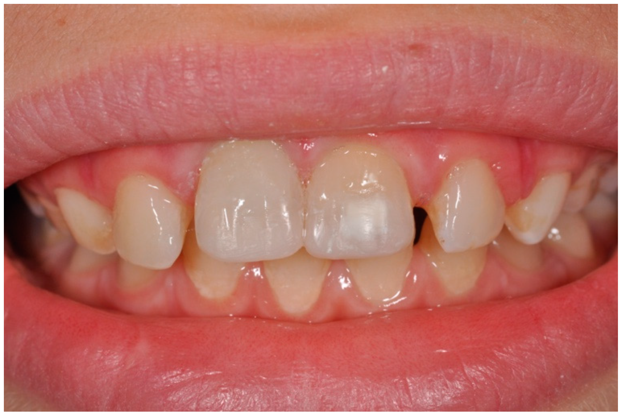
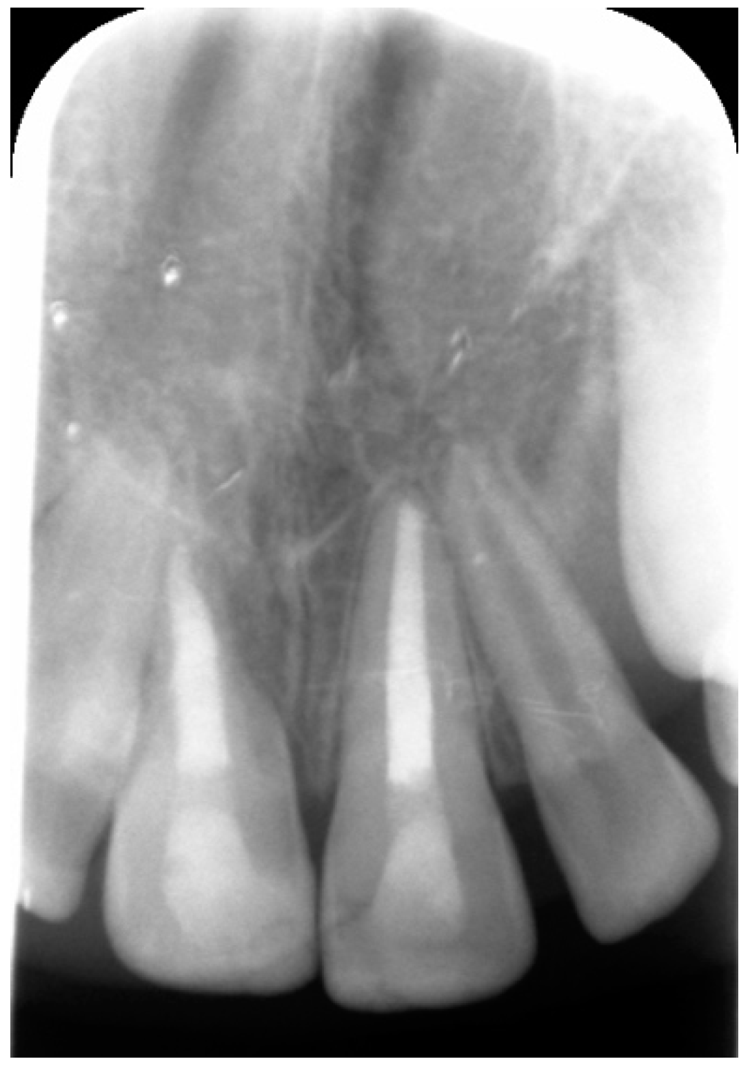
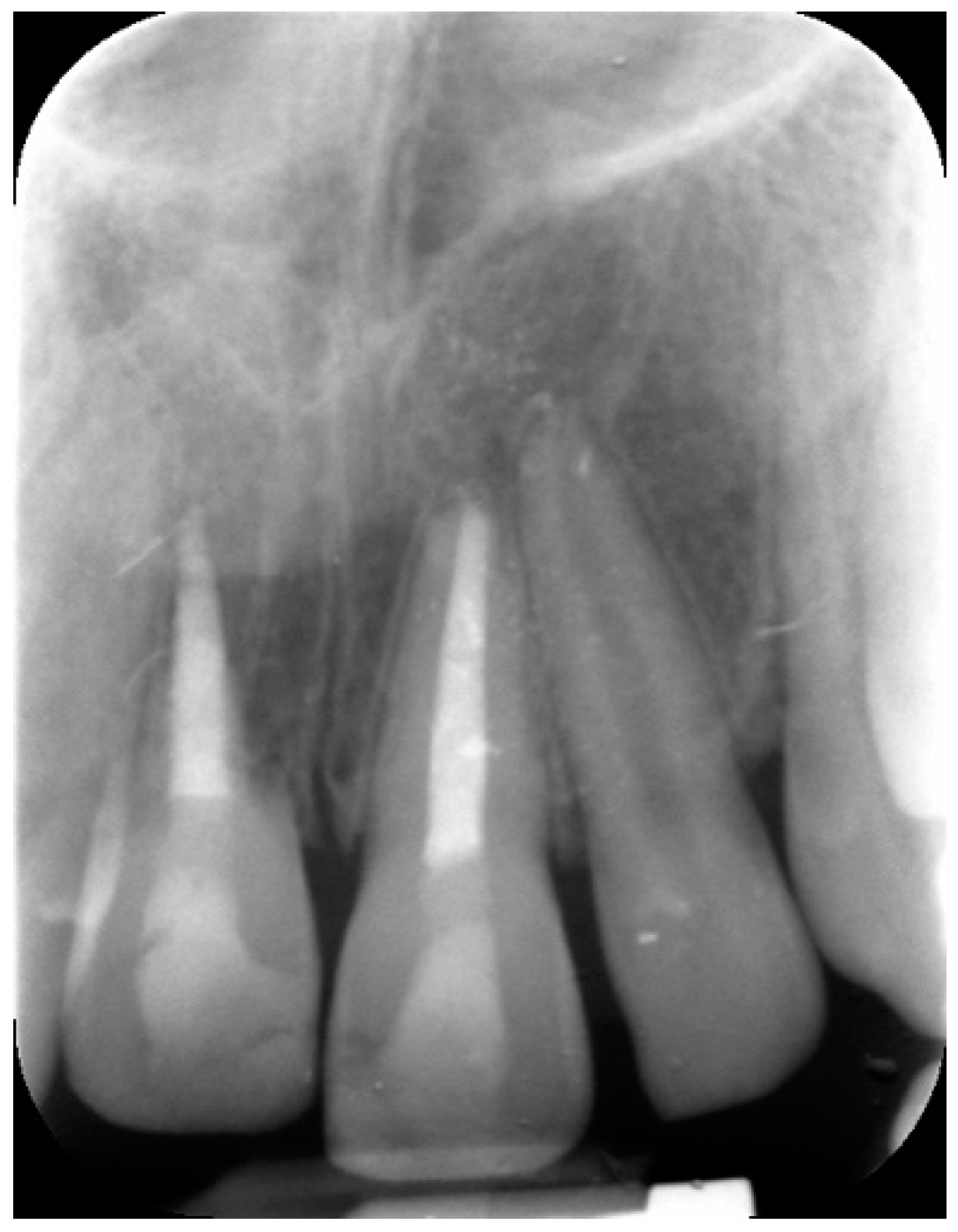
2. Case Report
3. Discussion
4. Conclusions
Author Contributions
Funding
Institutional Review Board Statement
Informed Consent Statement
Data Availability Statement
Conflicts of Interest
Appendix A

| Section/Topic | Item Number | Checklist Item | Reported on Page Number |
|---|---|---|---|
| Title | 1a | The words “case report(s)” must be included in the title | 1 |
| 1b | The area of interest (e.g., anatomy, disease, treatment) must be included briefly in the title | 1 | |
| Keywords | 2a | At least two relevant keywords, preferably MeSH terms, related to the content of the case report must be included | 1 |
| Abstract | 3a | The Introduction must contain information on how the report is novel and contributes to the literature, clinical practice and/or fills a gap(s) in knowledge | 1 |
| 3b | The Body must describe the main clinical findings, including symptoms and signs, if present | 1 | |
| 3c | The Body must describe the main radiographic/histological/ laboratory/diagnostic findings | 1 | |
| 3d | The Body must describe the main outcomes of treatment, if active treatment has been provided | 1 | |
| 3e | The Conclusion(s) must contain the main “take-away” lesson(s), sometimes referred to as key learning point(s) | 1 | |
| Introduction | 4a | A background summary of the case(s) with relevant information must be provided | 1–2 |
| Informed consent | 5a | A clear statement that informed, valid consent was obtained from the patient(s) must be provided | 4 |
| Case report information | 6a | The age of the patient(s) must be provided | 4 |
| 6b | The gender of the patient(s) must be provided | 4 | |
| 6c | The ethnicity of the patient(s) must be provided, if relevant | N/A | |
| 6d | The main concern, chief complaint or symptoms of the patient(s), if any, must be provided | N/A | |
| 6e | The medical history of the patient(s) must be provided, if relevant | 4 | |
| 6f | The dental history of the patient(s) must be provided, if relevant | N/A | |
| 6g | The family history of the patient if associated with the primary complaint must be provided, if relevant | N/A | |
| 6h | The psychosocial history of the patient if associated with the primary complaint must be provided, if relevant | N/A | |
| 6i | Genetic information, including details of relevant comorbidities and past interventions and their outcomes must be provided when possible, if relevant | N/A | |
| 6j | Extra-oral findings must be provided, if relevant | 4 | |
| 6k | restorations, periodontal condition, soft tissues, etc. | 4 | |
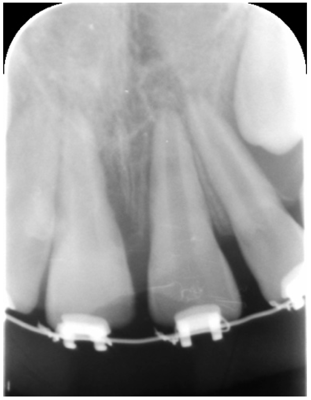
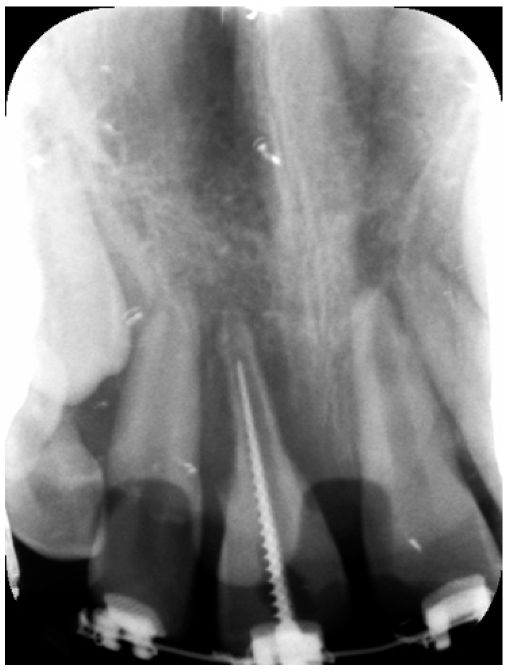
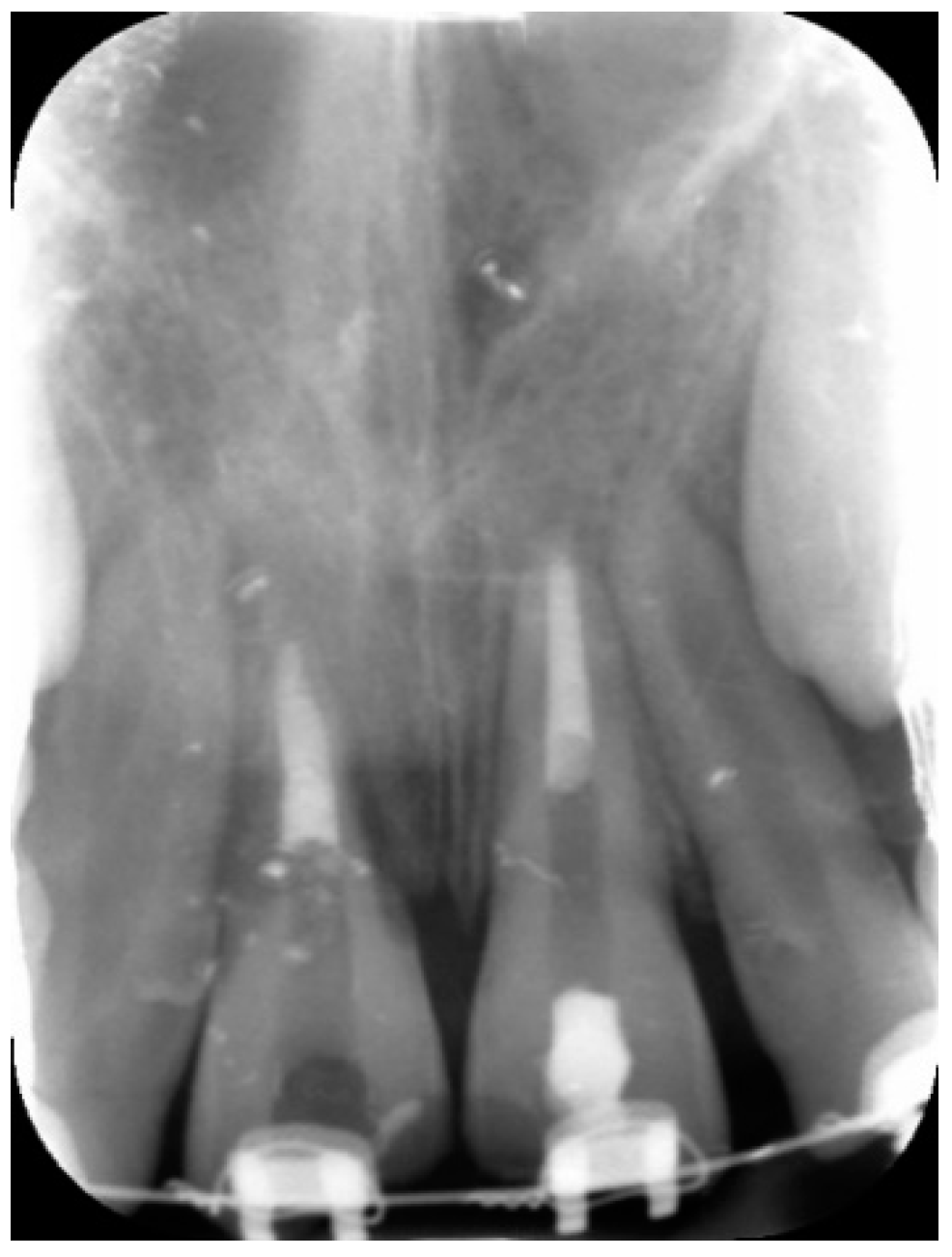
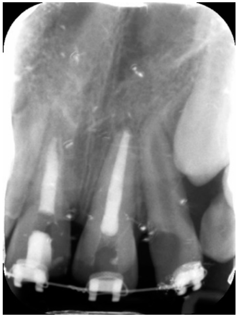
| 6l | Important/relevant dates and times (in the text, or a table or figure) must be provided in chronological order | 4–7 | |
| 6m | The diagnostic methods and the results for the specific tooth/teeth (e.g., pulp sensibility test, tenderness, mobility, periodontal probing depths, laboratory investigations, imaging techniques, or other special tests) must be provided | 4 | |
| 6n | The diagnostic challenges, if any, must be provided | N/A | |
| 6o | The diagnostic reasoning including other possible diagnoses that were considered must be provided | N/A | |
| 6p | The active treatment (s) or intervention(s) performed, if any, must be provided | 4–7 | |
| 6q | Any modifications to the proposed treatment(s) or intervention(s), if necessary, must be provided | N/A | |
| 6r | The assessment method(s) used to determine the clinician-assessed and patient-assessed treatment outcomes and their results must be provided | 6–7 | |
| 6s | Adverse and unanticipated events or consequences, if any, must be provided | 7 | |
| Discussion | 7a | The specific treatment(s) and intervention(s) (if any) must be discussed with reference to the relevant literature | 8 |
| 7b | The strengths of the case report and its importance must be discussed with reference to the relevant literature | 8 | |
| 7c | The limitations of the case report must be discussed | 8–9 | |
| 7d | The rationale for the conclusion(s) must be discussed | 8 | |
| Patient perspective | 8a | Feedback from the patient on the treatment and the care they received should be provided, if relevant | N/A |
| Conclusion | 9a | Explicit conclusion(s), i.e., the main “take-away” lessons must be provided | 9 |
| 9b | Implications for clinical practice or future research must be provided | 9 | |
| Funding details | 10a | Sources of funding and other support (such as supply of instruments, equipment) as well as the role of funders must be acknowledged and described | 9 |
| Conflict of interest | 11a | An explicit statement on conflicts of interest must be provided | 9 |
| Quality of images | 12a | Details of the equipment, software and settings used to acquire the image(s) must be described in the text or legend | N/A |
| 12b | The reason why the image(s) was acquired and the rationale for its inclusion in the manuscript must be provided in the text | N/A | |
| 12c | The circumstances (conditions) under which the image(s) were viewed and evaluated by the authors must be provided in the text | N/A | |
| 12d | The resolution and any magnification of the image(s) or any modifications/enhancements (e.g., adjustments for brightness, color balance, or magnification, image smoothing, staining, etc.) that were carried out must be described in the text or legend | N/A | |
| 12e | Patient(s) identifiers (names, patient numbers) must be removed to ensure they are anonymized | N/A | |
| 12f | An interpretation of the findings (meaning and implications) from the image (s) must be provided in the text | 4–8 | |
| 12g | The legend associated with each image must describe clearly what the subject is and what specific feature(s) it illustrates. Legends associated with images of patients must describe the age, gender and ethnicity of the person, if relevant | 4–8 | |
| 12h | Markers/labels must be used to identify the key information in the image(s) and be defined in the legend or as a footnote | N/A |
References
- Fouad, A.F.; Abbott, P.V.; Tsilingaridis, G.; Cohenca, N.; Lauridsen, E.; Bourguignon, C.; O’Connell, A.; Flores, M.T.; Day, P.F.; Hicks, L.; et al. International association of dental traumatology guidelines for the management of traumatic dental injuries: 2. Avulsion of permanent teeth. Dent. Traumatol. 2020, 36, 331–342. [Google Scholar] [CrossRef]
- Krastl, G.; Weiger, R.; Filippi, A.; Van Waes, H.; Ebeleseder, K.; Ree, M.; Connert, T.; Widbiller, M.; Tjäderhane, L.; Dummer, P.M.H.; et al. Endodontic management of traumatized permanent teeth: A comprehensive review. Int. Endod. J. 2021. [Google Scholar] [CrossRef]
- Nagy, M.M.; Tawfik, H.E.; Hashem, A.A.; Abu-Seida, A.M. Regenerative potential of immature permanent teeth with necrotic pulps after different regenerative protocols. J. Endod. 2014, 40, 192–198. [Google Scholar] [CrossRef]
- Yoshpe, M.; Einy, S.; Ruparel, N.; Lin, S.; Kaufman, A.Y. Regenerative endodontics: A potential solution for external root resorption (case series). J. Endod. 2020, 46, 192–199. [Google Scholar] [CrossRef]
- Souza, B.D.; Dutra, K.L.; Kuntze, M.M.; Bortoluzzi, E.A.; Flores-Mir, C.; Reyes-Carmona, J.; Felippe, W.T.; Porporatti, A.L.; Canto, G.D. Incidence of root resorption after the replantation of avulsed teeth: A meta-analysis. J. Endod. 2018, 44, 1216–1227. [Google Scholar] [CrossRef] [PubMed]
- Donaldson, M.; Kinirons, M.J. Factors affecting the time of onset of resorption in avulsed and replanted incisor teeth in children. Dent. Traumatol. 2001, 17, 205–209. [Google Scholar] [CrossRef] [PubMed]
- Aidos, H.; Diogo, P.; Santos, J.M. Root resorption classifications: A narrative review and a clinical aid proposal for routine assessment. Eur. Endod. J. 2018, 3, 134–145. [Google Scholar] [CrossRef] [PubMed]
- Spinas, E.; Generali, L.; Mameli, A.; Demontis, C.; Martinelli, D.; Giannetti, L. Delayed tooth replantation and inflammatory root resorption in childhood and adolescence. J. Biol. Regul. Homeost. Agents 2019, 33, 623–627. [Google Scholar] [PubMed]
- Ambu, E.; Fimiani, M.; Vigna, M.; Grandini, S. Use of bioactive materials and limited FOV CBCT in the treatment of a replanted permanent tooth affected by inflammatory external root resorption: A case report. Eur. J. Paediatr. Dent. 2017, 18, 51–55. [Google Scholar] [PubMed]
- Patel, S.; Foschi, F.; Condon, R.; Pimentel, T.; Bhuva, B. External cervical resorption: Part 2-management. Int. Endod. J. 2018, 51, 1224–1238. [Google Scholar] [CrossRef] [PubMed] [Green Version]
- Nagendrababu, V.; Chong, B.S.; McCabe, P.; Shah, P.K.; Priya, E.; Jayaraman, J.; Pulikkotil, S.J.; Setzer, F.C.; Sunde, P.T.; Dummer, P.M.H. PRICE 2020 guidelines for reporting case reports in Endodontics: A consensus-based development. Int. Endod. J. 2020, 53, 619–626. [Google Scholar] [CrossRef] [PubMed]
- Petti, S.; Glendor, U.; Andersson, L. World traumatic dental injury prevalence and incidence, a meta- analysis—one billion living people have had traumatic dental injuries. Dent. Traumatol. 2018, 34, 71–86. [Google Scholar] [CrossRef] [PubMed] [Green Version]
- Mazur, M.; Marasca, R.; Ottolenghi, L.; Vozza, I.; Covello, F.; Zupancich, A.; Cristiani, E.; Nava, A. Different resorptive patterns of two avulsed and replanted upper central incisors based on scanning electron microscopy and stereomicroscopic analysis: A case report. Appl. Sci. 2020, 10, 3551. [Google Scholar] [CrossRef]
- Andersson, L.; Bodin, I.; Sörensen, S. Progression of root resorption following replantation of human teeth after extended extraoral storage Endod. Dent. Traumatol. 1989, 5, 38–47. [Google Scholar] [CrossRef] [PubMed]
- Torabinejad, M.; Parirokh, M.; Dummer, P.M.H. Mineral trioxide aggregate and other bioactive endodontic cements: An updated overview-part II: Other clinical applications and complications. Int. Endod. J. 2018, 51, 284–317. [Google Scholar] [CrossRef] [PubMed]
- Marão, H.F.; Panzarini, S.R.; Aranega, A.M.; Sonoda, C.K.; Poi, W.R.; Esteves, J.C.; Silva, P.I. Periapical tissue reactions to calcium hydroxide and MTA after external root resorption as a sequela of delayed tooth replantation. Dent. Traumatol. 2012, 28, 306–313. [Google Scholar] [CrossRef] [PubMed]
- Lauridsen, E.; Andreasen, J.O.; Bouaziz, O.; Andersson, L. Risk of ankylosis of 400 avulsed and replanted human teeth in relation to length of dry storage: A re-evaluation of a long-term clinical study. Dent. Traumatol. 2020, 36, 108–116. [Google Scholar] [CrossRef] [PubMed]
- Giannetti, L.; Spinas, E.; Diago, A.M.D. Tooth avulsion with extra oral time in less than 60 minutes: Two different therapeutic protocols with 13-year follow-up. J. Biol. Regul. Homeost. Agents 2019, 33, 629–631. [Google Scholar] [PubMed]
- Spinas, E.; Aresu, M.; Canargiu, F.; Giannetti, L. Preventive treatment of post-traumatic dental infraocclusion: Study on the knowledge of dental decoronation in a sample of Italian dental students and dentists. Eur. J. Paediatr. Dent. 2015, 16, 279–283. [Google Scholar] [PubMed]
- Schmalz, G.; Widbiller, M.; Galler, K.M. Clinical Perspectives of Pulp Regeneration. J. Endod. 2020, 46, S161–S174. [Google Scholar] [CrossRef] [PubMed]


Publisher’s Note: MDPI stays neutral with regard to jurisdictional claims in published maps and institutional affiliations. |
© 2021 by the authors. Licensee MDPI, Basel, Switzerland. This article is an open access article distributed under the terms and conditions of the Creative Commons Attribution (CC BY) license (https://creativecommons.org/licenses/by/4.0/).
Share and Cite
Di Giorgio, G.; Salucci, A.; Sfasciotti, G.L.; Iaculli, F.; Bossù, M. External Root Resorption Management of an Avulsed and Reimplanted Central Incisor: A Case Report. Dent. J. 2021, 9, 72. https://0-doi-org.brum.beds.ac.uk/10.3390/dj9060072
Di Giorgio G, Salucci A, Sfasciotti GL, Iaculli F, Bossù M. External Root Resorption Management of an Avulsed and Reimplanted Central Incisor: A Case Report. Dentistry Journal. 2021; 9(6):72. https://0-doi-org.brum.beds.ac.uk/10.3390/dj9060072
Chicago/Turabian StyleDi Giorgio, Gianni, Alessandro Salucci, Gian Luca Sfasciotti, Flavia Iaculli, and Maurizio Bossù. 2021. "External Root Resorption Management of an Avulsed and Reimplanted Central Incisor: A Case Report" Dentistry Journal 9, no. 6: 72. https://0-doi-org.brum.beds.ac.uk/10.3390/dj9060072