Article

Rosemary Extract and Essential Oil as Drink Ingredients: An Evaluation of Their Chemical Composition, Genotoxicity, Antimicrobial, Antiviral, and Antioxidant Properties

by
Spyridoula D. Christopoulou
1,
Chrysa Androutsopoulou
2,
Panagiotis Hahalis
3,
Chrysoula Kotsalou
2,
Apostolos Vantarakis
2 and
Fotini N. Lamari
1,*
1
Laboratory of Pharmacognosy & Chemistry of Natural Products, Department of Pharmacy, University of Patras, 26504 Patras, Greece
2
Department of Public Health, Faculty of Medicine, University of Patras, 26504 Patras, Greece
3
Tentoura Castro-G.P. Hahalis Distillery, 26225 Patras, Greece
*
Author to whom correspondence should be addressed.
Submission received: 15 November 2021 / Revised: 9 December 2021 / Accepted: 15 December 2021 / Published: 18 December 2021

Abstract

:
Rosmarinus officinalis L. (rosemary) is in high demand in the food and drink industries due to its distinct organoleptic properties. With the aim of evaluating the rosemary leaves as drink ingredients, both the essential oil and alcoholic (38%, v/v) extract were studied in terms of chemical composition, genotoxicity, antimicrobial, antiviral, and antioxidant properties. GC–MS analysis showed that the main volatile compounds in the essential oil were eucalyptol (40.1%), camphor (12.4%), and α-pinene (12.9%). LC–MS analysis revealed gallocatechin and rosmarinic acid as the main extract ingredients. Both the essential oil and the extract were not genotoxic (Ames test) against TA98 and TA100 at the dilutions of 5% and 90%, respectively; those dilutions were selected as the maximum possible ones in the drink industry. Their activity was investigated against Escherichia coli, Salmonella enterica serovar Typhimurium, Staphylococcus aureus, Aspergillus niger, and Adenovirus 35. Both were effective against Adenovirus and A. niger, even the essential oil at 5% (v/v). The extract at dilutions of 25–90% had more pronounced activity against tested bacteria than the essential oil at the dilutions of 5–100%; the essential oil at the dilution of 5% inhibited S. aureus growth. The antioxidant activity was evaluated by the 2,2-diphenyl-1-picrylhydrazyl radical scavenging assay, the 2,2-azino-bis-3-ethylbenzothiazoline-6-sulfonic acid decolorization assay, and the ferric reducing antioxidant power assay. Both exhibited good antioxidant activity, but rosemary essential oil was far more effective than the extract. Our results demonstrate that rosemary essential oil and extract are safe and have beneficial biological properties. Therefore, they could serve as health-promoting ingredients in the drink industry.

1. Introduction

Rosmarinus officinalis L. (Lamiaceae family) is widely cultivated for its leaves. It has been used as a stimulant, as an analgesic, and against inflammatory diseases, physical and mental fatigue in traditional medicine [1]. The pharmacologically validated medicinal properties of rosemary include antibacterial and antioxidant [1,2], antiviral [3], anti-inflammatory [4], antifungal [5], and antiproliferative effects toward cancer cells, absence of genotoxicity, and the ability to prolong thrombin time [6]. The antidepressant properties have been studied in various models [7,8]; we have earlier shown that rosemary infusion has antidepressant/anxiolytic-like and anticholinesterase effects [9]. In 2018, an intervention study in healthy adults showed that rosemary water consumption had beneficial cognition effects, which agreed with those previously discovered for inhaling the scent of rosemary essential oil [10].
The Food and Drug Administration (FDA, Silver Spring, MD, USA) has listed many essential oils as “substances generally recognized as safe” for consumption in foods and beverages [11]. European Food Safety Authority (EFSA, Parma, Italy) has also authorized rosemary extracts as a food additive (E 392) and the range of margins of safety was 100–2000 and 200–3000 mg carnosol plus carnosic acid for children and adults, respectively [12]. Due to the high demand for it as a food additive and a food/drink ingredient, the rosemary extracts market was estimated at USD 215 Mn in 2019 and is anticipated to grow at an annual rate of 3.7 percent between 2020 and 2025 [13].
Apart from its distinct favorable organoleptic properties, many studies have demonstrated beneficial antimicrobial and antioxidant properties of the rosemary essential oils and extracts justifying their use as natural preservatives [14]. Specifically, their addition can effectively inhibit the oxidative processes in the processing and storage of meat products such as salami and fresh chicken and maintain/improve the product’s organoleptic properties [15]. However, their value as natural ingredients in the drink industry has not been thoroughly investigated, although there is a trend in producing bitter alcoholic beverages from aromatic and medicinal plants [16]; in those, their content can be high.
Antibacterial and antioxidant activities of the essential oil stem from certain volatile categories, i.e., monoterpenes ketones, hydrocarbons, and oxides [2,17]; 1,8-cineole, camphor, and α-pinene are mainly responsible for that bioactivity [17,18,19]. With regard to the rosemary extracts, antimicrobial and antioxidant activities are attributed to the phenolic acids, flavonoids, and terpenoids [20,21], and particularly to carnosic acid and rosmarinic acid [22,23]. Many studies have shown the great variability of chemical compositions among different rosemary genotypes, environments, cultivation conditions, harvest times, and extraction conditions [24], revealing the need for thorough extract and essential oil characterization and thereafter standardization.
The aim of this study was the evaluation of rosemary essential oil and extract as ingredients in the drink industry. Thus, we proceeded to the full chemical characterization of rosemary essential oil and ethanolic extract and the investigation of their efficacy against bacteria (E. coli, S. aureus, S. typhimurium), fungus (A. niger), virus (Adenovirus 35) in different dilutions relevant to their potential use in final alcoholic beverages. 2,2-Diphenyl-1-picrylhydrazyl (DPPH) radical scavenging activity, 2,2-azino-bis-3-ethylbenzothiazoline-6-sulfonic acid (ABTS) decolorization, and ferric reducing antioxidant power (FRAP) assays were used to evaluate their antioxidant capacity. Finally, their toxicity was determined with the AMES test.

2. Materials and Methods

2.1. Essential Oils and Extracts

The essential oil and the herbal extract were produced by the distillery “Tentoura Castro-G.P. Hahalis” located in Patras, Greece. Rosmarinus officinalis L. was cultivated in Patras and was collected in July–August 2019. In an experimental 10 L distillery, the essential oil was isolated using water-steam distillation. Specifically, 0.6 kg of fresh rosemary leaves were distilled for 3–4 h in 8 L of water. Maceration was used to create the herbal extracts. Glass and stainless-steel containers were used for extraction. Rosemary leaves were dried for 8 days before being extracted for 25 days in 40 L of a solvent containing 38 percent v/v ethanol at a temperature of 20–25 °C.

2.2. Gas Chromatography–Mass Spectrometry

An Agilent Technologies 6890N GC instrument equipped with a 5975B mass selective detector (MSD) was used in the electron impact (EI) mode of 70 eV at the Central Instrumental Analysis Laboratory of the University. The carrier gas was helium, and the capillary column was HP-5MS (30 m × 0.25 mm, 0.25 μm). GC/MSD Chemstation (Agilent Technologies, Inc., Santa Clara, CA, USA) and MestreNova v.6.0.2-5475 were used to analyze the data (Mestrelab Research S.L., Santiago de Compostela, Spain).
The initial GC oven was 50 °C for 2 min, which was then ramped at 2 °C/min to 80 °C for 1 min, at 8 °C/min to 100 °C for 1 min, at 10 °C/to 200 °C for 1 min. Finally, the oven warmed up to 300 °C at a rate of 10 °C/min and then kept at 300 °C for 5 min. The carrier gas was at a rate of 1.0 mL/min, in a splitless mode, and the mass range was m/z 40–1000.
The sample was diluted (1:40) in ethyl acetate, and the injection volume was 1 μL. n-Octane (98% purity) was utilized as an internal standard at a final concentration of 0.3 mg/mL. Alkanes (C8–C24) were examined under the same conditions and utilized as reference points for computing retention indices using the Van den Dool and Kratz equation [25]. By comparing the experimental retention indices (AIcal) and MS spectra to commercial databases [26,27] and the literature, the chemical components were identified. The standard compounds α-pinene (>99%), eucalyptol (>99%), linalool (>99%), and camphor (>99%) were purchased from Sigma-Aldrich (Steinheim, Germany) and were used for the verification of the identification. The results were expressed as a percentage of the ratio of each compound peak area to that of the internal standard using the program WSEARCH32 (Ver. 16/2005).

2.3. Liquid Chromatography–Mass Spectrometry

In this study, a single quadrupole LC/MS system, the LC/MSD1260 Infinity II (Agilent Technologies, Inc.), was used. The mass range of this system was m/z 100–1000, and it was equipped with an ESI ion source. Nitrogen was used as an ionization gas. A Poroshell 120 EC 18 column (4.6 × 100 mm, 2.7 μm) was used for separation (Agilent Technologies, Inc.). The following were the LC conditions: solvent A (0.1% formic acid) and solvent B (acetonitrile with 0.1% formic acid) and the gradient elution method was: 0–5 min 4% B; 5–15 min 4–15% B; 15–18 min 15% B; 18–23 min 15–20% B; 23–33 min 20% B; 33–53 min 20–58% B; 53–68 min 58% B; 68–80 min 58–95% B; 80–85 min 95% B; 85–89 min 95–4% B; 89–93 min 4% B. The injection volume was 20 μL, and the flow rate was 0.5 mL/min. Dilution in 1% formic acid to a final concentration of 2 mg raw plant material/mL was used to prepare samples for UPLC–ESI–MS.
The standards that were used for identification and/or quantification were rutin (HPLC > 99%) from Extrasynthese (Genay, France), quercetin 3-O-glucoside (HPLC > 98%) from Extrasynthese, kaempferol (HPLC > 96%) from Sigma Aldrich (Steinheim, Germany), rosmarinic acid (HPLC > 98%) from Extrasynthese, and carnosic acid (HPLC > 98%) from Extrasynthese. The rest of the compounds were identified by comparing their elution order and their mass spectra to those in the literature. The quantification of the flavonoid compounds was based on the rutin standard curve (3.125–100.000 μg/mL, y = 29,361x + 425,743, R2 = 0.9632), of the phenolic acids on the rosmarinic acid curve (2.50–100.00 μg/mL, y = 99,751x + 307,188, R2 = 0.9978), and of the diterpenes on the carnosic acid (1.00–25.00 μg/mL, y = 380,826x + 309,232, R2 = 0.9943).

2.4. Antioxidant Activity

The antioxidant activity was evaluated by the DPPH (1,1 diphenyl-2-picrylhydrazyl) radical method, the Ferric reducing antioxidant power (FRAP) assay and the ABTS [(2, 2′-azinobis-(3-ethylbenzothiazoline-6-sulfonate)] assay. Sample blanks (containing only the sample in the appropriate final volume) are used for every assay and their response was subtracted from that of the samples.

2.4.1. DPPH Radical Scavenging Assay

The scavenging of the DPPH radical by the samples was determined with a slight modification of previous protocols [28]; briefly, 5 μL of the test materials (standard or sample) were mixed with 195 μL of 0.1 mM methanolic DPPH solution. Butylhydroxytoluene (BHT) was used as a standard. After an incubation period of 30 min at 25 °C in the dark, the absorbance at 540 nm was recorded as Asample. The radical scavenging activity was expressed as percentage inhibition of DPPH and was calculated according to the formula IC (%) = [(Ao − At)/Ao] × 100, where Ao and At are the absorbance values of the blank sample and the test sample, respectively. The radical scavenging activity of the samples was expressed as mg BHT/mL and it was based on the BHT calibration curve (0.025–40 mg/mL, y = 31.605x + 44.512, R2 = 0.9924).

2.4.2. FRAP Assay

Based on a previous method [29], the samples’ ability to reduce ferric iron (Fe3+) was determined. Before each experiment, the FRAP reagent was freshly prepared and contained 300 mM of acetate buffer (pH = 3.6), 10 mM 2,4,6-tri(2-pyridyl)-1,3,5-triazine (TPTZ) in 40 mM HCl, and 20 mM FeCl3×6H2O in pure water in a ratio of 5:1:1. Solutions of FeSO4×7H2O were used as standards. 60 μL of test materials (standards/plant essential oil and extracts/blank) were mixed with 55 μL 300 mM of acetate buffer (pH = 3.6) and 80 μL of freshly FRAP reagent and incubated for 5 min. Absorbance was determined at 594 nm. The FRAP values were expressed as mg FeSO4×7H2O/mL of sample, and it was based on the FeSO4×7H2O standard curve (0.014–0.278 mg/mL, y = 2.9543x + 0.0353, R2 = 0.9917).

2.4.3. ABTS Decolorization Assay

The ABTS test was performed using the method Tzima et al. [28], with minimal changes. Reagent stock solution was prepared by mixing 88 μL of 2.45 mM K2S2O3 in water with 7 mM of a methanolic ABTS solution to a final volume of 5 mL. For 12–16 h, the freshly made reagent solution was stored at room temperature in the dark. The solution was then diluted in ethanol (approximately 1:30, v/v) to an absorbance of 0.700 at 754 nm before the experiment. Solutions of Trolox were used as standards. Then, 5 μL of diluted samples/standards/blank and 245 μL of ABTS reagent were mixed and incubated for 5 min in the dark. The absorbance was measured at 754 nm. ABTS radical scavenging activity of the samples was expressed as scavenging activity (%) = [(Asample − Acontrol)/Acontrol] × 100, where Acontrol is the absorbance of the blank control (ABTS solution without test sample), and Asample is the absorbance of the test sample. The ABTS values were expressed as mg Trolox/mL and it was based on Trolox calibration curve (0.025–5 mg/mL, y = 2.9543x + 0.0353, R2 = 0.9917).

2.5. Mutagenicity Assay

The mutagenic potential was determined with the Ames test [30,31]. The kit Ames Salmonella/mutagenicity assay was provided by EBPI (Mississauga, ON, Canada), and the experimental procedure was described by Androutsopoulou et al. [32]. In brief, Salmonella strains TA98 and TA100 were cultured in growth media containing Express Reagent V. Dilutions of rosemary essential oil up to 5% in DMSO and of the extracts up to 90% in distilled water were used. The determination of toxicity was colorimetric at 600 nm. All experiments were performed in triplicate.

2.6. Antibacterial and Antifungal Assay

Antibacterial activity was evaluated using the agar dilution technique against a group of bacterial strains, as recommended by the National Committee for Clinical Laboratory Standards [33]. Experiments were performed on Escherichia coli NCTC 9001, Salmonella typhimurium NCTC 12023, and Staphylococcus aureus NCTC 6571 (Sigma-Aldrich, St. Louis, MI, USA), as described by Androutsopoulou et al. [32]. Antifungal activity against Aspergillus niger (Sigma-Aldrich) was also performed by daily monitoring of the diameter of each colony as described by Androutsopoulou et al. [32]. All experiments were performed in triplicate at each concentration.

2.7. Antiviral Assay

The antiviral activity of the samples against human Adenovirus serotype 35 (AdV) at concentrations that were not cytopathic in A549 cells (Life Science Chemilab, Athens, Greece), P +92 generation, was determined as previously described [32]. In brief, AdV in serial dilutions (four replications of each dilution) or the samples in different concentrations were propagated in A549 cells and the effect on cell viability was recorded. For the antiviral activity, the cytotoxicity was observed by electron microscope, according to Saderi et al. (2011) [34]; the sample concentration that entirely suspends AdV35 cytopathic effect is recorded as efficient concentration, compared with virus control. All experiments were performed twice.

2.8. Data Analysis

All data are expressed as mean ± standard deviation of all replicates. The data were organized, analyzed, and visualized with Microsoft® Excel. If a difference is reported, it means that it is statistically significant at p < 0.05.

3. Results and Discussion

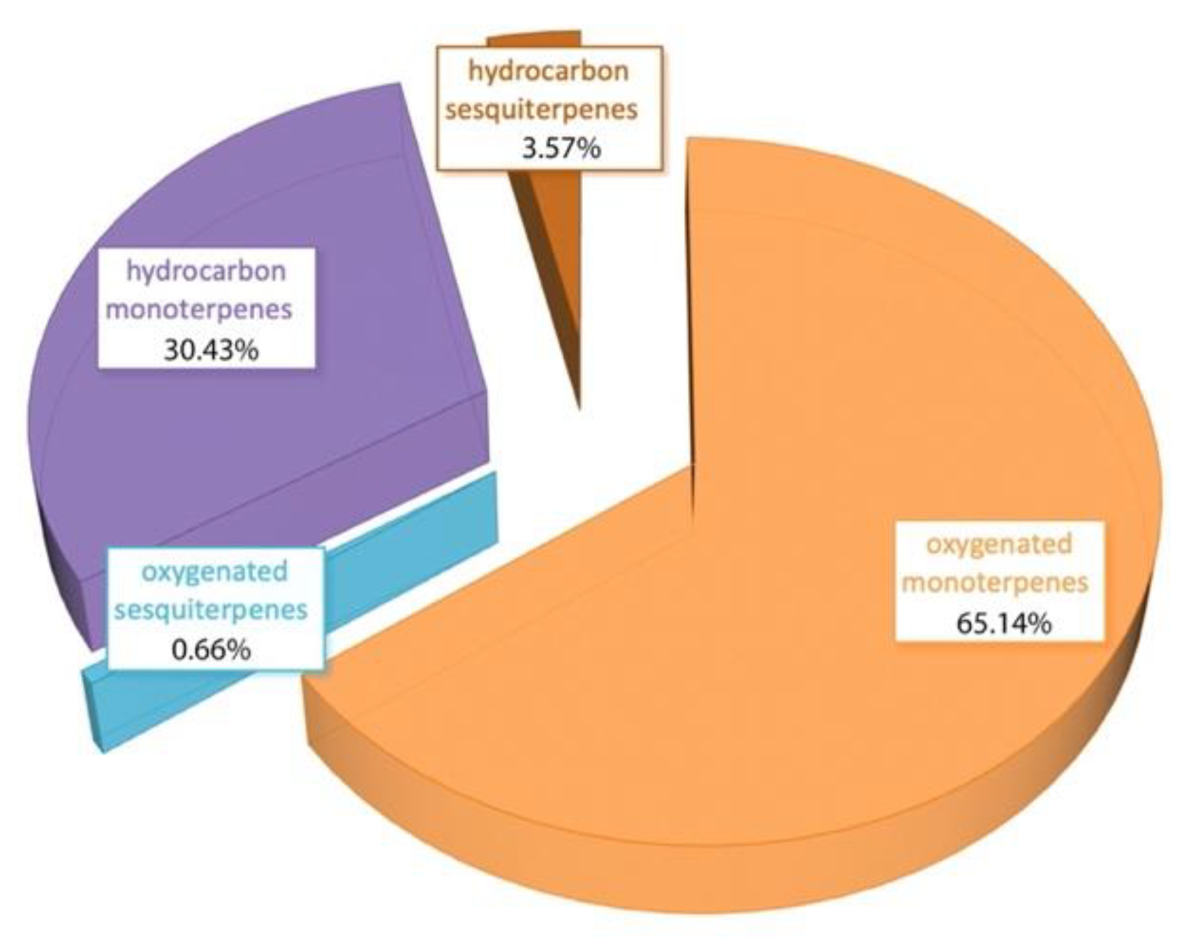
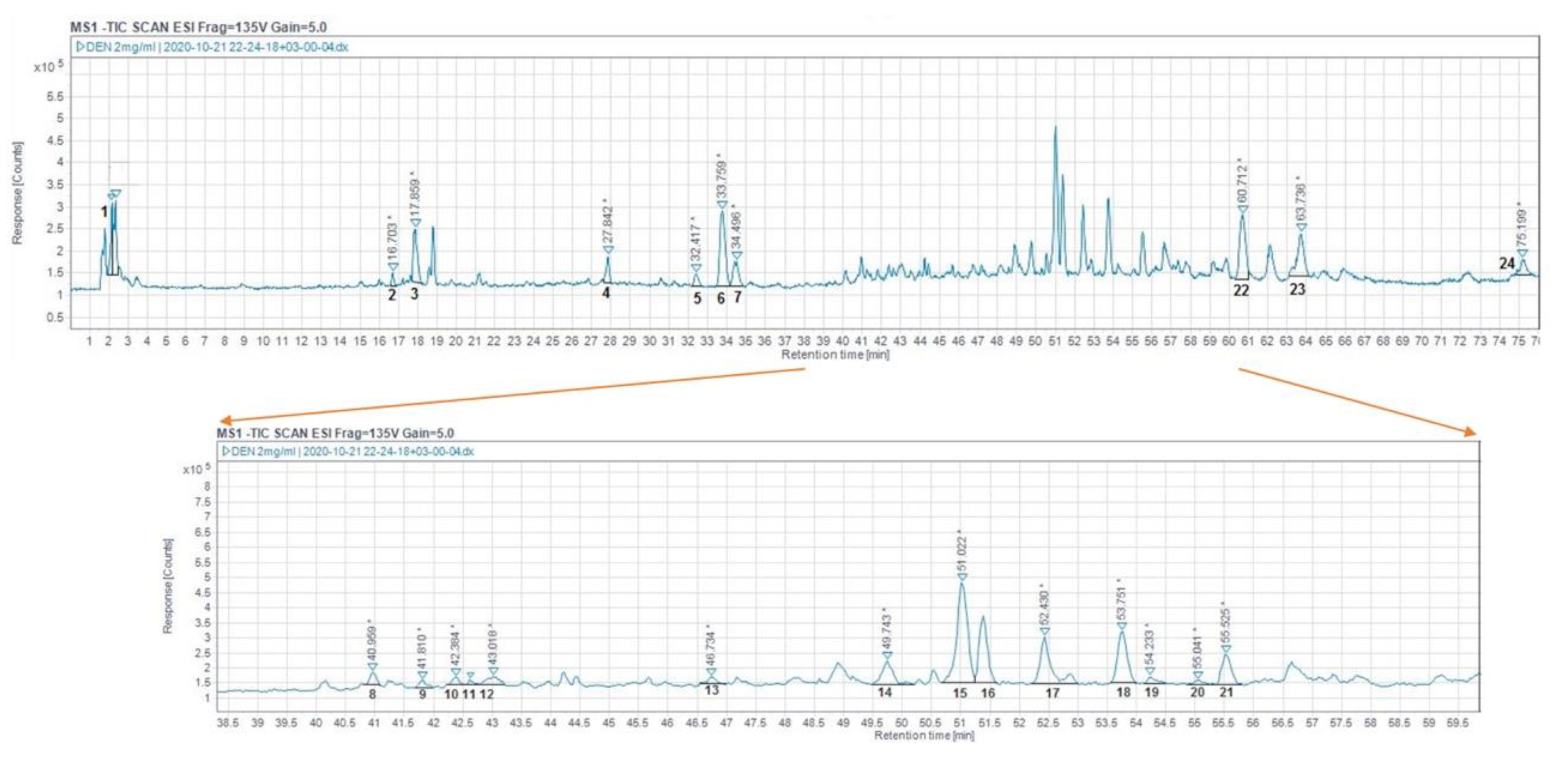
3.1. Chemical Analysis

In the essential oil of rosemary, 20 compounds were identified, representing 99.81% of the total oil (Table 1, Supplementary Materials Figure S1). The main components of the essential oil were eucalyptol (1,8-cineole) (40.10%), camphor (12.40%), α-pinene (12.94%), β-pinene (8.94%), and camphene (6.38%).
The great majority of essential oil compounds were monoterpenes (95.57%), while the sesquiterpenes amounted only to 4.24%, as can be seen in Figure 1. The results of our analysis are in line with previous studies of rosemary in Greece and abroad. Studies show great variability of the relative abundance of 1,8-cineole, camphor, and a-pinene suggesting the existence of 1,8-cineole and camphor-chemotypes, and two intermediate types, i.e., camphor/1,8-cineole/borneol type and 1,8-cineole/camphor type [35]. The sample analyzed herein clearly belongs to the 1,8-cineole chemotype.
The rosemary extract analysis showed 24 ingredients (Table 2, Figure 2). Compounds were identified by comparison of their retention time and mass spectra with those of the standards and the literature data; those compounds were terpenoids (peaks 14–21, 23, 24), flavonoids (peaks 3–5, 7–11, 13), and phenolic acids (peaks 1, 2, 6, 12), whereas peak 22 could not be identified. The flavonoids were the predominant category of compounds (54.4%) that were quantified, followed by the phenolic acids (26.7%) and the diterpenes (17.1%); triterpenes were not detected. In this study, nine flavonoids (compounds 3–5, 7–11 and 13) were identified in accordance with earlier studies [23,36,37,38]. In particular, peak 9 (luteolin-3-O-acetyl-β-glucuronide) has also been reported in R. οfficinalis from Serbia [37], peak 10 (luteolin) in R. οfficinalis from Spain [38], and peak 12 in an ethanolic extract from Mexico [29]. Finally, in this study, four phenolic acids were identified in line with Queralt et al. [39] and Perez-Mendoza et al. [29].
The main ingredients of the herbal extract were gallocatechin (26.4%), rosmarinic acid (18.0%), and luteolin-3-O-acetyl-O-glucuronide (14.2%). Carnosol was detected but could not be quantified, carnosic acid was not detected, and only 12-O-methyl carnosic acid was in low levels (0.5%). High seasonal variability in the content of carnosic acid was earlier shown by Fumiere Lemos et al. [24], which might be one reason for our findings, along with genotype and extraction parameters.

3.2. Antioxidant Activity

The evaluation of the antioxidant capacity with three different assays demonstrated that the essential oil had significantly (p < 0.001) higher antioxidant activity in all cases (Table 3).
In a previous comparative study [24], rosemary essential oil was not an efficient antioxidant in the DPPH assay, whereas the extract was a good one, but the composition in both essential oil and extract was different from that described herein especially in the content of 1,8-cineole and carnosic acid, showing the need for complex mixture characterization every time a biological property is studied. According to Bereta et al. [40], the difference in radical scavenging activity among rosemary essential oils is primarily due to the varying amounts of major compounds in the essential oils. According to Insawang et al. [41], 1,8-cineole, when compared with other monoterpenes, is the most effective at reducing reactive oxygen species. Moreover, Goze et al. [42] demonstrated that these terpenes have similar antioxidant properties to those obtained from phenolic mixtures. Rosemary essential oil with the same composition was an effective antioxidant in vitro and in vivo [43]; the per os administration of the essential oil to rats at a 10 mg/kg body weight for 7 days before the administration of CCl4 attenuated the hepatotoxicity markers, preserved renal function and the liver antioxidant status. Similar beneficial results of the rosemary essential oil were noted against hexavalent chromium toxicity [44].
Concerning the extracts, Kontogianni et al. have demonstrated that the antioxidant activities of commercial extracts depend on the concentration of phenolic abietane diterpenes, such as carnosic acid and its derivative carnosol and rosmanol isomers [23]. In our extract analysis, the terpenoids were the most abundant compounds. Yeddes et al. [45] reported that bioclimatic and season conditions (especially temperature) can influence the concentrations of rosmarinic acid and carnosic acid and thus the antioxidant potential of rosemary plant extracts; the highest antiradical activity of rosemary leaf extracts was obtained in summer. The plant material used in this study was collected in summer as well, and it had a good antioxidant activity with the DPPH assay (IC50 value of 12.32 mg/mL). Earlier studies in male hamsters that followed a high-fat diet supplemented with rosemary ethanolic extract demonstrated that rosemary was an efficient antioxidant in vivo, alleviating high-fat-induced oxidative damage, as well as enhancing the antioxidant enzyme activity, gene, and protein expression level via regulating the Nrf2 pathway [46]. More importantly, when rosemary in the form of dry powder (500 mg) was administered to healthy human volunteers twice a day for a month, plasma acetylcholinesterase was decreased, plasma total antioxidant activity was increased, and protein carbonylation was decreased in comparison with the values before treatment and the placebo group [47].
Conclusively, both essential oil and extract had good in vitro antioxidant activity; although the essential oil had higher antioxidant activity, it is used in lower percentages than the extract; in the scenario of essential oil usage at 0.5% and extract of 90% they could still have antioxidant activity, which would be comparable.

3.3. Antimutagenesis

The essential oil up to the concentration of 5% was able to inhibit mutations induced in TA98 and TA100 Salmonella strains as well as the extract did at concentrations up to 90% (Supplementary Materials Table S1). Therefore, they were both not genotoxic in the studied range of dilutions. Earlier, it was shown that rosemary essential oil was genotoxic in peripheral mononuclear cells in a dose-specific way [48]. In a study evaluating the frequency of micronuclei in different cell lines after exposure to the extract for 24 h, it was also shown that all tested concentrations did not stimulate DNA damage and thus were not mutagenic [49]. Razavi-Azarkhiavi et al. [50] also concluded that the constituents of the ethanol extract of rosemary prevented human lymphocyte oxidative DNA damage. Moreover, in a clinical study in 67 HIV patients receiving antiretroviral therapy and concomitant rosemary extracts, the administration of the extracts reduced the genomic instability in oral epithelial cells [51].

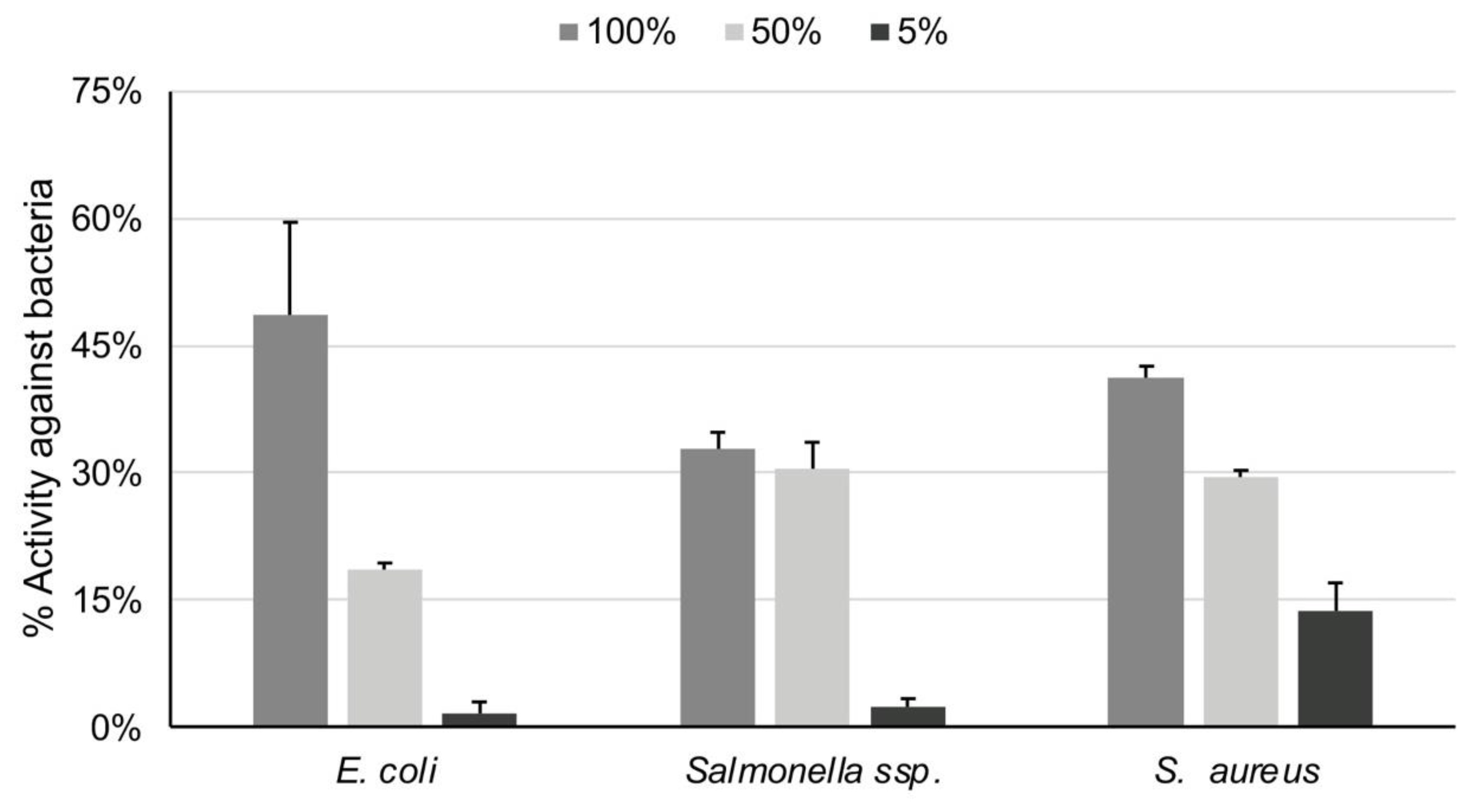
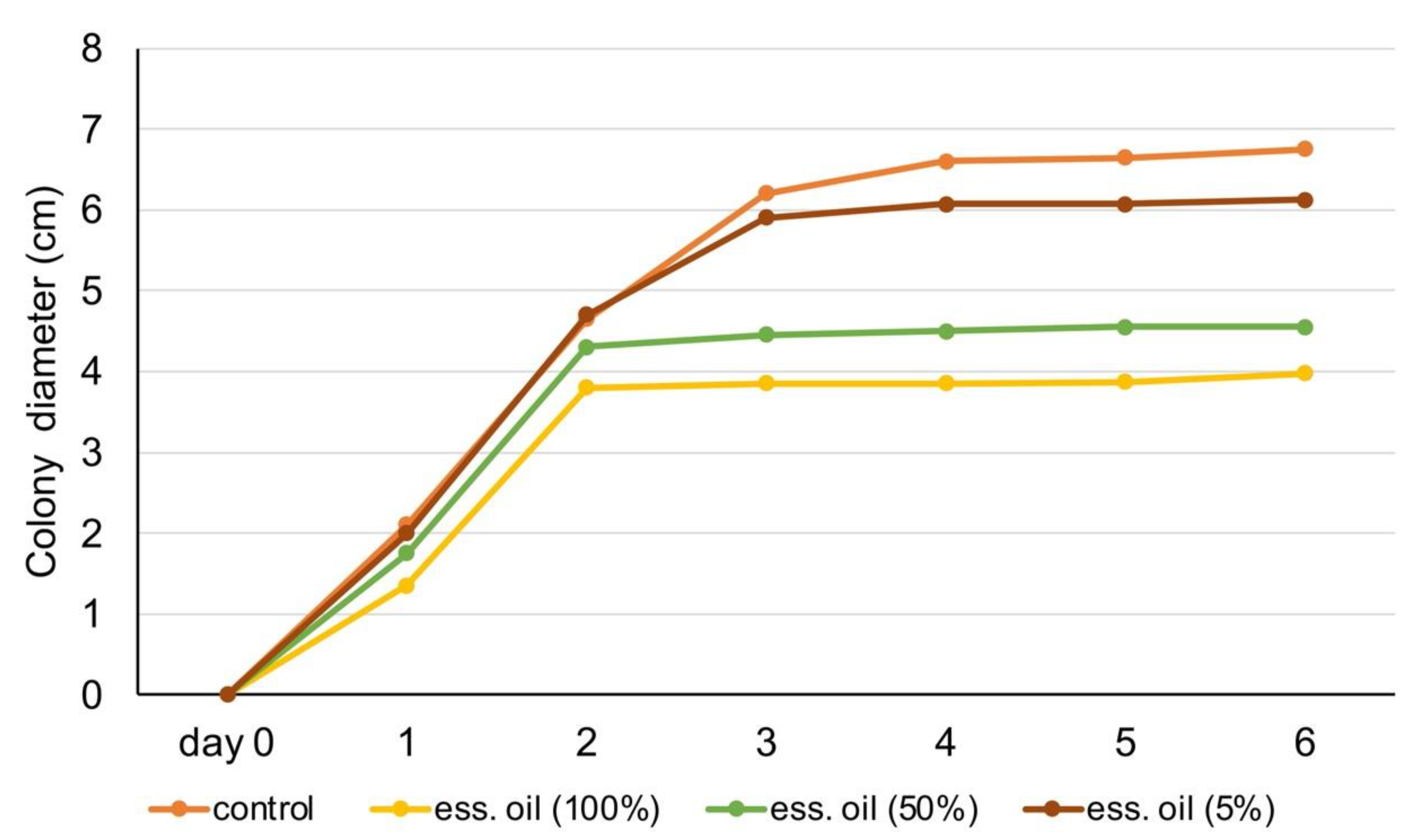
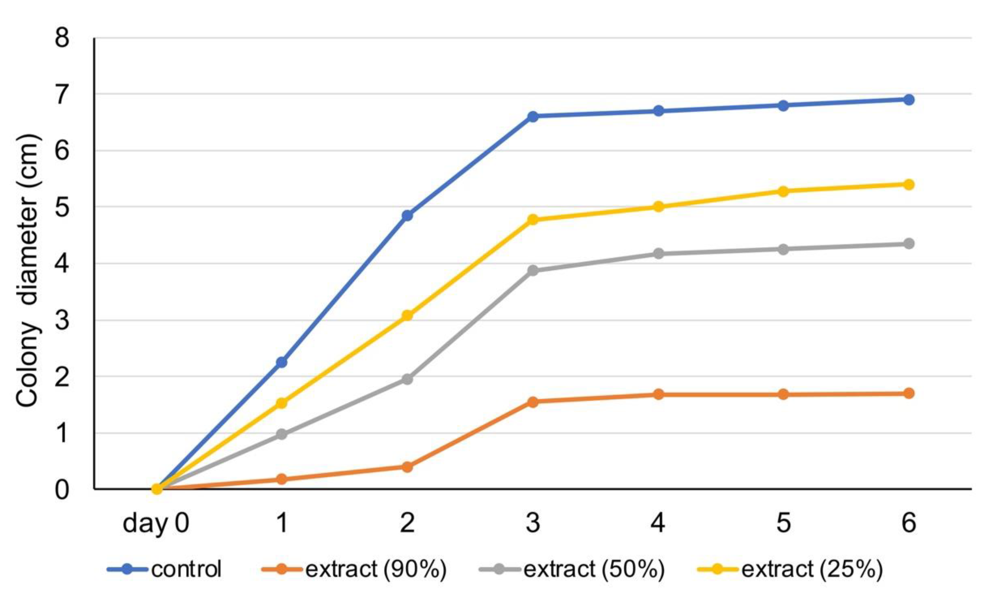
3.4. Antimicrobial and Antiviral Properties

The antibacterial activity of both the essential oil and the extract was dose dependent (Figure 3 and Figure 4). Rosmarinus officinalis essential oil revealed low-to-modest antibacterial activity against E. coli, in all trial concentrations; its ability in 100% concentration was 49%. The extract was more active (64% in the dilution of 90% and 55% in that of 50%) against E. coli. Regarding the antibacterial activity against S. aureus, the essential oil had a low activity even at the dilution of 5%, whereas the extract reached 74% in 90% concentration and 58% in 50%. Lastly, the essential oil also had low efficiency against Salmonella typhimurium; the extract on the other hand reached 59% in the concentration of 90%.
In Figure 5 and Figure 6, experimental data show the fungal colony diameter (in cm) over one week. Results show that rosemary essential oil (100% and 50%) inhibited fungus colony growth. Specifically, by day 3, the diameter of colony development stabilized when treated with those essential oil dilutions, whereas in the control, it increased until it occupied the entire Petri dish, slowing its growth rate even more in the last days (Figure 5). Figure 6 shows the results of experiments on the rosemary extract. All three dilutions of the extract were efficient in limiting the growth of fungus (in comparison with control), with 90% presenting the highest antifungal activity.
Cytotoxicity assay results are shown in Table 4 The essential oil in the concentration of 5% was not cytopathic, while the extract was not even at 100% concentration. The effects of essential oil (5%) and extract against Adenovirus at concentrations of 109 PFU/mL to 104 PFU/mL are shown in Table 5. The extract efficiently prevented the AdV35 cytopathic effect in all viral concentrations. In addition, the essential oil, despite its low concentration (5%), was effective against all Adenovirus concentrations.
Previous studies have demonstrated that rosemary essential oils have antimicrobial and antiviral activities [2,17,18,19,52,53,54,55]. Our results are in line with those studies and demonstrate, for the first time, the activity against Adenovirus 35. The essential oil studied herein with high 1,8-cineole content had a dose-dependent and modest antibacterial effect. Bajalan et al. [2] have demonstrated significant positive correlations of volatile oxides in rosemary essential oil and with the inhibition zone on S. aureus, and of ketones with that on E. coli. Moreover, in a previous study, 1,8-cineole was effective only against Gram-negative bacteria (E. coli, K. pneumoniae) inducing cell membrane disruption and a-pinene was against both Gram-positive (S. aureus, E. faecalis) and Gram-negative bacteria, whereas the effect of the rosemary essential oils was dependent on the composition [17]. Concerning the antifungal activity of the rosemary essential oil, our results confirm earlier studies [19,52]. Interestingly, in the study of Jianga et al. [19], the essential oil showed pronounced antifungal activity, compared with the pure compounds 1,8-cineole and α-pinene against the same fungus. With regard to the antiviral activity, we herein show the important anti-adenoviral activity of the essential oil. Accordingly, it has been earlier shown that rosemary 1,8-cineole chemotype significantly reduced the hepatitis A virus titers [53]. Due to the lipophilic nature of the volatiles in essential oils, they have the potential to infiltrate the lipid bilayer of the viral envelope [54]. Monoterpenes, the major compound class in rosemary oil, increase the fluidity and permeability of the cytoplasmic membrane, and disrupt the order of membrane-embedded proteins [55].
In our study, the rosemary extract was drastic against E. coli, S. typhimurium, A. niger, and Adenovirus. The rosemary extracts were potent against Gram-positive and Gram-negative bacteria in a study by Mahmood et al. [56]. In our experiment, rosemary extract displayed antifungal action against A. niger at concentrations ranging from 25 to 90%, which is consistent with the findings of Swari et al. [57], who demonstrated that rosemary ethanol extract had an inhibitory effect against Candida albicans. Rosemary extract had antiviral efficacy against HSV-1 and HSV-2, according to Al Mergin et al. [3]. Moreno et al. [20] proposed that the antibacterial efficiency of rosemary extracts was linked to their unique phenolic makeup, with antimicrobial action attributed mostly to carnosic acid and rosmarinic acid. The inhibitory effect of rosemary extract, according to Nieto et al. [1], is due to the activity of rosmarinic acid, rosmaridiphenol, carnosol, epirosmanol, carnosic acid, rosmanol, and isorosmanol. The primary components of rosemary extract in our investigation were rosmarinic acid and its derivatives. They interact with the cell membrane, altering the transport of electrons, causing changes in genetic material and nutrients, leakage of cellular components, and production changes in fatty acids [1].

4. Conclusions

This study provides, for the first time, an overall assessment of rosemary essential oil and extract in the concentrations that could be used in the drink industry in terms of safety, antioxidant, antimicrobial, and antiviral properties. Monoterpenes are the major compounds of rosemary essential oil, especially 1.8 cineole. The main components of the ethanolic rosemary extract belong to the class of flavonoids. The essential oil in dilutions of up to 5% and the extract in the range of 25–90% are not genotoxic, and confer antiviral, antifungal, and antioxidant properties; the extract also confers important antibacterial properties, whereas the essential oil is mainly effective against S. aureus. It can be concluded that those raw materials are safe for use in the drink industry and could impart important health benefits apart from their distinct sensory qualities.

Supplementary Materials

The following are available online at https://0-www-mdpi-com.brum.beds.ac.uk/article/10.3390/foods10123143/s1, Table S1: Results from toxicity experiments on essential oil and rosemary extracts, Figure S1: GC–MS chromatogram of rosemary essential oil.

Author Contributions

Conceptualization, P.H., F.N.L. and A.V.; methodology, P.H., F.N.L. and A.V.; validation, C.K., F.N.L. and A.V.; investigation, C.A., S.D.C., and P.H.; resources, P.H., F.N.L. and A.V.; writing—original draft preparation, C.A., S.D.C. and C.K.; writing—review and editing, F.N.L. and A.V.; supervision, F.N.L. and A.V.; funding acquisition, P.H., F.N.L. and A.V. All authors have read and agreed to the published version of the manuscript.

Funding

This research has been co-financed by the European Union and Greek national funds through the Regional Operational Program “Western Greece 2014–2020”, under the Call “Regional research and innovation strategies for smart specialization (RIS3) in Agri-Food” in the context of the project MIS 5040309 entitled “Development of new bitters from aromatic and medicinal herbs with health benefits (NatuBitters)”.

Institutional Review Board Statement

Not applicable.

Informed Consent Statement

Not applicable.

Data Availability Statement

Not applicable.

Conflicts of Interest

P.H. is an employee of Tentoura Castro-G.P. Hahalis Distillery. The company produced the raw materials under investigation (rosemary extract and essential oil) and provided information on the industrial production of bitter alcoholic beverages so as to determine the maximum concentrations of those in the experimental evaluation.

References

  1. Nieto, G.; Ros, G.; Castillo, J. Antioxidant and Antimicrobial Properties of Rosemary (Rosmarinus officinalis, L.): A Review. Medicines 2018, 5, 98. [Google Scholar] [CrossRef] [PubMed] [Green Version]
  2. Bajalan, I.; Rouzbahani, R.; Pirbalouti, A.G.; Maggi, F. Antioxidant and antibacterial activities of the essential oils obtained from seven Iranian populations of Rosmarinus officinalis. Ind. Crops Prod. 2017, 107, 305–311. [Google Scholar] [CrossRef]
  3. Al-Megrin, W.A.; Al Sadhan, N.A.; Metwally, D.M.; Al-Talhi, R.A.; El-Khadragy, M.F.; Abdel-Hafez, L.J.M. Potential antiviral agents of Rosmarinus officinalis extract against herpes viruses 1 and 2. Biosci. Rep. 2020, 40, BSR20200992. [Google Scholar] [CrossRef]
  4. Altinier, G.; Sosa, S.; Aquino, R.P.; Mencherini, P.; Loggia, R.D.; Tubaro, A. Characterization of Topical Antiinflammatory Compounds in Rosmarinus officinalis L. J. Agric. Food Chem. 2007, 55, 1718–1723. [Google Scholar] [CrossRef] [PubMed]
  5. Malvezzi de Macedo, L.; Mendes dos Santos, E.; Militão, L.; Tundisi, L.L.; Ataide, J.A.; Souto, E.B.; Mazzola, P.G. Rosemary (Rosmarinus officinalis L., syn Salvia rosmarinus Spenn.) and Its Topical Applications: A Review. Plants 2020, 9, 651. [Google Scholar] [CrossRef] [PubMed]
  6. Lamponi, S.; Baratto, M.C.; Miraldi, E.; Baini, G.; Biagi, M. Chemical Profile, Antioxidant, Anti-Proliferative, Anticoagulant and Mutagenic Effects of a Hydroalcoholic Extract of Tuscan Rosmarinus officinalis. Plants 2021, 10, 97. [Google Scholar] [CrossRef] [PubMed]
  7. Pangcong, D.; Liu, H. Research on the biological activity of rosemary extracts and its application in food. E3S Web Conf. 2021, 251, 02034–02041. [Google Scholar]
  8. Guo, Y.; Xie, J.; Li, X.; Yuan, Y.; Zhang, L.; Hu, W.; Luo, H.; Yu, H.; Zhang, R. Antidepressant Effects of Rosemary Extracts Associate with Anti-inflammatory Effect and Rebalance of Gut Microbiota. Front. Pharmacol. 2018, 9, 1126–1139. [Google Scholar] [CrossRef] [PubMed] [Green Version]
  9. Ferlemi, A.V.; Katsikoudi, A.; Kontogianni, V.G.; Kellici, T.F.; Iatrou, G.; Lamari, F.N.; Tzakos, A.G.; Margarity, M. Rosemary tea consumption results to anxiolytic- and anti-depressant like behavior of adult male mice and inhibits all cerebral area and livercholinesterase activity; phytochemical investigation and in silico studies. Chem. Biol. Interact. 2015, 237, 47–57. [Google Scholar] [CrossRef] [PubMed]
  10. Moss, M.; Smith, E.; Milner, M.; McCready, J. Acute ingestion of rosemary water: Evidence of cognitive and cerebrovascular effects in healthy adults. J. Psychopharmacol. 2018, 32, 1319–1329. [Google Scholar] [CrossRef] [PubMed]
  11. U.S. Food and Drug Administration. Available online: https://www.fda.gov/food/food-additives-petitions/food-additive-status-list#ftnR (accessed on 9 November 2021).
  12. EFSA Panel on Food Additives and Nutrient Sources Added to Food (EFSA ANS Panel); Younes, M.; Aggett, P.; Aguilar, F.; Crebelli, R.; Dusemund, B.; Filipič, M.; Frutos, M.J.; Galtier, P.; Gott, D.; et al. Refined Exposure Assessment of Extracts of Rosemary (E 392) from Its Use as Food Additive. EFSA J. 2018, 16, e05373. [Google Scholar]
  13. Industry ARC “Rosemary Extracts Market—Industry Analysis, Market Size, Share, Trends, Application Analysis, Growth and Forecast 2021–2026”. Available online: https://www.industryarc.com/Research/Rosemary-Extracts-Market-Research-504501 (accessed on 13 October 2021).
  14. Tzima, K.; Makris, D.; Nikiforidis, C.V.; Mourtzinos, I. Potential use of Rosemary, Propolis and Thyme as Natural Food Preservatives. J. Nutri. Health 2015, 1, 6–12. [Google Scholar]
  15. Kaur, R.; Gupta, T.B.; Bronlund, J.; Kaur, L. The potential of rosemary as a functional ingredient of meat products—A review. Food Rev. Int. 2021, 16, 1–21. [Google Scholar] [CrossRef]
  16. Tonutti, I.; Liddle, P. Aromatic plants in alcoholic beverages. A review. Flavour Fragr. J. 2010, 25, 341–350. [Google Scholar] [CrossRef]
  17. Ojeda-Sana, A.M.; van Baren, C.M.; Elechosa, M.A.; Juárez, M.A.; Moreno, S. New insights into antibacterial and antioxidant activities of rosemary essential oils and their main components. Food Control 2013, 31, 189–195. [Google Scholar] [CrossRef]
  18. Pitarokili, D.; Tzakou, O.; Loukis, A. Composition of the Essential oil of Spontaneous Rosmarinus officinalis from Greece and Antifungal Activity Against Phytopathogenic Fungi. J. Essent. Oil Res. 2007, 20, 457–459. [Google Scholar] [CrossRef]
  19. Jianga, Y.; Wua, N.; Fua, Y.J.; Wanga, W.; Luo, M.; Zhao, C.J.; Zua, Y.G.; Liua, X.L. Chemical composition and antimicrobial activity of the essential oil of Rosemary. Environ. Toxicol. Pharmacol. 2011, 32, 63–68. [Google Scholar] [CrossRef]
  20. Moreno, S.; Scheyer, T.; Romano, C.S.; Vojnov, A.A. Antioxidant and antimicrobial activities of rosemary extracts linked to their polyphenol composition. Free Radic. Res. 2005, 40, 223–231. [Google Scholar] [CrossRef]
  21. Oreopoulou, A.; Papavassilopoulou, E.; Bardouki, H.; Vamvakias, M.; Bimpilas, A.; Oreopoulou, V. Antioxidant recovery from hydrodistillation residues of selected Lamiaceae species by alkaline extraction. J. Appl. Res. Med. 2018, 8, 83–89. [Google Scholar] [CrossRef]
  22. Ekambaram, S.P.; Perumal, S.S.; Balakrishnan, A.; Marappan, N.; Srinivasan, S.; Viswanathan, G.V. Antibacterial synergy between rosmarinic acid and antibiotics against methicillin-resistant Staphylococcus aureus. J. Intercult. Ethnopharmacol. 2016, 5, 358–363. [Google Scholar] [CrossRef]
  23. Kontogianni, V.G.; Tomic, G.; Nikolic, I.; Nerantzaki, A.A.; Sayyad, N.; Stosic-Grujicic, S.; Stojanovic, I.; Gerothanassis, I.P.; Tzakos, A.P. Phytochemical profile of Rosmarinus officinalis and Salvia officinalis extracts and correlation to their antioxidant and anti-proliferative activity. Food Chem. 2013, 136, 120–129. [Google Scholar] [CrossRef]
  24. Lemos, M.F.; Lemos, M.F.; Pacheco, H.P.; Endringer, D.C.; Scherer, R. Seasonality modifies rosemary’s composition and biological activity. Ind. Crops Prod. 2015, 70, 41–47. [Google Scholar] [CrossRef]
  25. Van den Dool, H.; Kratz, P.D. A generalization of the retention index system including linear temperature programmed gas liquid partition chromatography. J. Chromatogr. 1963, 11, 463–471. [Google Scholar] [CrossRef]
  26. Adams, R.P. Identification of Essential oil Components by Gas Chromatography/Mass Spectrometry; Allured Publishing Corporation: Carol Stream, IL, USA, 2012; Volume 4. [Google Scholar]
  27. National Institute of Standards and Technology. NIST WebBook. Available online: http://webbook.nist.gov/chemistry/ (accessed on 9 September 2021).
  28. Tzima, K.; Brunton, N.P.; Rai, D.K. Evaluation of the impact of chlorophyll removal techniques on polyphenols in rosemary and thyme by-products. J. Food Biochem. 2020, 44, 13148–13169. [Google Scholar] [CrossRef] [PubMed]
  29. Berenice, M.; Mendoza, P.; Llorens-Escobar, L.; Vanegas-Espinoza, P.E.; Cifuentes, A.; Ibanez, E.; Del Villar-Martınez, A.A. Chemical characterization of leaves and calli extracts of Rosmarinus officinalis by UHPLC-MS. Electrophoresis 2019, 41, 1776–1783. [Google Scholar]
  30. Kristien, M.; Zeiger, E. The Ames Salmonella/microsome mutagenicity assay, Fundamental and Molecular Mechanisms of Mutagenesis. Mutat. Res. 2000, 455, 29–60. [Google Scholar]
  31. Taherkhami, T.; Zakaria, R.A.; Taherkharni, M. Mutagenic and Anti-mutagenic properties of the essential oil of Jurinea leptoloba DC by Ames Test. Cumhur. Univ. Fac. Sci. Sci. J. 2015, 36, 1682–1687. [Google Scholar]
  32. Androutsopoulou, C.; Christopoulou, S.D.; Hahalis, P.; Kotsalou, C.; Lamari, F.N.; Vantarakis, A. Evaluation of Essential oils and Extracts of Rose Geranium and Rose Petals as Natural Preservatives in Terms of Toxicity, Antimicrobial, and Antiviral Activity. Pathogens 2021, 10, 494. [Google Scholar] [CrossRef]
  33. Mudyiwa, M.; Muredzi, P.; Nyati, H. Isolation of Citric Acid Producing Asp. niger Strains; Lap Lambert Academic Publishing: Chisinau, Republic of Moldova, 2013. [Google Scholar]
  34. Horieh, S.; Abbasi, M. Evaluation of anti-adenovirus activity of some plants from Lamiaceae family grown in Iran in cell culture. Afr. J. Biotechnol. 2011, 10, 17546–17550. [Google Scholar]
  35. Lakusic, D.V.; Ristic, M.S.; Slavkovska, V.N.; Sinzar-Sekulic, J.B.; Lakusic, B.S. Environment-Related Variations of the Composition of the Essential Oils of Rosemary (Rosmarinus officinalis L.) in the Balkan Peninsula. Chem. Biodivers. 2012, 9, 1286–1302. [Google Scholar] [CrossRef] [PubMed]
  36. Borrás-Linares, I.; Stojanović, Z.; Quirantes-Piné, R.; Arráez-Román, D.; Švarc-Gajić, J.; Fernández-Gutiérrez, A.; Segura-Carretero, A. Rosmarinus officinalis Leaves as a Natural Source of Bioactive Compounds. Int. J. Mol. Sci. 2014, 15, 20585–20606. [Google Scholar] [CrossRef]
  37. Mena, P.; Cirlini, M.; Tassotti, M.; Herrlinger, K.A.; Dall’Asta, C.; Del Rio, D. Phytochemical Profiling of Flavonoids, Phenolic Acids, Terpenoids, and Volatile Fraction of a Rosemary (Rosmarinus officinalis L.) Extract. Molecules 2016, 21, 1576. [Google Scholar] [CrossRef] [PubMed]
  38. Santana-Méridas, O.; Polissiou, M.; Izquierdo-Melero, M.E.; Astraka, K.; Tarantilis, P.A.; Herraiz-Peñalver, D.; Sánchez-Vioque, R. Polyphenol composition, antioxidant and bioplaguicide activities of the solid residue from hydrodistillation of Rosmarinus officinalis L. Ind. Crops Prod. 2014, 59, 125–134. [Google Scholar] [CrossRef]
  39. Vallverdú-Queralt, A.; Regueiro, J.; Martínez-Huélamo, M.; Rinaldi Alvarenga, J.F.; Neto Leal, L.; Lamuela-Raventos, R.M. A comprehensive study on the phenolic profile of widely used culinary herbs and spices: Rosemary, thyme, oregano, cinnamon, cumin and bay. Food Chem. 2014, 154, 299–307. [Google Scholar] [CrossRef] [PubMed]
  40. Beretta, G.; Artali, R.; Maffei Facino, R.; Gelmini, F. An analytical and theoretical approach for the profiling of the antioxidant activity of essential oils: The case of Rosmarinus officinalis L. J. Pharm. Biomed. Anal. 2011, 55, 1255–1264. [Google Scholar] [CrossRef] [PubMed]
  41. Insawang, S.; Pripdeevech, P.; Tanapichatsakul, C.; Khruengsai, S.; Monggoot, S.; Nakham, T.; Artrod, A.; D’Souza, P.E.; Panuwete, P. Essential Oil Compositions and Antibacterial and Antioxidant Activities of Five Lavandula stoechas Cultivars Grown in Thailand. Chem. Biodivers. 2019, 16, e1900371–e1900383. [Google Scholar] [CrossRef] [PubMed]
  42. Göze, I.; Vural, N.; Ercan, N. Characterization of Essential oil and Antioxidant Activities of Some Species of Salvia in Turkey. Nat. Volatiles Essent. Oils 2016, 3, 1–7. [Google Scholar]
  43. Rašković, A.; Milanović, I.; Pavlović, N.; Ćebović, T.; Vukmirović, S.; Mikov, M. Antioxidant activity of rosemary (Rosmarinus officinalis L.) essential oil and its hepatoprotective potential. BMC Complement. Altern. Med. 2014, 14, 225. [Google Scholar] [CrossRef] [Green Version]
  44. El-Demerdash, F.M.; El-Sayed, R.A.; Abdel-Daim, M.M. Hepatoprotective potential of Rosmarinus officinalis essential oil against hexavalent chromium-induced hematotoxicity, biochemical, histological, and immunohistochemical changes in male rats. Environ. Sci. Pollut. Res. 2021, 28, 17445–17456. [Google Scholar] [CrossRef]
  45. Yeddes, W.; Chalghoum, A.; Aidi-Wannes, W.; Ksouri, R.; Tounsi, M.S. Effect of bioclimatic area and season on phenolics and antioxidant activities of rosemary (Rosmarinus officinalis L.) leaves. J. Essent. Oil Res. 2019, 31, 432–443. [Google Scholar] [CrossRef]
  46. Wang, H.; Cheng, J.; Yang, S.; Cuic, S.W.; Wang, M.; Haob, W. Rosemary extract reverses oxidative stress through activation of Nrf2 signaling pathway in hamsters fed on high fat diet and HepG2 cells. J. Funct. Foods 2020, 74, 1–10. [Google Scholar] [CrossRef]
  47. Dabaghzadeh, F.; Mehrabani, M.; Abdollahi, H.; Karami-Mohajeri, S. Antioxidant and anticholinergic effects of rosemary (Salvia rosmarinus) extract: A double-blind randomized controlled trial. Adv. Integr. Med. 2021, 3. in press. [Google Scholar] [CrossRef]
  48. Contini, A.; Di Bello, D.; Azzarà, A.; Giovanelli, S.; D’Urso, G.; Piaggi, S.; Pinto, B.; Pistelli, L.; Scarpatoa, R.; Testi, S. Assessing the cytotoxic/genotoxic activity and estrogenic/antiestrogenic potential of essential oils from seven aromatic plants. Food Chem. Toxicol. 2020, 138, 1112052–1112063. [Google Scholar] [CrossRef]
  49. De Oliveira, J.F.; de Jesus, D.; Figueira, L.W.; De Oliveira, F.D.; Soares, C.P.; Camargo, S.C.A.; Jorge, A.O.C.; De Oliveira, L.D. Biological activities of Rosmarinus officinalis L. (rosemary) extract as analyzed in microorganisms and cells. Exp. Biol. Med. 2017, 242, 625–634. [Google Scholar] [CrossRef] [PubMed] [Green Version]
  50. Razavi-Azarkhiavi, K.; Behravan, J.; Mosaffa, F.; Sehatbakhsh, S.; Shirani, K.; Karimi, G. Protective effects of aqueous and ethanol extracts of rosemary on H2O2-induced oxidative DNA damage in human lymphocytes by comet assay. J. Complement. Integr. Med. 2014, 11, 27–33. [Google Scholar] [CrossRef] [PubMed]
  51. Lazalde-Ramos, B.P.; Zamora-Perez, A.L.; Ortega-Guerrero, A.I.; Quintero-Fraire, S.Z.; Palacios-Lara, O.; Quirarte-Báez, S.M.; Galaviz-Hernández, C.; Sosa-Macías, M.; Ortiz-García, Y.M.; Morales-Velazquez, G. Genomic Instability Decreases in HIV Patient by Complementary Therapy with Rosmarinus officinalis Extracts. J. Med. Food 2020, 23, 1070–1076. [Google Scholar] [CrossRef] [PubMed]
  52. Salih, T.F.M.; Mohammed, L.M.A.; Qadar, K.O. Chemical Analysis and Growth Inhibitory Effect of Rosemary Plant on Aspergillus niger. Kurd. J. Appl. Res. (KJAR) 2017, 2, 151–154. [Google Scholar] [CrossRef] [Green Version]
  53. Battistini, R.; Rossini, I.; Ercolini, C.; Goria, M.; Callipo, M.R.; Maurella, C.; Pavoni, E.; Serracca, L. Antiviral Activity of Essential Oils Against Hepatitis A Virus in Soft Fruits. Food Environ. Virol. 2018, 11, 90–95. [Google Scholar] [CrossRef]
  54. Asif, M.; Saleem, M.; Saadullah, M.; Yaseen, H.S.; Al Zarzour, R. COVID 19 and therapy with essential oils having antiviral, antiinflammatory, and immunomodulatory properties. Inflammopharmacology 2020, 28, 1153–1161. [Google Scholar] [CrossRef] [PubMed]
  55. Schnitzler, P.; Astani, A.; Reichling, J. Antiviral Effects of Plant-Derived Essential Oils and Pure Oil Components. In Lipids and Essential Oils as Antimicrobial Agents; Thormar, H., Ed.; John Wiley & Sons Ltd.: Chichester, UK, 2011; pp. 239–254. [Google Scholar]
  56. Mahmood, H.M.; Khudhair, I.A.; Nasir, G.A.; Abdulla, A.S.; Shujairi, A. In vitro Scavenging Activity of Rosemary Extract and its Activity against Some Pathogenic Microorganisms. Ann. Trop. Med. Public Health 2020, 23, 232–239. [Google Scholar] [CrossRef]
  57. Swari, D.A.M.A.; Santika, I.W.M.; Aman, I.G.M. Antifungal activities of ethanol extract of rosemary leaf (Rosmarinus officinalis L.) against Candida albicans. J. Pharm. Sci. Appl. 2020, 2, 28–35. [Google Scholar]
Figure 1. Percentage of each category of volatile compounds in rosemary essential oil.
Foods 10 03143 g001
Figure 2. Total ion chromatogram for LC–MS analysis in negative ionization mode of rosemary leaves extract. The whole chromatogram is shown in the upper panel and a zoomed-in section is presented in the lower panel. Peaks are numbered as in Table 2.
Foods 10 03143 g002
Figure 3. Antibacterial activity of rosemary essential oil against E. coli, Salmonella typhimurium, S. aureus in three different concentrations (5, 50 and 100%).
Foods 10 03143 g003
Figure 4. Antibacterial activity of rosemary extracts (90%, 50%, 25%) against E. coli, Salmonella typhimurium, and S. aureus.
Foods 10 03143 g004
Figure 5. Antifungal activity of rosemary essential oil (100%, 50%, 5%) against A. niger.
Foods 10 03143 g005
Figure 6. Antifungal activity of rosemary extract (90%, 50%, 25%) against A. niger.
Foods 10 03143 g006
Table 1. List of volatile compounds in the essential oil of the rosemary leaves. Their % content is presented along with the experimental and literature retention indices.
NumberVolatile Compounds AIcallit% Peak Area/IS Area
1Tricyclene9189210.22 ± 0.00
2α-Pinene st92993212.94 ± 0.49
3Camphene9459466.38 ± 0.12
4β-Pinene9749748.94 ± 0.19
5Myrcene9919881.37 ± 0.02
63-Carene100810080.32 ± 0.00
7p-Cymene 102210200.17 ± 0.00
8Eucalyptol (1,8-cineole) st1032102640.10 ± 0.65
9Terpinolene108510860.08 ± 0.00
10Linalool st109210951.41 ± 0.05
11Camphor st1151114112.40 ± 0.27
12Borneol117011655.31 ± 0.21
13Terpinen-4-ol117911741.31 ± 0.08
14α-terpineol119511863.07 ± 0.15
15Myrtenol11951194nq
16Carvone 12491239nq
17Bornyl acetate 128812841.54 ± 0.06
18E-Caryophyllene141414173.45 ± 0.07
19α-Humulene146114520.13 ± 0.01
20Caryophyllene oxide 157815820.66 ± 0.11
number of components 20
% total identified 99.81 ± 2.50
Notes: IS: internal standard, nq: not quantified, st: the respective standard compound was used for the identification. Results are presented as mean ± standard deviation from triplicate analysis. AIcal: Experimental retention index on the HP-5MS column. AIlit: Literature retention indices on apolar column from Adams et al. (2012) [26].
Table 2. LC–MS identification and quantification of metabolites of the extract of rosemary leaves. Concentration is presented as mean ± standard deviation derived from triplicate analysis.
Peak No.Rt (min)Negative Ionization (m/z)Positive Ionization (m/z)M.W.Molecular FormulaTentative
Identification
Concentration (μg/mL)
12.1341 [M − H]
387 [M + FA−H]
179 [caffeic − H]
683 [2M − H]
365 [M + Na]+342C15H18O9 Caffeic acid hexoside [39]14.47 ± 0.44
216.6325 [M − H]
651 [2M − H]
163 [p-coumaric − H]
349 [M + Na]+
365 [M + K]+
691 [2M + K]+
183 [M + H + K]2+
326C15H18O8 Coumaric acid hexoside [39]nq
317.8305 [M − H]307 [M + H]+
329 [M + Na]+
345 [M + K]+
306C15H14O7 Gallocatechin [29,36]163.77 ± 0.83
427.8477 [M − H]
955 [2M − H]
315 [M – H − 162(hexose)]
479 [M + H]+
501 [M + Na]+
979 [2M + Na]+
478C22H22O12Isorhamnetin 3-O-hexoside [35,36]43.47 ± 1.22
532.4461 [M − H]
923 [2M − H]
299 [M – H – 162 (hexose)]
463 [M + H]+
485 [M + Na]+
947 [2M + Na]+
462C22H22O11Homoplantaginin (Hispidulin-7-glucoside) [23,36,37]nq
633.7359 [M − H]
719 [2M − H]
383 [M + Na]+
743 [2M + Na]+
360C18H16O8 Rosmarinic acid st 111.75 ± 1.24
734.5461 [M − H]
923 [2M − H]
285 [luteolin − H]
463 [M + H]+462C21H18O12Luteolin -3-O-acetyl-O- glucuronide [37]88.57 ± 1.85
841.0503 [M − H]
1007 [2M − H]
285 [luteolin − H]
390
443
505 [M + H]+504C23H20O13Luteolin-3-O-(O-acetyl)-β-D-glucuronide isomer I [36,37]16.27 ± 2.94
941.8623 [M − H]625 [M + H]+
647 [M + Na]+
624C28H32O16Isorhamnetin-rutinosidenq
1042.3285 [M − H]
571 [2M − H]
287 [M + H]+286C15H10O6Luteolin [37,38]5.02 ± 2.23
1142.6315 [M − H]317 [M + H]+316C16H12O7Isorhamnetin [29,37]nq
1243.0207 [M − H]209 [M + H]+
231 [M + Na]+
208 Trihydroxy cinnamic acid derivative [29]35.06 ± 0.06
1346.7329 [M − H]353 [M + Na]+
683 [2M + Na]+
330C17H14O7Cirsiliol11.77 ± 1.2
1449.7345 [M − H]
691 [2M − H]
347 [M + H]+
715 [2M + Na]+
346C20H26O5Rosmanol [23,29,36,37,38]6.52 ± 0.1
1551.0345 [M−H]
691 [2M − H]
347 [M + H]+
715 [2M + Na]+
346C20H26O5Rosmanol isomer [23,36,37]40.25 ± 0.11
1652.4345 [M − H]
691 [2M − H]
369 [M + Na]+
715 [2M + Na]+
346C20H26O5Epirosmanol [23,36,37]14.05 ± 0.1
1752.6359 [M − H]393 [M + Na]+360C18H16O8 Epirosmanol methyl ether [36,37]nq
1853.7343 [M − H]
389 [M + FA − H]
367 [M + Na]+
711 [2M + Na]+
344C20H24O5Rosmadial [23,36,38] 15.77 ± 0.12
1954.2359 [M − H]361 [M + H]+
383 [M + Na]+
360C18H16O8 Epirosmanol methyl ether [37]1.31 ± 0.12
2055.0329 [M − H]
375 [M + FA − H]
331 [M + H]+
353 [M + Na]+
683 [2M + Na]+
330C20H26O4Carnosol [37,39]nq
2155.5359 [M − H]361 [M + H]+
383 [M + Na]+
360C18H16O8 Rosmanol methyl ether isomer [37]9.72 ± 0.13
2260.7403 [M + H]427 [M + Na]+
831 [2M + Na]+
404 Unknown 27.97 ± 0.34 #
2363.7373 [M − H]375 [M + H]+
397 [M + Na]+
374C22H30O511,12-Dimethyl rosmanol [37]12.66 ± 3.83
2475.2345 [M − H]369 [M + Na]+
715 [2M + Na]+
346C20H26O512-O-Methyl carnosic acid [37]3.1 ± 0.11
Notes: nq: not quantified, st: standard compound used for identification, FA: formic acid. #: this compound, although unknown, was quantified with rosmarinic acid curve. The superscript numbers refer to the previous studies on leaves of Rosmarinus officinalis that mention the same ingredient.
Table 3. Antioxidant activity of rosemary samples expressed as equivalent standard compounds with different antioxidant capacity assays (DPPH, ABTS, FRAP).
RosemaryDPPHABTSFRAP
Samplemg BHT/mLmg Trolox/mL mg FeSO4 × 7H2O/mL
Essential Oil15.10 ± 4.752.21 ± 0.1122.84 ± 2.32
Extract1.04 ± 0.060.25 ± 0.020.52 ± 0.05
Note: BHT: butylhydroxytoluene.
Table 4. Cytotoxicity of rosemary essential oil and extract on A549 cells.
Essential Oil
Concentration
EffectExtract
Concentration
Effect
100%Cytotoxic100%Non-cytotoxic
5%Non-cytotoxic90%Non-cytotoxic
Table 5. Effectiveness of rosemary essential oil and extract against various concentrations of Adenovirus.
Essential Oil AdV 109 PFU/mLAdV 108 PFU/mLAdV 107 PFU/mLAdV 106 PFU/mLAdV 105 PFU/mLAdv 104 PFU/mL
5%++++++
Extract
90%++++++
50%++++++
25%++++++
Note: +: Effect against Adenovirus.
Publisher’s Note: MDPI stays neutral with regard to jurisdictional claims in published maps and institutional affiliations.

