Article

Antimicrobial Susceptibility of Lactobacillus delbrueckii subsp. lactis from Milk Products and Other Habitats

1
Competence Division Methods Development and Analytics, Agroscope, Schwarzenburgstrasse 161, 3003 Bern, Switzerland
2
Interfaculty Bioinformatics Unit and SIB Swiss Institute of Bioinformatics, University of Bern, Baltzerstrasse 6, 3012 Bern, Switzerland
3
Research Division Food Microbial Systems, Agroscope, Schwarzenburgstrasse 161, 3003 Bern, Switzerland
*
Author to whom correspondence should be addressed.
Submission received: 15 November 2021 / Revised: 13 December 2021 / Accepted: 15 December 2021 / Published: 18 December 2021

Abstract

:
As components of many cheese starter cultures, strains of Lactobacillus delbrueckii subsp. lactis (LDL) must be tested for their antimicrobial susceptibility to avoid the potential horizontal transfer of antibiotic resistance (ABR) determinants in the human body or in the environment. To this end, a phenotypic test, as well as a screening for antibiotic resistance genes (ARGs) in genome sequences, is commonly performed. Historically, microbiological cutoffs (MCs), which are used to classify strains as either ‘sensitive’ or ‘resistant’ based on the minimal inhibitory concentrations (MICs) of a range of clinically-relevant antibiotics, have been defined for the whole group of the obligate homofermentative lactobacilli, which includes LDL among many other species. This often leads to inaccuracies in the appreciation of the ABR status of tested LDL strains and to false positive results. To define more accurate MCs for LDL, we analyzed the MIC profiles of strains originating from various habitats by using the broth microdilution method. These strains’ genomes were sequenced and used to complement our analysis involving a search for ARGs, as well as to assess the phylogenetic proximity between strains. Of LDL strains, 52.1% displayed MICs that were higher than the defined MCs for kanamycin, 9.9% for chloramphenicol, and 5.6% for tetracycline, but no ARG was conclusively detected. On the other hand, all strains displayed MICs below the defined MCs for ampicillin, gentamycin, erythromycin, and clindamycin. Considering our results, we propose the adaptation of the MCs for six of the tested clinically-relevant antibiotics to improve the accuracy of phenotypic antibiotic testing.

Graphical Abstract

1. Introduction

The thermophilic lactic acid bacterium (LAB) Lactobacillus delbrueckii is important in many traditional fermented foods prepared worldwide (Table 1), and three subspecies (bulgaricus, delbrueckii, and lactis) have a long history of safe use [1]. To date, six subspecies have been described: L. delbrueckii subsp. bulgaricus (LDB), L. delbrueckii subsp. delbrueckii (LDD), L. delbrueckii subsp. indicus (LDI), L. delbrueckii subsp. jakobsenii (LDJ), L. delbrueckii subsp. lactis (LDL), and L. delbrueckii subsp. sunkii (LDS). In dairy fermentations, LDB is mainly used in yogurt making, whereas LDL is traditionally used in the production of cooked cheeses, owing its tolerance to the high temperatures during the early phases of cheese manufacturing. Both subspecies seem to have developed a similar adaptation towards optimized utilization of milk resources through reductive evolution and limited acquisition of particular functions. In its evolutionary path, LDB lost more functions than LDL and has thus further diverged from their common ancestor [2].
In yogurt fermentation, the so-called protocooperation between Streptococcus salivarius subsp. thermophilus and LDB, both of which grow in a mutualistic interaction by taking advantage of each other’s metabolism, has long been described [3]. Recent findings have indicated that in cheese, a similar mutualistic interaction may take place between S. salivarius subsp. thermophilus and LDL [4]. The production of semi-hard (e.g., Appenzeller® and Tête de Moine PDO), hard (e.g., Emmentaler PDO, Le Gruyère PDO, and Comté PDO), and extra-hard cooked cheeses (e.g., Parmigiano Reggiano PDO, Grana Padano PDO, and Sbrinz PDO) relies on the acidification of milk by thermophilic starter cultures containing a combination of thermophilic LAB. A recent metagenomics study has revealed the presence of S. salivarius subsp. thermophilus, Lactobacillus helveticus and LDL in natural whey cultures used to produce Le Gruyère PDO [5]. Defined starter cultures are relatively commonly exclusively composed of S. salivarius subsp. thermophilus and LDL. The latter is responsible for the second phase of lactic acid fermentation given its lower pH optimum and its ability to ferment galactose besides glucose. LDL is thus of primary importance in the production of most types of cooked cheese [6]. Given its considerable peptidase complement and its cell envelope-associated proteinase activity, it contributes significantly to proteolysis. These characteristics, together with its propensity to undergo autolysis, confers LDL the ability to influence the development of flavor and texture during cheese ripening [7,8,9].
Similar to most LAB that constitute starter cultures, new strains of LDL and LDB used in food or feed applications are exempt of a full safety assessment, due to their qualified presumption of safety (QPS) status [10]. Nonetheless, several safety aspects must still be monitored, one of them being the absence of acquired resistance to clinically relevant antimicrobials [11]. The spread of antibiotic resistance genes (ARGs) has become a major issue in the last decades, threatening the efficacy of antibiotics used for medical purposes. Thus, limiting the dissemination of ARGs is an important measure, besides others, to prevent the development of antibiotic resistant bacteria [12]. It is in this respect that the bacteria introduced into the food chain through fermented foods become a concern. Indeed, fermented foods serve as a vehicle for bacteria to enter the body, where the exchange of genes, including ARGs, has been reported to occur [13,14,15]. To prevent the spread of ARGs through fermented foods, the European Food Safety Authority (EFSA) requires that strains deliberately introduced into the food chain should be devoid of acquired transferable antibiotic resistance (ABR) determinants [16]. Consequently, producers of bacterial cultures have to determine the ABR status of the strains they bring onto the market, even those belonging to species with a QPS status and meant for food production [10].
The EFSA Panel on Additives and Products or Substances used in Animal Feed (FEEDAP) recommends testing bacteria intended to be introduced into the food chain for nine antibiotics selected for their human and veterinary importance. To this end, minimal inhibitory concentrations (MICs) of these antibiotics should be determined using a standardized and internationally recognized phenotypic test [17]. For each antibiotic, a microbiological cutoff (MC) has been defined for several LAB taxa based on the available data. MCs are used as a basis to distinguish the strains with acquired antibiotic resistance from the susceptible ones. Although the absence of ARGs is already a good indication of a strain’s safety in terms of ABR, phenotypic tests allow for the detection of unknown ABR mechanisms or of common mechanisms that could not be detected with genomic assessment due, e.g., to potential methodological issues.
Because no MCs have been defined specifically for L. delbrueckii or its subspecies, MICs obtained for strains of LDL have to be compared with the MCs defined for obligate homofermentative lactobacilli, which include dozens of different phylogenetically related species [18]. The necessity of grouping all these species to define their MCs probably stems from the scarcity of ABR data for L. delbrueckii and other obligate homofermentative species; however, this approach could eventually lead to inaccuracies. For culture producers, this may have serious consequences, such as putting aside their assortment strains with interesting technological properties or introducing into the market strains with undetected ABR [19].
This study aimed to challenge the current MCs for L. delbrueckii, more specifically for LDL. To this end, we assessed antibiotic susceptibility at the genomic and phenotypic levels for 100 strains of L. delbrueckii isolated from various habitats. Based on the measured MICs and our calculations, we propose the revision of the recommended MCs for LDL for several antimicrobials.
Table 1. Subspecies of Lactobacillus delbrueckii isolated from various fermented food products (reviewed by [7,20,21]).
ProductSubstrateSubspecies of Lactobacillus delbrueckiiCountry
CheeseAnimal milkL. delbrueckii subsp. delbrueckii, L. delbrueckii subsp. lactisWorldwide
DahiCow/buffalo milk, starter cultureL. delbrueckii subsp. indicusIndia, Nepal, Sri Lanka, Bangladesh, Pakistan
Misti dahi (mishti doi, lal dahi, payodhi)Cow/buffalo milkL. delbrueckii subsp. bulgaricusIndia, Bangladesh
Tarag, Khoormog, Airag, KumysCow/yak/goat/mare milkL. delbrueckii subsp. bulgaricusMongolia
YogurtAnimal milkL. delbrueckii subsp. bulgaricusEurope, Australia, America
IdliRice, black gram, or other dehusked pulsesL. delbrueckiiIndia, Sri Lanka, Malaysia, Singapore
Poto potoMaizeL. delbrueckiiCongo
SourdoughRye, wheatL. delbrueckiiEurope, Australia, America
KimchiCabbage, green onion, hot pepper, gingerL. delbrueckiiKorea
Kha Nhom JeenRiceL. delbrueckiiThailand
SoibumBamboo shootL. delbrueckiiIndia
SunkiTurnipL. delbrueckiiJapan
TarhanaWheat flour, yogurt, vegetables, spicesL. delbrueckiiTurkey
TaucoSoybeanL. delbrueckiiIndonesia
TsukemonoPickled vegetableL. delbrueckiiJapan

2. Materials and Methods

2.1. Bacterial Strains and Culture Conditions

A total of 101 strains of L. delbrueckii were selected for analysis (Table 2). Identification using matrix-assisted laser desorption–ionization time of flight (MALDI-TOF) and genome comparison based on the calculated average nucleotide identity (ANI) values (see Identification using MALDI-TOF and Average Nucleotide Identities) revealed inaccuracies in the taxonomic assignments. One strain assigned to another species was excluded from this study. After being re-assigned at the subspecies level, the remaining 100 L. delbrueckii strains (80 LDL, 17 LDB, and three LDS) were further analyzed. Seventy-eight strains obtained from various dairy products or dairy starter cultures, two from distilleries, one from human urine, one from human saliva, one from dried calf stomachs, one from fermented vegetables (sunki), and 16 others were of unknown origin. Six strains were constituents of commercial starter cultures used in the cheese industry.
The strains were stored at −80 °C in De Man, Rogosa, Sharpe (MRS) [22] broth with Tween 80 (Biolife Italiana Srl, Milan, Italy) containing sterile low-fat milk as the cryoprotectant. They were routinely cultured in MRS broth with Tween 80 at 37 °C under aerobic conditions. The absence of contaminants was confirmed by plating 10-fold serial dilutions in 10 mL 0.9% NaCl onto MRS agar plates and by observing the colony morphologies and the cells under a microscope.

2.2. Species-Level Identification

The identities of the strains were verified using MALDI-TOF on a MicroFlex™ LT/SH MS (Bruker Daltonics, Bremen, Germany), as described previously [23]. Data were acquired with FlexControl v. 3.4.105. Spectra were analyzed by the MBT Compass software v.1.4 (Bruker Daltonics, Inc., Billerica, MA, USA) and by Realtime Classification Biotyper MBT RUO 3.1 with BDAL v11.0 library.

2.3. Genome Sequences of the L. delbrueckii Strains

Available assemblies were downloaded from the National Center for Biotechnology Information (NCBI). Strains with unavailable assemblies in open databases were sequenced de novo. Their DNA was extracted as described previously [24]. Quality control assessment of the extracted DNA, library generation, and sequencing runs were performed on the Next Generation Sequencing Platform, University of Bern, Switzerland. In brief, the libraries were prepared using a TruSeq DNA PCR-free Library Prep kit (Illumina, 20015963, San Diego, CA, USA) in combination with TruSeq DNA UD Indexes (Illumina, 20022370) according to Illumina’s guidelines. Pooled DNA libraries were sequenced by paired-end sequencing (2 × 150 bp or 2 × 250 bp) either on an HiSeq 3000 instrument, or using a shared Illumina NovaSeq 6000 S Prime (SP) Reagent Kit (20029137; 500 cycles; Illumina, San Diego, CA, USA) on an Illumina NovaSeq 6000 instrument, generating an average of 4.3 million reads/library. ConFindr v.0.7.2 was used with raw reads to check the intra-species bacterial contamination [25]. Quality control of raw data was performed with FastQC v.0.11.7 [26]. Adaptor removal and trimming of raw data were performed using fastp v.0.20.0 [27]. The trimmed reads were assembled with SPAdes v.3.14.0 [28] in the --isolate mode, and contigs shorter than 200 bp were removed from the final assembly. QUAST v.4.6 [29] and BUSCO v.4.0.6 [30] were used in the --auto-lineage-prok mode to assess the quality of the assemblies. Newly obtained assemblies were uploaded to NCBI. Taxonomic affiliation of the assemblies was performed at the species level using the GTDB-Tk v.0.3.2 ‘classify’ workflow and reference data version r89 to confirm the previous identification. The Whole Genome Shotgun projects of the strains sequenced in this study have been deposited at GenBank under BioProject PRJNA777018 with GenBank WGS accessions JAJNSH000000000-JAJNVX000000000. The version described in this paper is version 01.

2.4. Average Nucleotide Identities (ANIs)

The assignment of the strains at the subspecies level was checked by calculating ANI values using fastANI [31]. Assemblies of the type strains of LDD (DSM 20074 T GenBank assembly accession GCA_001908495.1), LDS (JCM 17838 T, GenBank assembly accession GCA_001888965.1), LDI (JCM 15610 T, GenBank assembly accession GCA_001908415.1), and LDJ (DSM 26046 T, GenBank assembly accession GCA_001888925.1) were included in the analysis. Visualization was done using the package gplots v.3.1.1 [32] using RStudio Pro v.1.4.1103-4 [33] and the R software v.4.0.3 [34].

2.5. Strains’ Phylogeny

Phylogenetic relationships of the isolates were assessed using PhaME v.1.0.2 [35] and IQ-TREE multicore v.2.0.3 [36], as described previously [19]. LDL and LDB were assessed separately. The resulting trees were visualized with the Interactive Tree Of Life (iTOL) v.5 [37]. Pairs of isolates with fewer than 50 single nucleotide polymorphisms (SNPs) in the SNP pairwise matrix from PhaME were considered to be possibly identical. In such cases, their origins and years of isolation were taken as an additional information to determine whether they should be considered different ‘strains in the taxonomic sense’ [38].

2.6. Screening Assemblies for ARGs

The genome assemblies were screened for known transferrable ABR genes with ABRicate [39] using the default filtering parameters (minimum DNA %identity = 75 and minimum DNA %coverage = 0) and with the databases NCBI AMRFinderPlus [40], CARD 2017 [41,42], ARG-ANNOT v.4 [43], and Resfinder v.3.0 [44]. The genome assembly of P. acidilactici FAM 13875 (GenBank assembly accession GCA_009789085.1), which harbors the ABR genes tetM and ermA [23], was included as positive control.

2.7. Antibiotic Susceptibility Testing and MC Determination

The existing MCs were evaluated based on the Standard Operating Procedure (SOP) 10.1 of the EUCAST [45]. The antibiotic susceptibility of the strains was tested at the Culture Collection of Switzerland (CCOS, Wädenswil, Switzerland) using a broth microdilution susceptibility method following the standard procedure of the International Organization for Standardization ISO 10932|IDF 223:2010 [46], as recommended by the FEEDAP [17]. In brief, the strains were propagated in MRS broth with Tween 80 (Biolife Italiana Srl, Milan, Italy) at 37 °C for 24 h and plated on MRS agar with Tween 80 (Biolife Italiana Srl, Milan, Italy). Standardized inocula were obtained by suspending colonies in a sterile saline solution to a turbidity of McFarland 1. The suspensions were then diluted 1000 times and transferred into microplates containing LAB susceptibility medium (LSM) [47] and different antibiotics of varying concentrations: ampicillin (AMP, 0.03–16 μg/mL), vancomycin (VAN, 0.25–128 μg/mL), gentamycin (GEN, 0.5–256 μg/mL), kanamycin (KAN, 2–1024 μg/mL), streptomycin (STR, 0.5–256 μg/mL), erythromycin (ERY, 0.016–8 μg/mL), clindamycin (CLI, 0.03–16 μg/mL), tetracycline (TET, 0.12–64 μg/mL), and chloramphenicol (CHL, 0.12–64 μg/mL). The microdilution plates were incubated at 37 °C for 48 h under anaerobic conditions. Growth was assessed visually.
The MIC distributions of the antibiotics were processed in the ECOFFinder program v2.1 (available at http://www.eucast.org/fileadmin/src/media/PDFs/EUCAST_files/MIC_distributions/ECOFFinder_XL_2010_v2.1_web_version.xlsm (accessed on 28 July 2021) [48] and the MCs were defined as the 99.0% ECOFF values. The MIC distributions and the MCs were visualized with the ggplot2 package v. 3.3.3 [49] using RStudio Pro v. 1.4.1103-4 [33] with the R software v. 4.0.3 [34].

3. Results

3.1. Taxonomic Assignment and ANIs

At the species level, the identity of most strains was confirmed using MALDI-TOF and GTDB-tk. Only strain CIRM BIA 879 was assigned to L. johnsonii and was excluded from the subsequent analyses.
Based on the ANI values, LDB formed a well-defined cluster, with pairs of strains displaying ANI similarities at 98.83–100% (median 99.19%), and it was evidently distinct from the other L. delbrueckii subspecies (Figure S1). By contrast, the LDL strains formed a quite inhomogeneous cluster (ANI 97.59–100%, median 98.79%), although it was still evidently divergent from the other L. delbrueckii subspecies.
The taxonomic assignment at the subspecies level was re-evaluated for three strains (Table 2): CIP 101810 and CIRM BIA 266 (both originally assigned to LDL) displayed a high similarity (98.27% and 98.61%, respectively) with the LDS type strain DSM 24966T, and CIRM BIA 1375 (originally LDB) displayed similarity with LDL DSM 20072T (98.53%). NCIMB 702468 and NCIMB 702469, referenced in the NCIMB database as LDS and LDD, respectively, were assigned to LDL.

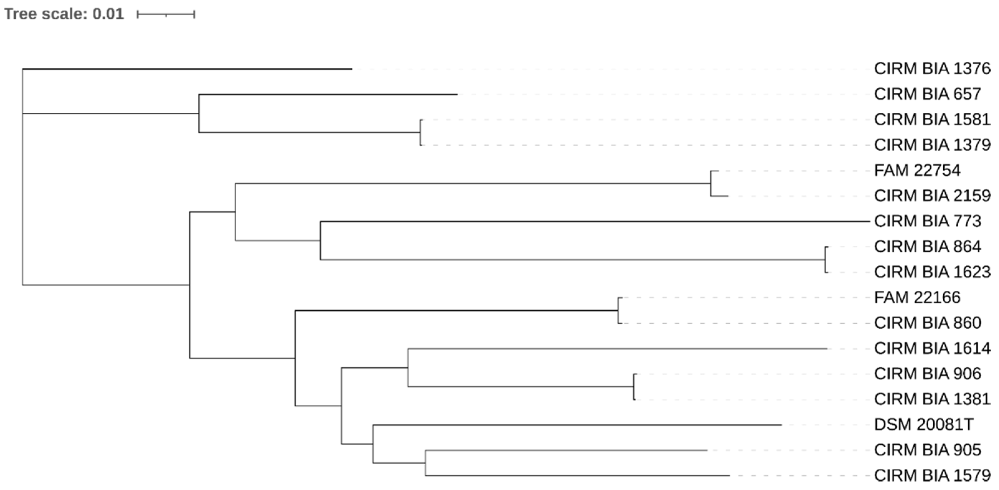
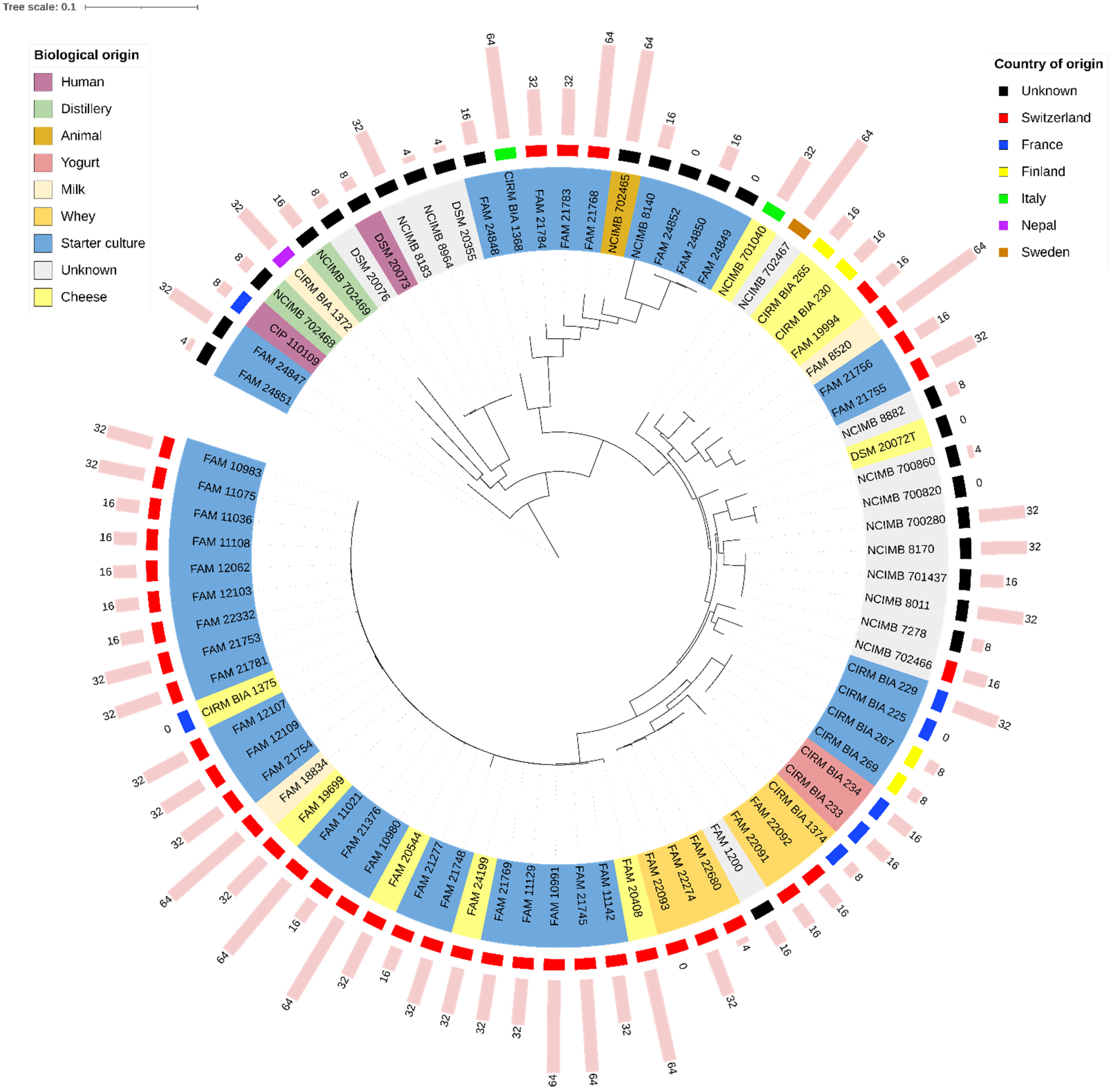
3.2. SNP-Based Phylogenetic Relationships of the Strains

The SNP-based analysis revealed that the LDB strains were all phylogenetically distinct (Figure 1). The closest pairs of strains displayed 34 (CIRM BIA 1379 and CIRM BIA 1581) and 94 (CIRM BIA 864 and CIRM BIA 1623) SNPs. As the origins of these isolates were different, they were considered closely related but different strains.
A more complex pattern was obtained for the LDL strains (Figure 2). Twenty-eight strains formed a homogeneous phylogenetic group. All pairs of strains in this group, however, displayed more than 120 SNPs (median 1067 SNPs). All but one (CIRM BIA 1375) were isolated from Swiss dairy products, mainly from cheese or from undefined mixed starter cultures, mostly obtained from different cheese factories. Several other groups consisted of similar, though different, strains: FAM 1200, FAM 22091, FAM 22092, FAM 22093, FAM 22274, and FAM 22680 displayed more than 368 SNPs. These strains were isolated from Swiss whey (no information available for FAM 1200). NCIMB 700820 and NCIMB 700860 displayed more than 2000 SNPs. Both CIRM BIA 230 and CIRM BIA 265 were isolated from Finnish Emmental cheese in 1968, but they were considered different strains, as they displayed 138 SNPs. CIRM BIA 233 and CIRM BIA 234 were both isolated from a French yogurt factory in 1984, and they displayed 12 SNPs. Given the strong suspicion that they belong to the same strain, CIRM BIA 234 was not included in the antibiotic susceptibility profiles. CIRM BIA 269 displayed 12 SNPs when compared with CIRM BIA 233 and CIRM BIA 234, but it was still regarded as a different strain given its different origin. CIRM BIA 267 had the same origin as CIRM BIA 269, but these isolates displayed more than 150 SNPs. NCIMB 8183, NCIMB 8964, DSM 20355, DSM 20073, and DSM 20076 all displayed more than 100 SNPs. NCIMB 7278 and NCIMB 701437 displayed 36 SNPs. As information about their origin was unavailable, they were considered identical, and only NCIMB 701437 was included in the antibiotic susceptibility profiles. NCIMB 8011 and NCIMB 8170, although considerably closely related to NCIMB 701437, were still considered different strains, as they displayed more than 50 SNPs with respect to the latter strain and between each other. Finally, FAM 24849, FAM 24850, and FAM 24852 all displayed fewer than 12 SNPs. As these strains were isolated from different commercial starter cultures but from the same company, there is a high probability that the same strain was used in all three starter cultures. Therefore, only FAM 24850 was included in the susceptibility profiles.

3.3. ARGs in the Genome Assemblies

The search for ARGs in the strain assemblies revealed a single match, namely strain FAM 22754. In the assembly of this strain, an ermB gene was detected in a small, low coverage, scaffold (763 bp). To confirm/infirm the presence of this gene in the strain FAM 22754, we performed a specific PCR targeting ermB, as described previously [50]. The presence of ermB could neither be confirmed using the DNA extracts used for sequencing, nor using new DNA extracts from the strain, and we concluded that its presence in the assembly was due to a contamination during DNA extraction, library preparation, or sequencing.

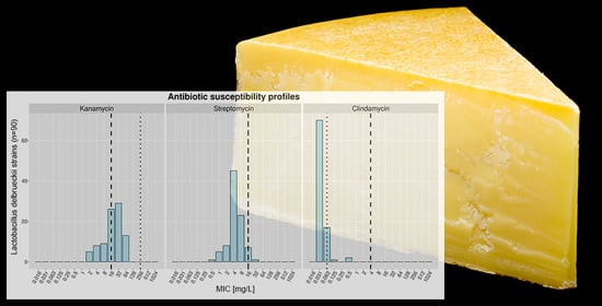
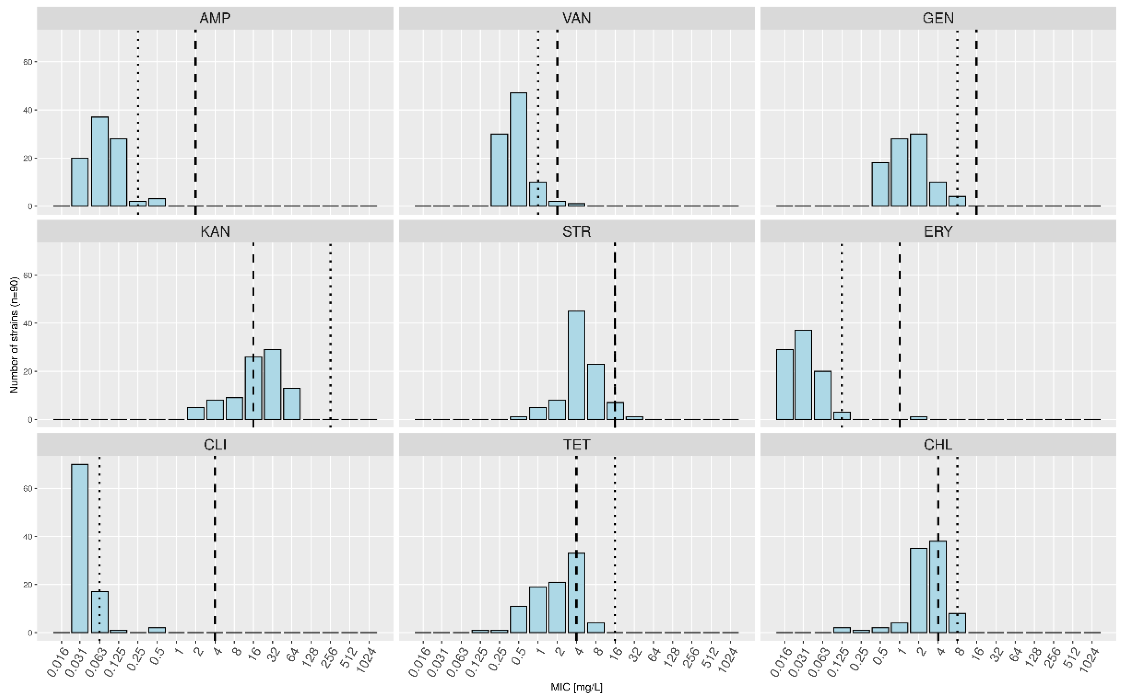
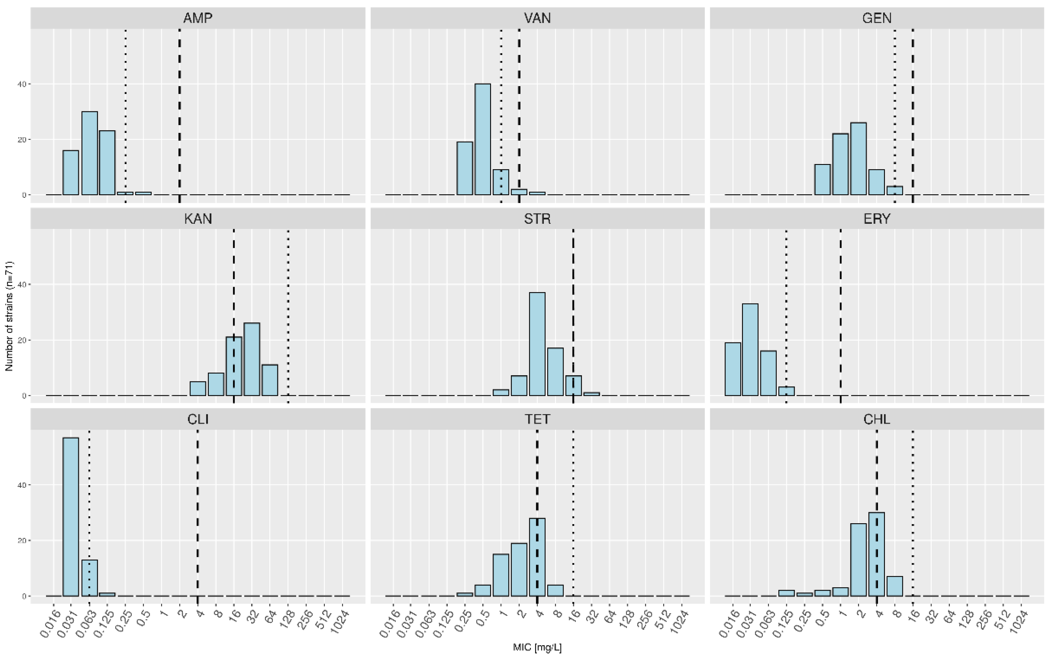
3.4. Antibiotic Susceptibility Profiles and Determination of MCs

Six strains (five LDL and one LDB) did not grow in the test medium (i.e., LSM): FAM 22093, CIRM BIA 225, NCIMB 700820, CIRM BIA 1375, DSM 20072 T and CIRM BIA 1381. Thus, they were excluded from subsequent analyses.
Among the 90 remaining L. delbrueckii strains, 42 (46.7%) displayed MICs higher than the FEEDAP MC for KAN, eight (8.9%) for CHL, four (4.4%) for TET, and one (1.1%) each for VAN, STR, and ERY (Table 3, Figure 3). The MCs for all the investigated subspecies were higher than the FEEDAP MCs for KAN (256 mg/L instead of 16 mg/L), TET (16 mg/L instead of 4 mg/L), and CHL (8 mg/L instead of 4 mg/L).
As for LDL (Table 3, Figure 4), 37 strains out of 71 (52.1%) displayed MICs higher than the FEEDAP MC for KAN, seven (9.9%) for CHL, four (5.6%) for TET, and one (1.4%) each for VAN and STR. The MCs calculated based solely on this subspecies were higher than the FEEDAP MCs for KAN (128 mg/L instead of 16 mg/L), TET (16 mg/L instead of 4 mg/L), and CHL (16 mg/L instead of 4 mg/L). Conversely, MCs were lower for AMP (0.25 mg/L instead of 2 mg/L), GEN (8 mg/L instead of 16 mg/L), ERY (0.125 mg/L instead of 1 mg/L), CLI (0.125 mg/L instead of 4 mg/L), and VAN (1 mg/L instead of 2 mg/L).
No link could be drawn between the levels of resistance to antibiotics and the origins of the strains or their position in the SNP-based phylogenetic tree (Figure 2).

4. Discussion

Used alongside S. salivarius subsp. thermophilus, LDL is the major L. delbrueckii subspecies added as a thermophilic starter in the production of cooked Swiss cheese varieties. The development of starter or adjunct cultures for the cheese market involves a thorough screening of strain characteristics. MIC values determined through broth microdilution testing are commonly used as an easy criterion for the early acceptance or rejection of candidate strains prior to the in-depth characterization of their properties to determine their suitability for cheese production. In the case of LDL, the ABR status of new candidates is often subject to debate, as values regularly exceed the defined FEEDAP MCs, particularly that for KAN (Agroscope, data not shown). The FEEDAP MCs have been defined for the entire group of obligate homofermentative lactobacilli, consisting of at least 31 species [18]. Here, we questioned the relevance of the current FEEDAP MCs for L. delbrueckii, more specifically for LDL.
An unambiguous identification of a strain is a prerequisite for any further safety assessment [17]. Controlling the taxonomic affiliation of our strain assortment led to the re-assignment of one and five strains at the species and subspecies levels, respectively. In addition, during the preliminary selection of strains suitable for this study, six strains in the Agroscope Culture Collection were taxonomically re-assigned at the subspecies level (data not shown). Here, we assessed the taxonomic affiliation at the subspecies level based on the ANI values. According to previous research, strains whose genomes display an ANI of ≥96.5% and an alignment fraction of ≥0.6 can be grouped into a single species [51]. At the subspecies level, however, no similar cutoffs have been defined. We therefore assessed visually the positions of the strains and their similarities in the ANI-based tree relative to different type strains. The subspecies of strains in some culture collections have often been defined based solely on physiological properties, and modern molecular techniques have not been systematically applied. Today, as NGS has become an affordable approach, taxonomic affiliations can be reassessed in the light of whole genome comparisons. Considering the taxonomic assessment performed here and the subsequent re-classifications, we strongly recommend that the taxonomic affiliation of the examined strains, even those deposited in culture collections, be systematically questioned. Comparisons at the genomic level are a good approach; however, more rapid methods that require less computational analysis (e.g., MALDI-TOF) may also provide a good evaluation of certain taxa. In our study, MALDI-TOF allowed for the discrimination of LDL and LDB, but not of LDS, as this subspecies is not yet referenced in the BDAL v11.0 library used here.
Most strains selected for analysis in our study were dairy isolates (obtained from milk, dairy products, natural whey, and other starter cultures) from Switzerland, as most strains were provided by the Agroscope Culture Collection, which includes strains mainly obtained from Swiss dairy products. To determine the MICs of the non-dairy isolates, we broadened the range of origins by including strains obtained from other habitats, such as human saliva, calf stomachs, or distilleries. Moreover, care was taken in expanding the strain selection to include those obtained from heterogeneous dairy products and those with different geographical origins. Furthermore, the selected strains were isolated within a period covering at least 69 years (1950–2019), within which the golden age of antibiotics and the subsequent dissemination of ABR determinants is included [52]. On the one hand, our strain selection allowed to avoid potential habitat-related biases. Although strains in some environments may be more exposed to selective pressure exerted by antibiotics or other antimicrobials, the spread of ABR is less likely to occur between habitats than within a single habitat [53,54]. Therefore, the overall elevated resistance towards KAN observed here is most likely an intrinsic characteristic of L. delbrueckii rather than a resistance that has been transmitted across habitats and time. On the other hand, selecting strains of different origins increases the chance of getting as many different strains as possible.
MCs were calculated using MIC distributions based on the susceptibility profiles of distinct strains. Here, potential duplicate strains were discarded after empirically examining the strains’ relationships in a SNP-based phylogenetic tree and after determining the number of SNPs between pairs of strains. This step is crucial to avoid duplicated MIC results that would lead to biased MIC distributions. The number of SNPs below which two strains were considered identical was defined subjectively based on previous SNPs analyses of different strains (data not shown) and may be adapted. Although the use of SNPs alone to discriminate between different strains is not standard, the threshold defined in this study is relatively consistent with results of other studies investigating clinical isolates [55,56,57]. Furthermore, the defined threshold of 50 SNPs appears reasonable considering the origins of the analyzed strains.
No association between the origins of the strains and their antimicrobial susceptibility patterns could be evidenced, suggesting that no particular antibiotic susceptibilities were selected in various ecological niches. Similarly, no link could be drawn between the year of isolation and the antimicrobial susceptibility patterns, suggesting that the selection pressure exerted by antibiotics used in veterinary medicine and farming towards L. delbrueckii has remained relatively moderate.
In this study, we postulated that the observed phenotypic resistances in LDL are actually false positives due to the inaccurate MCs. Our results tend to confirm our hypothesis. Indeed, although more than half of the tested LDL strains were phenotypically defined as ‘resistant’ to KAN according to the FEEDAP criteria, none of them displayed any known ARG. Similar observations were previously reported, with, e.g., 25% of tested LDB having MICs of KAN above the EFSA MC but no detected ARG [58]. The proportion of L. delbrueckii strains with MICs above the MCs for chloramphenicol (8.9%) in our study is higher than previously reported [58,59]. Conversely, the proportion of TET-resistant strains was lower in our study (4.4%) than in a previous study carried out on 11 L. delbrueckii strains [59]. The range of MICs of TET seems to be relatively broad by L. delbrueckii, as shown here, but also considering results from other studies. Indeed, the proportion of TET-resistant strains found here was lower (4.4%) than in a previous study carried out on 11 L. delbrueckii strains [59] but higher than in another one based on 4 LDL strains from natural whey starters, where no resistance to GEN, ERY, TET, and VAN was detected [60]. In addition to the fact that we detected no ARGs, all MIC distributions of LDL were unimodal, while a bimodal distribution would be expected in case of an acquired antibiotic resistance. On the other hand, the high susceptibilities to AMP, ERY, and CLI observed in most of our investigated strains represent a common feature of Lactobacillus delbrueckii that has already been reported by others [58,59,61]. For these three antibiotics, it rather seems that the MCs have been defined too high, which may open the door to false negative results and consequently the potential introduction of antibiotic resistant strains into the food chain.
Based on our observations, we therefore conclude that the MCs defined for the entire group of homofermentative lactobacilli should be redefined at the species or even at the subspecies level. Our strain selection may be too narrow in order that new MCs could be proposed for the entire species, as we have not tested some of the subspecies for their antimicrobial susceptibilities (LDD, LDI, and LDJ). However, our calculations based on strains of the subspecies bulgaricus, lactis, and sunkii suggest that the MCs for KAN, TET and CHL for L. delbrueckii should be increased, whereas the MCs for AMP, ERY and CLI should be decreased. For LDL, we suggest that the MCs for KAN, TET and CHL be increased to at least 128, 16, and 16 mg/L, respectively. Conversely, the MCs for AMP, ERY and CLI could be reduced to 0.25, 0.125, and 0.063 mg/L, respectively.
Recently, Stefanska et al. detected 32% of KAN-resistant strains in a wider range of LAB species using broth microdilution [62]. Of these strains, only three (4.6%) carried aph(3′′)-IIIa, a gene conferring resistance to this antibiotic. The issue raised in our study and that has already been reported for other species may thus be widespread in other LAB species, too [19,23].
The facts that a non-negligible number of strains were unable to grow in the test medium and that some variations occur in the phenotypic measures support the dual approach proposed by the FEEDAP, that is, phenotypic testing complemented by a search of the WGS for known ARGs [17]. The phenotypic approach may allow for the detection of yet unknown ABR mechanisms and is relatively advantageous in terms of cost and time. It can thus be used as a primary screening approach, along with the use of accurate MCs to avoid false positives that may drastically reduce the number of candidates [19]. Meanwhile, the WGS approach provides an appropriate method to evaluate strains that do not grow on test media and to confirm the resistances observed using the phenotypic test.

Supplementary Materials

The following are available online at https://0-www-mdpi-com.brum.beds.ac.uk/article/10.3390/foods10123145/s1, Figure S1: Average Nucleotide Identities of the Lactobacillus delbrueckii strains.

Author Contributions

Conceptualization: N.S. and H.-P.B.; formal analysis: N.S. and S.O.; writing—original draft preparation: N.S. and H.-P.B.; writing—review and editing: H.-P.B., R.S.S., H.B. and S.O.; supervision: H.-P.B. and H.B.; resources: H.B. and R.S.S.; funding acquisition: H.-P.B. and R.S.S. All authors have read and agreed to the published version of the manuscript.

Funding

This research received no external funding.

Institutional Review Board Statement

Not applicable.

Informed Consent Statement

Not applicable.

Data Availability Statement

Publicly available datasets were analyzed in this study. Genomic data generated in this study have been deposited at GenBank under BioProject PRJNA777018 with GenBank WGS accessions JAJNSH000000000-JAJNVX000000000. Other publicly available genomic data analysed in this study can be found at GenBank under following Genbank WGS accessions: GCA_005864055.1, GCA_005864125.1, GCA_002278095.1, GCA_000056065.1, GCA_001888965.1, GCA_001908495.1, GCA_001888965.1, GCA_001908415.1, GCA_001888925.1, and GCA_009789085.1.

Acknowledgments

We thank the Next Generation Sequencing Platform, University of Bern, for performing the high-throughput sequencing experiments and the Culture Collection of Switzerland (CCOS) for the antibiotic susceptibility testing experiments. We also thank Daniel Marzohl and Pascal Bätscher for their valuable technical help.

Conflicts of Interest

The authors declare no conflict of interest. The funders had no role in designing this study; in the collection, analyses, or interpretation of data; in manuscript writing; or in the decision to publish the results.

References

  1. Bourdichon, F.; Alper, I.; Bibiloni, R.; Dubois, A.; Laulund, S.; Miks, M.; Morelli, L.; Yao, S. Inventory of Microbial Food Cultures with Safety Demonstration in Fermented Food Products; Update of the Bulletin of the IDF 455–2012; Bulletin of the International Dairy Federation: Brussels, Belgium, 2018; Volume 495, pp. 1–74. [Google Scholar]
  2. El Kafsi, H.; Binesse, J.; Loux, V.; Buratti, J.; Boudebbouze, S.; Dervyn, R.; Kennedy, S.; Galleron, N.; Quinquis, B.; Batto, J.-M.; et al. Lactobacillus delbrueckii ssp. lactis and ssp. bulgaricus: A chronicle of evolution in action. BMC Genom. 2014, 15, 1–12. [Google Scholar] [CrossRef] [Green Version]
  3. Sieuwerts, S.; de Bok, F.A.M.; Hugenholtz, J.; van Hylckama Vlieg, J.E.T. Unraveling Microbial Interactions in Food Fermentations: From Classical to Genomics Approaches. Appl. Environ. Microbiol. 2008, 74, 4997–5007. [Google Scholar] [CrossRef] [PubMed] [Green Version]
  4. Somerville, V.; Berthoud, H.; Schmidt, R.S.; Bachmann, H.-P.; Meng, Y.H.; Fuchsmann, P.; von Ah, U.; Engel, P. Functional strain redundancy and persistent phage infection in Swiss hard cheese starter cultures. ISME J. 2021, 1–12. [Google Scholar] [CrossRef]
  5. Schmid, M.; Muri, J.; Melidis, D.; Varadarajan, A.R.; Somerville, V.; Wicki, A.; Moser, A.; Bourqui, M.; Wenzel, C.; Eugster-Meier, E.; et al. Comparative Genomics of Completely Sequenced Lactobacillus helveticus Genomes Provides Insights into Strain-Specific Genes and Resolves Metagenomics Data Down to the Strain Level. Front. Microbiol. 2018, 9, 63. [Google Scholar] [CrossRef]
  6. Gobbetti, M.; Di Cagno, R.; Calasso, M.; Neviani, E.; Fox, P.F.; De Angelis, M. Drivers that establish and assembly the lactic acid bacteria biota in cheeses. Trends Food Sci. Technol. 2018, 78, 244–254. [Google Scholar] [CrossRef]
  7. Ghosh, T.; Beniwal, A.; Semwal, A.; Navani, N.K. Mechanistic insights into probiotic properties of lactic acid bacteria associated with ethnic fermented dairy products. Front. Microbiol. 2019, 10, 1–19. [Google Scholar] [CrossRef] [Green Version]
  8. Liu, M.; Bayjanov, J.R.; Renckens, B.; Nauta, A.; Siezen, R.J. The proteolytic system of lactic acid bacteria revisited: A genomic comparison. BMC Genom. 2010, 11, 36. [Google Scholar] [CrossRef] [PubMed] [Green Version]
  9. Parente, E.; Cogan, T.M.; Powell, I. Starter cultures: General aspects, chemistry, physics and microbiology. In Cheese, 4th ed.; McSweeney, P.L.H., Fox, P.F., Cotter, P.D., Everett, D.W., Eds.; Academic Press: London, UK, 2017; pp. 201–226. [Google Scholar] [CrossRef]
  10. European Food Safety Authority Panel on Biological Hazards (BIOHAZ); Koutsoumanis, K.; Allende, A.; Alvarez-Ordóñez, A.; Bolton, D.; Bover-Cid, S.; Chemaly, M.; Davies, R.; De Cesare, A.; Hilbert, F.; et al. Update of the list of QPS-recommended biological agents intentionally added to food or feed as notified to EFSA 14: Suitability of taxonomic units notified to EFSA until March 2021. EFSA J. 2021, 19, e06689. [Google Scholar] [CrossRef] [PubMed]
  11. European Food Safety Authority Panel on Biological Hazards (BIOHAZ); Andreoletti, O.; Budka, H.; Buncic, S.; Colin, P.; Collins, J.D.; De Koeijer, A.; Griffin, J.; Havelaar, A.; Hope, J.; et al. The maintenance of the list of QPS microorganisms intentionally added to food or feed—scientific opinion of the Panel on Biological Hazards. EFSA J. 2008, 6, 923. [Google Scholar] [CrossRef]
  12. Ben, Y.; Fu, C.; Hu, M.; Liu, L.; Wong, M.H.; Zheng, C. Human health risk assessment of antibiotic resistance associated with antibiotic residues in the environment: A review. Environ. Res. 2018, 169, 483–493. [Google Scholar] [CrossRef] [PubMed]
  13. Huddleston, J.R. Horizontal gene transfer in the human gastrointestinal tract: Potential spread of antibiotic resistance genes. Infect. Drug Resist. 2014, 7, 167–176. [Google Scholar] [CrossRef] [Green Version]
  14. Lerner, A.; Matthias, T.; Aminov, R. Potential Effects of Horizontal Gene Exchange in the Human Gut. Front. Immunol. 2017, 8, 1630. [Google Scholar] [CrossRef] [Green Version]
  15. Thumu, S.C.R.; Halami, P.M. Conjugal transfer of erm(B) and multiple tet genes from Lactobacillus spp. to bacterial pathogens in animal gut, in vitro and during food fermentation. Food Res. Int. 2018, 116, 1066–1075. [Google Scholar] [CrossRef]
  16. Herman, L.; Chemaly, M.; Cocconcelli, P.S.; Fernandez, P.; Klein, G.; Peixe, L.; Prieto, M.; Querol, A.; Suarez, J.E.; Sundh, I.; et al. The qualified presumption of safety assessment and its role in EFSA risk evaluations: 15 years past. FEMS Microbiol. Lett. 2018, 366. [Google Scholar] [CrossRef]
  17. European Food Safety Authority Panel on Additives and Products or Substances used in Animal Feed (FEEDAP); Rychen, G.; Aquilina, G.; Azimonti, G.; Bampidis, V.; Bastos, M.d.L.; Bories, G.; Chesson, A.; Cocconcelli, P.S.; Flachowsky, G.; et al. Guidance on the characterisation of microorganisms used as feed additives or as production organisms. EFSA J. 2018, 16, 5206. [Google Scholar]
  18. Hammes, W.P.; Hertel, C. Lactobacillus. In Bergey’s Manual of Systematics of Archaea and Bacteria; Whitman, W.B., DeVos, P., Dedysh, S., Hedlund, B., Kämpfer, P., Rainey, F., Trujillo, M.E., Bowman, J.P., Brown, D.R., Glöckner, F.O., et al., Eds.; John Wiley & Sons, Inc.: Hoboken, NJ, USA, Bergey’s Manual Trust: New York, NY, USA, 2015; pp. 1–76. [Google Scholar]
  19. Shani, N.; Oberhaensli, S.; Arias-Roth, E. Antibiotic Susceptibility Profiles of Pediococcus pentosaceus from Various Origins and Their Implications for the Safety Assessment of Strains with Food-Technology Applications. J. Food Prot. 2020, 84, 1160–1168. [Google Scholar] [CrossRef] [PubMed]
  20. Ashaolu, T.J.; Reale, A. A Holistic Review on Euro-Asian Lactic Acid Bacteria Fermented Cereals and Vegetables. Microorganisms 2020, 8, 1176. [Google Scholar] [CrossRef]
  21. Tamang, J.P.; Watanabe, K.; Holzapfel, W.H. Review: Diversity of Microorganisms in Global Fermented Foods and Beverages. Front. Microbiol. 2016, 7, 377. [Google Scholar] [CrossRef] [Green Version]
  22. De Man, J.C.; Rogosa, M.; Sharpe, M.E. A Medium for the Cultivation of Lactobacilli. J. Appl. Bacteriol. 1960, 23, 130–135. [Google Scholar] [CrossRef]
  23. Lüdin, P.; Roetschi, A.; Wüthrich, D.; Bruggmann, R.; Berthoud, H.; Shani, N. Update on Tetracycline Susceptibility of Pediococcus acidilactici Based on Strains Isolated from Swiss Cheese and Whey. J. Food Prot. 2018, 81, 1582–1589. [Google Scholar] [CrossRef]
  24. Dreier, M.; Berthoud, H.; Shani, N.; Wechsler, D.; Junier, P. SpeciesPrimer: A bioinformatics pipeline dedicated to the design of qPCR primers for the quantification of bacterial species. PeerJ 2020, 8, e8544. [Google Scholar] [CrossRef] [Green Version]
  25. Low, A.J.; Koziol, A.G.; Manninger, P.A.; Blais, B.; Carrillo, C.D. ConFindr: Rapid detection of intraspecies and cross-species contamination in bacterial whole-genome sequence data. PeerJ 2019, 7, e6995. [Google Scholar] [CrossRef]
  26. Andrews, S. FastQC: A Quality Control Tool for High Throughput Sequence Data. 2010. Available online: http://www.bioinformatics.babraham.ac.uk/projects/fastqc/ (accessed on 1 June 2021).
  27. Chen, S.; Zhou, Y.; Chen, Y.; Gu, J. fastp: An ultra-fast all-in-one FASTQ preprocessor. Bioinformatics 2018, 34, i884–i890. [Google Scholar] [CrossRef] [PubMed]
  28. Bankevich, A.; Nurk, S.; Antipov, D.; Gurevich, A.A.; Dvorkin, M.; Kulikov, A.S.; Lesin, V.M.; Nikolenko, S.I.; Pham, S.; Prjibelski, A.D.; et al. SPAdes: A new genome assembly algorithm and its applications to single-cell sequencing. J. Comput. Biol. 2012, 19, 455–477. [Google Scholar] [CrossRef] [PubMed] [Green Version]
  29. Gurevich, A.; Saveliev, V.; Vyahhi, N.; Tesler, G. QUAST: Quality assessment tool for genome assemblies. Bioinformatics 2013, 29, 1072–1075. [Google Scholar] [CrossRef] [PubMed]
  30. Seppey, M.; Manni, M.; Zdobnov, E.M. BUSCO: Assessing Genome Assembly and Annotation Completeness. In Gene Prediction; Kollmar, M., Ed.; Methods in Molecular Biology; Humana: New York, NY, USA, 2019; Volume 1962, pp. 227–245. [Google Scholar]
  31. Jain, C.; Rodriguez-R, L.M.; Phillippy, A.M.; Konstantinidis, K.T.; Aluru, S. High throughput ANI analysis of 90K prokaryotic genomes reveals clear species boundaries. Nat. Commun. 2018, 9, 5114. [Google Scholar] [CrossRef] [Green Version]
  32. Warnes, G.R.; Bolker, B.; Bonebakker, L.; Gentleman, R.; Huber, W.; Liaw, A.; Lumley, T.; Maechler, M.; Magnusson, A.; Moeller, S.; et al. gplots: Various R Programming Tools for Plotting Data. Available online: https://CRAN.R-project.org/package=gplots (accessed on 12 October 2021).
  33. R StudioTeam. RStudio: Integrated Development for R; RStudio, PBC: Boston, MA, USA, 2020; Available online: http://www.rstudio.com/ (accessed on 12 October 2021).
  34. R Core Team. R: A Language and Environment for Statistical Computing; R Foundation for Statistical Computing: Vienna, Austria, 2020; Available online: https://www.R-project.org/ (accessed on 12 October 2021).
  35. Shakya, M.; Ahmed, S.A.; Davenport, K.W.; Flynn, M.C.; Lo, C.-C.; Chain, P.S.G. Standardized phylogenetic and molecular evolutionary analysis applied to species across the microbial tree of life. Sci. Rep. 2020, 10, 1–15. [Google Scholar] [CrossRef]
  36. Minh, B.Q.; Schmidt, H.A.; Chernomor, O.; Schrempf, D.; Woodhams, M.D.; von Haeseler, A.; Lanfear, R. IQ-TREE 2: New Models and Efficient Methods for Phylogenetic Inference in the Genomic Era. Mol. Biol. Evol. 2020, 37, 1530–1534. [Google Scholar] [CrossRef] [Green Version]
  37. Letunic, I.; Bork, P. Interactive Tree of Life (iTOL) v5: An online tool for phylogenetic tree display and annotation. Nucleic Acids Res. 2021, 49, W293–W296. [Google Scholar] [CrossRef] [PubMed]
  38. Dijkshoorn, L.; Ursing, B.; Ursing, J. Strain, clone and species: Comments on three basic concepts of bacteriology. J. Med Microbiol. 2000, 49, 397–401. [Google Scholar] [CrossRef] [PubMed] [Green Version]
  39. Seemann, T. Abricate. 2020. Available online: https://github.com/tseemann/abricate (accessed on 25 February 2020).
  40. Feldgarden, M.; Brover, V.; Haft, D.H.; Prasad, A.B.; Slotta, D.J.; Tolstoy, I.; Tyson, G.H.; Zhao, S.; Hsu, C.-H.; McDermott, P.F.; et al. Validating the AMRFinder Tool and Resistance Gene Database by Using Antimicrobial Resistance Genotype-Phenotype Correlations in a Collection of Isolates. Antimicrob. Agents Chemother. 2019, 63. [Google Scholar] [CrossRef] [Green Version]
  41. Jia, B.; Raphenya, A.R.; Alcock, B.; Waglechner, N.; Guo, P.; Tsang, K.K.; Lago, B.A.; Dave, B.M.; Pereira, S.; Sharma, A.N.; et al. CARD 2017: Expansion and model-centric curation of the comprehensive antibiotic resistance database. Nucleic Acids Res. 2017, 45, D566–D573. [Google Scholar] [CrossRef]
  42. McArthur, A.G.; Waglechner, N.; Nizam, F.; Yan, A.; Azad, M.A.; Baylay, A.J.; Bhullar, K.; Canova, M.J.; De Pascale, G.; Ejim, L.; et al. The Comprehensive Antibiotic Resistance Database. Antimicrob. Agents Chemother. 2013, 57, 3348–3357. [Google Scholar] [CrossRef] [Green Version]
  43. Gupta, S.; Padmanabhan, B.R.; Diene, S.M.; Lopez-Rojas, R.; Kempf, M.; Landraud, L.; Rolain, J.-M. ARG-ANNOT, a New Bioinformatic Tool to Discover Antibiotic Resistance Genes in Bacterial Genomes. Antimicrob. Agents Chemother. 2013, 58, 212–220. [Google Scholar] [CrossRef] [Green Version]
  44. Zankari, E.; Hasman, H.; Cosentino, S.; Vestergaard, M.; Rasmussen, S.; Lund, O.; Aarestrup, F.M.; Larsen, M.V. Identification of acquired antimicrobial resistance genes. J. Antimicrob. Chemother. 2012, 67, 2640–2644. [Google Scholar] [CrossRef]
  45. The European Committee on Antimicrobial Susceptibility Testing. Breakpoint Tables for Interpretation of MICs and Zone Diameters. Version 11.0. 2021. Available online: http://www.eucast.org (accessed on 28 July 2021).
  46. International Organization for Standardization (ISO). Milk and Milk Products—Determination of the Minimal Inhibitory Concentration (MIC) of Antibiotics Applicable to Bifidobacteria and Non-Enterococcal Lactic Acid Bacteria (LAB); ISO 10932|IDF 223:2010; International Organization for Standardization: Geneva, Switzerland, 2010. [Google Scholar]
  47. Klare, I.; Konstabel, C.; Müller-Bertling, S.; Reissbrodt, R.; Huys, G.; Vancanneyt, M.; Swings, J.; Goossens, H.; Witte, W. Evaluation of New Broth Media for Microdilution Antibiotic Susceptibility Testing of Lactobacilli, Pediococci, Lactococci, and Bifidobacteria. Appl. Environ. Microbiol. 2005, 71. [Google Scholar] [CrossRef] [PubMed] [Green Version]
  48. Turnidge, J.; Kahlmeter, G.; Kronvall, G. Statistical characterisation of bacterial wild-type MIC value distributions and the determination of epidemiological cut-off values. Clin. Microbiol. Infect. 2006, 12, 418–425. [Google Scholar] [CrossRef] [PubMed]
  49. Wickham, H. ggplot2: Elegant Graphics for Data Analysis, 2nd ed.; Springer International Publishing: New York, NY, USA, 2016. [Google Scholar]
  50. Böckelmann, U.; Dörries, H.-H.; Ayuso-Gabella, M.N.; de Marçay, M.S.; Tandoi, V.; Levantesi, C.; Masciopinto, C.; Van Houtte, E.; Szewzyk, U.; Wintgens, T.; et al. Quantitative PCR Monitoring of Antibiotic Resistance Genes and Bacterial Pathogens in Three European Artificial Groundwater Recharge Systems. Appl. Environ. Microbiol. 2009, 75, 154–163. [Google Scholar] [CrossRef] [PubMed] [Green Version]
  51. Varghese, N.J.; Mukherjee, S.; Ivanova, N.; Konstantinidis, K.T.; Mavrommatis, K.; Kyrpides, N.C.; Pati, A. Microbial species delineation using whole genome sequences. Nucleic Acids Res. 2015, 43, 6761–6771. [Google Scholar] [CrossRef]
  52. Davies, J.; Davies, D. Origins and Evolution of Antibiotic Resistance. Microbiol. Mol. Biol. Rev. 2010, 74, 417–433. [Google Scholar] [CrossRef] [PubMed] [Green Version]
  53. Martinez, J.L.; Coque, T.M.; Baquero, F. What is a resistance gene? Ranking risk in resistomes. Nat. Rev. Genet. 2014, 13, 116–123. [Google Scholar] [CrossRef]
  54. Pal, C.; Bengtsson-Palme, J.; Kristiansson, E.; Larsson, D.G.J. The structure and diversity of human, animal and environmental resistomes. Microbiome 2016, 4, 54. [Google Scholar] [CrossRef] [PubMed] [Green Version]
  55. Ksibi, B.; Ktari, S.; Othman, H.; Ghedira, K.; Maalej, S.; Mnif, B.; Abbassi, M.S.; Fabre, L.; Rhimi, F.; Le Hello, S.; et al. Comparison of conventional molecular and whole-genome sequencing methods for subtyping Salmonella enterica serovar Enteritidis strains from Tunisia. Eur. J. Clin. Microbiol. Infect. Dis. 2020, 40, 597–606. [Google Scholar] [CrossRef]
  56. Schürch, A.; Arredondo-Alonso, S.; Willems, R.; Goering, R. Whole genome sequencing options for bacterial strain typing and epidemiologic analysis based on single nucleotide polymorphism versus gene-by-gene–based approaches. Clin. Microbiol. Infect. 2018, 24, 350–354. [Google Scholar] [CrossRef] [Green Version]
  57. Taylor, A.J.; Lappi, V.; Wolfgang, W.J.; Lapierre, P.; Palumbo, M.J.; Medus, C.; Boxrud, D. Characterization of Foodborne Outbreaks of Salmonella enterica Serovar Enteritidis with Whole-Genome Sequencing Single Nucleotide Polymorphism-Based Analysis for Surveillance and Outbreak Detection. J. Clin. Microbiol. 2015, 53, 3334–3340. [Google Scholar] [CrossRef] [PubMed] [Green Version]
  58. Guo, H.; Zhang, W.; Kwok, L.-Y.; Menghe, B. In vitro evalution of antibiotic resistance of Lactobacillus bulgaricus strains isolated from traditional dairy products. Czech J. Food Sci. 2019, 37, 36–43. [Google Scholar] [CrossRef]
  59. Cho, G.-S.; Cappello, C.; Schrader, K.; Fagbemigum, O.; Oguntoyinbo, F.A.; Csovcsics, C.; Sch, N.R.; Kabisch, J.; Neve, H.; Bockelmann, W.; et al. Isolation and Characterization of Lactic Acid Bacteria from Fermented Goat Milk in Tajikistan. J. Microbiol. Biotechnol. 2018, 28, 1834–1845. [Google Scholar] [CrossRef]
  60. Rossetti, L.; Carminati, D.; Zago, M.; Giraffa, G. A Qualified Presumption of Safety approach for the safety assessment of Grana Padano whey starters. Int. J. Food Microbiol. 2009, 130, 70–73. [Google Scholar] [CrossRef]
  61. D’Aimmo, M.R.; Modesto, M.; Biavati, B. Antibiotic resistance of lactic acid bacteria and Bifidobacterium spp. isolated from dairy and pharmaceutical products. Int. J. Food Microbiol. 2007, 115, 35–42. [Google Scholar] [CrossRef]
  62. Stefańska, I.; Kwiecień, E.; Jóźwiak-Piasecka, K.; Garbowska, M.; Binek, M.; Rzewuska, M. Antimicrobial susceptibility of lactic acid bacteria strains of potential use as feed additives—The basic safety and usefulness criterion. Front. Vet. Sci. 2021, 9, 687071. [Google Scholar] [CrossRef]
Figure 1. SNP-based phylogenetic tree of Lactobacillus delbrueckii subsp. bulgaricus strains assessed in this study. The scale shows the rate of single nucleotide polymorphisms.
Foods 10 03145 g001
Figure 2. SNP-based phylogenetic tree of the Lactobacillus delbrueckii subsp. lactis strains assessed in this study. Country and biological origin, as well as the defined minimal inhibitory concentration of kanamycin (MICs, pink bars, along with their numerical values expressed in mg/L) are displayed. For strains that did not grow in the test medium or that have not been tested, MIC of kanamycin is indicated with ‘0′. The scale shows the rate of single nucleotide polymorphisms.
Foods 10 03145 g002
Figure 3. Combined minimal inhibitory concentration (MIC) distributions for Lactobacillus delbrueckii subsp. lactis and L. delbrueckii subsp. bulgaricus. The dashed lines indicate the current microbiological cutoffs (MCs) as defined by the European Food Safety Authority, and the dotted lines represent the calculated MCs based on the MICs measured in this study.
Foods 10 03145 g003
Figure 4. Minimal inhibitory concentration (MIC) distributions for Lactobacillus delbrueckii subsp. lactis only. The dashed lines indicate the current microbiological cutoffs (MCs) as defined by the European Food Safety Authority, and the dotted lines represent the calculated MCs based on the MICs measured in this study.
Foods 10 03145 g004
Table 2. Bacterial strains and their taxonomic affiliation, origin, and year of isolation.
No.Subspecies bStrain IDCulture Collection aOriginYear of IsolationOriginal Depositor; Strain DesignationGenBank Assembly Accessions d
1LDLFAM 1200ACCUnknownUnknownFAM 1200GCA_021135575.1
2LDLFAM 8520ACCSwiss milkUnknownJimeno, J.; FM 1GCA_021135595.1
3LDLFAM 10980ACCSwiss undefined mixed starter culture1979Isolini, D.; Lb 101.07GCA_021135555.1
4LDLFAM 10983ACCSwiss undefined mixed starter culture1984Isolini, D.; Lb 101.10GCA_021135485.1
5LDLFAM 10991ACCSwiss undefined mixed starter culture1980Isolini, D.; Lb 104.80GCA_021135515.1
6LDLFAM 11021ACCSwiss undefined mixed starter culture1982Isolini, D.; Lb 115.53GCA_021135535.1
7LDLFAM 11036ACCSwiss undefined mixed starter culture1979Isolini, D.; Lb 119.17GCA_021135475.1
8LDLFAM 11075ACCSwiss undefined mixed starter culture1978Isolini, D.; Lb 150.14GCA_021135455.1
9LDLFAM 11108ACCSwiss undefined mixed starter culture1983Isolini, D.; Lb 157.02GCA_021135435.1
10LDLFAM 11129ACCSwiss undefined mixed starter culture1983Isolini, D.; Lb 164.35GCA_021135405.1
11LDLFAM 11142ACCSwiss undefined mixed starter culture1988Isolini, D.; Lb 202.02GCA_021135395.1
12LDLFAM 12062ACCSwiss undefined mixed starter culture1983Isolini, D.; Lb 302.01GCA_021135375.1
13LDLFAM 12103ACCSwiss undefined mixed starter culture1986Isolini, D.; Lb 325.13GCA_021135355.1
14LDLFAM 12107ACCSwiss undefined mixed starter culture1988Isolini, D.; Lb 202.07GCA_021135335.1
15LDLFAM 12109ACCSwiss undefined mixed starter culture1988Isolini, D.; Lb 202.09GCA_021135275.1
16LDLFAM 18834ACCSwiss milk2005Unknown; C1GCA_021135295.1
17LDLFAM 19699ACCSwiss Emmental cheese1989–1990Isolini, D.; 23.10GCA_021135315.1
18LDLFAM 19994ACCSwiss Emmental cheese1989–1990Isolini, D.; 43.13GCA_021135255.1
19LDLFAM 20408ACCSwiss hard cheese1989–1990Isolini, D.; LDELA 871-33GCA_021135235.1
20LDLFAM 20544ACCSwiss hard cheese1989–1990Isolini, D.; LDEBU 927-84GCA_021135205.1
21LDLFAM 21277ACCSwiss undefined mixed starter cultureUnknownMeyer, J.; 101/100GCA_005864055.1
22LDLFAM 21376ACCSwiss undefined mixed starter cultureUnknownMeyer, J.; 169/126GCA_021135175.1
23LDLFAM 21745ACCSwiss undefined mixed starter culture1979Isolini, D.; Lb 101.01GCA_021135195.1
24LDLFAM 21748ACCSwiss undefined mixed starter culture1979Isolini, D.; Lb 101.56GCA_021135155.1
25LDLFAM 21753ACCSwiss undefined mixed starter culture1981Isolini, D.; Lb 124.49GCA_021135095.1
26LDLFAM 21754ACCSwiss undefined mixed starter culture1982Isolini, D.; Lb 153.48GCA_021135135.1
27LDLFAM 21755ACCSwiss undefined mixed starter culture1982Isolini, D.; Lb 153.08GCA_021135105.1
28LDLFAM 21756ACCSwiss undefined mixed starter culture1982Isolini, D.; Lb 153.09GCA_021135075.1
29LDLFAM 21768ACCSwiss undefined mixed starter culture1982Isolini, D.; Lb 153.20GCA_021135055.1
30LDLFAM 21769ACCSwiss undefined mixed starter culture1978Isolini, D.; Lb 150.10GCA_021135025.1
31LDLFAM 21781ACCSwiss undefined mixed starter culture1981Isolini, D.; Lb 124.09GCA_021135015.1
32LDLFAM 21783ACCSwiss undefined mixed starter culture1982Isolini, D.; Lb 153.27GCA_021134955.1
33LDLFAM 21784ACCSwiss undefined mixed starter culture1982Isolini, D.; Lb 153.28GCA_005864125.1
34LDLFAM 22091ACCSwiss natural whey culture1973A50.2GCA_021134995.1
35LDLFAM 22092ACCSwiss natural whey culture1977A56.1GCA_021134975.1
36LDLcFAM 22093ACCSwiss natural whey culture1977A66.5GCA_021134935.1
37LDLFAM 22274ACCSwiss natural whey culture1968A44.2GCA_021134915.1
38LDLFAM 22332ACCSwiss undefined mixed starter cultureUnknownWeishaupt, C.; 313.1GCA_021134885.1
39LDLFAM 22680ACCSwiss natural whey culture1967A77.5GCA_021134855.1
40LDLFAM 24199ACCSwiss Tomme cheese2017Shani, N.; 55/8GCA_021134875.1
41LDS (LDL)CIP 101810CIPUnknownUnknownUnknownGCA_021134835.1
42LDLCIP 110109CIPHuman urine, France1976Vandekerkove; 103-76GCA_021134765.1
43LDL cCIRM BIA 225CIRMArtisanal lactic starter (for Gruyère de Comté cheese making), Franche-Comté, France1963Accolas, J.P.; P12GCA_021134795.1
44LDLCIRM BIA 229CIRMArtisanal lactic starter (for Gruyère de Comté cheese making), Franche-Comté, France1964Accolas, J.P.; H5aGCA_021134815.1
45LDLCIRM BIA 230CIRMCheese (Emmental), Finland1968CNRZ331GCA_021134745.1
46LDLCIRM BIA 233CIRMProbably a French yoghurt factory1984Cluzel, P.J.; LT4-G2NGCA_021134735.1
47LDLCIRM BIA 234CIRMProbably a French yoghurt factory1984Cluzel, P.J.; LT4-G18GCA_021134695.1
48LDLCIRM BIA 265CIRMCheese (Emmental), Finland1968CNRZ330GCA_021134675.1
49LDS (LDL)CIRM BIA 266CIRMFermented milk (kefir), Russia1971Accolas, J.P.; KFA1GCA_021134645.1
50LDLCIRM BIA 267CIRMLactic starter (for Emmental cheese making), Finland1974Tybeck, E., LKT; VALIOGCA_021134615.1
51LDLCIRM BIA 269CIRMArtisanal lactic starter (for Emmental cheese making), FinlandUnknownTybeck, E.; ISL 19GCA_021134635.1
52LDLCIRM BIA 1368CIRMStarter (for Grana Padano cheese making), Piedmont, Italy1988IMPC AlGCA_021134595.1
53LDLCIRM BIA 1372CIRMYak milk, Nepal1996Quenee, P.; Np 5tGCA_021134505.1
54LDLCIRM BIA 1374CIRMWhey (from Comté), Franche-Comté, France1993CML10GCA_021134545.1
55LDLcDSM 20072 TDSMZEmmental cheese (country of origin unknown)Before 22.08.1990Snog-Kjaer, A. (Orla-Jensen, S., Thermobacterium lactis No. 10)GCA_002278095.1
56LDLDSM 20073DSMZSaliva (country of origin unknown)Before 22.08.1990Williams, N.; 14-1GCA_021134535.1
57LDLDSM 20076DSMZUnknownBefore 22.08.1990Fred, E.B.; F 59GCA_021134495.1
58LDLDSM 20355DSMZUnknownBefore 22.08.1990McCoy; Ld 5GCA_021134475.1
59LDLNCIMB 7278NCIMBUnknownBefore 01.01.1950Dorner, W.; 39 E/KGCA_021134455.1
60LDLNCIMB 8011NCIMBUnknownBefore 07.02.1950DornerGCA_021134375.1
61LDLNCIMB 8140NCIMB“Ga” starter cultureBefore 01.09.1956“““Ga”””GCA_021134435.1
62LDLNCIMB 8170NCIMBUnknownBefore 1999Merck & Co., Inc.; MB 367GCA_021134415.1
63LDLNCIMB 8183NCIMBUnknownBefore 30.11.1950McCoy, E.; 326GCA_021134395.1
64LDLNCIMB 8882NCIMBUnknownBefore 01.07.1957Winkler, K.C.; 1175GCA_021134335.1
65LDLNCIMB 8964NCIMBUnknownBefore 01.12.1958Galloway & Barton-WrightGCA_021134315.1
66LDLNCIMB 700280NCIMBUnknownBefore 01.01.1954244GCA_021134355.1
67LDLcNCIMB 700820NCIMBUnknownBefore 01.01.1954C808/5GCA_021134295.1
68LDLNCIMB 700860NCIMBUnknownBefore 01.01.195618/40GCA_021134275.1
69LDLNCIMB 701040NCIMBItalian hard cheeseBefore 01.01.1957C14/8GCA_021134235.1
70LDLNCIMB 701437NCIMBUnknownUnknownSnog-Kjaer, A.; LI 1GCA_021134215.1
71LDLNCIMB 702465NCIMBDried calve stomachs for Gruyere cheeseUnknownL24, C57GCA_021134255.1
72LDLNCIMB 702466NCIMBSwitzerlandUnknownRitter, P.; L26, H14, 1304GCA_021134175.1
73LDLNCIMB 702467NCIMBSwedenBefore 01.01.1981Swartling, P.; L27, L39 (WL39)GCA_021134185.1
74LDL (LDS)NCIMB 702468NCIMBDistilleryBefore 01.01.1961Sharpe, M.E.; M2/2GCA_021134155.1
75LDL (LDD)NCIMB 702469NCIMBDistilleryBefore 01.01.1961Sharpe, M.E.; LE8, M2/3GCA_021134135.1
76LDLFAM 24847NCCommercial cheese starter culture2019naGCA_021134095.1
77LDLFAM 24848NCCommercial cheese starter culture2019naGCA_021134115.1
78LDLFAM 24849NCCommercial cheese starter culture2019naGCA_021134075.1
79LDLFAM 24850NCCommercial cheese starter culture2019naGCA_021134035.1
80LDLFAM 24851NCCommercial cheese starter culture2019naGCA_021134055.1
81LDLFAM 24852NCCommercial cheese starter culture2019naGCA_021134005.1
82LDBFAM 22166ACCSwiss natural whey culture1968A135.3GCA_021133995.1
83LDBFAM 22754ACCYoghurt1952A171.1GCA_021133975.1
84LDBCIRM BIA 657CIRMFermented milk, Crete, Greece1987Zourari, A.; ZL071B1GCA_021133935.1
85LDBCIRM BIA 773CIRMLactic starter (for yoghurt making), Île-de-France, France1963Chevalier, R.; LT1GCA_021133955.1
86LDBCIRM BIA 860CIRMLactic starter (for yoghurt making), Rhône-Alpes, France1971Accolas, J.P.; LAY 1GCA_021133915.1
87LDBCIRM BIA 864CIRMFermented milk, FranceUnknownIL1609GCA_021133885.1
88Lactobacillus johnsonii (LDB)CIRM BIA 879CIRMTarag (yoghurt), Mongolia1974Accolas, J.P.na
89LDBCIRM BIA 905CIRMCheese (Boulettes d’Avesne), Nord-Pas-de-Calais, France1998Quenee, P.; AV2GCA_021133875.1
90LDBCIRM BIA 906CIRMYak milk, Nepal1996Quenee, P.; NP 2TGCA_021133835.1
91LDL (LDB) cCIRM BIA 1375CIRMCheese (Morbier, raw milk), Jura, France199310F10GCA_021133855.1
92LDBCIRM BIA 1376CIRMLactic starter (for yoghurt making), Bulgaria1978Bouillanne, C. 5GCA_021133815.1
93LDBCIRM BIA 1379CIRMYoghurt (ewe milk), Crete, Greece1987Zourari, A.; ZL023A1GCA_021133775.1
94LDBcCIRM BIA 1381CIRMYoghurt, Bali, Indonesia1990CNRZ1493GCA_021133785.1
95LDBCIRM BIA 1579CIRMLactic starter, USA1974Reinbold, G.W.; LB CGCA_021133715.1
96LDBCIRM BIA 1581CIRMArtisanal yoghurt, Crete, Greece1987Zourari, A.K; ZL031B4GCA_021133685.1
97LDBCIRM BIA 1614CIRMYoghurt, Île-de-France, FranceUnknownChevalier, R.; LY3GCA_021133675.1
98LDBCIRM BIA 1623CIRMYoghurt, the NetherlandsUnknownNIZO IbGCA_021133735.1
99LDBCIRM BIA 2159CIRMYoghurt (Bulgarian), Belgium1960Accolas, J.P.; RM7GCA_021133755.1
100LDBDSM 20081 TDSMZBulgarian yoghourt (country of origin unknown)Before 22.08.1990Orla-Jensen, S.; 14GCA_000056065.1
101LDSDSM 24966 TDSMZSunki, Japan2004K. Watanabe; YIT 11221GCA_001888965.1
a ACC: Agroscope Culture Collection, Agroscope, Bern, Switzerland; CIRM: Centre International de Ressources Microbiennes, Bactéries d’Intérêt Alimentaire, Institut National de la Recherche Scientifique (INRA), Rennes, France; DSMZ: German Collection of Microorganisms and Cell Cultures GmbH, Braunschweig, Gemany; NC: not communicated (private company); NCIMB: National Collection of Industrial Food and Marine Bacteria, Aberdeen, Scotland, UK. b Taxon as determined in this study. LDL: Lactobacillus delbrueckii subsp. lactis; LDB: L. delbrueckii subsp. bulgaricus; LDD: L. delbrueckii subsp. delbrueckii; LDS: L. delbrueckii subsp. sunkii. If the assignment of the strains in the present study diverged from the official denomination, the taxon as referenced in the collections is indicated in brackets. c No growth in test medium LSM. d Bolded accession numbers indicate the strains that have been sequenced in this study (description in the Section 2). T Type strain. na: not available.
Table 3. Distributions of the minimal inhibitory concentrations of the analyzed strains. The bolded numbers indicate the number of strains for all subspecies analyzed (Lactobacillus delbrueckii subsp. lactis, bulgaricus and sunkii). The numbers of strains for each subspecies are indicated in brackets in this order: lactis, bulgaricus, sunkii. The microbiological cutoffs proposed by the European Food Safety Authority are indicated in brackets beside each antibiotic and expressed in mg/L. AMP: ampicillin; VAN: vancomycin; GEN: gentamycin; KAN: kanamycin; STR: streptomycin; ERY: erythromycin; CLI: clindamycin; TET: tetracycline; CHL: chloramphenicol.
Minimal Inhibitory Concentrations (mg/L)
Antibiotic0.0160.030.060.120.250.512481632641282565121024
AMP (2) 20
(16/4/0)
37
(30/6/1)
28
(23/3/2)
2
(1/1/0)
3
(1/2/0)
00000
VAN (2) 30
(19/10/1)
47
(40/5/2)
10
(9/1/0)
2
(2/0/0)
1
(1/0/0)
00000
GEN (16) 18
(11/7/0)
28
(22/3/3)
30
(26/4/0)
10
(9/1/0)
4
(3/1/0)
00000
KAN (16) 5
(0/5/0)
8
(5/3/0)
9
(8/1/0)
26
(21/3/2)
29
(26/2/1)
13
(11/2/0)
0000
STR (16) 1
(0/1/0)
5
(2/3/0)
8
(7/1/0)
45
(37/5/3)
23
(17/6/0)
7
(7/0/0)
1
(1/0/0)
000
ERY (1)29
(19/9/1)
37
(33/4/0)
20
(16/2/2)
3
(3/0/0)
0001
(0/1/0)
00
CLI (4) 70
(57/11/2)
17
(13/4/0)
1
(1/0/0)
02
(0/1/1)
00000
TET (4) 1
(0/1/0)
1
(1/0/0)
11
(4/7/0)
19
(15/4/0)
21
(19/1/1)
33
(28/3/2)
4
(4/0/0)
000
CHL (4) 2
(2/0/0)
1
(1/0/0)
2
(2/0/0)
4
(3/1/0)
35
(26/8/1)
38
(30/6/2)
8
(7/1/0)
000
Publisher’s Note: MDPI stays neutral with regard to jurisdictional claims in published maps and institutional affiliations.

