Review

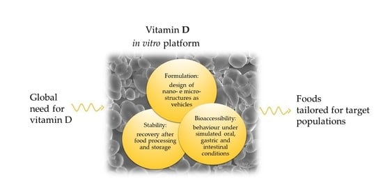
Vitamin D Incorporation in Foods: Formulation Strategies, Stability, and Bioaccessibility as Affected by the Food Matrix

Department of Food, Environmental and Nutritional Sciences (DeFENS), University of Milan, 20133 Milan, Italy
*
Author to whom correspondence should be addressed.
Submission received: 13 July 2021 / Revised: 20 August 2021 / Accepted: 23 August 2021 / Published: 25 August 2021

Abstract

:
Inadequate intake of vitamin D is a global health issue related to severe diseases, mainly involving subjects with dark skin pigmentation, patients affected by malnutrition, malabsorption syndromes, or obesity, and elderly people. Some foods fortified with vitamin D have been tested in vivo, but fortification strategies with a global outreach are still lacking. This review is focused on food fortification with vitamin D, with the aim to collect information on (a) formulation strategies; (b) stability during processing and storage; and (c) in vitro bioaccessibility. Approaches to add vitamin D to various foods were analyzed, including the use of free vitamin D, vitamin D loaded in simple and double nanoemulsions, liposomes, casein micelles, and protein nanocapsules. Numerous studies were reviewed to elucidate the impact of food technologies on vitamin D’s stability, and mechanisms that lead to degradation were identified—namely, acid-catalyzed isomerization, radical-induced oxidation, and photo-oxidation. There is, however, a lack of kinetic data that allow for the prediction of vitamin D’s stability under industrial processing conditions. The roles that lipids, proteins, fibers, and antioxidants play in vitamin bioaccessibility have been clarified in various studies, while future needs include the design of specific food matrices that simultaneously achieve a balance between the long-term stability, bioaccessibility and, ultimately, in vivo functionality of vitamin D.

Graphical Abstract

1. Introduction

Vitamin D performs essential functions for human health, and concerns about its inadequate dietary intake have been expressed worldwide [1,2]. Vitamin D has two prevalent forms, i.e., cholecalciferol (vitamin D3) and ergocalciferol (vitamin D2). These vitamers are formed through the UV conversion of precursor compounds—namely, 7-dehydrocholesterol present in vertebrates for vitamin D3, and ergosterol present in fungi and some invertebrates for vitamin D2. The equivalence of the in vivo effectiveness of vitamin D2 and vitamin D3 has not been fully elucidated. In fact, some studies showed the same biological activity for both vitamers [3,4], while other studies suggested a higher bioavailability for vitamin D3 than for vitamin D2 [5,6,7]. Upon ingestion, both vitamin D2 and vitamin D3 are converted into the same biologically active form, i.e., 1,25-dihydroxyvitamin D (1,25(OH)2D). This latter compound binds to a nuclear receptor and modulates gene expression. Interestingly, the vitamin D receptor was found to be present in most cells in the body, and this finding supports the involvement of vitamin D in the prevention of multiple diseases [8,9]. In fact, vitamin D performs a fundamental role in the maintenance of skeletal calcium and phosphate balance, thus preventing rickets in children and osteomalacia, osteoporosis, and bone fractures in adults. Moreover, it has been associated with protection from the risk of common cancers, autoimmune diseases, hypertension, and infectious diseases [2]. The recommended dietary intake of vitamin D is 10, 15, and 20 µg/d for infants (<1 year), subjects aged 1–70 years, and subjects aged ˃ 70 years, respectively [10]. Observational studies led to the conclusion that vitamin D supplementation should be 2–3 times higher for obese subjects and 1.5 times higher for overweight subjects, relative to normal-weight subjects [11]. Moreover, for people at risk of vitamin D deficiency, the recommended daily dose of vitamin D is 50 µg/d or higher, depending on age [10]. The recognized biomarker for vitamin D status is serum 25-hydroxyvitamin D (25(OH)D) level. Some panels of experts indicate that vitamin D deficiency corresponds to a (25(OH)D) level below 50 nmol/L, vitamin D insufficiency to a level between 50 and 75 nmol/L, and vitamin D toxicity to levels above 500 nmol/L [10]. However, other panels of experts indicated that the cutoff below which the risk of clinical vitamin D deficiency increases corresponds to a serum level of 25(OH)D < 30 nmol/L [12]. Populations at risk of vitamin D deficiency include subjects with dark skin pigmentation, subjects affected by malnutrition, malabsorption syndromes, or obesity, and elderly people [9]. Vitamin D insufficiency is also common. In fact, a survey throughout Europe has shown that the intake of vitamin D is inadequate for 77–100% of adults (19–64 years old) and for 55–100% of elderly adults (>64 years old) [13]. Compared to the USA, Vitamin D intake from food in Europe is lower [14], which can be attributed to the higher availability of foods fortified with vitamin D in the USA than in Europe [1]. Indeed, food fortification with vitamin D is considered a fundamental strategy to fight vitamin D malnutrition and its associated diseases [1,3,15].
Foods fortified with vitamin D—especially bread and dairy products—have been assessed in vivo for their efficacy in raising the level of 25(OH)D and improving health in different target populations (Table 1). In both low-fiber white bread and high-fiber sourdough rye bread, fortification with a water-dispersible form of vitamin D3 providing approximately 10 µg/d was as effective as vitamin D3 supplements in increasing serum 25(OH)D concentration in healthy women aged 25–45 years [16]. The production of bread with UV-irradiated yeast as a source of 25 µg/d of vitamin D2 did not affect serum 25(OH)D concentration in healthy women aged 20–37 years, while supplements of both vitamin D2 and vitamin D3 resulted in a positive effect [17]. Conversely, vitamin D2 from UV-irradiated mushroom formulated in a soup was as effective as vitamin D2 supplements in increasing serum 25(OH)D concentration in healthy subjects younger than 45 years, but the amount provided was high, equal to 700 µg/d [18]. In Ca++-enriched orange juice, water-dispersible forms of either vitamin D2 or vitamin D3 in a dose of 25 µg/d were equally bioavailable as the respective supplements in adult subjects aged 18–84 years [4]. The impact of Ca++ enrichment along with vitamin D2 was not investigated, because a control without Ca++ addition was not included [4].
Reduced-fat milk enriched with Ca++ was an effective matrix for vitamin D3 fortification, when administered at a level of 20 µg/d to healthy male subjects aged 50–87 years, causing increased serum 25(OH)D concentration and reduced bone loss, but no information was provided on the incorporated form of vitamin D3 [19]. In a subsequent study, fortification of Ca++-enriched milk with a non-specified form of vitamin D3, as well as fortification of Ca++-enriched orange juice, were designed for healthy children aged 9–12 years, and were found to be effective in increasing serum 25(OH) concentration, while serum osteocalcin and intact parathyroid hormone were not affected [20]. However, in these latter two studies, foods enriched in Ca++ and vitamin D were also considered, but the impact of Ca++ enrichment was not investigated [19,20].
Contradictory results were obtained by applying a water-dispersible form of vitamin D3 in cheese, but the supplied amounts were different [21,22]. In fact, in one study involving healthy, ≥60-year-old subjects, no effects on 25(OH)D, parathyroid hormone, or osteocalcin serum concentrations were observed upon the consumption of vitamin-D3-fortified cheese supplying 15 µg/d [21], while in another study involving healthy subjects aged 18–60 years, an increase in serum 25(OH)D concentration was observed along with a decreased serum parathyroid hormone upon consumption of either regular Cheddar cheese (obtained from milk having 3.8% fat content) or reduced-fat cheese (obtained from skim milk having 0.8% fat content), both supplying 100 µg/d of vitamin D3 [22].
Table 1. In vivo studies on the effects of vitamin-D-fortified foods.
Fortified FoodSubjects and DurationDose
(µg/d)
OutcomeRef.
Water-dispersible vitamin D3
in low-fiber wheat bread
25–45 years, healthy women
n = 41, 1 month
10.8Both vitamin-D3-fortified breads
increased serum 25(OH)D as effectively as vitamin D3 supplement.
[16]
Water-dispersible vitamin D3
in high-fiber sourdough rye bread
12.3
Vitamin D2 from UV-irradiated yeast
in bread
20–37 years, healthy women
n = 33, 2 months
25Vitamin D2 from UV-irradiated yeast in bread did not raise serum 25(OH)D.[17]
Vitamin D2 from UV-irradiated mushrooms in soup<45 years, healthy
n = 26, 1 month
700Vitamin-D2-fortified soup increased serum 25(OH)D as effectively as vitamin D2 supplement.[18]
Water-dispersible vitamin D3
in Ca++-enriched orange juice
(Ca++ dose: 350 mg/d)
18–84 years, healthy
n = 105, 3 months
25Vitamin D2 and vitamin D3 were equally bioavailable in Ca++-enriched orange juice and capsules.[4]
Water-dispersible vitamin D2
in Ca++-enriched orange juice
(Ca++ dose: 350 mg/d)
Vitamin D3 non-specified
in Ca++-enriched reduced-fat milk
(Ca++ dose: 1000 mg/d)
50–87 years, healthy men
n = 149, 2 years
20Vitamin-D3- and Ca++-fortified milk increased serum 25(OH)D and reduced bone loss.[19]
Vitamin D3 non-specified
in Ca++-enriched orange juice
(Ca++ dose: 500 mg/d)
9–12 years, healthy
n = 410, 3 months
2.5Vitamin-D3-fortified milk and orange juice increased serum 25(OH). Serum OC and PHT were not affected.[20]
Vitamin D3 non-specified
in Ca++-enriched milk
(Ca++ dose: 500 mg/d)
Water-dispersible vitamin D3
in cheese
≥60 years, healthy
n = 110, 2 months
15Vitamin-D3-fortified cheese had no effect on serum 25(OH)D, OC and PHT.[21]
Water-dispersible vitamin D3
in Cheddar cheese
18–60 years, healthy
n = 30, 2 months
100 *Both vitamin-D3-fortified cheeses increased serum 25(OH)D and decreased serum PHT as vitamin D3 supplement[22]
Vitamin D3 non-specified
in regular yogurt
(Ca++ dose: 150 mg/d)
30–60 years, diabetic
n = 90, 3 months
25Both vitamin-D3-fortified regular yogurt and Ca++-enriched yogurt increased serum 25(OH)D and improved glycemic status.[23]
Vitamin D3 non-specified
in Ca++ enriched yogurt
(Ca++ dose: 250 mg/d)
Vitamin D3 non-specified
in Ca++ enriched yogurt
(Ca++ dose: 800 mg/d)
≥65 years, healthy women
n = 20, 3 months
10Vitamin-D3-fortified yogurt increased serum 25(OH)D, and maintained cognitive performance.[24]
Water-dispersible vitamin D3
in yogurt
37–47 years, pre-diabetic
n = 60, 3 months
25Vitamin-D3-fortified yogurt increased serum 25(OH)D as effectively as vitamin D3 supplement, and improved serum lipid profile.[25]
Vitamin D3 in casein micelles
in low-fat yogurt
18–61 years, healthy
n = 87, single intake
1250Both vitamin D3 carriers in yogurt increased serum 25(OH)D.[26]
Vitamin D3 in emulsion
in low-fat yogurt
*: Equivalent to a daily dose (fortified food was ingested in one weekly dose); PHT: parathyroid hormone; OC: osteocalcin.
Various approaches have been applied for yogurt fortification with vitamin D3. In one study, a non-specified form of vitamin D3 was added alone or in combination with Ca++ in yogurt, and both formulations supplying 25 µg/d were effective in raising serum 25(OH)D levels and improving the glycemic status of diabetic subjects aged 30–60 years. Hence, the combined effect of Ca++ and vitamin D enrichment of the food matrix seemed not to be relevant [23]. Positive effects on serum 25(OH)D level and in the maintenance of cognitive performance were also observed upon consumption of yogurt enriched with Ca++ and 10 µg/d of a non-specified form of vitamin D3 by a population of healthy elderly women (≥65 years), while vitamin D enrichment of yogurt with regular Ca++ content was not considered [24]. In another approach, the water-dispersible form of vitamin D3 was used to fortify yogurt, and proved to be effective in increasing serum 25(OH)D levels and improving the serum lipid profiles of pre-diabetic subjects aged 37–47 years [25], when administered at a dose of 25 µg/d. Casein micelles or polysorbate/Tween-80 emulsions were designed as carriers to formulate vitamin D3 in low-fat yogurt, and both formulations were found to be effective in raising serum 25(OH)D in healthy subjects aged 18–61 years, but the dose administered was equal to 1250 µg/d [26]—well above the recommended daily dose.
The above-reported in vivo studies cannot be compared because of the different target populations involved, daily doses administered, and durations of the interventions. However, in general they support the efficacy of fortification of various food matrices as a strategy to manage inadequate vitamin D intake. Nevertheless, in order to apply a vitamin D fortification strategy, further issues need to be considered. Firstly, the form in which vitamin D was administered is generally unknown or lacks relevant information, and the dosage applied was higher than the recommended daily dose. Hence, no information on the cost-effectiveness of the fortification strategy can be derived. Moreover, in most in vivo studies, food fortification was performed by adding vitamin D to the processed product just before the administration, while no information is provided on vitamin D stability when addition is carried out prior to food processing, packaging, and long-term storage. In vitro modelling studies can provide in-depth information on vitamin D’s stability and bioaccessibility. Hence, this review article is focused on food fortification with vitamin D, with the aim to collect information on (a) the formulation strategies of vitamin D in foods; (b) the stability of vitamin D in fortified foods during processing and storage; and (c) the in vitro bioaccessibility of vitamin D from the fortified foods. The ultimate aim of this study is to support the design of an efficient technology for the vitamin D fortification of foods.

2. Vitamin D Fortified Foods: Formulation Strategies

Vitamin D3 obtained at the industrial level is generally used for food fortification. The free form of vitamin D3 has likely been incorporated in the food matrices evaluated in some in vivo studies, although this is not specifically indicated [19,20,23,24]. Free vitamin D3 has also been used in various in vitro studies [27,28,29,30,31,32,33], while free vitamin D2 has been considered in only a few in vitro studies [31].
Irradiated yeasts and mushrooms have been used directly as a source of vitamin D2 in fortified foods for in vivo [17,18] and in vitro studies [30]. The application of the unprotected compound in food formulation has the major drawbacks of non-homogeneous dispersion in the matrix, uncontrolled stability, and fast release. Moreover, the application of vitamin D2 entrapped in yeast cells was not effective in vivo [17], while that entrapped in mushrooms was effective in vivo only at very high dosages [18].
Vitamin D is a large sterol molecule. Considering its low water solubility and susceptibility to oxidative or heat degradation, suitable delivery systems have been studied for food fortification (Table 2). In brief, water-dispersible vitamin premixes—often containing an emulsifier—and oil-based systems have been proposed, depending on the characteristics of the target food. A commercial water-soluble form of vitamin D containing propylene glycol and polysorbate 80 (Vitex-D, Kingsway Chocolate Co. Ltd., Bunge Foods, Mississauga, ON, Canada) was used in one in vivo [22] and in some in vitro studies [34,35], but no information was provided on the formulation characteristics, such as its particle size distribution, which affects its bioaccessibility [36]. Other in vivo [4,16,20,25] or in vitro [37,38,39] studies reported the use of water-dispersible forms of vitamin D3 or vitamin D2, but no details were provided on composition or particle size distribution.
A strategy to develop value-added fortified foods is the design of specific nano- or micro-structures (Table 2) that enable the homogeneous distribution of bioactive compounds in the food matrix and improve stability and bioavailability [40]. It is worth noting that nano- and micro-structures can potentially be affected by pH, ionic strength, and heat treatments; hence, it is fundamental to test their stability during food processing [41].
It must be underlined that many of the carrier systems developed for vitamin D delivery look promising, but have not found application at the industrial level so far. This review only focuses on studies including a food product application—namely, lipid-based encapsulation systems [42,43,44,45,46,47,48,49,50], which were considered in vitro studies, and protein complexes, which were considered in studies performed both in vivo [26] and in vitro [51,52].
Lipid-based encapsulation systems used for food fortification with vitamin D include oil-in-water (O/W) nanoemulsions [36,43,45,46,47]; double water-in-oil-in-water (W1/O/W2) emulsions [48], and liposomes [50]. All of these structures can be affected by chemical instability due to lipid oxidation and physical instability due to their tendency to phase separation.
O/W emulsions are structures with a lipophilic core where a lipophilic bioactive compound is entrapped, a hydrophilic shell (aqueous phase), and an amphiphilic interface consisting of the emulsifier/surfactant [53]. Emulsion particles move randomly in the presence of gravitational and Brownian motion forces acting on the droplets in the medium. The main instability processes are flocculation, coalescence, and creaming (or sedimentation), which ultimately lead to complete separation of the two constituting phases. In nanoemulsions, small particle size (d < 200 nm) creates low susceptibility to gravitational separation due to the dominant Brownian forces [53]. The zeta potential (ζ-potential) of dispersed oil droplets is also related to emulsion stability, because levels close to zero promote aggregation due to the absence of charge repulsions [54]. In one approach, vitamin D2 emulsions were prepared using corn oil, Tween® 80, and phosphate buffer; the amount of vitamin D2 in the oil phase was 50 mg/100 g of oil. By blending these components at a high speed (10,000 rpm), a large emulsion was obtained with a volume-weighted mean diameter of 14.5 μm and ζ-potential of −13.8 mV; by passing the large emulsion through a Microfluidizer operating at 6000 psi, a medium emulsion was obtained with a volume-weighted mean diameter of 0.53 μm, and further processing at 15,000 psi resulted in a small emulsion with a volume-weighted mean diameter of 0.11 μm, with little variation in the ζ-potential [36]. Hence, it was possible to modulate emulsions’ nanostructure by varying the energy input of the process. The droplets should have had a low net charge because they were stabilized by a non-ionic surfactant; however, a negative surface charge was observed and attributed to the presence of anionic impurities such as free fatty acids in either the surfactant or the oil phase [36]. The negative charge is expected to inhibit particle aggregation, but the emulsions were not tested in food matrices [36]. In other studies, emulsions were prepared using vitamin D3, flaxseed oil, calcium caseinate, and distilled water, +/− soy lecithin, but the amount of vitamin D3 was not specified. No information was provided on the particle size distribution and ζ-potential; however, physical stability was proven up to 6 d of storage in the absence of soy lecithin and 15 d of storage in the presence of soy lecithin; afterwards, the appearance of serum indicated that casein–casein interactions occurred. The adsorption of lecithin to the oil–water interface reduced the interactions between proteins and led to a slow gelation of the emulsion, which was then added to cheese [43]. In another approach, a nanoemulsion of vitamin D3 was created by combining vitamin D3 in corn oil and the surfactant (quillaja saponin) into phosphate buffer solution (5 mM, pH 7); the amount of vitamin D3 was 4 g/100 g of the oil phase. The mean particle diameter of the vitamin-enriched nanoemulsions was 0.14 μm, the pH was 4.74, and the surface charge was negative (–74.1 mV). Due to the strong repulsions between the negative charges on the surface, the nanoemulsion was stable for 7 d of storage, and was found to be suitable for the fortification of almond milk [47].
The W1/O/W2 double emulsions have a water-continuous system containing oil droplets with smaller water droplets dispersed within. Double emulsions can simultaneously encapsulate both hydrophobic and hydrophilic compounds. Moreover, double emulsions can have a reduced fat content compared to O/W emulsions. As with the single emulsions, double emulsions can undergo flocculation, coalescence, and creaming (or sedimentation); additional instability processes include swelling and rupture of the inner droplets induced by osmotic transport between the two aqueous phases. Hence, there should be a balance between the osmotic pressure of the inner and outer aqueous regions [54]. In one study, W1/O/W2 double emulsions containing vitamin D3 were considered; the W1 phase was prepared by adding black chokeberry phenolic extract, folic acid, vitamin B12, and vitamin C; the O phase consisted of polyglycerol polyricinoleate dispersed in rapeseed oil containing vitamin D3 and vitamin A; and the W2 phase was a milk protein solution. The emulsification process included the preparation of the primary emulsion at a high speed (15,000 rpm) and the preparation of the secondary emulsion at a low speed (11,000 rpm). The amount of vitamin D3 was 7 mg/100 g of oil, and encapsulation efficiency was 98.52%. The resulting W1/O/W2 emulsion had a monomodal particle size distribution with a volume moment-weighted mean diameter of 37.65 µm, and was physically stable for 30 d [48]. This W1/O/W2 double emulsion was then added to yogurt [49].
Liposomes are vesicles with an aqueous core surrounded by one or more amphiphilic molecules—such as polar lipids—that produce a bilayer structure. Hydrophobic substances can be entrapped within the hydrophobic bilayer [55]. Phospholipids are often used as amphiphilic molecules for liposome preparation. The small radius of the curvature of the vesicles generates instability because it disrupts the regular packing of the phospholipid bilayer and imposes constraints on the phospholipid molecules [56]. One of the key parameters for the liposome structure is the transition temperature, where the liposomes lose their ordered packing structure due to melting of the hydrocarbon chains, i.e., they undergo a phase transition from gel to liquid-crystalline. Since natural phospholipids contain hydrocarbon chains that differ in length, phase transition occurs over a wide temperature range. Physical instability of liposomes may arise from merging, which results in increased particle size. Cholesterol is generally added to the phospholipid structure at a volume of over 20% because it decreases the transition temperature and increases stability [55]. Lipophilic bioactive compounds that are positioned in the lipid bilayer of liposomes, such as vitamin D, can alter the interactions between hydrocarbon chains and, hence, affect bilayer stability. In one study, liposomes loaded with vitamin D3 were added to milk intended for producing fortified cheese, but a commercial liposome premix was used and no information was provided on liposome structure [57]. In another study, liposomes loaded with vitamin D3 were applied in chocolate. Phospholipids, cholesterol, and vitamin D3 were used in a 3:1:1 ratio; hence, vitamin D3 constituted 20 g/100 g of the lipid phase. Liposomes were obtained by film dispersion–homogenization. Loading efficiency for vitamin D3 was 68.2%. The resulting liposomes had a monomodal distribution, with an equivalent volume diameter at 50% cumulative volume of 354.91 nm and a ζ-potential of −27.62 mV; however, no information on stability was provided [50].
Hydrophobic molecules can also bind to proteins via hydrophobic interactions. A consensus exists on the role of some well-structured proteins, such as milk and soy proteins, as natural nanocarriers of hydrophobic bioactive compounds [58,59,60,61,62,63]. However, the loading capacity of these structures is likely low, being targeted at the naturally occurring levels of bioactive compounds. Thus, protein-based nanocarriers have been developed as vehicles of vitamin D at the target levels of fortification in food, with casein being the most frequently involved protein.
Complexation of vitamin D with whey proteins proved to increase the stability of the vitamin under light and prolonged storage, compared to the free form, but no data obtained on real foods were given [59,60]. Compared to native casein micelles, sodium or calcium caseinates are more water soluble, and single casein molecules still maintain the capacity for self-assembly due to their amphiphilic nature. On this basis, the so-called reassembled casein micelles (rCMs) have been studied and shown to represent microencapsulation structures that potentially guarantee enhanced stability of vitamin D during subsequent food processing. The vitamin-loading capacity of rCMs essentially depends on the content of calcium, phosphate, and citrate added in the formulation. By optimizing these parameters, a loading of 1.38–1.46 mg/100 mg casein was achieved for vitamin D, with particles of rCMs showing a monomodal distribution and average volume-weighted particle sizes of 16–19 µm, depending on phosphate concentration [61]. However, aqueous suspensions of the rCMs showed D(0.5) (diameter where 50% of the particle size distribution is above and 50% is below) values around 12–13 µm, i.e., much larger than those of native casein micelles (50–700 nm), indicating that rCMs may merge into large particles due to the high phosphate content (4.9 mM). Other authors [26], who prepared vitamin-D-loaded rCMs with the same approach, achieving a loading of 1.6 mg vitamin D3/100 mg casein, adopted a homogenization step of the mixture, and obtained a bimodal volume-weighted particle size distribution, with a large population that had an average volume moment-weighted diameter of 89 nm, and a smaller population around 277 nm in diameter. The efficacy of casein complexes and rCMs in protecting the associated vitamin D2 from degradation was compared in fluid milk processed at the laboratory scale [51]. Disruption and subsequent reassembly of casein micelles with increased affinity for vitamin D was also attained using high-pressure (> 400 MPa) treatments at selected temperatures [62]. Large rCMs (278 nm average hydrodynamic diameter) with a vitamin D2 loading of 10.4 µg/mg casein were obtained by treating a dispersion of native micelles at 600 MPa at 50 °C. These structures were claimed to be suitable for the enrichment of low-fat dairy products, since they maintain the original properties [62].
Table 2. Formulation of vitamin D for food fortification.
Structures
and Vitamin D Loading
ComponentsPSD and Average Sizeζ-PotentialRef.
O/W emulsions
S, small;
M, medium;
L, large
50 mg vitamin D2/100 g oil
Vitamin D2, corn oil, Tween® 80, phosphate bufferMonomodal
S: 0.11 μm,
M: 0.53 μm,
L: 14.5 μm (volume moment-weighted mean diameter)
S: −12.4 mV, M: −14.1 mV,
L: −13.8 mV
[36]
O/W nanoemulsion
4 g vitamin D3 /100 g oil
Vitamin D3, corn oil, quillaja saponin, phosphate bufferMonomodal
0.14 µm (mean particle diameter)
−36.4 mV[47]
W1/O/W2 double emulsion
7 mg vitamin D3/100 g oil
Vitamin D3, vitamin A, vitamin B12, vitamin C, chokeberry extract, folic acid, rapeseed oil, polyglycerol polyricinoleate, milk protein, sodium chloride, waterMonomodal
37.65 µm (volume moment-weighted mean diameter)
[48]
Liposomes
20 g vitamin D3/100 g lipid phase
Vitamin D3, phospholipids, cholesterol in ethanol, distilled waterMonomodal
354.91 nm (equivalent volume diameter at 50% cumulative volume)
−27.62 mV[50]
rCMs: reassembled casein micelles
1.38–1.46 mg vitamin D3/100 mg casein
Vitamin D3 in ethanol, sodium caseinate in water, dipotassium hydrogen phosphate, potassium citrate, calcium chlorideMonomodal
from 16.10 to 19.04 µm (average volume-weighted particle sizes)
from −17.3 to −17.8 mV[61]
rCMs: reassembled casein micelles
1.6 mg vitamin D3/100 mg casein
Vitamin D3 in ethanol, sodium caseinate in water, dipotassium hydrogen phosphate, tripotassium citrate, calcium chlorideBimodal
89 nm and 277 nm (volume moment-weighted mean diameter)
[26]
rCMs: reassembled casein micelles
1 µg vitamin D2/100 mg casein
Vitamin D2 in ethanol, micellar casein in sweet whey permeateMonomodal
from 145 to 303 nm (mean hydrodynamic diameter)
[62]
β-Conglycinin nanoparticles
10 µg vitamin D3/100 mg β-conglycinin
Vitamin D3 in ethanol, β-conglycinin, phosphate bufferBimodal
31 nm and 120 nm (volume weighted average particle diameter)
[63]
Zein nanocapsules
vitamin D3 loading not specified
Vitamin D3 in ethanol, zein, Tween® 80Monomodal
185.7 ± 2.10 nm (Z-average size)
24.5 mV[52]
PSD: particle size distribution.
Due to their wider consumer acceptance and lower costs compared to animal proteins, plant proteins were also studied as vehicles for numerous bioactive compounds. Vitamin D3 was encapsulated in soybean β-conglycinin nanoparticles, leading to a bimodal distribution: a large population (90% of the particle volume) with an average volume moment-weighted diameter of 31 nm, and the remaining 10% with a diameter around 120 nm [63]. These nanoparticles were claimed to be suitable for the fortification of both foods and clear beverages [63]. Zein—a major protein from corn—was successfully used to produce nanoparticles that were loaded with vitamin D3, attaining a 97.27% encapsulation efficiency [52]. In this case, nanoparticles had a Z-average size of 185.7 nm and, upon observation via transmission electron microscopy (TEM), showed a smooth surface. The ζ-potential of 24.5 mV was attributed to the cationic character of zein, because the value was similar (21.2 mV) in the unloaded zein nanoparticles. A jelly based on the pulp of Acca sellowiana, xylitol, and pectin was then fortified with the nanoparticles [52].
The formulations described in Table 2 were generally tested in model foods: the O/W emulsions were added to cheese [43] and almond milk [47], the W1/O/W2 double emulsions were added to yogurt [49], liposomes were added to cheese [56] and chocolate [50], the rCMs were added to milk [51], and the zein nanocapsules were assessed in a model jelly food [52]. Although these model foods were prepared at the laboratory scale, they proved that the designed formulations could persist after processing and, thus, pave the way for applications in real food systems.

3. Vitamin D in Fortified Foods: Yield and Stability

The cis-triene configuration of vitamin D makes it potentially sensitive to isomerization and oxidation. Nevertheless, purified vitamin D3 was found to be stable at neutral pH, in air, at ambient temperature [64]. Accordingly, a differential calorimetric study performed on the purified vitamins D2 and D3 revealed that the onset temperatures for their thermal decomposition are about 166 and 156 °C, respectively, and their activation energy values are 141 and 131 kJ mol−1, respectively [65]. However, vitamin D’s stability can be affected by the food matrix components (Table 3).
In the presence of acids, vitamin D3 isomerizes, and its acid-catalyzed isomerization product—i.e., isotachysterol—is very oxygen-sensitive [63,66]. Moreover, vitamin D3 degradation is triggered by unsaturated lipid oxidation [29,67]. The reaction pattern for isomerization and oxidation of vitamin D3 does not involve the side chain that differs between vitamin D2 and D3; hence, these reactions most likely occur in vitamin D2 as well.
Considering that vitamin D is often formulated as an oil phase before being used in food processing, some studies have investigated vitamin D’s stability in bulk oils. Soybean oil packed in polyethylene terephthalate bottles was used as a model matrix to study the effects of oxidative status, antioxidant content (α-tocopherol), and exposure to light and oxygen on vitamin D3 degradation at 30 °C. This approach confirmed that the degradation of vitamin D3 occurs via oxidation, and the factors affecting degradation were, in decreasing order: storage time, exposure to light, and initial oxidative status of the oil, whereas α-tocopherol content played a protective role. As a result, vitamin D3 retention after storage at 30 °C for 50 d varied in the range 32–76% [29]. Sunflower oil was also used as a medium to study both vitamin D3 and vitamin D2 degradation at 100 and 210 °C, and both isomers were found to degrade at a similar rate upon heating [31].
Formulation of vitamin D3 in lipids with a low degree of unsaturation led to higher stability. In fact, both free vitamin D3 and liposomes made with vitamin D3, phospholipids, and cholesterol added to white chocolate after conching resulted in total recovery of the vitamin in chocolate upon 120 d of storage at 25 °C [50]. In contrast, a short-term stability of liposomes was observed in cheese upon ripening, leading to a progressive degradation of the loaded vitamin D [57].
Whole wheat flour was also considered for vitamin D3 incorporation, which is a strategy to develop various fortified bakery products [68]. In dry and intermediate-moisture foods, chemical stability is critically dependent on water activity (aw). For oxygen-sensitive compounds, stability decreases with increasing aw above the monomolecular moisture content, due to a decrease in the viscosity of the food matrix and increase in the molecular mobility, accelerating oxidation [69]. Accordingly, the half-times for vitamin D3 added to whole wheat flour at 25 °C were found to be 173, 169, and 116 d at aw levels of 0.33, 0.63, and 0.93, respectively. The stability of vitamin D3 greatly decreases with increasing temperature to 45 °C, with half-times for degradation of 87, 77, and 63 d at aw of 0.33, 0.63, and 0.93, respectively [68].
Dry irradiated mushroom powder could represent a source of vitamin D2 for a variety of foods [70]; in this matrix, vitamin D2 stability was also found to decrease with increasing aw from 0.11 to 0.75 [71]. The activation energy found for vitamin D2 degradation in mushroom powder at aw 0.33 was 36.7 kJ/mol, which is close to the activation energy for oxygen diffusion in unsaturated lipids—i.e., 24 kJ/mol—and to the activation energies found for the free radical chain reactions in triglycerides—i.e., 34.0–37.3 kJ/mol [72]; hence, autoxidation was assumed to be the mechanism by which vitamin D2 was lost [71]. The half-life of vitamin D2 at aw 0.33, calculated on the basis of the activation energy provided by this latter study, was 175 d at 25 °C, which is the same as that found for vitamin D3 in whole wheat flour under the same temperature and aw conditions [68]. Conversely, in fresh irradiated mushrooms, vitamin D2′s half-life was approximately 7 d at both 4 °C and −14 °C [73]; hence, storage at −20 °C is necessary to preserve vitamin D2 in fresh mushroom [74].
Table 3. Recovery of vitamin D in fortified foods after processing and storage.
Fortified FoodVitamin D
Content (μg/100 g)
Main Processing StepsVitamin D Yield
(%)
Ref.
Free vitamin D3
in soybean oil
700Storage in PET bottles at 30 °C for 50 d in the dark56–76 (S)[29]
Storage in PET bottles at 30 °C for 50 d under natural light32–39 (S)
Free vitamin D3
in sunflower oil
14.5Heating at 110 °C for 30 min85 (P)[31]
Heating at 210 °C for 10 min79 (P)
Free vitamin D2
in sunflower oil
11.2Heating at 110 °C for 30 min89 (P)
Heating at 210 °C for 10 min76 (P)
Vitamin D3 in liposomes
in white chocolate
30Three-step tempering process (33 °C–35 °C, 24 °C–25 °C, and 25 °C–26 °C),
storage at 25 °C for 120 d
100 (S)[50]
Free vitamin D3
in white chocolate
99 (S)
Free vitamin D3
in whole wheat flour
ndStorage at aw 0.33 at 25 °C for 173 d50 (S)[68]
Storage at aw 0.93 at 25 °C for 116 d50 (S)
Storage at aw 0.33 at 45 °C for 87 d50 (S)
Storage at aw 0.93 at 45 °C for 63 d50 (S)
Vitamin D2
in dry mushroom powder
4420Storage at aw 0.33 at 25 °C for 175 d50 (S)[71]
Storage at aw 0.33 at 30 °C for 136 d50 (S)
Storage at aw 0.33 at 45 °C for 55 d50 (S)
Free vitamin D3
in wheat bread
10.8Baking at 170 °C for 60 min85 (P)[28]
Free vitamin D3
in rye bread
8.769 (P)
Vitamin D2 yeast
in wheat bread
9.585 (P)
Vitamin D2 yeast
in rye bread
8.573 (P)
Free vitamin D3
in wheat bread
40.2Baking at 200 °C for 20 min40.2 (P)[68]
Free vitamin D3
in cookies
64Baking at 200–205 °C for 12 min65.3 (P)
Free vitamin D3
in milk
12Steam injection (95 °C), spray-drying at 149 °C, fluid-bed finish at 107 °C100 (P)[27]
Water-dispersible vitamin D3
in milk
100Pasteurization at 73 °C for 15 s, homogenization at 13.8/3.4 MPa, storage at 4 °C for 21 d100 (P, S)[37]
Water-dispersible vitamin D2
in milk
2Pasteurization at 63 °C for 30 min, storage in glass bottles at 4°C for 7 d in the dark100 (P, S)[39]
Pasteurization at 63 °C for 30 min, storage in glass bottles at 4 °C for 32 h under light100 (P, S)
Pasteurization at 63 °C for 30 min, storage in PET pouches at 4 °C for 7 d in the dark~ 90 (P, S)
Pasteurization at 63 °C for 30 min, storage in PET pouches at 4 °C for 32 h under light~ 90 (P, S)
Sterilization at 121 °C for 15 min100 (P)
Vitamin D2 in casein complexes
in cow and buffalo milk (1:1)
1.25Pasteurization at 63 °C for 30 min, storage in glass bottles at 4 °C for 7 d under light~ 95 (P),
~ 70 (S)
[51]
Pasteurization at 63 °C for 30 min, storage in LDPE pouches at 4 °C for 7 d under light~ 95 (P),
~ 65 (S)
Sterilization at 121 °C for 15 min~ 75 (P)
Free vitamin D2
in cow and buffalo milk (1:1)
1.25Pasteurization at 63 °C for 30 min, storage in glass bottles at 4 °C for 7 d under light90 (P),
45 (S)
Pasteurization at 63 °C for 30 min, storage in LDPE pouches at 4 °C for 7 d under light90 (P),
45 (S)
Sterilization at 121 °C for 15 min67 (P)
Water-dispersible vitamin D3
in Cheddar (milk fat 3.9%)
ndStarter addition, rennet addition, salting, pressing, vacuum packaging, storage at 4° C for 84 d90.4 (P)
100 (S)
[34]
Free vitamin D3
in Cheddar (milk fat 3.9%)
86.7 (P)
93.7% (S)
Water-dispersible vitamin D3
in Cheddar (milk fat 3.8%)
2085Milk pasteurization 72°C for 16 s, starter addition, rennet addition, salting, pressing, vacuum packaging, storage at 4 °C for 365 d91 (P)
100 (S)
[35]
Water-dispersible vitamin D3
in low-fat cheese (milk fat 0.8%)
169055 (P)
100 (S)
Vitamin D3 in nanoemulsion
in cheese (milk fat 2%)
1Milk acidification with lactic acid, rennet addition, cutting, centrifugation, storage at 4 °C for 90 d91 (P)
100 (S)
[43]
Free vitamin D3
in cheese (17 MPa, 0.9 P/F ratio)
488Skimmed milk powder added with anhydrous milk fat and water at specific protein/fat (P/F) ratio, homogenization at 17–150 MPa, calcium chloride addition, coagulation at 85 °C, salting, pressing52 (P)[32]
Free vitamin D3
in cheese (150 MPa, 2 P/F ratio)
49949.3 (P)
Water-dispersible vitamin D3
in yogurt (milk fat 3.9%)
ndStarter inoculation, fermentation, storage at 4 °C for 21 d97.8 (P)
100 (S)
[34]
Free vitamin D3
in yogurt (milk fat 3.9%)
96.6 (P)
100 (S)
Vitamin D3 in W/O emulsion
in yogurt
12.5Addition to fresh yogurt, storage at 4 °C for 20 d15.97 (S)[45]
Vitamin D3 in W/O emulsion
in yogurt + goji berry extract
74.90 (S)
Water-dispersible vitamin D3
in yogurt (milk fat 1.5%)
2.25Starter inoculation, fermentation, storage in opaque containers at 4 °C for 21 d100 (S)[38]
Vitamin D3 in oil
in yogurt (milk fat 1.5%)
99 (S)
Water-dispersible vitamin D3
in yogurt (milk fat 1.5%)
2.25Starter inoculation, fermentation, storage in transparent containers at 4 °C for 21 d86 (S)
Vitamin D3 in oil
in yogurt (milk fat 1.5%)
80 (S)
Vitamin D3 in W1/O/W2 emulsion
in yogurt (milk fat 6.15%)
10Starter inoculation, fermentation, storage at 4 °C for 20 d50 (S)[49]
Free vitamin D3
in yogurt (milk fat 6.15%)
94 (S)
(P, S): recovery after processing and storage; (P): recovery after processing; (S): recovery after storage; nd: not reported.
A bread model provided evidence of the effects of acid compounds on vitamin D2 degradation. In fact, in rye and wheat bread fortified with vitamin D2 from dry yeast, the retention after baking at 170 °C for 60 min was 73% and 85%, respectively. The same retention percentages were observed when free vitamin D3 was applied to fortify rye and wheat bread. Rye bread has a lower pH than wheat bread, which was the explanation for the lower retention of vitamin D, due to the acid-catalyzed isomerization to isotachysterol [28]. In another fortification study of bread fortified with free vitamin D3, retention was only 40%, but baking was carried out at a higher temperature, i.e., 200 °C for 20 min [68]. For cookies baked at 200–205 °C for a shorter time (12 min), the retention of vitamin D3 was 65% [68].
Milk intended for direct use or further processing in dairy products was also considered as a matrix to incorporate either vitamin D3 or vitamin D2. Nevertheless, contradictory results have been reported in literature regarding vitamin D’s stability in milk upon processing and storage. Natural vitamin D3 present in milk was reported to be unstable upon pasteurization and sterilization [57]. Conversely, free vitamin D3 added directly to milk before steam injection at 95 °C, spray-drying at 149 °C, and fluid-bed finishing at 107 °C was completely recovered in the dried milk powder [27]. In another study, the percentages of recovery for free vitamin D2 in milk after pasteurization at 63 °C for 30 min and sterilization at 121 °C for 15 min were 90 and 67%, respectively [51]. In some applications, both vitamin D3 and vitamin D2 were previously formulated in water-dispersible forms before addition to milk [37,39]. Pasteurization at 73 °C for 15 s, homogenization at 13.8/3.4 MPa, and storage in opaque plastic containers (with no further specifications) at 4 °C for 21 d did not affect the content of water-dispersible vitamin D3 added to milk [37]. Complete retention of vitamin D3 was also observed upon UHT processing (138 °C for 2 s) of chocolate milk with 2% added fat [37]. Similarly, water-dispersible vitamin D2 was found to be stable in milk upon pasteurization at 63 °C for 30 min and storage at 4 °C for 7 d in glass bottles in the dark, or for 32 h under light (1485–4455 lux), while vitamin D2 retention was ~90% when polyethylene pouches were used as a packaging material, regardless of light exposure during storage. This phenomenon was attributed to absorption of vitamin D2 by the polymer [39]. Sterilization at 121 °C for 15 min also resulted in total recovery of water-dispersible vitamin D2, while the retention during the storage of sterilized milk was not investigated [39].
In another approach, vitamin D2 complexed with casein in rCMs was added to milk and found to have improved stability with respect to the free vitamin D2 after pasteurization and sterilization, with recovery percentages up to 95 and 76%, respectively [51]. This latter study also reported the effect of storage on vitamin D2′s stability in milk packed in glass bottles or low-density polyethylene pouches, which was 70 and 65%, respectively, for vitamin D2 in casein complexes, and approximately 45% for free vitamin D2 regardless of the packaging material used [51]. Loss of vitamin D2 in milk packed in polyethylene pouches was attributed to the porous and hydrophobic nature of this material, which promotes absorption of vitamin D2 [39,51]. Moreover, during exposure of milk to light, singlet oxygen is produced, which causes degradation of vitamin D. It is worth noting that, in milk, riboflavin acts as a photosensitizer, thus accelerating the formation of singlet oxygen and leading to the photo-oxidation of vitamin D [75,76].
In cheese, the retention of vitamin D is a key aspect to optimize. In fact, during cheese-making, the fat-soluble vitamin D3 could be entrapped in the fat fraction that mostly precipitates in the curd. However, vitamin D3 possesses high affinity towards both β-lactoglobulin [77] and α-lactalbumin [78]; hence, it could also be lost in whey [34]. Cheddar cheese obtained from vitamin-D3-fortified full-fat milk (3.9% fat, w/w) showed approximately 90% recovery, regardless of the form in which vitamin D3 (free or water-dispersible) was added. The addition of vitamin D3 nanoemulsion to partially defatted milk (2.0% fat, w/w) also resulted in 91% recovery in cheese [43]. Conversely, when water-dispersible vitamin D3 was added to reduced-fat milk (0.78%, w/w), and further processed to Cheddar cheese, the recovery yield was only 55% [35]. Low recovery of vitamin D3, equal to 50%, was also observed upon the application of a model cheese-making process, based on heat-induced coagulation at 82 °C in the presence of calcium chloride, regardless of the pressure applied (17–150 MPa) or the protein-to-fat ratio (0.9–2) [32]. During storage, vitamin D3′s stability was high for up to 1 year in both full-fat and reduced-fat cheese [35].
Full-fat yogurt was fortified with both free and water-dispersible vitamin D3 and stored at 4 °C for 21 d, achieving almost complete recovery with both approaches [34]. Low-fat yogurt was also fortified with both water-dispersible vitamin D3 and oil-dispersible vitamin D3, and vitamin recovery was found to be complete after 21 d of storage at 4 °C when opaque packaging material was used, while approximately 80% recovery was observed when transparent packaging material was used [38]. However, in another fortification study, a O/W nanoemulsion of vitamin D3 made with coconut oil, soybean lecithin, Tween 80, and NaCl was added to yogurt before packaging (packaging material was not specified), and only 15.97% of vitamin D3 was recovered after storage at 4 °C for 20 d, unless goji berry antioxidants were added, which allowed 74.90% of vitamin D3 to be recovered [45]. Similarly, in a subsequent study, the addition of free vitamin D3 to yogurt led to a recovery of 50% after storage (packaging material was not specified), while the addition of a W1/O/W2 double emulsion made of refined rapeseed oil, polyglycerol polyricinoleate, milk protein concentrate, sodium chloride, and sucrose led to 94% recovery [49]. One possible mechanism for free vitamin D3 degradation in yogurt observed in this latter study could be oxidation, triggered by the high fat content (6.5%), whereas the emulsion protected vitamin D3 from oxidation.

4. Vitamin D in Fortified Foods: Bioaccessibility

Vitamin D is dissolved within the hydrophobic domains of the food matrices—such as bulk oils, fat droplets, or liposomes—or it is associated with proteins [79,80]. The metabolism of vitamin D begins in the stomach, where pepsin plays a role by releasing the fraction of vitamin D associated with proteins, and the gastric lipase hydrolyses vitamin D esters at least partially.
The fundamental step of vitamin D metabolism occurs in the duodenum, where proteases and lipases carry on its release from the food matrix and allow its solubilization in the mixed micelles formed from lipid digestion products, such as free fatty acids, monoglycerides, bile salts, and phospholipids. Then, vitamin D absorption by the epithelial cells takes place, which is mainly protein-mediated at low dietary concentrations and passive at high, pharmacological concentrations of vitamin D [79].
Hence, as for the other fat-soluble vitamins, the bioavailability of vitamin D depends on three phenomena: bioaccessibility (release from the lipid phase of the food matrix and solubilization within mixed micelles); transformation (chemical and biochemical conversion); and transport and uptake (migration through the mucus layer to the surfaces of the intestinal lumen and absorption by epithelial cells) [80]. Among these phenomena, bioaccessibility is the most critical in terms of governing bioavailability [81].
The effect of the food matrix on vitamin D bioavailability is not fully understood. The presence of lipids could improve vitamin D bioavailability by several mechanisms: providing a hydrophobic phase where it can be solubilized; stimulating biliary secretion and, consequently, micelle production; and contributing to micelle formation with their lipid digestion products, inducing chylomicron synthesis, which enhances vitamin D transport outside the enterocytes [79]. Nevertheless, in vivo studies available to date neglect the hypothesis that high amounts of fat in meals improve vitamin D’s bioavailability [44,79].
Dietary fiber seems to impair vitamin D’s bioavailability by affecting micelle formation and by increasing the viscosity of the chime, thus limiting the diffusion of vitamin-D-containing micelles to the brush border [79]. However, in vivo studies do not provide enough data to conclude on the effects of dietary components on vitamin D’s bioavailability.
As discussed previously, data on vitamin D’s bioavailability from food matrices obtained in various studies are difficult to compare because of the different study plans, including subjects involved, amount of vitamin D administered, and duration of the intervention [79]. In particular, vitamin D’s bioavailability is thought to depend on human-associated factors (age, disease, surgery, obesity, genetic variation, etc.) [45]. In vitro studies can provide insights on the effect of the food matrix on vitamin D’s release in the gastrointestinal tract and, hence, support the design of fortified foods (Table 4).
Bread was studied as a food matrix for vitamin D incorporation. Vitamin D2 from UV-treated yeast was used for both white and whole bread production, and the resulting bioaccessibility percentages were 15 and 9.9%, respectively. However, the bioaccessibility of free vitamin D2 in bread was higher (38%). It was concluded that the yeast cells entrapped vitamin D2 and hindered its incorporation into the mixed micelles [30]. As reported above, an in vivo study also led to the conclusion that bread made with UV-irradiated yeast as a source of vitamin D2 did not raise serum levels of 25(OH) D, while supplements of both vitamin D2 and vitamin D3 resulted in a positive effect [18].
Various milk and dairy matrices were used for vitamin D3 incorporation. Skimmed milk, partially defatted milk (2% fat), whole milk (3.25% fat), and a standard infant formula powder (4% fat) were fortified with vitamin D3 and studied for vitamin D3 bioaccessibility [30]. Results showed that, with respect to the initial content, the recovery of vitamin D3 in the intestinal digesta was only 40% for skimmed milk, 88% for partially defatted milk, 75% for whole milk, and 77% for infant formula. In general, only a part of vitamin D3 found in the digesta was effectively incorporated in the mixed micelles, equal to 70% for whole milk and infant formula and 80% for partially defatted milk and skimmed milk. Hence, considering bioaccessibility as the percentage of vitamin D3 recovered in the micellar phase after in vitro digestion compared to the amount of vitamin D3 in food, it can be calculated that bioaccessibility was 32, 70, 53, and 54% for skimmed milk, partially defatted milk, whole milk, and infant formula, respectively. It was concluded that bovine milk is an efficient delivery vehicle for vitamin D3′s bioaccessibility, even if the optimal amount of lipid for in vivo bioavailability of vitamin D3 requires further investigation [30], as observed in vivo [79].
Digestion models have also been tailored to specific populations, because the functioning of the gastrointestinal tract is known to depend on factors such as age and health status [80]. Three in vitro elderly models—namely, oral altered conditions; oral and gastric altered conditions; and oral, gastric, and intestinal altered conditions—were applied in parallel with a healthy adult model to investigate the digestibility of dairy products [82]. Interestingly, vitamin D3 naturally present in milk, yogurt, fresh cheese, and aged cheese was equally bioavailable for both the healthy and unhealthy gastrointestinal tract models considered, suggesting that age does not alter vitamin D3′s bioaccessibility. On the other hand, vitamin D3′s bioaccessibility from milk was found to be 30%, which is lower than that reported previously [30]. Moreover, vitamin D’s bioaccessibility was reported to be greatly dependent on the dairy matrix, being on average 30, 38, 24, and 21% for milk, yogurt, fresh cheese, and aged cheese, respectively [82].
Despite model cheeses not being optimal for vitamin D3 supply, gel-like matrices such as cheese can be modulated depending on formulation and processing conditions, leading to different behavior during digestion. In fact, in cheese matrices, aggregated casein micelles create a microstructural network with entrapped solid fat globules and serum, and their digestion is directly related to cheese microstructure [83]. Detailed information on the impact of gel structure on vitamin D3′s bioaccessibility was obtained by comparing model acid-coagulated fresh cheeses varying in protein-to-fat ratios (0.9, 1.3, 1.7, and 2) and in the particle size of the cheese milk emulsion due to different homogenization pressures applied (17–150 MPa) [32]. To prepare these model cheeses, milk was fortified by adding free vitamin D3 previously dissolved in ethanol and then in melted anhydrous milk fat. Interestingly, vitamin D3′s bioaccessibility decreased from 64.51 to 41.56% when the protein-to-fat ratio was increased from 0.9 to 2. An increase in milk homogenization pressure from 17 to 150 MPa also caused a decrease in vitamin D3′s bioaccessibility from 64.51 to 27.17%. Both increase in protein content and increase in homogenization pressure caused the formation of a stronger protein network that entrapped smaller fat particles, leading to the hypothesis that changes in the gel microstructure were responsible for the lower release of fat-soluble components during digestion.
Table 4. Bioaccessibility of vitamin D from various fortified foods.
Fortified FoodVitamin D Content
(μg/100 g)
Bioaccessibility
(%)
Ref
Yeast vitamin D2
in white wheat bread
4.6715[30]
Yeast vitamin D2
in whole wheat bread
4.879.9
Free vitamin D2 (control)
in whole wheat bread
3.938
Free vitamin D3
in skimmed milk
1.1232[30]
Free vitamin D3
in partially defatted milk (2% fat)
1.0270
Free vitamin D3
in whole milk (3.25% fat)
1.2853
Free vitamin D3
in infant powder formula (4% fat)
10.9454
Natural vitamin D3
in whole milk
3.9730[82]
Natural vitamin D3
in yogurt
3.138
Natural vitamin D3
in fresh cheese
13.824
Natural vitamin D3
in aged cheese
21.721
Free vitamin D3
in cheese (17 MPa, protein/fat ratio 0.9),
48864[32]
Free vitamin D3
in cheese (17 MPa, protein/fat ratio 2),
49851
Free vitamin D3
in cheese (150 MPa, protein/fat ratio 2)
49927
Vitamin D3 in W1/O/W2 emulsion
in high-protein yogurt
10100[49]
Vitamin D3 in W1/O/W2 emulsion
in high-protein yogurt
10100
Vitamin D3 nanoemulsion
in oat milk or almond milk
40020[46]
Vitamin D3 W/O nanoemulsion
in almond milk + CaCl2 or CaCO3
nd<20[47]
Vitamin D3 in refined olive oil
in beef
16.75[33]
Vitamin D3 in refined olive oil
in semolina or in chickpeas
3.025
Vitamin D3 in canola oil
in brownies
nd65.2[84]
Oily vitamin D3 in canola oil + tert-butylhydroquinone
in brownies
nd98
Vitamin D3 in zein nanocapsules
in a fruit (Acca sellowiana) jelly
15981[52]
nd: not reported.
The concept of combining vitamin D3 fortification of food with increase in protein content is particularly relevant to address the nutritional demands of older people [85]. Consistent with this aim, a high-protein (10%) yogurt fortified with a W1/O/W2 double emulsion containing berry polyphenols and water-soluble vitamins (C, B9 and B12) in the inner water phase and lipid-soluble vitamins (A and D) in the oil phase was developed [49]. The ingredients used to formulate the double emulsion were refined rapeseed oil, polyglycerol polyricinoleate, milk protein concentrate, sodium chloride, and sucrose. Free vitamin D3 was also added directly to the oil as a control. The results showed that the fortification of yogurt with vitamins loaded into the double emulsion did not hinder the release of vitamins during digestion, which was 100% for both the encapsulated vitamin D3 and the free oily vitamin D3 at the end of the intestinal phase. However, the kinetics of digestion were different since the release of oily vitamin D3 at the end of the gastric phase was considerably higher compared to the release of encapsulated vitamin D3.
Plant-based “milk” types were investigated as matrices to incorporate various forms of vitamin D3—namely, vitamin D3 in O/W nanoemulsion made with corn oil, and vitamin D3 nanoemulsion with added TiO2 or nanocellulose powders [46]. The vitamin bioaccessibility was around 20% in all of the fortified plant-based milks, indicating that neither type of nanoparticle (inorganic or organic) had a major impact on the bioaccessibility of vitamin D. The reasons for the observed low bioaccessibility were hypothesized to be either a saturation of the mixed micelles or the aggregation and precipitation of the majority of vitamin-loaded mixed micelles due to some of the constituents in the plant-based milks. In a subsequent study, vitamin D3 was encapsulated in a nanoemulsion made with corn oil quillaja saponin before addition to almond milk, but this approach also resulted in only 20% vitamin bioaccessibility [47]. Moreover, since plant-based milks are deficient in calcium, either soluble (CaCl2) or insoluble (CaCO3) calcium salts were added to vitamin-D3-fortified almond milk, but the addition of calcium further reduced vitamin D’s bioaccessibility [47]. A negative effect of Ca++ on vitamin D3′s bioaccessibility had also been observed in a previous study, in which Ca++ and vitamin D3 had been co-encapsulated in different W1/O/W2 double emulsions. This effect was attributed to insoluble calcium soap formation in the small intestine step of digestion [86].
In meat, semolina, and chickpeas, vitamin D3 was applied after solubilization in refined olive oil, and the resulting bioaccessibility was 5, 25, and 25%, respectively. Low bioaccessibility in meat was attributed to intense oxidation during digestion [33]. Semolina and chickpea antioxidants protected vitamin D3 from oxidative degradation; however, despite high vitamin D3 content being found in the intestinal digesta of these meals, the incorporation of vitamin D3 into the mixed micelles was hindered by tannins, fibers, and saponins [33]. The decrease in vitamin D3 bioaccessibility due to the occurrence of oxidation during digestion has also been supported by a previous study, which reported increased bioaccessibility of vitamin D3 in brownies fortified with both vitamin D3 and tert-butylhydroquinone, compared to a control fortified with vitamin D3 alone. On the other hand, α-tocopherol and butylated hydroxytoluene were not efficient [84].
A fruit jelly model proved to be an effective matrix for vitamin D3 fortification. In fact, vitamin D3 encapsulated in zein nanoparticles was used as an ingredient in jelly, which presented 81% bioaccessibility for vitamin D3 [52].

5. Conclusions and Future Perspectives

For the effective delivery of vitamin D through fortified foods, a careful formulation into nano- and micro-structures to be dispersed in the food matrix is a promising strategy. Due to the complexity and diversity of the food structures, the application of different encapsulation strategies could better address the need to develop carrier systems that simultaneously provide long-term chemical and physical stability, bioaccessibility and, ultimately, in vivo functionality of the vitamin.
However, information on vitamin D’s stability during processing is still fairly correlated with the form in which the vitamin was added to the food matrix. Moreover, knowledge concerning vitamin D’s stability during processing is limited to only a few processing steps, while a better understanding of the kinetic parameters of vitamin D3 and D2 degradation—i.e., rate constant and activation energy, could allow the prediction of vitamin D content in fortified foods and, thus, enable process optimization under industrial conditions.
Bioaccessibility studies provided insight into the potential efficacy of various vitamin-D-fortified foods, addressing the role of lipids, proteins, fibers, antioxidants, and Ca++ ions. The main challenge in future studies will be to acquire advanced knowledge of the interactions between vitamin D and specific molecules—especially single proteins and fibers—in order to take advantages from possible synergism. Moreover, validated in vitro models to assess vitamin D’s bioaccessibility in various target populations—such as the elderly, obese subjects, and subjects affected by malabsorption diseases—could assist a proper fortified food design, since a large variability in response to vitamin D supplementation has also been observed among individuals.

Author Contributions

Conceptualization, V.L.; methodology, V.L. and P.D.; investigation, data curation, original draft preparation, V.L., P.D. and L.P.; writing—review and editing, V.L., L.P. and P.D.; supervision, V.L. All authors have read and agreed to the published version of the manuscript.

Funding

This research received no external funding.

Acknowledgments

The authors are grateful to their colleague John A. Hogenboom for kindly revising the manuscript.

Conflicts of Interest

The authors declare no conflict of interest.

References

  1. Milešević, J.; Samaniego, L.; Kiely, M.; Glibetić, M.; Roe, M.; Finglas, P. Specialized food composition dataset for vitamin D content in foods based on European standards: Application to dietary intake assessment. Food Chem. 2018, 240, 544–549. [Google Scholar] [CrossRef] [PubMed]
  2. Grant, W.B.; Anouti, F.A.; Moukayed, M. Targeted 25-hydroxyvitamin D concentration measurements and vitamin D3 supplementation can have important patient and public health benefits. Eur. J. Clin. Nutr. 2020, 74, 366–376. [Google Scholar] [CrossRef] [PubMed]
  3. Holick, M.F.; Chen, T.C. Vitamin D deficiency: A worldwide problem with health consequences. Am. J. Clin. Nutr. 2008, 87, 1080S–1086S. [Google Scholar] [CrossRef] [PubMed] [Green Version]
  4. Biancuzzo, R.M.; Young, A.; Bibuld, D.; Cai, M.H.; Winter, M.R.; Klein, E.K.; Ameri, A.; Reitz, R.; Salameh, W.; Chen, T.C.; et al. Fortification of orange juice with vitamin D2 or vitamin D3 is as effective as an oral supplement in maintaining vitamin D status in adults. Am. J. Clin. Nutr. 2010, 91, 1621–1626. [Google Scholar] [CrossRef] [Green Version]
  5. Khazai, N.B.; Judd, S.E.; Jeng, L.; Wiolfenden, L.L.; Stecenko, A.; Ziegler, T.R.; Tangpricha, V. Treatment and prevention of vitamin D insufficiency in cystic fibrosis patients: Comparative efficacy of ergocalciferol, cholecalciferol, and UV light. J. Clin. Endocr. Metab. 2009, 94, 2037–2043. [Google Scholar] [CrossRef] [Green Version]
  6. Romagnoli, E.; Mascia, M.L.; Cipriani, C.; Fassino, V.; Mazzei, F.; D’Erasmo, E.; Carnevale, V.; Scillitani, A.; Minisola, S. Short and long-term variations in serum calciotropic hormones after a single very large dose of ergocalciferol (vitamin D2) or cholecalciferol (vitamin D3) in the elderly. J. Clin. Endocr. Metab. 2008, 93, 3015–3020. [Google Scholar] [CrossRef]
  7. Jakobsen, J.; Andersen, E.A.W.; Christensen, T.; Andersen, R.; Bügel, S. Vitamin D vitamers affect vitamin D status differently in young healthy males. Nutrients 2018, 10, 12. [Google Scholar] [CrossRef] [Green Version]
  8. Holick, M.F.; Biancuzzo, R.M.; Chen, T.C.; Klein, E.K.; Young, A.; Bibuld, D.; Reitz, R.; Salameh, W.; Ameri, A.; Tannenbaum, A.D. Vitamin D2 is as effective as vitamin D3 in maintaining circulating concentrations of 25-hydroxyvitamin D. J. Clin. Endocr. Metab. 2008, 93, 677–681. [Google Scholar] [CrossRef]
  9. Holick, M.F. The vitamin D deficiency pandemic: Approaches for diagnosis, treatment and prevention. Rev. Endocr. Metab. Disord. 2017, 18, 153–165. [Google Scholar] [CrossRef]
  10. Holick, M.F.; Binkley, N.C.; Bischoff-Ferrari, H.A.; Gordon, C.M.; Hanley, D.A.; Heaney, R.P.; Murad, M.H.; Weaver, C.M. Evaluation, treatment, and prevention of vitamin D deficiency: An Endocrine Society clinical practice guideline. J. Clin. Endocrinol. Metab. 2011, 96, 1911–1930. [Google Scholar] [CrossRef] [Green Version]
  11. Ekwaru, J.P.; Zwicker, J.D.; Holick, M.F.; Giovannucci, E.; Veugelers, P.J. The importance of body weight for the dose response relationship of oral vitamin D supplementation and serum 25-hydroxyvitamin D in healthy volunteers. PLoS ONE 2014, 9, 111265. [Google Scholar] [CrossRef] [Green Version]
  12. Cashman, K.D.; Kiely, M.E.; Andersen, R.; Grønborg, I.M.; Madsen, K.H.; Nissen, J.; Tetens, I.; Tripkovic, L.; Lanham-New, S.A.; Toxqui, L.; et al. Individual participant data (IPD)-level meta-analysis of randomized controlled trials with vitamin D-fortified foods to estimate Dietary Reference Values for vitamin D. Eur. J. Nutr. 2021, 60, 939–959. [Google Scholar] [CrossRef]
  13. Roman Viñas, B.; Ribas Barba, L.; Ngo, J.; Gurinovic, M.; Novakovic, R.; Cavelaars, A.; de Groot, L.C.P.G.M.; van’t Veer, P.; Matthys, C.; Serra Majem, L. Projected prevalence of inadequate nutrient intakes in Europe. Ann. Nutr. Metab. 2011, 59, 84–95. [Google Scholar] [CrossRef]
  14. Moore, C.E.; Radcliffe, J.D.; Liu, Y. Vitamin D intakes of adults differ by income, gender and race/ethnicity in the USA, 2007 to 2010. Public Health Nutr. 2013, 17, 1–8. [Google Scholar] [CrossRef] [Green Version]
  15. O’Mahony, L.; Stepien, M.; Gibney, M.J.; Nugent, A.P.; Brennan, L. The potential role of vitamin D enhanced foods in improving vitamin D status. Nutrients 2011, 3, 1023–1041. [Google Scholar] [CrossRef] [Green Version]
  16. Natri, A.M.; Salo, P.; Vikstedt, T.; Palssa, A.; Huttunen, M.; Karkkainen, M.U.; Salovaara, H.; Piironen, V.; Jakobsen, J.; Lamberg-Allardt, C.J. Bread fortified with cholecalciferol increases the serum 25-hydroxyvitamin D concentration in women as effectively as a cholecalciferol supplement. J. Nutr. 2006, 136, 123–127. [Google Scholar] [CrossRef] [Green Version]
  17. Itkonen, S.T.; Skaffari, E.; Saaristo, P.; Saarnio, E.M.; Erkkola, M.; Jakobsen, J.; Cashman, K.D.; Lamberg-Allardt, C. Effects of vitamin D2-fortified bread v. supplementation with vitamin D2 or D3 on serum 25-hydroxyvitamin D metabolites: An 8-week randomised-controlled trial in young adult Finnish women. Br. J. Nutr. 2016, 115, 1232–1239. [Google Scholar] [CrossRef] [Green Version]
  18. Urbain, P.; Singler, F.; Ihorst, G.; Biesalski, H.K.; Bertz, H. Bioavailability of vitamin D2 from UV-B-irradiated button mushrooms in healthy adults deficient in serum 25 hydroxyvitamin D: A randomized controlled trial. Eur. J. Clin. Nutr. 2011, 65, 965–971. [Google Scholar] [CrossRef] [Green Version]
  19. Daly, R.M.; Brown, M.; Bass, S.; Kukuljan, S.; Nowson, C. Calcium- and vitamin D3-fortified milk reduces bone loss at clinically relevant skeletal sites in older men: A 2-year randomized controlled trial. J. Bone Miner. Res. 2006, 21, 397–405. [Google Scholar] [CrossRef]
  20. Neyestani, T.R.; Hajifaraji, M.; Omidvar, N.; Nikooyeh, B.; Eshraghian, M.R.; Shariatzadeh, N.; Kalayi, A.; Khalaji, N.; Zahedirad, M.; Abtahi, M.; et al. Calcium-vitamin D-fortified milk is as effective on circulating bone biomarkers a fortified juice and supplement but has less acceptance: A randomised controlled school-based trial. J. Human Nutr. Diet. 2014, 27, 606–616. [Google Scholar] [CrossRef]
  21. Johnson, J.L.; Mistry, V.V.; Vukovich, M.D.; Hogie-Lorenzen, T.; Hollis, B.W.; Specker, B.L. Bioavailability of vitamin D from fortified process cheese and effects on vitamin D status in the elderly. J. Dairy Sci. 2005, 88, 2295–2301. [Google Scholar] [CrossRef]
  22. Wagner, D.; Sidhom, G.; Whiting, S.J.; Rousseau, D.; Vieth, R. The bioavailability of vitamin D from fortified cheeses and supplements is equivalent in adults. J. Nutr. 2008, 138, 1365–1371. [Google Scholar] [CrossRef] [PubMed]
  23. Nikooyeh, B.; Neyestani, T.R.; Farvid, M.; Alavi-Majd, H.; Houshiarrad, A.; Kalayi, A.; Shariatzadeh, N.; Gharavi, A.; Heravifard, S.; Tayebinejad, N.; et al. Daily consumption of vitamin D− or vitamin D+ calcium-fortified yogurt drink improved glycemic control in patients with type 2 diabetes: A randomized clinical trial. Am. J. Clin. Nutr. 2011, 93, 764–771. [Google Scholar] [CrossRef] [PubMed]
  24. Beauchet, O.; Launay, C.P.; Galery, K.; Vilcocq, C.; Dontot-Payen, F.; Rousseau, B.; Benoit, V.; Allali, G. Effects of vitamin D and Calcium fortified yogurts on gait, cognitive performances, and serum 25-hydroxyvitamin D concentrations in older community-dwelling females: Results from the GAit, MEmory, Dietary and vitamin D (GAME-D2) randomized controlled trial. Nutrients 2019, 11, 2880. [Google Scholar] [CrossRef] [Green Version]
  25. Mostafaia, R.; Nachvakc, S.M.; Mohammadi, R.; Rocha, R.S.; da Silva, M.C.; Esmerinod, E.A.; Nascimento, K.O.; Cruz, A.G.; Mortazavian, A.M. Effects of vitamin D-fortified yogurt in comparison to oral vitamin D supplement on hyperlipidemia in pre-diabetic patients: A randomized clinical trial. J. Func. Foods 2019, 52, 116–120. [Google Scholar] [CrossRef]
  26. Levinson, Y.; Ish-Shalom, S.; Segal, E.; Livney, Y.D. Bioavailability, rheology and sensory evaluation of fat-free yogurt enriched with VD3 encapsulated in re-assembled casein micelles. Food Funct. 2016, 7, 1477–1482. [Google Scholar] [CrossRef] [Green Version]
  27. Indyk, H.; Littlejohn, V.; Woollard, D. Stability of vitamin D3 during spray-drying of milk. Food Chem. 1996, 57, 283–286. [Google Scholar] [CrossRef]
  28. Jakobsen, J.; Knuthsen, P. Stability of vitamin D in foodstuffs during cooking. Food Chem. 2014, 148, 170–175. [Google Scholar] [CrossRef]
  29. Hemery, Y.M.; Fontan, L.; Moench-Pfanner, R.; Laillou, A.; Berger, A.; Renaud, J.C.; Avallone, S. Influence of light exposure and oxidative status on the stability of vitamins A and D3 during the storage of fortified soybean oil. Food Chem. 2015, 184, 90–98. [Google Scholar] [CrossRef]
  30. Lipkie, T.E.; Ferruzzi, M.G.; Weaver, C.W. Low bioaccessibility of vitamin D2 from yeast fortified bread compared to crystalline D2 bread and D3 from fluid milks. Food Funct. 2016, 7, 4589–4596. [Google Scholar] [CrossRef]
  31. Ložnjak, P.; Jakobsen, J. Stability of vitamin D3 and vitamin D2 in oil, fish and mushrooms after household cooking. Food Chem. 2018, 254, 144–149. [Google Scholar] [CrossRef] [Green Version]
  32. Castaneda, N.; Lee, Y. Microstructure of a model fresh cheese and bioaccessibility of vitamin D3 using in vitro digestion. Gels 2019, 5, 16. [Google Scholar] [CrossRef] [Green Version]
  33. Antoine, T.; Icard-Verniere, C.; Scorrano, G.; Salhi, A.; Halimi, C.; George, S.; Carriere, F.; Mouquet-Rivier, C.; Reboul, E. Evaluation of vitamin D bioaccessibility and mineral solubility from test meals containing meat and/or cereals and/or pulses using in vitro digestion. Food Chem. 2021, 347, 128621. [Google Scholar] [CrossRef]
  34. Kazmi, S.A.; Vieth, R.; Rosseau, D. Vitamin D3 fortification and quantification in processed dairy products. Int. Dairy J. 2007, 17, 753–759. [Google Scholar] [CrossRef]
  35. Wagner, D.; Rousseau, E.; Sidhom, G.; Pouliot, M.; Audet, P.; Vieth, R. Vitamin D3 fortification, quantification, and long-term stability in Cheddar and low-fat cheeses. J. Agric. Food Chem. 2008, 56, 7964–7969. [Google Scholar] [CrossRef]
  36. Salvia-Trujillo, L.; Fumiaki, L.B.; Park, J.; McClements, D.J. The influence of lipid droplet size on the oral bioavailability of vitamin D2 encapsulated in emulsions: An in vitro and in vivo study. Food Funct. 2017, 8, 767–777. [Google Scholar] [CrossRef]
  37. Hanson, A.L.; Metzger, L.E. Evaluation of increased vitamin D fortification in high-temperature, short-time–processed 2% milk, UHT-processed 2% fat chocolate milk, and low-fat strawberry yogurt. J. Dairy Sci. 2010, 93, 801–807. [Google Scholar] [CrossRef] [Green Version]
  38. Jafari, T.; Askari, G.; Mirlohi, M.; Javanmard, S.H.; Faghihimani, E.; Fallah, A.A. Stability of Vitamin D3 in fortified yoghurt and yoghurt drink (Doogh). Adv. Biomed. Res. 2016, 5, 52. [Google Scholar] [CrossRef]
  39. Kaushik, R.; Sachdeva, B.; Arora, S. Vitamin D2 stability in milk during processing, packaging and storage. LWT—Food Sci. Technol. 2014, 56, 421–442. [Google Scholar] [CrossRef]
  40. Lavelli, V. Circular food supply chains—Impact on value addition and safety. Trends Food Sci. Technol. 2021, 114, 323–332. [Google Scholar] [CrossRef]
  41. McClements, D.J.; Decker, E.A.; Park, Y.; Weiss, J. Structural design principles for delivery of bioactive components in nutraceuticals and functional foods. Crit. Rev. Food Sci. Nutr. 2009, 49, 577–606. [Google Scholar] [CrossRef] [PubMed]
  42. Stratulat, I.; Britten, M.; Salmieri, S.; Fustier, P.; St-Gelais, D.; Champagne, C.P.; Lacroix, M. Enrichment of cheese with bioactive lipophilic compounds. J. Func. Foods 2014, 6, 48–59. [Google Scholar] [CrossRef] [Green Version]
  43. Stratulat, I.; Britten, M.; Salmieri, S.; Fustier, P.; St-Gelais, D.; Champagne, C.P.; Lacroix, M. Enrichment of cheese with vitamin D3 and vegetable omega-3. J. Func. Foods 2015, 13, 300–307. [Google Scholar] [CrossRef] [Green Version]
  44. Maurya, V.K.; Aggarwal, M. Factors influencing the absorption of vitamin D in GIT: An overview. J. Food Sci. Technol. 2017, 54, 3753–3765. [Google Scholar] [CrossRef]
  45. Maurya, V.K.; Aggarwal, M. Impact of Aqueous/ethanolic goji Berry (Lycium barbarum) fruit extract supplementation on vitamin D stability in yoghurt. Int. J. Curr. Microbiol. App. Sci. 2017, 6, 2016–2029. [Google Scholar] [CrossRef] [Green Version]
  46. Zhou, H.; Liu, J.; Dai, T.; Mundo, J.L.M.; Tan, Y.; Bai, L.; McClements, D.J. The gastrointestinal fate of inorganic and organic nanoparticles in vitamin D-fortified plant-based milks. Food Hydrocoll. 2021, 112, 106310. [Google Scholar] [CrossRef]
  47. Zhou, H.; Zheng, B.; Zhang, Z.; Zhang, R.; He, L.; McClements, D.J. Fortification of plant-based milk with calcium may reduce vitamin D bioaccessibility: An in vitro digestion study. J. Agric. Food Chem. 2021, 69, 4223–4233. [Google Scholar] [CrossRef]
  48. Kersien, M.; Jasutien, I.; Eisinait, V.; Venskutonis, P.R.; Leskauskait, D. Designing multiple bioactives loaded emulsions for the formulations for diets of elderly. Food Funct. 2020, 11, 2195–2207. [Google Scholar] [CrossRef]
  49. Kersien, M.; Jasutien, I.; Eisinait, V.; Pukalskien, M.; Venskutonis, P.R.; Damulevicien, G.; Knasiene, J.; Lesauskait, V.; Leskauskait, D. Development of a high-protein yoghurt-type product enriched with bioactive compounds for the elderly. LWT—Food Sci. Technol. 2020, 131, 109820. [Google Scholar] [CrossRef]
  50. Didar, Z. Inclusion of vitamin D3 (free or liposome) into white chocolate and an investigation of its stability during storage. J. Food Process. Preserv. 2021, 45, 15231. [Google Scholar] [CrossRef]
  51. Syama, M.A.; Arora, S.; Gupta, C.; Sharma, A.; Sharma, V. Enhancement of vitamin D2 stability in fortified milk during light exposure and commercial heat treatments by complexation with milk proteins. Food Biosci. 2019, 29, 17–23. [Google Scholar] [CrossRef]
  52. Zapelini de Melo, A.P.; Gonçalves da Rosa, C.; Montanheiro Noronha, C.; Heck Machado, M.; Sganzerla, W.G.; da Cunha Bellinati, V.N.; Verruck, S.; Schwinden Prudencio, E.; Manique Barreto, P.L. Nanoencapsulation of vitamin D3 and fortification in an experimental jelly model of Acca sellowiana: Bioaccessibility in a simulated gastrointestinal system. LWT—Food Sci. Technol. 2021, 145, 111287. [Google Scholar] [CrossRef]
  53. Ozturk, B. Nanoemulsions for food fortification with lipophilic vitamins: Production challenges, stability, and bioavailability. Eur. J. Lipid Sci. Technol. 2017, 119, 1500539. [Google Scholar] [CrossRef]
  54. Muschiolik, G.; Dickinson, E. Double emulsions relevant to food systems: Preparation, stability, and applications. Compr. Rev. Food Sci. Food Saf. 2017, 16, 532–555. [Google Scholar] [CrossRef] [Green Version]
  55. Taylor, T.M.; Weiss, J.; Davidson, P.M.; Bruce, B.D. Liposomal nanocapsules in food science and agriculture. Crit. Rev. Food Sci. Nutr. 2005, 45, 587–605. [Google Scholar] [CrossRef]
  56. Taylor, T.M.; Morris, R.M. Thermal analysis of phase transition behavior in liposomes. Thermochim. Acta 1995, 248, 289–301. [Google Scholar] [CrossRef]
  57. Banville, C.; Vuillemard, J.C.; Lacroix, C. Comparison of different methods for fortifying Cheddar cheese with vitamin D. Int. Dairy J. 2000, 10, 375–382. [Google Scholar] [CrossRef]
  58. Tang, C.H. Assembly of food proteins for nano-encapsulation and delivery of nutraceuticals (a mini-review). Food Hydrocoll. 2021, 117, 106710. [Google Scholar] [CrossRef]
  59. Diarrassouba, F.; Garrait, G.; Remondetto, G.; Alvarez, P.; Beyssac, E.; Subirade, M. Improved bioavailability of vitamin D3 using a β-lactoglobulin-based coagulum. Food Chem. 2015, 172, 361–367. [Google Scholar] [CrossRef]
  60. Abbasi, A.; Emam-Djomeh, Z.; Mousavi, M.A.E.; Davoodi, D. Stability of vitamin D3 encapsulated in nanoparticles of whey protein isolate. Food Chem. 2014, 143, 379–383. [Google Scholar] [CrossRef]
  61. Loewen, A.; Chan, B.; Li-Chan, E.C. Optimization of vitamins A and D3 loading in re-assembled casein micelles and effect of loading on stability of vitamin D3 during storage. Food Chem. 2018, 240, 472–481. [Google Scholar] [CrossRef]
  62. Menéndez-Aguirre, O.; Kessler, A.; Stuetz, W.; Grune, T.; Weiss, J.; Hinrichs, J. Increased loading of vitamin D2 in reassembled casein micelles with temperature-modulated high pressure treatment. Food Res. Int. 2014, 64, 74–80. [Google Scholar] [CrossRef]
  63. Levinson, Y.; Israeli-Lev, G.; Livney, Y.D. Soybean β-conglycinin nanoparticles for delivery of hydrophobic nutraceuticals. Food Biophys. 2014, 9, 332–340. [Google Scholar] [CrossRef]
  64. Jin, X.; Yang, X.; Yang, L.; Liu, Z.L.; Zhang, F. Autoxidation of isotachysterol. Tetrahedron 2004, 60, 2881–2888. [Google Scholar] [CrossRef]
  65. Tsai, S.-Y.; Lin, H.-Y.; Hong, W.-P.; Lin, C.-P. Evaluation of preliminary causes for vitamin D series degradation via DSC and HPLC analyses. J. Therm. Anal. Calorim. 2017, 130, 1357–1369. [Google Scholar] [CrossRef]
  66. Mahmoodani, F.; Perera, C.O.; Fedrizzi, B.; Abernethy, G.; Chen, H. Degradation studies of cholecalciferol (vitamin D3) using HPLC-DAD, UHPLC-MS/MS and chemical derivatization. Food Chem. 2017, 219, 373–381. [Google Scholar] [CrossRef]
  67. Scott, K.; Latshaw, J. Effects of commercial processing on the fat-soluble vitamin content of menhaden fish oil. J. Am. Oil Chem. Soc. 1991, 68, 234–236. [Google Scholar] [CrossRef]
  68. Bajaj, S.R.; Singhal, R.S. Fortification of wheat flour and oil with vitamins B12 and D3: Effect of processing and storage. J. Food Compos. Anal. 2021, 96, 103703. [Google Scholar] [CrossRef]
  69. Corey, M.E.; Kerr, W.L.; Mulligan, J.H.; Lavelli, V. Phytochemical stability in dried apple and green tea functional products as related to moisture properties. LWT—Food Sci. Technol. 2011, 44, 67–74. [Google Scholar] [CrossRef]
  70. Lavelli, V.; Proserpio, C.; Gallotti, F.; Laureati, M.; Pagliarini, E. Circular reuse of bio-resources: The role of Pleurotus spp. in the development of functional foods. Food Funct. 2018, 9, 1353–1372. [Google Scholar] [CrossRef] [Green Version]
  71. Pedrali, D.; Gallotti, F.; Proserpio, C.; Pagliarini, E.; Lavelli, V. Kinetic study of vitamin D2 degradation in mushroom powder to improve its applications in fortified foods. LWT—Food Sci. Technol. 2020, 125, 109248. [Google Scholar] [CrossRef]
  72. Zhu, J.; Sevilla, M.D. Kinetic analysis of free-radical reactions in the low temperature autoxidation of triglycerides. J. Phys. Chem. 1990, 94, 1447–1452. [Google Scholar] [CrossRef]
  73. Salemi, S.; Saedisomeolia, A.; Azimi, F.; Zolfigol, S.; Mohajerani, E.; Mohammadi, M.; Yaseri, M. Optimizing the production of vitamin D in white button mushrooms (Agaricus bisporus) using ultraviolet radiation and measurement of its stability. LWT—Food Sci. Technol. 2021, 137, 110401. [Google Scholar] [CrossRef] [PubMed]
  74. Mattila, P.; Ronkainen, R.; Lehikoinen, K.; Piironen, V. Effect of household cooking on the vitamin D content in fish, eggs, and wild mushrooms. J. Food Composit. Anal. 1999, 12, 153–160. [Google Scholar] [CrossRef]
  75. Renken, J.D.; Warthesen, J.J. Vitamin D stability in milk. J. Food Sci. 1993, 58, 552–556. [Google Scholar] [CrossRef]
  76. Limbo, S.; Pellegrino, L.; D’Incecco, P.; Gobbi, S.; Rosi, V.; Fracassetti, D. Storage of pasteurized milk in clear PET bottles combined with light exposure on a retail display case: A possible strategy to define the shelf life and support a recyclable packaging. Food Chem. 2020, 329, 127116. [Google Scholar] [CrossRef]
  77. Forrest, S.A.; Yada, R.Y.; Rousseau, D. Interactions of vitamin D3 with bovine β-lactoglobulin A and β-casein. J. Agricul. Food Chem. 2005, 53, 8003–8009. [Google Scholar] [CrossRef]
  78. Delavari, B.; Saboury, A.A.; Atri, M.S.; Ghasemi, A.; Bigdeli, B.; Khammari, A.; Goliaei, B. Alpha-lactalbumin: A new carrier for vitamin D3 food enrichment. Food Hydrocoll. 2015, 45, 124–131. [Google Scholar] [CrossRef]
  79. Borel, P.; Caillaud, D.; Cano, N. Vitamin D bioavailability: State of the art. Crit. Rev. Food Sci. Nutr. 2015, 55, 1193–1205. [Google Scholar] [CrossRef]
  80. Tan, Y.; McClements, D.J. Improving the bioavailability of oil-soluble vitamins by optimizing food matrix effects: A. review. Food Chem. 2021, 348, 129148. [Google Scholar] [CrossRef]
  81. Dima, C.; Assadpour, E.; Dima, S.; Jafari, S.M. Bioavailability of nutraceuticals: Role of the food matrix, processing conditions, the gastrointestinal tract, and nanodelivery systems. Compr. Rev. Food Sci. Food Saf. 2020, 19, 954–994. [Google Scholar] [CrossRef] [PubMed]
  82. Hernandez-Olivas, E.; Munoz-Pina, S.; Sanchez-Garcia, J.; Andres, A.; Heredia, A. Understanding the role of food matrix on the digestibility of dairy products under elderly gastrointestinal conditions. Food Res. Int. 2020, 137, 109454. [Google Scholar] [CrossRef] [PubMed]
  83. Lamothe, S.; Corbeil, M.-M.; Turgeon, S.L.; Britten, M. Influence of cheese matrix on lipid digestion in a simulated gastro-intestinal environment. Food Funct. 2012, 3, 724–731. [Google Scholar] [CrossRef] [PubMed]
  84. Goebel, S.; Avallone, S.; Detchewa, P.; Prasajak, P.; Sriwichai, W. Natural and synthetic antioxidants prevent the degradation of vitamin D3 fortification in canola oil during baking and in vitro digestion. Appl. Sci. Eng. Prog. 2021, 14, 247–258. [Google Scholar] [CrossRef]
  85. Nowson, C.; O’Connell, S. Protein requirements and recommendations for older people: A review. Nutrients 2015, 7, 6874–6899. [Google Scholar] [CrossRef] [Green Version]
  86. Dima, C.; Dima, S. Bioaccessibility study of calcium and vitamin D3 co-microencapsulated in water-in-oil-in-water double emulsions. Food Chem. 2020, 303, 125416. [Google Scholar] [CrossRef]
Publisher’s Note: MDPI stays neutral with regard to jurisdictional claims in published maps and institutional affiliations.

