Article

Recombinase Polymerase Amplification (RPA) Combined with Lateral Flow Immunoassay for Rapid Detection of Salmonella in Food

1
Zhejiang Provincial Key Laboratory of Biometrology and Inspection & Quarantine, China Jiliang University, Hangzhou 310018, China
2
Department of Plant Biology, Uppsala BioCenter, Linnean Centre for Plant Biology, Swedish University of Agricultural Science (SLU), P.O. Box 7080, SE-75007 Uppsala, Sweden
*
Author to whom correspondence should be addressed.
Submission received: 31 October 2019 / Revised: 18 December 2019 / Accepted: 20 December 2019 / Published: 26 December 2019
(This article belongs to the Special Issue Advances in Foodborne Pathogen Analysis)

Abstract

:
Salmonella can cause serious foodborne diseases. We have developed a lateral flow immunoassay combined with recombinase polymerase amplification (LFD-RPA) for detection of Salmonella in food. The conserved fragment (fimY) was selected as the target gene. Under an optimal condition (37 °C, 10 min), the sensitivity was 12 colony-forming units (CFU)/mL in a pure culture. Testing with 16 non-Salmonella strains as controls revealed that LFD-RPA was specific to the fimY gene of Salmonella. The established assay could detect Salmonella at concentrations as low as 1.29 × 10² CFU/mL in artificially contaminated samples. This detection was at a slightly higher level than that for a pure bacterial culture. Combined with the test strip reader, the LFD-RPA is a feasible method for quantitative detection of Salmonella based on the test line intensity, which was the ratio for the test line and control line of the reflected light. The method could be a potential point-of-care test in limited resource areas and provides a new approach and technical support for the diagnosis of food safety.

1. Introduction

With the acceleration of economic globalization and trade liberalization, food safety has been a global public health issue prompting widespread concern [1]. Salmonella is an important food-borne pathogen causing food poisoning, which can transmit along the food production chain from farm to an retail chain, leading to severe foodborne diseases [2,3,4]. Salmonella is widespread in the natural environment, and can grow under ambient temperature conditions. To date, there are more than 80 million cases of foodborne salmonellosis worldwide [5]. The World Health Organization lists Salmonella as a moderately to seriously hazardous foodborne pathogen [6]. Salmonella is found in a wide variety of food products, including poultry, vegetables, eggs, and soybean products, and contamination is common in retail meats [7,8]. Accuracy is essential for a detection technique. Development of rapid and quantitative detection is desired for the diagnosis and control of the disease.
The conventional culture method is used as national standard for Salmonella detection [9]. The counting based on the method in the inspection and quarantine system has updated in 2016. Although a gold standard, the traditional method is time-consuming and laborious, taking around 5 days to complete the isolation culture and serotyping identification. There are limitations in terms of detection specificity and sensitivity. The complexity of the sample greatly affects not only the biochemical reactions between bacteria in enterobacteriaceae, but also the morphology of the bacterial colony [10]. With the advancements in molecular biotechnology, rapid amplification based on nucleic acid targets is now a common method in the laboratory. For the past few decades, polymerase chain reaction (PCR) has been the most widely used technique for DNA amplification. However, it requires precision temperature-controlled instruments with highly trained staff. The industry standard for import and export has used real-time PCR to detect Salmonella [11]. These restrict the application in the field, where there are resource limitations [12].
On the basis of understanding the principle of nucleic acid amplification, isothermal nucleic acid amplification has gradually become an alternative protocol. Combined with the cognition of recombinant technology, the researchers have paid more attention to developing point-of-care and rapid technology. As a new technology of nucleic acid isothermal amplification, recombinase polymerase amplification (RPA) has shown greater advantages in detection for its great sensitivity, low-cost, and high speed, and detection without complex instruments in resource-poor laboratories and the outdoors, which are typically lacking traditional molecular detection methods [13]. This technique mainly locates homologous sequences in double-stranded DNA by combining the recombinase with a protein-DNA complex, which is formed by binding of the primer. The exponential amplification is carried out on the target fragment by initiating a chain exchange reaction to form DNA synthesis [14]. The amplified products can typically be detected after operating only 5–10 min optimally at 37–40 °C. Since the first report in 2006, RPA has been emerging in medical diagnosis [15,16,17], foodborne pathogens [18,19], and genetically modified crops [20,21], as well as research on viruses [22,23]. For the detection of Salmonella, the fimY gene has been reported as unique to the Salmonella species and hence is suitable to use as an appropriate target gene for detecting Salmonella [24]. The fimY gene is involved in regulating type 1 fimbrial expression, which are the most common fimbriae in the Salmonella species [25]. Additionally, the amino acid sequence of fimY shares very little homology with other known prokaryotic proteins in the GenBank database [24]. Thus, the fimY gene was selected as the target gene in this study.
A portable, compact, homoiothermal instrument was considered for visual inspection of the RPA amplification product. In view of the market prospects, RPA, in combination with a lateral flow dipstick (LFD), is the ideal choice for point-of-care detection in a resource-limited setting [26]. The LFD is a simple device for endpoint testing with an antibody specific to an antigen, in which colloidal gold is commonly used for labelling. If the amplification succeeds, a red line is produced on the test wire due to the accumulation of colloidal gold. In the absence of target DNA, no red band results in a test line. The result is directly observable by the naked eye in a short time. In the present study, we describe the development and application of the LFD-RPA assay aiming to amplify the target fragment of Salmonella, which could provide better efficiency and sensitivity. The test strip reader (TSR-100, Allsheng Instruments Co. Ltd., Hangzhou, China) was used to scan strip results. After determining the analytical sensitivity and specificity, we applied this method to actual food samples. By comparison with the real-time quantitative PCR (TaqMan probe) method, we conclude that the LFD-RPA assay is fast, handy, efficient, and suitable for rapid detection of Salmonella.

2. Materials and Methods

2.1. Bacterial Culture and DNA Template Preparation

Eighteen bacterial strains in total were prepared to determine the specificity and sensitivity testing, including two Salmonella strains and 16 non-Salmonella strains (Table 1). The Salmonella standard strain GIMCC1.345 was recovered from −80 °C, and then streaked in Salmonella chromogenic agar (Hopebio, China) at 37 °C. After incubation for 16 h, a single colony was picked up in selenite cystine broth (SC, Hopebio). The single colony was grown at 37 °C for 18 h with shaking (200 rpm). After incubating at 37 °C for 16 h, the colony-forming units (CFU) of each plate were determined. Non-Salmonella strains were cultured in Luria–Bertani broth (Sangon, China) under the same conditions. Bacterial concentration was determined by plate colony counting. The genomic DNA of Salmonella was isolated using a bacterial DNA extraction kit (Sangon, China) according to the manufacturer’s protocol. The DNA concentration was determined by a spectrophotometer (DU730, Beckman Coulter, USA). The purified DNA extractions were stored at −20 °C to evaluate the analytical sensitivity and specificity testing.
When making metagenomic DNA from spiked and actual food samples, 10 mg/mL lysozyme (Bioteke corporation, China) was added to homogenized samples firstly at 37 °C for 5 min, and then 20 mg/mL proteinase K (Bioteke corporation, China) was added at 60 °C for 15 min to help digest proteins in the matrix and lyse the cell wall of the bacteria. The metagenomic DNA was obtained using a bacterial DNA extraction kit (Bioteke corporation, China) according to the manufacturer’s protocol.

2.2. Primer Design and Reaction Protocols for Recombinase Polymerase Amplification (RPA)

The primers and probes were manually designed based on the sequence of the conservative segments of the fimY gene (GenBank Accession No: JQ665438.1). Based upon the detailed analysis and comparison, primers and probes of RPA were designed using parameters according to the instruction manual (TwistDx) and Primer-BLAST [27] combining Primer 5 and BLAST (Basic Local Alignment Search Tool, National Center for Biotechnology Information) global alignment. All primers (Table 2) were synthesized by Invitrogen Biotechnology Co. Ltd (Shanghai, China).
The purified DNA extractions were used to construct a standard plasmid (the primers listed in Table 2). The PCR primers were used to amplify a 249 bp fragment of the fimY gene, which contains RPA primer pairs and a probe-specific binding sequence. Then, the fragment was cloned into the pMD19-T vector to construct a standard plasmid.
The assay was performed with a TwistAmp nfo kit (TwistDX, Cambridge, UK). All reaction mixtures were prepared in a final volume of 50 μL containing 0.4 μM of each primer, 0.1 μM of probe, 14 mM magnesium acetate, lyophilized enzyme pellets, reaction buffer and DNA template. The reaction was incubated at 37 °C for 20 min and then placed on ice. Negative controls (NC) contained nuclease free water instead of DNA. Afterwards, the RPA reactions were quenched by adding 50 μL P/C buffer (trichloromethane and phenol mixed in equal volumes), which removed proteins. The samples were then briefly mixed by flicking and centrifuging at 12,000 rpm for 1 min. The 10 μL supernatant (amplification products) was used for gel electrophoresis. It was performed at 90 V for 30 min. The 2 μL RPA amplification products was mixed with 98 μL of Tris buffer (pH 8.0) to the lateral flow dipsticks and samples migrate by capillary action. The stable incubation time of test strip is 5 min. Finally, the test line intensity was scanned and calculated by the test strip reader.

2.3. Optimization of the Lateral Flow Immunoassay with Recombinase Polymerase Amplification (LFD-RPA) Assay

To optimize the assay, different temperature, incubation time, and salt concentrations of magnesium ions were used. The assay was conducted under isothermal conditions between 30 and 50 °C, and the incubation time was extended from 2.5 to 25 min, respectively. In addition, the different concentration of magnesium ions was carried, which ranged from 2.8–16.8 mM. To determine the optimal conditions, 1 μL positive standard plasmid of 10 ng/μL concentration was used as the target template.

2.4. Sensitivity and Specificity of the LFD-RPA Assay

The specificity was assessed using DNA extracted from 105 CFU/mL of two Salmonella strains and sixteen non-Salmonella strains. The sensitivity of the LFD-RPA assay was performed with the extracted DNA of Salmonella at concentrations ranging from 1.29 × 10⁷ to 1 CFU/mL. All tests were independently repeated thrice.

2.5. Method for Comparison

A TaqMan-based qPCR assay was established, targeting the fimY gene. Primers and a probe sequence were designed by Beacon Designer 7.9 (Table 2). The reaction mixture was added in a total volume of 20 µL, and 2.5 µL of each DNA template was added following the monitor by using the instrumentation (IQTM5 optical System, Bio-Rad, USA) for qPCR assay. It contained 10 µL of SensiFAST Probe Lo-ROX (Bioline Inc., London, U.K.), 0.4 µM of primer sets, 0.1 µM of probe and the rest filled with ddH2O. The thermal cycle program was described as follows: 95 °C for 2 min, followed by 40 cycles of 95 °C for 10 s and 60 °C for 26 s.

2.6. Sample Processing and Artificial Contamination

Tomato, cabbage, and broccoli were obtained from local supermarkets in Hangzhou, China. The samples were verified to be negative for Salmonella by bacteriological analytical manual (BAM, Chapter 5, formulated by the Food and Drug Administration), which were used as artificially contaminated samples. Samples (25.0 g ± 0.1 g) were weighed out under sterile conditions with pre-sterilized instruments. To each sample was added 225 mL of universal preenrichment (UP) broth and they were homogenized by using a homogenizer (Bioprep-24, Allsheng Instruments Co. Ltd., Hangzhou, China) at 8000 rpm for 1 min under sterile conditions. They were spiked with Salmonella cells with 104, 103, 102, and 101 CFU/mL food homogenate as final concentrations. Immediately, 500 μL of the inoculated samples were collected, and the DNA of each sample was extracted. For the DNA extraction method refer to Section 2.1.

2.7. Practical Sample Testing

Actual samples were categorized into two groups based on the source: one group was purchased randomly from the local market, while the others were acquired from the Zhoushan Entry–Exit Inspection and Quarantine Bureau. Each sample (25.0 g ± 0.1 g) was added to 225 mL of buffered peptone water universal (BPW) and mix well by swirling under sterile conditions. Then, it was enriched at 37 °C with shaking at 200 rpm for 16 h after being fully homogenized. After enrichment, 500 μL of each sample for DNA extracting and testing. DNA extraction method refer to Section 2.1. Vegetables samples (tomato, cabbage, and broccoli) were processed according to Section 2.6. All of the samples were subject to by LFD-RPA and qPCR assays, and a traditional method of FDA was utilized in parallel for a comparison.

2.8. Statistical Analysis

Data collected from LFD-RPA reactions were analyzed by AS174TestStripReader software (Allsheng Instruments Co. Ltd., Hangzhou, China). Data collected from qPCR reactions including standard curves were analyzed by Bio-Rad iQ5 2.0 software and Microsoft Excel software (Microsoft Inc., USA). Results of qPCR were judged as positive when Cq value ≤35. The standard curve of the LFD-RPA assay was plotted according to T/C value and logarithm of bacterial cultures concentration. Each contamination level of samples was replicated 6 times and each strip scanned by test strip reader three times. The average was used to calculate the recovery rate (the ratio of the actual measured concentration to the concentration of the artificial concentration).

3. Results

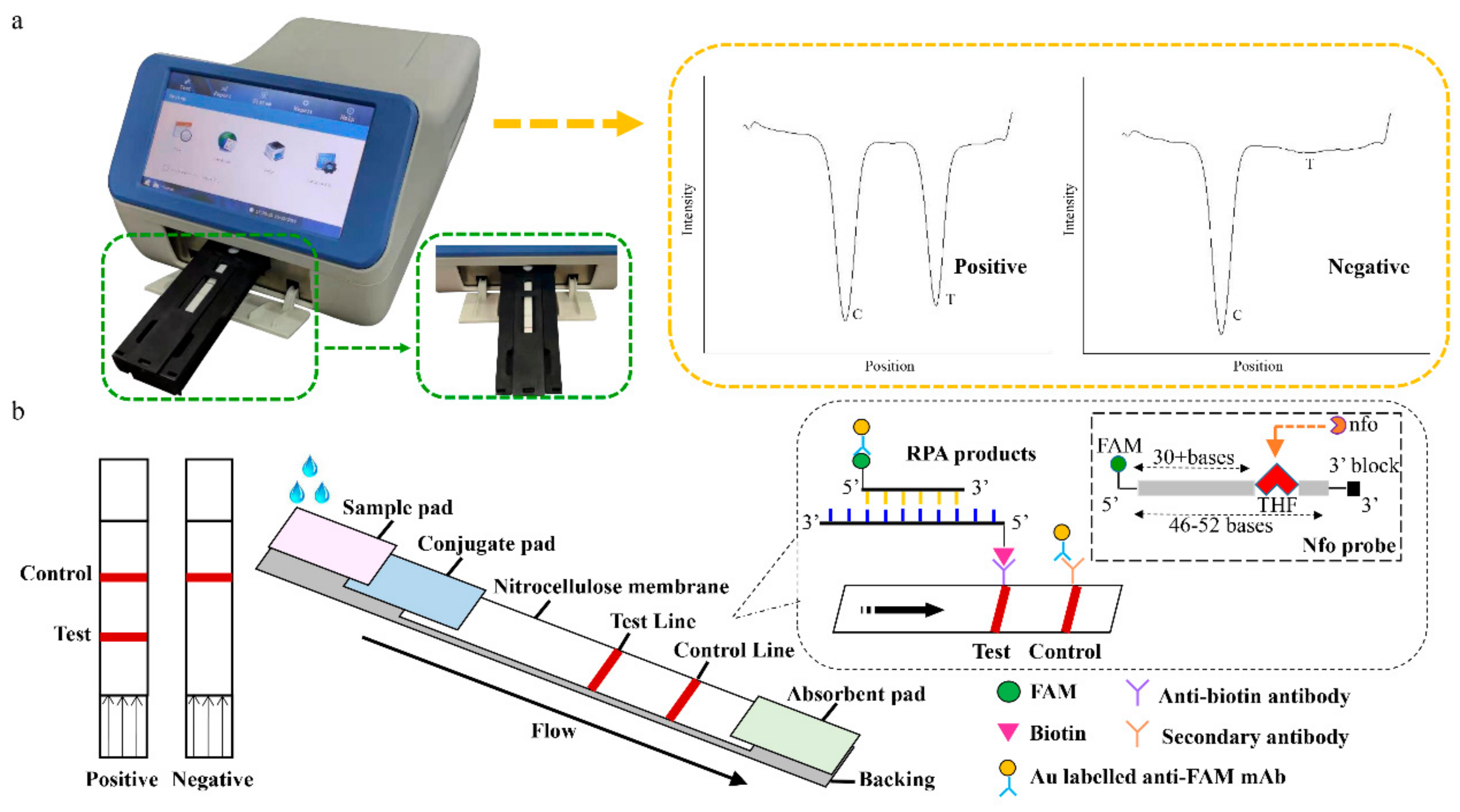
3.1. Assay Principle

The LFD-RPA assay used a pair of specific antibodies to identify the antigen containing two fixed labels for detection. The 5’ end biotin modified primer and the complementary sequences guaranteed the effective amplification of the target binding with the probe. The nfo probe consists of a FAM (carboxyfluorescein) antigenic label at its 5’ end, a tetrahydrofuran (THF) in the middle, and a C3-spacer at the 3’ end. The THF was the abasic-site mimic. The probe’s sequence was homologous to the overlapped region of the primer. It identifies the THF site in the extended double-stranded sequence and shears to form a 3’ hydroxyl substrate catalyzed by polymerase Bsu to extend the remaining portion of the probe extending to the remainder of the probe. Finally, amplicons containing biotin and FAM were obtained. The experimental results can be identified by the lateral flow dipstick. The amplification products were run by a capillary force along the nitrocellulose membrane. If the amplification succeeds, a red line is produced on the test wire due to the accumulation of colloidal gold. The uncaptured gold particles flowed through and were immobilized by the second antibody on the control line. In the absence of the target DNA, no red band is observed on the test line (Figure 1b). The entire procedure took approximately 5 min. In addition, the intensity of the test line and control line were scanned by the test strip reader (Figure 1a). The test strip reader converted the received light signals from test (T) line and control (C) line into electrical signals with “T value” and “C value”, and the results were described as the ratio of T to C values (T/C value).

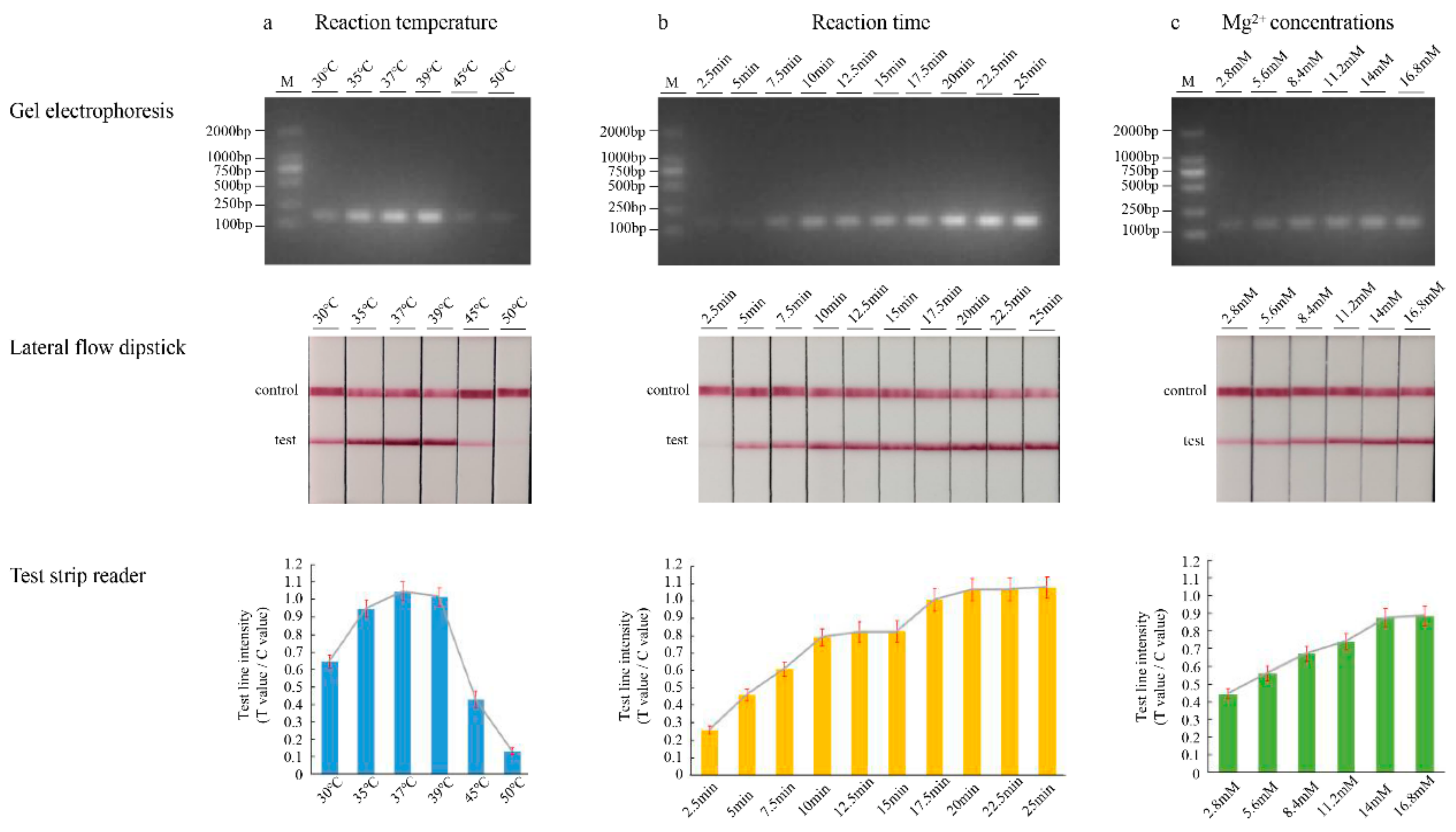
3.2. Optimization of the LFD-RPA Conditions

In order to achieve the optimal amplification effect, we adjusted the amount of pivotal component and reaction parameters in the reaction system. Firstly, six temperature gradients of 30, 35, 37, 39, 45, and 50 °C were conducted to determine the optimal RPA reaction temperature. We assessed this range of temperatures using an incubation time of 20 min and analyzed the amplification products using LF strips. As shown in Figure 2a, strong test lines were obtained at 35, 37, and 39 °C, and no obvious differences were directly observed in the range. When the temperature exceeded 39 °C, as the temperature increased, the stripes gradually became weak. Simultaneously, the test strips were placed into the test strip reader, followed by recording of the intensity of the reflected light for the test line and control line. Consequently, 37 °C was selected as the reaction temperature for subsequent experiments. Next, we tested the effect of incubation time on amplification efficiency. Ten time step gradients of 2.5, 5, 7.5, 10, 12.5, 15, 17.5, 20, 22.5, and 25 min were performed at 37 °C. As the amplification time increased, the brightness of the strip increased. There were no differences between the times of 10 and 15 min (Figure 2b). When the amplification time was 20 or 25 min, the band was brighter, but the reaction time was longer. This result indicates that 10 min is the appropriate reaction time. In addition, both in temperature- and time-optimization experiments, a concentration of 14 mM magnesium ion was used. Subsequently, six concentration gradients of 2.8, 5.6, 8.4, 11.2, 14, and 16.8 mM were tested. We assessed this range of magnesium ion concentrations using an incubation time of 10 min. The luminance of the RPA product analyzed by agarose gel electrophoresis and LFD remained relatively unchanged with concentration, from 14 to 16.8 mM (Figure 2c). Thus, 14 mM magnesium acetate was found to provide an optimal performance for the LFD-RPA assay in this study.

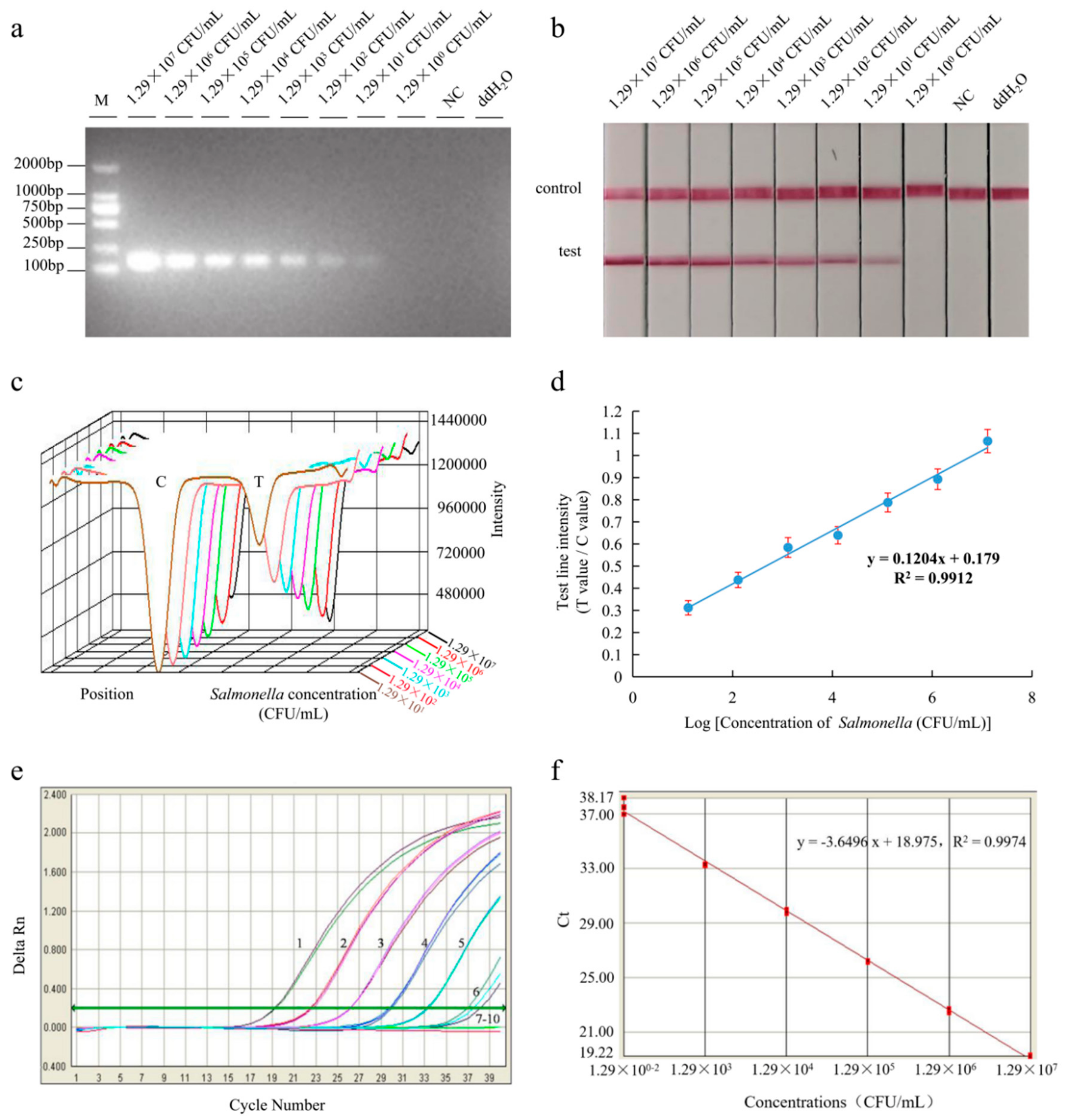
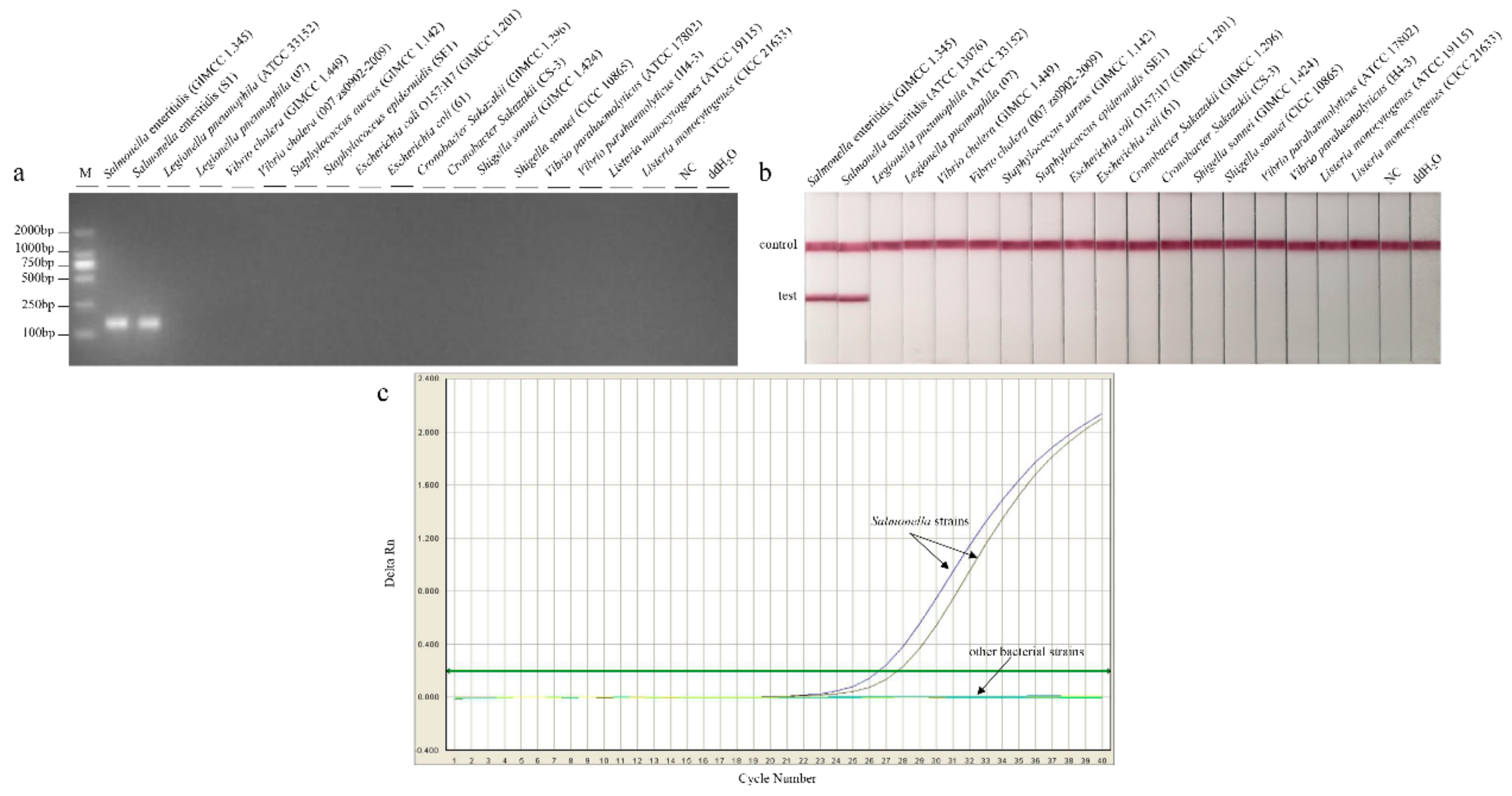
3.3. Sensitivity and Specificity

The LFD-RPA assay was applied to pure cultures with different Salmonella concentrations ranging from 1.29 × 10⁷ to 1 CFU/mL. The detection sensitivity of the medium was 1.29 × 101 CFU/mL, and the limit of detection of qPCR was 1.29 × 10² CFU/mL (Figure 3).
As for specificity, the two Salmonella strains were identified with a clear visual test line on the strip, whereas the 16 non-Salmonella strains only produced a visual band at the control line (Figure 4). No cross-reactions were observed. It revealed that LFD-RPA, based on specific primers, was a reliable assay.

3.4. Application to Food Samples

To analyze how different samples affected the LFD-RPA assay, the developed method was evaluated with tomato, cabbage, and broccoli samples spiked with Salmonella at concentrations ranging from 101 to 104 CFU/mL. Before artificial contamination, all samples were confirmed to be negative for Salmonella using BAM methods. Samples of each produce commodity that were not inoculated were used as negative controls. As shown in Table 3, the recovery of Salmonella in the range of (97.13 ± 0.62)% to (102.17 ± 1.32)% were obtained in three spiked samples. The detection limit of Salmonella in tomato, cabbage, and broccoli were at the same level. The assay has a slightly higher detection limit for Salmonella in food samples than that in pure cultures.

3.5. Detection of Practical Products

For practical product testing, 67 different sample types were analyzed (Table 4). Salmonella was clearly tested for in all samples using BAM, LFD-RPA, and qPCR assays. The positive detection rates of samples from the local market and Zhoushan Entry–Exit Inspection and Quarantine Bureau were 4% and 0%, respectively.

4. Discussion

With the rapid development of foreign trade, national food safety awareness is gradually improving. Food regulators and food producers are facing the same challenges. It is necessary not only to ensure the quality of detection, but also to accelerate the speed and shorten the testing process. The testing of Salmonella is very important for finding foodborne pathogens. The detection of Salmonella is developing towards the direction of being rapid, simple, of high sensitivity, strong specificity, and low cost. The application of isothermal amplification technology has made microbial detection free from dependence on expensive instruments. It is suitable for field testing and economically underdeveloped areas.
Recently, techniques like polymerase chain reaction (PCR), real-time PCR (qPCR), enzyme-linked immunosorbent assay (ELISA), and gene chip technology for detecting pathogens have developed rapidly and maturely. However, these assays have several drawbacks. Professionals are needed in some steps and the reaction reagents are expensive and unaffordable in rural areas. Therefore, the establishment of rapid and effective detection approaches is crucial. Recombinase polymerase amplification could amplify target fragments in a shorter time (10 min). By contrast, a longer time (40 min) is required for a Loop-mediated isothermal amplification (LAMP) assay, and approximately 40 cycles (40 min) are required in the qPCR assay [1,13,28]. Clearly, the RPA assay is more efficient. The RPA reaction is performed at an ambient constant temperature. Consequently, it evades the need for a high-cost thermo-cycler. In addition, RPA was able to tolerate the impure samples. The tomato, cabbage, and broccoli matrices had little effect on the analytical sensitivity of RPA, and the limit of detection was at a similar level to the pure bacterial culture in our study.
In general, the false-positive results were more likely to occur due to unexpected amplification or primer dimers [14]. An appropriate RPA primer/probe combination could avoid false positives or low sensitivity [29]. Furthermore, studies found that probes played an important role in RPA. In contrast, the change of primers did not have a significant impact [30]. Thus, we referred to the relevant literature in the design process [31]. The nucleotide composition, sequence length, and interplay between primers and probe were carefully considered.
Until now, the utility of isothermal amplification outside the laboratory has been restricted by the lack of signal transduction that is easy-to-use, low cost, and specific [32]. The RPA reaction could be undertaken in a water bath where the temperature is slightly higher than the surrounding temperature [33]. The lateral flow dipstick was free of expensive instruments, and it could be directly observed by the naked eye at room temperature. The LFD-RPA is suitable for testing in the field, and could provide low-cost and instrument-free nucleic acid detection in remote areas. In the previous reports of literatures, LFD-RPA assay was applied to detect Salmonella in shellfish with a detection limit of 5 CFU/mL in 8 min at 40 °C [1]. The testing of Salmonella typhimurium in milk could successfully reach as low as 1.95 CFU/mL in 10 min at 40–42 °C [29]. Commercially available kits could obtain a reaction result within 15 min at 40 °C [34]. Both were performed with one target fragment in a short time. In this paper, the sensitivity of LFD-RPA assay was 12 CFU/mL within 10 min at 37 °C. Although there is no obvious improvement in terms of reaction time and sensitivity, the artificial reading errors could have been minimaxed by using the reading instrument. The portable TSR-100 test strip reader was not only sensitive, but also could be applied to scan the test strips to decrease the man-made influence and increase the correctness of the diagnosis in this study. By using modern photoelectric technology and a photo-diode to detect the intensity of the reflected light, the reader can calculate the concentration. Such a reader can avoid reading mistakes by eye and perform with rapidity and high sensitivity in order to detect the color change of the test strip. Furthermore, the LFD-RPA assay with the test strip reader has the potential of rapid and quantitative detection in field. However, a few problems need to be solved. To date, LFD cannot distinguish more than two nucleotide amplicons in one RPA reaction system [25]. The challenges for the high-throughput detection of foodborne pathogens will be the focus in future research.

5. Conclusions

This study successfully combined recombinase polymerase amplification with a lateral flow immunoassay for rapid detection of Salmonella in food. The assay was easy to operate and could be completed in 10 min. It also showed a good specificity and high sensitivity. The reliability was verified by comparing the results of qPCR and BAM. The LFD-RPA method for the detection of Salmonella is not only time saving and easy to perform, but also generates specific and sensitive results for routine applications. This method does not require any complex equipment, and can be used as a rapid quantitative detection method with a portable strip reader. The fast and easy-to-read system could be useful as a rapid point-of-care-test for Salmonella in food. In summary, the LFD-RPA has practical significance for rapid screening in ports, and detection in grassroots areas or economically underdeveloped areas. However, future essential improvements will reduce potential contamination risks arising when testing many samples in the field.

Author Contributions

M.Z., X.Y. and C.S. designed the experiments. J.L., B.M., J.F., A.Z., E.C. and Y.X. conducted experiments and analyzed data. J.L. and B.M. wrote the main manuscript text and prepared all figures and tables. All authors reviewed the manuscript. All authors have read and agreed to the published version of the manuscript.

Funding

The work was supported by the National Key Research and Development Program of China (2017YFF0210203, 2018YFF0215205), the Public Projects of Zhejiang Province (LGC19C200006).

Acknowledgments

We also thank the researchers who facilitated the completion of this study.

Conflicts of Interest

The authors declare no conflict of interest.

References

  1. Gao, W.; Huang, H.; Zhu, P.; Yan, X.; Fan, J.; Jiang, J.; Xu, J. Recombinase polymerase amplification combined with lateral flow dipstick for equipment-free detection of Salmonella in shellfish. Bioprocess. Biosyst. Eng. 2018, 41, 603–611. [Google Scholar] [CrossRef] [PubMed]
  2. Li, R.; Lai, J.; Wang, Y.; Liu, S.; Li, Y.; Liu, K.; Shen, J.; Wu, C. Prevalence and characterization of Salmonella species isolated from pigs, ducks and chickens in Sichuan Province, China. Int. J. Food Microbiol. 2013, 163, 14–18. [Google Scholar] [CrossRef] [PubMed]
  3. Choi, S.W.; Ha, J.S.; Kim, B.Y.; Lee, D.H.; Park, J.K.; Youn, H.N.; Hong, Y.H.; Lee, S.B.; Lee, J.B.; Park, S.Y.; et al. Prevalence and characterization of Salmonella species in entire steps of a single integrated broiler supply chain in Korea. Poult. Sci. 2014, 93, 1251–1257. [Google Scholar] [CrossRef] [PubMed]
  4. Rukambile, E.; Sintchenko, V.; Muscatello, G.; Kock, R.; Alders, R. Infection, colonization and shedding of Campylobacter and Salmonella in animals and their contribution to human disease: A review. Zoonoses Public Health 2019, 66, 562578. [Google Scholar] [CrossRef]
  5. Jarvis, N.A.; O’Bryan, C.A.; Dawoud, T.M.; Park, S.H.; Kwon, Y.M.; Crandall, P.G.; Ricke, S.C. An overview of Salmonella thermal destruction during food processing and preparation. Food Control 2016, 68, 280–290. [Google Scholar] [CrossRef]
  6. Fang, J.; Wu, Y.; Qu, D.; Ma, B.; Yu, X.; Zhang, M.; Han, J. Propidium monoazide real-time loop-mediated isothermal amplification for specific visualization of viable Salmonella in food. Lett. Appl. Microbiol. 2018, 67, 79–88. [Google Scholar] [CrossRef]
  7. Nair, D.V.T.; Venkitanarayanan, K.; Kollanoor Johny, A. Antibiotic-Resistant Salmonella in the Food Supply and the Potential Role of Antibiotic Alternatives for Control. Foods 2018, 7, 167. [Google Scholar] [CrossRef] [Green Version]
  8. Colello, R.; Ruiz, M.J.; Padín, V.M.; Rogé, A.D.; Leotta, G.; Padola, N.L.; Etcheverría, A.I. Detection and Characterization of Salmonella Serotypes in the Production Chain of Two Pig Farms in Buenos Aires Province, Argentina. Front. Microbiol. 2018, 9, 1370. [Google Scholar] [CrossRef] [Green Version]
  9. GB.4789.4-2016. National Food Safety Standard for Salmonella in Food Microbiological Examination; Standards Press of China: Beijing, China, 2016. [Google Scholar]
  10. Nassib, T.A.; El-Din, M.Z.; El-Sharoud, W.M. Assessment of the presence of Salmonella spp. in Egyptian dairy products using various detection media. Lett. Appl. Microbiol. 2003, 37, 405–409. [Google Scholar] [CrossRef] [Green Version]
  11. SN/T 1059.7-2010. Detection of Salmonella in Food for Import and Export—Real-Time PCR Method; Standards Press of China: Beijing, China, 2010. [Google Scholar]
  12. Zhang, X.; Guo, L.; Ma, R.; Cong, L.; Wu, Z.; Wei, Y.; Xue, S.; Zheng, W.; Tang, S. Rapid detection of Salmonella with Recombinase Aided Amplification. J. Microbiol. Methods 2017, 139, 202–204. [Google Scholar] [CrossRef]
  13. Kim, J.Y.; Lee, J.L. Rapid detection of Salmonella enterica serovar enteritidis from eggs and chicken meat by real-time recombinase polymerase amplification in comparison with the two-step real-time PCR. J. Food Saf. 2016, 36, 402–411. [Google Scholar] [CrossRef]
  14. Piepenburg, O.; Williams, C.H.; Stemple, D.L.; Armes, N.A. DNA detection using recombination proteins. PLoS Biol. 2006, 4, e204. [Google Scholar] [CrossRef] [PubMed]
  15. Liu, W.; Liu, H.X.; Zhang, L.; Hou, X.X.; Wan, K.L.; Hao, Q. A Novel Isothermal Assay of Borrelia burgdorferi by Recombinase Polymerase Amplification with Lateral Flow Detection. Int. J. Mol. Sci. 2016, 17, 1250. [Google Scholar] [CrossRef]
  16. Ma, B.; Fang, J.; Wang, Y.; He, H.; Dai, M.; Lin, W.; Su, W.; Zhang, M. Isothermal Method of a Recombinase Polymerase Amplification Assay for the Detection of Most Common High-Risk Human Papillomavirus Type 16 and Type 18 DNA. Clin. Lab. 2017, 63, 27–38. [Google Scholar] [CrossRef]
  17. Ng, B.Y.C.; Wee, E.J.H.; Woods, K.; Anderson, W.; Antaw, F.; Tsang, H.Z.H.; West, N.P.; Trau, M. Isothermal Point Mutation Detection: Toward a First-Pass Screening Strategy for Multidrug-Resistant Tuberculosis. Anal. Chem. 2017, 89, 9017–9022. [Google Scholar] [CrossRef]
  18. Choi, G.; Jung, J.H.; Park, B.H.; Oh, S.J.; Seo, J.H.; Choi, J.S.; Kim, D.H.; Seo, T.S. A centrifugal direct recombinase polymerase amplification (direct-RPA) microdevice for multiplex and real-time identification of food poisoning bacteria. Lab Chip 2016, 16, 2309–2316. [Google Scholar] [CrossRef]
  19. Ji, Y.K.; Lee, J.L. Development of a multiplex real-time recombinase polymerase amplification (RPA) assay for rapid quantitative detection of Campylobacter coli, and jejuni, from eggs and chicken products. Food Control 2017, 73, 1247–1255. [Google Scholar]
  20. Xu, C.; Li, L.; Jin, W.; Wan, Y. Recombinase polymerase amplification (RPA) of CaMV-35S promoter and nos terminator for rapid detection of genetically modified crops. Int. J. Mol. Sci. 2014, 15, 18197–18205. [Google Scholar] [CrossRef] [Green Version]
  21. Wang, R.; Zhang, F.; Wang, L.; Qian, W.; Qian, C.; Wu, J.; Ying, Y. Instant, Visual, and Instrument-Free Method for On-Site Screening of GTS 40-3-2 Soybean Based on Body-Heat Triggered Recombinase Polymerase Amplification. Anal. Chem. 2017, 89, 4413–4418. [Google Scholar] [CrossRef]
  22. Magro, L.; Jacquelin, B.; Escadafal, C.; Garneret, P.; Kwasiborski, A.; Manuguerra, J.C.; Monti, F.; Sakuntabhai, A.; Vanhomwegen, J.; Lafaye, P.; et al. Paper-based RNA detection and multiplexed analysis for Ebola virus diagnostics. Sci. Rep. 2017, 7, 1347. [Google Scholar] [CrossRef]
  23. Tan, K.K.; Azizan, N.S.; Yaacob, C.N.; Che Mat Seri, N.A.A.; Samsudin, N.I.; Teoh, B.T.; Sam, S.S.; AbuBakar, S. Operational utility of the reverse-transcription recombinase polymerase amplification for detection of dengue virus. BMC Infect. Dis. 2018, 18, 169. [Google Scholar] [CrossRef] [PubMed]
  24. Yeh, K.S.; Chen, T.H.; Liao, C.W.; Chang, C.S.; Lo, H.C. PCR amplification of the Salmonella typhimurium fimY gene sequence to detect the Salmonella species. Int. J. Food Microbiol. 2002, 78, 227–234. [Google Scholar] [CrossRef]
  25. Tinker, J.K.; Clegg, S. Characterization of FimY as a coactivator of type 1 fimbrial expression in Salmonella enterica serovar Typhimurium. Infect. Immun. 2000, 68, 3305–3313. [Google Scholar] [CrossRef] [PubMed] [Green Version]
  26. Zhang, L.; Du, X.; Chen, Z.; Chen, C.; Gong, N.; Song, Y.; Song, Y.; Han, Q.; Xia, X.; Luo, H.; et al. Instrument-Free and Visual Detection of Salmonella Based on Magnetic Nanoparticles and an Antibody Probe Immunosensor. Int. J. Mol. Sci. 2019, 20, 4645. [Google Scholar] [CrossRef] [PubMed] [Green Version]
  27. Primer-BLAST. Available online: https://0-www-ncbi-nlm-nih-gov.brum.beds.ac.uk/tools/primer-blast/index.cgi (accessed on 23 December 2019).
  28. Mei, X.R.; Zhai, X.W.; Lei, C.W.; Ye, X.L.; Kang, Z.Z.; Wu, X.; Xiang, R.; Wang, Y.L.; Wang, H.N. Development and application of a visual loop-mediated isothermal amplification combined with lateral flow dipstick (LAMP-LFD) method for rapid detection of Salmonella strains in food samples. Food Control 2019, 104, 9–19. [Google Scholar] [CrossRef]
  29. Hu, J.; Huang, R.; Sun, Y.; Wei, X.; Wang, Y.; Jiang, C.; Geng, Y.; Sun, X.; Jing, J.; Gao, H.; et al. Sensitive and rapid visual detection of Salmonella Typhimurium in milk based on recombinase polymerase amplification with lateral flow dipsticks. J. Microbiol. Methods 2019, 158, 25–32. [Google Scholar] [CrossRef]
  30. Liu, X.Q.; Yan, Q.Y.; Huang, J.F.; Chen, J.; Guo, Z.Y.; Liu, Z.D.; Cai, L.; Li, R.S.; Wang, Y.; Yang, G.W.; et al. Influence of design probe and sequence mismatches on the efficiency of fluorescent RPA. World J. Microbiol. Biotechnol. 2019, 35, 95. [Google Scholar] [CrossRef]
  31. Higgins, M.; Ravenhall, M.; Ward, D.; Phelan, J.; Ibrahim, A.; Forrest, M.S.; Clark, T.G.; Campino, S. PrimedRPA: primer design for recombinase polymerase amplification assays. Bioinformatics 2019, 35, 682–684. [Google Scholar] [CrossRef] [Green Version]
  32. Phillips, E.A.; Moehling, T.J.; Bhadra, S.; Ellington, A.D.; Linnes, J.C. Strand Displacement Probes Combined with Isothermal Nucleic Acid Amplification for Instrument-Free Detection from Complex Samples. Anal. Chem. 2018, 90, 6580–6586. [Google Scholar] [CrossRef] [Green Version]
  33. Lillis, L.; Lehman, D.; Singhal, M.C.; Cantera, J.; Singleton, J.; Labarre, P.; Toyama, A.; Piepenburg, O.; Parker, M.; Wood, R.; et al. Non-instrumented incubation of a recombinase polymerase amplification assay for the rapid and sensitive detection of proviral HIV-1 DNA. PLoS ONE 2014, 9, e108189. [Google Scholar] [CrossRef] [Green Version]
  34. TwistFlow® Manuals. Available online: https://www.twistdx.co.uk/en/support/manuals/twistflow-manuals (accessed on 17 December 2019).
Figure 1. The working principle of the recombinase polymerase amplification combined with lateral flow dipstick (LFD-RPA) assay. (a) The test strip reader and the positive/negative test results. (b) Schematic diagram of the recombinase polymerase amplification combination with lateral flow dipstick. FAM: carboxyfluorescein, THF: tetrahydrofuran.
Foods 09 00027 g001
Figure 2. Optimization of the recombinase polymerase amplification reaction temperature (a), incubation time (b) and the concentration of magnesium ions (c) using target DNA. Gel electrophoresis verifying amplification (top), LFD test results (middle), and LFD test line quantification (bottom). Test line intensity was the ratio of the reflected light for the test line and control line.
Foods 09 00027 g002
Figure 3. Reaction sensitivity of LFD-RPA and qPCR assays for the Salmonella strain culture. The amplified products could be observed with the naked eye by using gel electrophoresis (a) and lateral flow dipstick (b). The intensity (c) was used for quantitative analysis, and it shows a linear correlation (d) with the concentration of pure cultures. The qPCR assay (e) test concentrations ranged from 1.29 × 10⁷ to 1.29 colony-forming units (CFU)/mL, and the standard curve (f) was generated with the Bio-Rad iQTM5 optical System software. 1–8: 1.29 × 10⁷, 1.29 × 10⁶, 1.29 × 10⁵, 1.29 × 10⁴, 1.29 × 10³, 1.29 × 10², 1.29 × 101, and 1.29 × 100 CFU/mL; 9: Negative control; 10: ddH2O. Test line intensity was the ratio of the reflected light for the test line and control line.
Foods 09 00027 g003
Figure 4. Specificity test of the recombinase polymerase amplification (a) combination with lateral flow dipstick (b) and qPCR (c) assays for targeting Salmonella.
Foods 09 00027 g004
Table 1. Information of bacterial strains used for specificity tests in the study.
SpeciesID of StrainsResult of RPAResult of qPCR
Salmonella EnteritidisGIMCC 1.345++
Salmonella EnteritidisATCC 13076++
Legionella pneumophilaATCC 33152
Legionella pneumophila07 *
Vibrio choleraeGIMCC 1.449
Vibrio cholerae007 zs0902-2009 *
Staphylococcus aureusGIMCC 1.142
Staphylococcus epidermidisSE1 *
Escherichia coli O157:H7GIMCC 1.201
Escherichia coli
O157:H7
61 *
Cronobacter SakazakiiGIMCC 1.296
Cronobacter SakazakiiCS-3 *
Shigella sonneiGIMCC 1.424
Shigella flexneriCICC 10865
Vibrio parahaemolyticusATCC 17802
Vibrio parahaemolyticusH4-3 *
Listeria monocytogenesATCC 19115
Listeria monocytogenesCICC 21633
RPA: recombinase polymerase amplification; qPCR: real-time quantitative polymerase chain reaction; GIMCC: Guangdong Microbiology Culture Center, Guangdong, China; ATCC: American Type Culture Collection, Virginia, USA; CICC: China Center of Industrial Culture Collection, Shanghai, China; * Afforded by Zhoushan Entry-exit Inspection and Quarantine Bureau, Zhejiang, China; +: positive result; −: negative result.
Table 2. Sequences of Salmonella enteritidis primers and probes.
Primer NameSequenceTarget GeneFragment Length
PCR PrimersfimY
Sal-F5′-CGGTTGGTATCATTTCTC-3’249 bp
Sal-R5′-GCAGGTAAATCTGGATATC-3′
qPCR Primers and Probe
Sal-qF5′-CGGTTGGTATCATTTCTC-3′249 bp
Sal-qR5′-GCAGGTAAATCTGGATATC-3′
Sal-qP5′-FAM-ATTACAACTGACAACTACCTCGGCT-BQH1-3’
RPA Primers and Probe
Sal RPA FP15′-TATCAGATAAAACCTCCGCTATAACACAGT-3’131 bp
Sal RPA RP15’-Biotin-CTTTCCGATAAGCGAGGTTTGGAGGCTGAT-3’
Sal RPA P1
nfo Probe
5′-FAM-TCAGATAAAACCTCCGCTATAACACAGTTT[THF]ACGATGGCTGGGCGTT-C3-Spacer
Table 3. Recovery of Salmonella in artificially contaminated produce commodities by recombinase polymerase amplification combined with lateral flow dipstick (LFD-RPA) assay.
Samples (n = 6 Each)StrainInoculation Level (CFU/mL)LFD-RPA Detected Concentration (CFU/mL)Recovery (%)
tomatoGIMCC1.3451.29 × 104+ (1.269 ± 0.014) × 10498.37 ± 1.09
1.29 × 10³+ (1.304 ± 0.010) × 103101.08 ± 0.78
1.29 × 10²+ (1.253 ± 0.008) × 10297.13 ± 0.62
1.29 × 101
cabbage1.29 × 104+ (1.282 ± 0.021) × 10499.38 ± 1.63
1.29 × 10³+ (1.264 ± 0.016) × 10397.98 ± 1.24
1.29 × 10²+ (1.298 ± 0.011) × 102100.62 ± 0.85
1.29 × 101
broccoli1.29 × 104+ (1.312 ± 0.012) × 104101.71 ± 0.93
1.29 × 10³+ (1.318 ± 0.017) × 103102.17 ± 1.32
1.29 × 10²+ (1.258 ± 0.019) × 10297.51 ± 1.48
1.29 × 101
+, Salmonella positive by the method. −, Salmonella negative by the method.
Table 4. Detection of practical samples by LFD-RPA assay compared with qPCR and bacteriological analytical manual (BAM) method.
ResourceSamplesNumber of SamplesPositive Number
LFD-RPAqPCRBAM
Local marketShrimp11111
Tomato6000
Cabbage6000
Cod7111
Meretrix4000
Broccoli6000
Chicken2000
Shellfish7000
Dried squid1000
Total50222
Positive detection rate (%)/4.04.04.0
Zhoushan Entry–Exit Inspection and Quarantine BureauDried squid17000
Positive detection rate (%)/000

