Article

Enhanced Uptake of Arsenic Induces Increased Toxicity with Cadmium at Non-Toxic Concentrations on Caenorhabditis elegans

1
Information Materials and Intelligent Sensing Laboratory of Anhui Province, Institutes of Physical Science and Information Technology, Anhui University, Hefei 230601, China
2
Key Laboratory of High Magnetic Field and Ion Beam Physical Biology, Chinese Academy of Sciences; Anhui Province Key Laboratory of Environmental Toxicology and Pollution Control Technology, High Magnetic Field Laboratory, Hefei Institutes of Physical Science, Chinese Academy of Sciences, Hefei 230031, China
*
Authors to whom correspondence should be addressed.
Submission received: 17 February 2022 / Revised: 3 March 2022 / Accepted: 7 March 2022 / Published: 10 March 2022
(This article belongs to the Section Ecotoxicology)

Abstract

:
Cadmium (Cd) and arsenic (As) are widely distributed pollutants that co-exist in the environment; however, their joint toxicity on living organisms is still largely unknown. In this study, we explored the joint toxicity of concurrent exposure to Cd and different As species at low concentrations on Caenorhabditis elegans (C. elegans) in comparison to single exposures. Endpoints such as germ cell apoptosis, the number of oocytes, brood size, and the life span were employed to evaluate the combined effects of Cd and As on exposed C. elegans from L3 or L4 stages. Our results showed that concurrent exposure to non-toxic concentrations of Cd and As caused the synergy of reproductive and developmental toxicity. The presence of Cd promoted the accumulation of As in both germline and intestine detected by laser ablation inductively coupled plasma mass spectrometry (LA-ICP-MS). Although a conversion of As(III) to As(V) was detected as dependent on pH according to the microenvironment of the intestine in the worm, there was no significant difference of toxicity in C. elegans concurrently exposed to Cd and different As species. Using loss-of-function mutant strains, As was deemed responsible for the enhanced joint toxicity, and in which gcs-1 played a key protective role. These data help to better evaluate the comprehensive adverse effects of concurrent exposure of heavy metals at low concentrations on living organisms in the environment.

1. Introduction

Heavy metal pollutants are widely distributed in the natural environment due to local geological background and intensified industrialization [1]. Cadmium (Cd) and arsenic (As) receive the most attention due to their threat to the surrounding ecology and human health [2,3]. Both Cd and As have been recognized as carcinogens with high toxicity [4]. Cadmium is well known as the cause of “itai-itai disease” in Japan [5], and trace amounts of Cd entering the human body may lead to kidney, liver, and bone damage [6]. Cadmium decreases DNA polymerase fidelity, inhibits DNA repair [7], and significantly increases the mutation rate in mammalian cells [8]. Injection of CdCl2 into mice causes kidney damage and alters testicular sperm expression in mice [9]. It could also affect the motility of Caenorhabditis elegans (C. elegans) with a significant decrease in crawling speed and bending frequency [10]. Studies showed that Cd inhibited SOD and AChE enzyme activities in C. elegans, and caused reproductive and developmental toxicity [11]. Bovio et al. [12] reported that the nematode mortality rose in a dose-dependent manner to Cd concentration due to the loss of enzymatic activity of SOD1. YeonRoh et al. [13] demonstrated that the mtl-2 (gk125) had the lowest LC50 among mutant strains when exposed to Cd, showing significantly less egg production and increased expression of mtl-2 gene. Arsenic has multiple species with different toxicity in As(III) > As(V) > Dimethylarsinic Acid (DMA) > Monomethylarsonic Acid (MMA) order, indicating a higher risk from inorganic As than organic As [14,15]. It has a significant impact on human health, causing cardiovascular diseases, neurobehavioral disorders, hematological disorders, and various types of diseases [16], by blocking DNA repair and apoptosis, stimulating angiogenesis, causing cell cycle arrest, and inducing aneuploidy [17,18]. Arsenic also threatens various organisms in the environment, inducing tumors in mice, elevating oxidative stress in chicks, and reducing the pigment contents in plants [19,20,21]. Arsenic exposure may cause increased amounts of GST-4, HSP-4, and HSP-70 in C. elegans, producing more reactive oxygen species (ROS) and thus causing toxic effects [22]. Yu et al. [23] found that the mRNA levels of transcriptional makers of aging (hsp-16.1, hsp-16.49, and hsp-70) increased in worms under arsenite exposure, accelerating senescence of the nematodes. By parental exposure alone to As, C. elegans was found to show decrease in the number of eggs laid in both the parental and offspring generations, and further investigation suggested that the intergenerational reproductive toxicity was due to the decrease of spr-5 mRNA and the increase of H3K4me2 level in the parental generation [24].
The environmental toxicity of Cd and As is mainly due to their alarming pollution levels in soil and water. The worldwide background value of soil Cd is about 0.35 mg/kg, while the concentration in polluted areas far exceeds this level [25]. In Bangladesh, Cd pollution in water and soil are tens of times higher than the global background value [26]. Cadmium is also considered the most heavily polluted heavy metal in China [26,27,28]. According to the 2014 National Soil Pollution Survey Bulletin, the total exceedance rate of Cd in the soil is 16.1%, of which the highest exceedance rate is 7%, and the exceedance values are tens or even hundreds of times higher than the environmental background value [29]. The world has been exposed to high concentrations of As for a long time [30]. There are more than 10,000 As-contaminated sites in Australia [31]. In Bangladesh, As levels in cultivated soils of Faridpur can reach four times the world limit [27]. In China, the “12th Five-Year Plan for heavy metal pollution control” [32] has recognized arsenic as a first-class heavy metal pollution that needs urgent prevention and control. Arsenic pollution in Xinjiang, Inner Mongolia, Shanxi, and other provinces and regions in China is relatively serious. For example, As concentration in the surface soil of Xi’an reached 14.5 mg/kg, approximately three times more than the world soil background value [33]. As multiple heavy metal pollutants co-exist in the environment, it is hard to reveal the true picture of their hazardous effects by single pollutant exposure, while the joint toxicity of heavy metal exposure urges the need for comprehensive investigation.
The combined toxic effects of two or more compounds on an organism simultaneously or sequentially is defined as joint toxicity [34]. The concurrent exposure effects can be categorized as antagonistic effects, synergistic effects, and additive effects [35,36]. Andrade et al. [37] investigated the neurological and behavioral effects of nickel (Ni) and zinc (Zn) co-exposure on rats, and found that Zn attenuated the motor impairment caused by Ni, indicating antagonistic effects. It was found that co-exposure of mercury (Hg) and Cd inhibited the development of follicles in the human ovary, thereby reducing the number of human oocytes, indicating synergistic effects [38]. Lanier et al. [39] used edible kale and white clover as models to study the chronic toxic effects of Cd and lead (Pb), and the data showed that the concurrent exposure slowed the growth of roots of white clover and caused DNA damage, indicating synergistic effects. Meng et al. [40] found the chlorophyll content of wheat seedlings by co-exposure of Cd and Pb lying between single exposures, indicating an additive effect. Therefore, the different joint toxicity of heavy metals may be influenced by the type of metals involved, the existing form of pollutants, the accumulation of heavy metals in the organisms, and the test models.
C. elegans is a nematode ubiquitously distributed in the environment, such as soil and water. It is sensitive to the toxicity of pollutants, and has short life cycle, large number of offspring, transparent body, simple organ composition, clear genetic background, and various mutant strains, showing remarkable advantages as an environment-related model organism [41]. In addition, C. elegans genome has 60–80% of human homologs, which may reveal health implication based on its toxicological research [42,43]. It has been widely used in the study of both single and joint toxicity of various heavy metals so far [11,38]. With the newly developed analytical tools such as laser ablation-inductively coupled plasma-mass spectrometry (LA-ICP-MS), we can further explore the accumulation of heavy metals in different organs of C. elegans, providing basis for determining the underlying mechanism of possible toxicity [44,45]. In this study, we explored the joint toxicity of Cd and As at low concentrations on C. elegans. The accumulation and distribution of heavy metals in C. elegans, the transformation of As in the intestinal microenvironment, and the potential mechanism based on mutant strains were explored to explain the co-toxic effects. This study could provide important information on the potential hazards of heavy metal co-exposure in living organisms, and thus laying basis to devise regulations to better manage the hazards of heavy metals.

2. Materials and Methods

2.1. Preparation of Test Solutions

We used CdCl2 (99% purity, AR, Aladdin Group Chemical Reagent Co., Ltd., Shanghai, China) and NaAsO2 (>90% purity, AR, Aladdin Group Chemical Reagent Co., Ltd., Shanghai, China) as reference materials to prepare the stock solutions by dissolving precisely weighted powder samples in Milli-Q water to 1 mM. Arsenic(V) oxide hydrate reference material of 1000 mg/L (99%, Inorganic Ventures, Lakewood, NJ, USA) and the stock solutions of CdCl2 and NaAsO2 were stored in a refrigerator at 4 °C until use. Different working concentrations were prepared by diluting the stock solutions with K-medium [KM, 51 mM NaCl (99.5%purity, AR, Sinopharm Chemical Reagent Co., Ltd., Shanghai, China) + 32 mM KCl (99.5%purity, AR, Sinopharm Chemical Reagent Co., Ltd., Shanghai, China)] before use.

2.2. Test Organism

According to the standard procedure described by Brenner [46], all worm strains are cultured on nematode growth medium (NGM) at 20 °C in dark, with Escherichia coli (E. coli) OP50 seeded as food. Strains including N2 (wild-type Bristol), mtl-2 (gk125), and gcs-1 (ok436) are provided by Caenorhabditis Genetics Center (CGC). Synchronized worms can be obtained when gravid hermaphrodites were lysed in an alkaline hypochlorite solution, and the harvested eggs are hatched overnight at 20 °C without food to remain at L1 stage.

2.3. Toxicity Experiments

Germ cell apoptosis: According to the standard procedure described by Gartner [47], apoptotic germ cells were detected by vital staining with acridine orange (AO) (Shenggong Bioengineering Co., Ltd., Shanghai, China). C. elegans at L4 stage were exposed to designated working solutions on Costar 24-well plates. After exposure, the solutions containing C. elegans were aspirated into centrifuge tubes, and the worms were collected after natural settling, then placed in 400 μL of 75 g/mL AO with OP50 for an incubation at 20 °C for 60 min. After repeated elution, C. elegans were moved to NGM plates for a 60 min recovery on bacterial lawns. The purpose of recovery is to remove AO from the intestine of the worms and make it easier for subsequent detection of apoptotic germ cells. The worms were then anaesthetized with sodium azide 2% (NaN3) (>99.5%, Sigma-Aldrich, St. Louis, MO, USA) and examined under an Axioscope 5 fluorescence microscope (1769-608, Zeiss, Germany) at 20×. After AO staining, the apoptotic cells were yellow-green, indicating increased DNA fragmentation, while all intact cells were uniformly green. Numbers of apoptotic cells were obtained from at least 20 worms for each group.
Oocyte assay: The C. elegans oocyte count was referred to the method of Ruan et al. [48]. Following the same exposure scenario as germ cell apoptosis, 20 treated C. elegans from each group were placed on agar plate and anaesthetized with a drop of 2% NaN3. The number of oocytes was counted from the spermatheca to the gonad loop per gonad arm under an optical microscope.
Brood-size assay: According to the method of Ruan et al. [48], 15 worms were randomly selected and transferred to 3 mm NGM plates seeded with OP50 after 24 h of exposure from L3 stage. Adults C. elegans were transferred to new NGM plates every 24 h until oviposition ceased (approximately 5 days), and eggs laid on the NGM plates were counted under a microscope (K series, Motic, Guangzhou, China).
Life-span assay: According to the method of Du et al. [49], lifespan assays were conducted in 96-well plates containing designated working concentrations, 20 μg/mL 5-fluoro-2′-deoxyuridine (5-FudR, >99%, Sigma-Aldrich, St. Louis, MO, USA) to prevent egg-laying, and concentrated OP50 in KM with a total volume of 200 μL. A total of 20 C. elegans grown synchronously to L3 stage were transferred to the plates, and one worm was placed in each well. The C. elegans were observed daily under a microscope, and worms with a lack of response to physical stimulation were deemed dead. Survival curves were recorded by continuously exposing C. elegans to test solutions until all worms were dead.

2.4. Cd and As Accumulation in C. elegans

Synchronized L1 C. elegans were placed on NGM plates for a cultivation of 48 h and collected. Since the working concentrations were low, we needed to raise a large number of C. elegans for the test. After being exposed in 9 mm Petri dishes for 24 h, C. elegans were washed at least 3 times with Milli-Q water in order to remove the exposure solutions on the nematodes. After washing and removing the supernatant, the worms were frozen at −20 °C in a refrigerator and freeze-dried for at least 48 h until constant weight was reached. Dried worms were precisely weighted, then digested in HNO3 (99.5%purity, GR, Sinopharm Chemical Reagent Co., Ltd., Shanghai, China) by electric heating at 150 °C. The contents of Cd and As in C. elegans were determined by inductively coupled plasma-mass spectrometry (ICP-MS, NexION 2000, Perkin Elmer, Waltham, MA, USA) and atomic fluorescence spectrometer (AFS-930, Titan Instruments, Beijing Jitian Instrument Co., Ltd., Beijing, China), respectively, based on dry weight.

2.5. Cd and As Distribution in C. elegans

The distribution of Cd and As in the C. elegans was determine by LA-ICP-MS at the Mineral Deposit and Exploration Center (ODEC) of Hefei University of Technology, using a laser ablation system (LA) (Photon Machines Analyte HE with a 193 nm ArF Excimer, Omaha, NE, USA), coupled to Quadrupole-based ICP-MS (Agilent 7900, Santa Clara, CA, USA). L4 worms were treated for 24 h and rinsed three times with Milli-Q water. Randomly selected worms were picked to droplets of Milli-Q water on microscopic slides. Single separated C. elegans were obtained after the droplets have been air-dried. The slides were then loaded onto the LA system according to the method of Dai et al. [50], for a scanning with a beam size of 8 μm at a speed of 8 μm/s, laser energy of 2 J/min, laser frequency at 10 Hz, carrier gas He flow rate of 0.9 L/min, and the atomizer Ar flow rate of 0.87 L/min. Mapping images were generated using software lims 5.0.

2.6. As Species Alteration Corresponding to pH

We adjusted the pH of KM by HCl and NaOH (95% purity, AR, Sinopharm Chemical Reagent Co., Ltd., Shanghai, China) to 3.6, 4, 5, and 6 to simulate the microenvironment in the intestine of C. elegans. After pH adjustment, KM solutions of different pH were stocked in centrifuge tubes and marked. Cadmium, As(III), and As(V) at 1000 μg/mL of 4 μL, respectively, were added to 10 mL KM of different pH and mixed for 24 h. Arsenic species change was detected by atomic fluorescence spectroscopy (LC-AFS) (AFS-930, Beijing Jitian Instrument Co., Ltd., Beijing, China) at Instruments Center for Physical Science, University of Science and Technology of China. The carrier gas flow rate was set at 400 mL/min and the shielding gas flow rate at 600 mL/min. The results were processed by LC-AFS Data Workstation V5.5.3.

2.7. Statistical Analysis

All data of toxicity assays in this study are expressed as mean ± standard deviation, based on at least three independent experiments. The difference between groups is analyzed by One-Way ANOVA with Graphpad prism 8, and p < 0.05 is considered significant.

3. Results

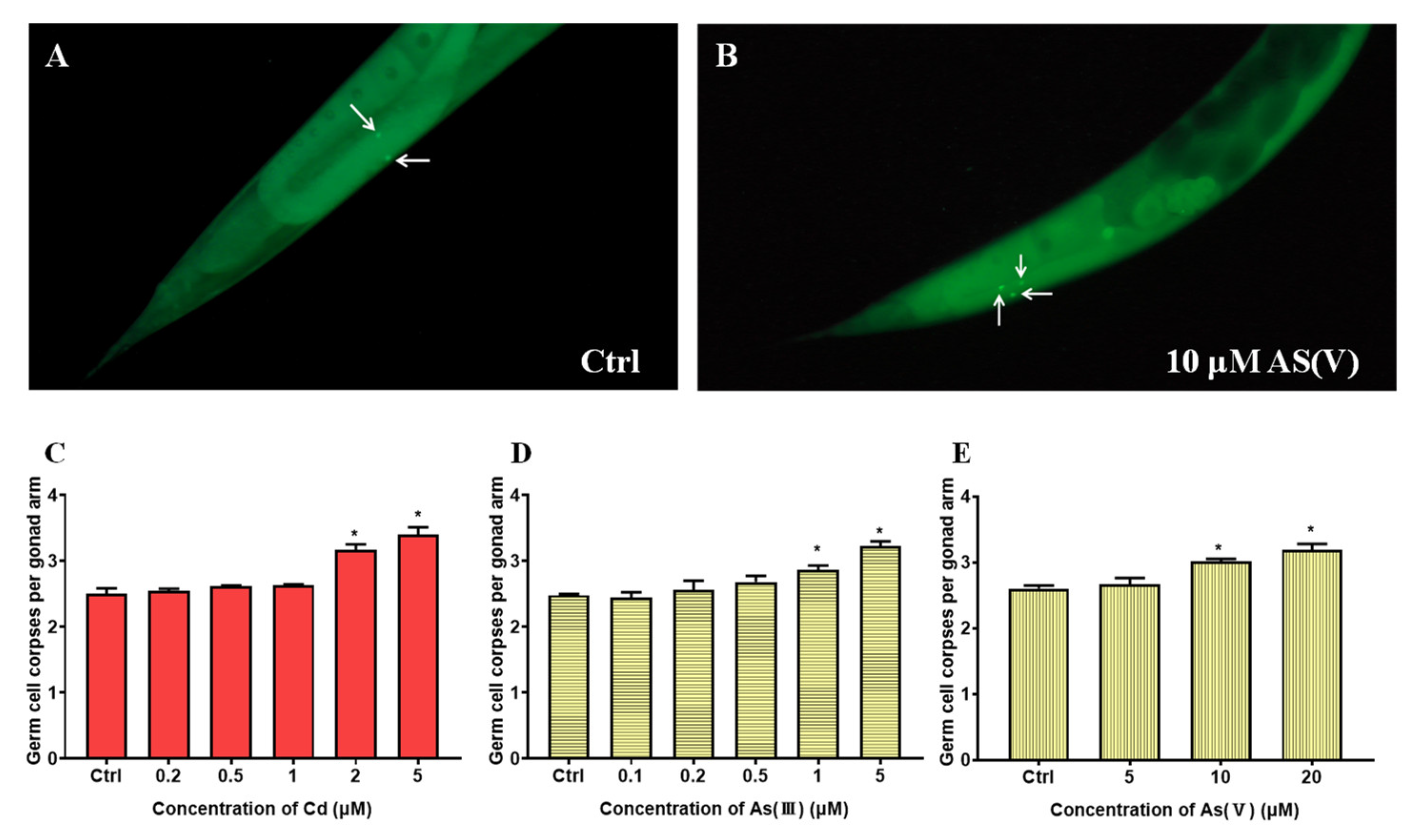
3.1. Joint Toxicity of Cd and As at Non-Toxic Concentrations on C. elegans

The maximum non-toxic concentrations of Cd, As(III), and As(V) are determined based on germ cell apoptosis. As shown in Figure 1, when Cd, As(III), and As(V) reached 2, 1, and 10 μM, respectively, significant differences from control groups were observed, deeming 1, 0.5, and 5 μM as the maximum non-toxic concentrations for Cd, As(III), and As(V), respectively. The following toxicity assays were all carried out using these concentrations. To explore the joint toxicity of Cd and As, the following experiments are carried out on a control group and six test groups including the single exposure of Cd, As(III), and As(V), and co-exposure of Cd-As(III), Cd-As(V), and Cd-As(III)-As(V) at their maximum non-toxic concentrations, respectively. Reproductive toxicity is evaluated by three end points including germ cell apoptosis, brood size, and the number of oocytes. As shown in Figure 2A, the number of apoptotic germ cells increased significantly in co-exposure groups compared to single exposure groups. Brood size and the number of oocytes showed similar results (Figure 2B,C), except that Cd-As(III) co-exposure did not significantly decrease eggs laid (Figure 2B). Co-exposure significantly reduced the lifespan of C. elegans to 16.4 ± 0.30 days for Cd-As(III), 15.9 ± 0.43 days for Cd-As(V), and 15.8 ± 1.06 days for Cd-As(III)-As(V) from 20 ± 0.50 days for the control group, in agreement with reproductive toxicity (Figure 3). These data indicated that concurrent exposure to Cd and As caused synergies in both reproductive and development toxicity.

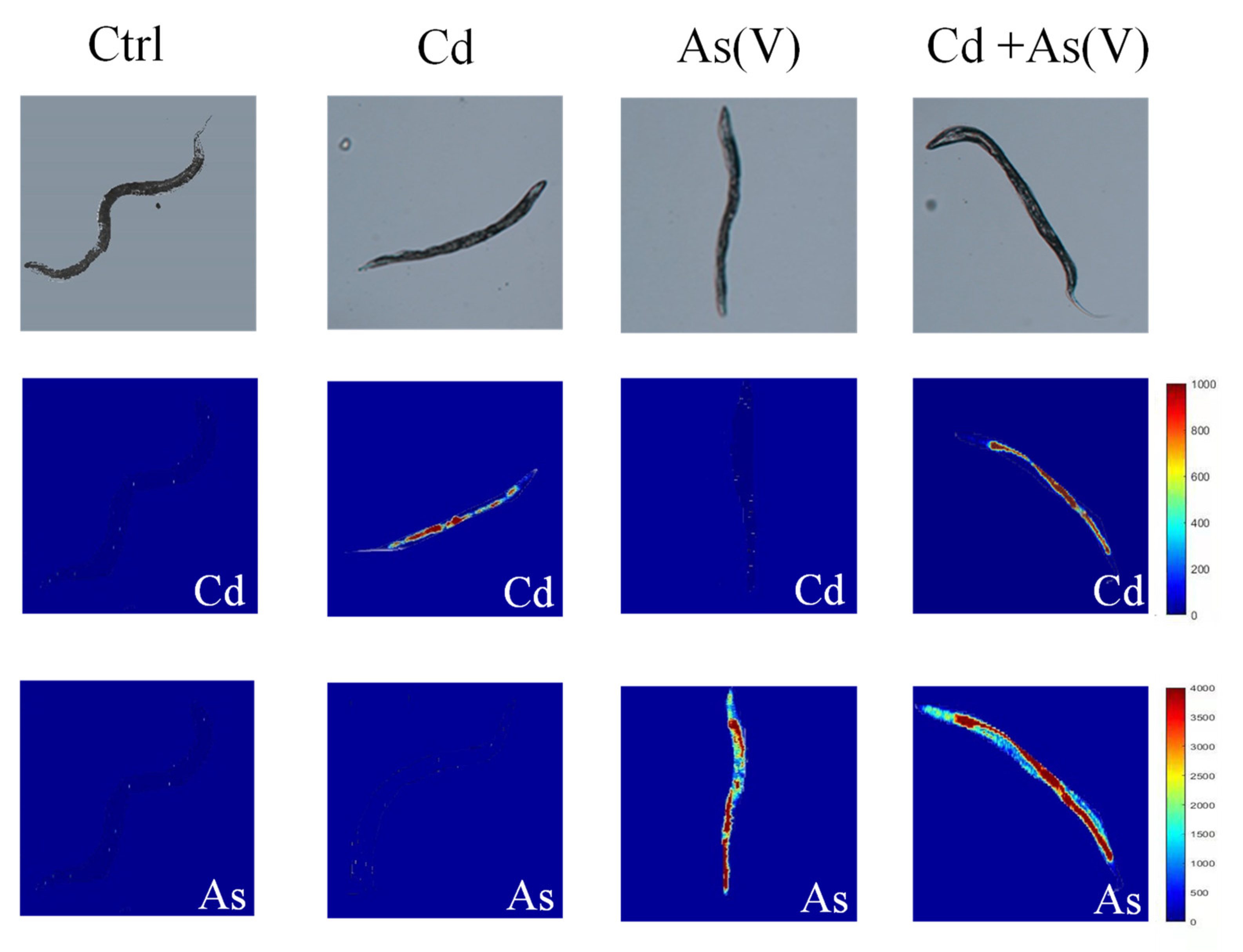
3.2. The Accumulation and Distribution of Cd and As in C. elegans

To explore the mechanism behind the synergies of Cd and As, we analyzed the accumulation of the heavy metals in C. elegans using ICP-MS. Since the non-toxic concentrations were too low to carry out measurements on As species, As accumulation was only determined as total As concentrations in Cd-As(III) and Cd-As(V) groups. The results showed that the mean value of As in the single As(III) group was 0.7 ± 0.15 μg/g, and the mean value of As in the Cd-As(III) group was 0.84 ± 0.04 μg/g, about 1.11 times more than the single exposure group. Similarly, As concentration was 310 ± 29.1 μg/g in the single As(V) group, and the mean value of As was 370 ± 31.2 μg/g when Cd and As(V) were co-exposed, which was 1.17 times more than the single exposure group. Cd content in the C. elegans did not show significant change in single and concurrent exposure groups. LA-ICP-MS was further employed to explore the distribution of Cd and As in C. elegans. Since the non-toxic concentration of As(III) was too low for LA-ICP-MS analysis, the distribution of Cd and As in C. elegans was only measured in the Cd-As(V) group (Figure 4). Randomly selected C. elegans were scanned, and from the visual result of mapping, we found a higher accumulation of As after co-exposure than that of As alone, and the accumulation was mainly in the intestine and gonad. LA-ICP-MS is a semi-quantitative analysis, the accumulation of As can be calculated based on every pixel of measured worms in relative calorific units. The value of single As(V) exposure was 2017.53 and the value of Cd-As(V) co-exposure was 2739.47, in agreement with ICP-MS results.

3.3. The Influence of Intestinal Microenvironment (pH) on As Species

Since As has multiple valence states with different toxicity, ingested As may alter in species in the intestine of C. elegans due to the microenvironment. Considering the complexity of the intestinal microenvironment, we chose pH here to simulate C. elegans intestine, hoping to detect the physicochemical changes of As. The C. elegans gut has a pH range from 6 in the anterior pharynx to 3.6 in the posterior gut [44]. We adjusted the pH of the KM solution to 3.6, 4, 5, and 6, and both As species were added for an incubation of 24 h. The concentrations of As(III) and As(V) were determined by LC-AFS. Apart from the data of pH = 6, As(V) was found to increase as pH decreased, while As(III) decreased, indicating part of As(III) was converted to As(V) (Figure 5).

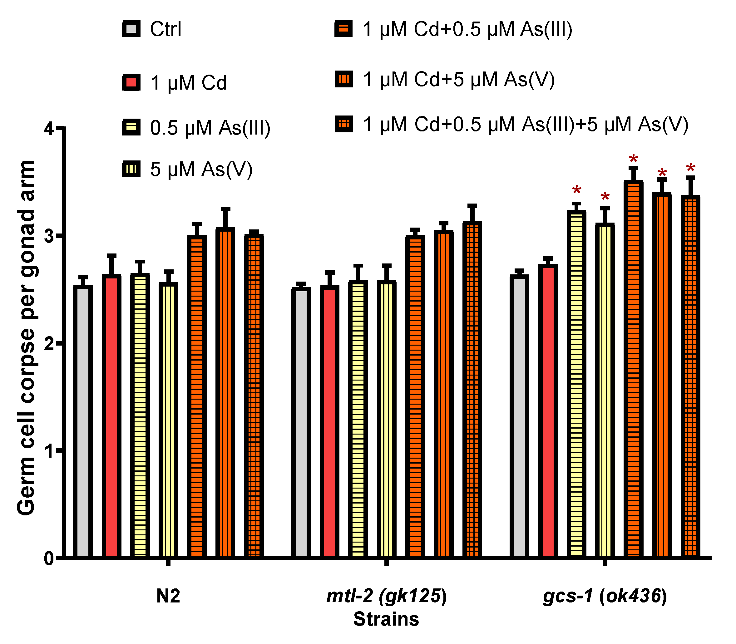
3.4. The Effects of mtl-2 and gcs-1 Gene on the Joint Toxicity of Cd and As

We observed changes in the amount of germ cell apoptosis by exposing the mutant strains mlt-2 (gk125) and gcs-1 (ok436) to the same working solutions. mlt-2 (gk125) showed no significant change in the amount of germ cell apoptosis with N2, while gcs-1 (ok436) exhibited more germ cell apoptosis in both single and co-exposure groups of As (Figure 6).

4. Discussion

To cope with the increasingly serious pollution caused by heavy metals, various managements were conducted, and strict regulations were applied. For example, the content of Cd in rice was limited to less than 0.2 mg/kg, and total arsenic in food was not allowed to exceed 0.6 mg/kg in China [51]. However, pollutants naturally co-exist in the environment, and the extra possible effects derived from the interaction of multiple pollutions at low concentrations should not be overlooked [49,52,53,54]. Therefore, it is environmentally reasonable to explore the combined toxicity of heavy metals at non-toxic levels. The gonad is one of the most important organs in C. elegans [50,55], and germ cell apoptosis is a highly sensitive endpoint for evaluating toxicity [51,56]. The mechanism of germ cell apoptosis under Cd and As exposure has been sufficiently discussed in previous studies, showing the dominant role played by the core apoptosis pathway when exposed to As [57,58], and DNA damage-induced apoptosis under Cd exposure [59]. We determined the non-toxic concentrations of Cd, As(III), and As(V) through germ cell apoptosis and conducted the following joint toxicity analysis based on these concentrations. When Cd and As were co-exposed at maximum non-toxic concentrations, the number of apoptotic germ cells was significantly increased compared to the single exposure groups (Figure 2A). Brood size, the number of oocytes (Figure 2B,C), and the lifespan of the worms (Figure 3) showed similar results, indicating that the co-exposure caused synergies in both reproductive and development toxicity. Similar observation on the synergic effects of Cd and As was reported in wheat and B. pilosa L [60,61]. Driessnack et al. [62] found that co-exposure to Cuprum (Cu) and Ni damaged the ovaries of fish and caused reproductive toxicity, and further studies revealed that co-exposure increased the accumulation of Cu and Ni in the body. Cadmium and As co-exposure resulted in greater joint toxicity on wheat seedling growth than when Cd and As were exposed alone, increasing the accumulation of Cd and As in vivo [63]. Tremaroli et al. [64] found that the transformation of As species in mice after the entry of As into the body generated different toxic effects. Thus, the synergistic effect may be attributed to the increase in the accumulation of heavy metals after co-exposure and the transformation of heavy metals in the organisms. We then analyzed the accumulation and distribution of Cd and As in C. elegans and the possible transformation of As species in the intestine, expecting to explain the synergistic effect through physicochemical mechanisms.
The accumulation and distribution of heavy metals in organisms is the basis for their subsequent toxicity, and different accumulation of heavy metals can lead to different effects [65]. Our results showed that As content in C. elegans after the compound exposure was elevated compared to the content in the single exposure groups, while Cd accumulation remained the same, indicating the presence of Cd promoted the accumulation of As in C. elegans. Száková et al. [66] reported that co-exposure to Cd and As significantly inhibited body weight gain in rats compared to the single exposure group, and As added to the diet promoted the accumulation of Cd in the testes. Cao et al. [61] found a significant increase in As accumulation in wheat after combined exposure to Cd and As compared to single exposure. The distribution of Cd and As in C. elegans was further obtained by LA-ICP-MS, a method that allows in situ observation of the distribution of heavy metals in organisms with high resolution [67], which has been successfully applied to C. elegans [45,68]. Our results showed that As accumulated mainly in the intestine as well as in the gonads of C. elegans, and the accumulation in selected nematodes scanned by LA-ICP-MS showed the same trend as the total accumulation measured by ICP-MS, further demonstrating that Cd promoted the uptake of As by C. elegans (Figure 4). Arsenic exposure can produce increased germ cell apoptosis due to oxidative stress [69]. Arsenite promotes the production of ROS and lipofuscin accumulation in the C. elegans intestine, which causes damage to the digestive tract [24,70]. When Cd and As were exposed in combination, the enhanced As accumulation in the gonad and intestine could be the cause for the synergistic effect.
The intestine is the first biological barrier against exogenous pollutants, whose microenvironment may influence the effects of heavy metals [71,72]. As one of the main target organs of As accumulation in C. elegans indicated by LA-ICP-MS, the pH of the intestinal lumen of C. elegans ranged from 6 in the anterior pharynx to 3.6 in the end of the intestine [56]. When As enters the body, the metabolic pathway of As(III) is converted to As(V) or methylation, while pH may be one of the important factors affecting As species [73]. Our results showed that as the pH decreased along the intestine, part of As(III) converted to the less toxic As(V) (Figure 5). Previous studies have reported higher toxicity caused by As(III) than As(V) in various test organisms [74,75]. Although As undergoes transformation in the intestine, there is no significant difference between different species of As when exposed with Cd. This is probably due to the fact that although As species conversion was detected, the magnitude is not enough to cause a distinguishable difference in toxicity.
We further investigated the potential mechanism of synergistic effects by the gk125 mutant strain (mtl-2 metallothionein gene defective type) and ok436 mutant strain (gcs-1 gene defective type). The main physiological function of metallothionein is involved in detoxification of heavy metals in organisms, including Cd [15]. Thus, mtl-2 in C. elegans also plays an important role in protecting against toxic effects caused by Cd detoxification [76]. Wang et al. [58] found that Cd activates caspases through JNK and p38 MAPK signaling pathways, leading to an increased amount of germ cell apoptosis in C. elegans. Cadmium exposure causes inflammatory responses and cellular damage in the human intestine, dysbiosis of intestinal flora, and damage to the intestinal tract [77]. Wang et al. [78] reported that pcs-1 (mt1748) exhibited a higher amount of apoptotic germ cells compared to N2 when exposed to the same level of Cd, indicating that the pcs-1 gene is involved in the toxic regulation of Cd in C. elegans. No difference was found in the amount of germ cell apoptosis between mtl-2 (gk125) and N2, and this is likely due to the similar Cd accumulation at a non-toxic level in single and co-exposure with As. It also suggests that even enhanced As accumulation does not respond to mtl-2. Wang et al. [22] found insulin-like growth factor-1 (IGF-1) networks and their target protein DAF-16/FOXO, known as key regulators of energy metabolism and growth, played important roles in arsenite-induced apoptosis. Glutathione (GSH) is also known to be an important endogenous antioxidant against exogenous contaminants, interfering with toxic substances by altering the rate of metal uptake as well as clearance, thus protecting cells from metal toxicity [76]. Arsenic readily reacts with thiol-containing molecules (GSH, Cys) [79], and intracellular GSH protects cells from arsenite-induced cytotoxicity in vitro [80,81,82]. Yu et al. [83] found that the gcs-1 gene is essential for GSH synthesis in C. elegans cells, so we used the gcs-1 gene defective phenotype to study the toxicity caused by As after co-exposure. It was found that when gcs-1 (ok436) was exposed to Cd alone, there was no significant change in the amount of germ cell apoptosis, showing that non-toxic concentration of Cd does not respond to the gcs-1 gene. After exposure to As(III) or As(V) alone, the amount of germ cell apoptosis in gcs-1 (ok436) increased significantly compared to N2, indicating when gcs-1 was defective in vivo, the worm will be unable to synthesize GSH to protect against the toxic effects caused by As, resulting in increased toxicity. Thus, we believe the higher joint toxicity of Cd-As co-exposure in gcs-1 (ok436) resulted from the increased accumulation of As in vivo. Therefore, the synergistic effect of Cd-As co-exposure was mainly caused by the increased toxicity of As (Figure 6).

5. Conclusions

Our results showed a synergistic effect of Cd and As at non-toxic concentrations on the reproduction and development of C. elegans. It was revealed that Cd promoted As uptake by C. elegans, increasing As accumulation in the gonad and intestine. Our simulation experiments indicated that the decrease of pH in the intestine may result in the conversion of As(III) to As(V) along the digestive tract, but was not enough to cause a significant difference in joint toxicity. We found that germ cell apoptosis in the mtl-2 (gk125) mutant strain was not significantly different from N2, while gcs-1 (ok439) showed higher reproductive damage, confirming that increased As toxicity dominated the synergistic effect of Cd and As. However, due to the low concentration of exposed As(III) and lack of extraction method, As(III) and As(V) cannot be distinguished concurrently in C. elegans, forcing us to rely on the simulation based on the microenvironment of the worm’s intestine. We plan to develop a proper extraction method for As species especially for biological samples, or introduce other in situ detection means such as μ-XANES to address the possible transformation of As(III) and As(V) in vivo. In addition, although the roles of Cd and As in the joint toxicity were determined based on mutant strain studies, the underlying toxicological mechanisms were not fully elucidated. Further exploration focusing on the molecular mechanisms will be carried out in the future. This study indicates co-exposure to heavy metals at non-toxic levels is a potential threat to the ecosystem, which needs further evaluation of other heavy metal combinations and systematical research on the mechanisms involved.

Author Contributions

Conceptualization, A.X. and Y.N.; methodology, C.P.; validation, Y.Z., S.N. and L.W.; formal analysis, C.P.; investigation, C.P.; resources, Y.N., L.W. and A.X.; data curation, L.S.; writing—original draft preparation, C.P. and Y.N.; writing—review and editing, Y.N., L.W. and A.X.; visualization, C.P.; supervision, Y.N. and A.X.; project administration, Y.N. and A.X.; funding acquisition, Y.N., L.W. and A.X. All authors have read and agreed to the published version of the manuscript.

Funding

This work was funded by Anhui Provincial Key Research and Development Programs (202004i07020015, 202004i07020016), Open Fund of Key Laboratory of High Magnetic Field and Ion Beam Physical Biology, Institute of Technical Biology and Agriculture Engineering, Hefei Institutes of Physical Science, CAS (2019-3), the National Key Research and Development Program of China (2020YFC1808204).

Institutional Review Board Statement

Not applicable.

Informed Consent Statement

Not applicable.

Data Availability Statement

Not applicable.

Acknowledgments

Fangyue Wang from Hefei University of Technology and Chao Ji from University of Science and Technology of China are acknowledged for their assistance in the experiments. Two anonymous reviewers are acknowledged for their valuable comments and suggestions. We also thank Hua Du from Hefei Institutes of Physical Science, Chinese Academy of Sciences for his aid in revising the manuscript.

Conflicts of Interest

The authors declare no conflict of interest.

References

  1. Hu, H.; Jin, Q.; Kavan, P. A study of heavy metal pollution in China: Current status, pollution-control policies and countermeasures. Sustainability 2014, 6, 5820–5838. [Google Scholar] [CrossRef] [Green Version]
  2. Fowler, B.A. Monitoring of human populations for early markers of cadmium toxicity: A review. Toxicol. Appl. Pharmacol. 2009, 238, 294–300. [Google Scholar] [CrossRef] [PubMed]
  3. Abdul, K.S.M.; Jayasinghe, S.S.; Chandana, E.P.; Jayasumana, C.; De Silva, P.M.C. Arsenic and human health effects: A review. Environ. Toxicol. Pharmacol. 2015, 40, 828–846. [Google Scholar] [CrossRef] [PubMed]
  4. Vardhan, K.H.; Kumar, P.S.; Panda, R.C. A review on heavy metal pollution, toxicity and remedial measures: Current trends and future perspectives. J. Mol. Liq. 2019, 290, 111197. [Google Scholar] [CrossRef]
  5. Genchi, G.; Carocci, A.; Lauria, G.; Sinicropi, M.S.; Catalano, A. Nickel: Human health and environmental toxicology. Int. J. Environ. Res. Public Health 2020, 17, 679. [Google Scholar] [CrossRef] [Green Version]
  6. Bernhoft, R.A. Cadmium toxicity and treatment. Sci. World J. 2013, 2013, 394652. [Google Scholar] [CrossRef]
  7. Cheng, C.-H.; Ma, H.-L.; Deng, Y.-Q.; Feng, J.; Jie, Y.-K.; Guo, Z.-X. Oxidative stress, cell cycle arrest, DNA damage and apoptosis in the mud crab (Scylla paramamosain) induced by cadmium exposure. Chemosphere 2021, 263, 128277. [Google Scholar] [CrossRef] [PubMed]
  8. Lee, W.-K.; Thévenod, F. Cell organelles as targets of mammalian cadmium toxicity. Arch. Toxicol. 2020, 94, 1017–1049. [Google Scholar] [CrossRef] [Green Version]
  9. Ren, Y.; Shao, W.; Zuo, L.; Zhao, W.; Qin, H.; Hua, Y.; Lu, D.; Mi, C.; Zeng, S.; Zu, L. Mechanism of cadmium poisoning on testicular injury in mice. Oncol. Lett. 2019, 18, 1035–1042. [Google Scholar] [CrossRef] [PubMed] [Green Version]
  10. Wang, S.; Chu, Z.; Zhang, K.; Miao, G. Cadmium-induced serotonergic neuron and reproduction damages conferred lethality in the nematode Caenorhabditis elegans. Chemosphere 2018, 213, 11–18. [Google Scholar] [CrossRef] [PubMed]
  11. Jiang, Y.; Chen, J.; Wu, Y.; Wang, Q.; Li, H. Sublethal toxicity endpoints of heavy metals to the nematode Caenorhabditis elegans. PLoS ONE 2016, 11, e0148014. [Google Scholar] [CrossRef] [PubMed] [Green Version]
  12. Bovio, F.; Sciandrone, B.; Urani, C.; Fusi, P.; Forcella, M.; Regonesi, M.E. Superoxide dismutase 1 (SOD1) and cadmium: A three models approach to the comprehension of its neurotoxic effects. Neurotoxicology 2021, 84, 125–135. [Google Scholar] [CrossRef] [PubMed]
  13. Roh, J.-Y.; Park, Y.-J.; Choi, J. A cadmium toxicity assay using stress responsive Caenorhabditis elegans mutant strains. Environ. Toxicol. Pharmacol. 2009, 28, 409–413. [Google Scholar] [CrossRef]
  14. Rahaman, M.S.; Rahman, M.M.; Mise, N.; Sikder, M.T.; Ichihara, G.; Uddin, M.K.; Kurasaki, M.; Ichihara, S. Environmental arsenic exposure and its contribution to human diseases, toxicity mechanism and management. Environ. Pollut. 2021, 289, 117940. [Google Scholar] [CrossRef] [PubMed]
  15. Moyson, S.; Vissenberg, K.; Fransen, E.; Blust, R.; Husson, S.J. Mixture effects of copper, cadmium, and zinc on mortality and behavior of Caenorhabditis elegans. Environ. Toxicol. Chem. 2018, 37, 145–159. [Google Scholar] [CrossRef] [PubMed]
  16. Sodhi, K.K.; Kumar, M.; Agrawal, P.K.; Singh, D.K. Perspectives on arsenic toxicity, carcinogenicity and its systemic remediation strategies. Environ. Technol. Innov. 2019, 16, 100462. [Google Scholar] [CrossRef]
  17. Jang, Y.; Somanna, Y.; Kim, H. Source, distribution, toxicity and remediation of arsenic in the environment—A review. Int. J. Appl. Environ. Sci. 2016, 11, 559–581. [Google Scholar]
  18. Rahman, M.M.; Chowdhury, U.K.; Mukherjee, S.C.; Mondal, B.K.; Paul, K.; Lodh, D.; Biswas, B.K.; Chanda, C.R.; Basu, G.K.; Saha, K.C. Chronic arsenic toxicity in Bangladesh and West Bengal, India review and commentary. J. Toxicol. Clin. Toxicol. 2001, 39, 683–700. [Google Scholar] [CrossRef]
  19. Khairul, I.; Wang, Q.Q.; Jiang, Y.H.; Wang, C.; Naranmandura, H. Metabolism, toxicity and anticancer activities of arsenic compounds. Oncotarget 2017, 8, 23905. [Google Scholar] [CrossRef] [PubMed] [Green Version]
  20. Singh, A.P.; Goel, R.K.; Kaur, T. Mechanisms pertaining to arsenic toxicity. Toxicol. Int. 2011, 18, 87. [Google Scholar] [PubMed] [Green Version]
  21. Rahman, Z.; Singh, V.P. The relative impact of toxic heavy metals (THMs) (arsenic (As), cadmium (Cd), chromium (Cr)(VI), mercury (Hg), and lead (Pb)) on the total environment: An overview. Environ. Monit. Assess. 2019, 191, 419. [Google Scholar] [CrossRef]
  22. Wang, S.; Teng, X.; Wang, Y.; Yu, H.Q.; Luo, X.; Xu, A.; Wu, L. Molecular control of arsenite-induced apoptosis in Caenorhabditis elegans: Roles of insulin-like growth factor-1 signaling pathway. Chemosphere 2014, 112, 248–255. [Google Scholar] [CrossRef] [PubMed]
  23. Yu, C.-W.; How, C.M.; Liao, V.H.-C. Arsenite exposure accelerates aging process regulated by the transcription factor DAF-16/FOXO in Caenorhabditis elegans. Chemosphere 2016, 150, 632–638. [Google Scholar] [CrossRef]
  24. Yu, C.-W.; Liao, V.H.-C. Transgenerational reproductive effects of arsenite are associated with H3K4 dimethylation and SPR-5 downregulation in Caenorhabditis elegans. Environ. Sci. Technol. 2016, 50, 10673–10681. [Google Scholar] [CrossRef] [PubMed]
  25. Mikhailenko, A.V.; Ruban, D.A.; Ermolaev, V.A.; van Loon, A. Cadmium pollution in the tourism environment: A literature review. Geosci. J. 2020, 10, 242. [Google Scholar] [CrossRef]
  26. Hossain, S.; Latifa, G.A.; Prianqa, P.; Al Nayeem, A. Review of cadmium pollution in Bangladesh. J. Health Pollut. 2019, 9, 190913. [Google Scholar] [CrossRef] [PubMed] [Green Version]
  27. Chen, N.C.; Zheng, Y.J.; Xiao-Feng, H.E.; Xiao-Fei, L.I.; Zhang, X.X. Analysis of the Report on the national general survey of soil contamination. J. Agro Environ. Sci. 2017, 36, 1689–1692. [Google Scholar]
  28. Abbas, G.; Murtaza, B.; Bibi, I.; Shahid, M.; Niazi, N.K.; Khan, M.I.; Amjad, M.; Hussain, M.; Natasha. Arsenic uptake, toxicity, detoxification, and speciation in plants: Physiological, biochemical, and molecular aspects. Int. J. Environ. Res. Public Health 2018, 15, 59. [Google Scholar] [CrossRef] [Green Version]
  29. Carocci, A.; Catalano, A.; Lauria, G.; Sinicropi, M.S.; Genchi, G. Lead Toxicity, Antioxidant Defense and Environment. Rev. Environ. Contam. Toxicol. 2015, 238, 45–67. [Google Scholar]
  30. Mawia, A.M.; Hui, S.; Zhou, L.; Li, H.; Tabassum, J.; Lai, C.; Wang, J.; Shao, G.; Wei, X.; Tang, S.; et al. Inorganic Arsenic Toxicity and Alleviation Strategies in Rice. J. Hazard. Mater. 2020, 408, 124751. [Google Scholar] [CrossRef] [PubMed]
  31. Gu, Z.; de Silva, S.; Reichman, S.M. Arsenic concentrations and dietary exposure in rice-based infant food in Australia. Int. J. Environ. Res. Public Health 2020, 17, 415. [Google Scholar] [CrossRef] [Green Version]
  32. Medunić, G.; Fiket, Ž.; Ivanić, M. Arsenic contamination status in Europe, Australia, and other parts of the world. In Arsenic in Drinking Water and Food; Springer: Berlin/Heidelberg, Germany, 2020; pp. 183–233. [Google Scholar]
  33. Shi, Y.-L.; Chen, W.-Q.; Wu, S.-L.; Zhu, Y.-G. Anthropogenic cycles of arsenic in mainland China: 1990–2010. Environ. Sci. Technol. 2017, 51, 1670–1678. [Google Scholar] [CrossRef] [PubMed]
  34. Zimeng, L.; Xiaoqian, L.; ZHANG, W.; Yining, J.; Siyu, S.; Sujing, W.; Qirui, A.; Pengyang, L.; ZHENG, N. Research progress on the effects of heavy metal compound pollution on organisms. Environ. Chem. 2021, 40, 3331–3343. [Google Scholar]
  35. Martin, O.; Scholze, M.; Ermler, S.; McPhie, J.; Bopp, S.K.; Kienzler, A.; Parissis, N.; Kortenkamp, A. Ten years of research on synergisms and antagonisms in chemical mixtures: A systematic review and quantitative reappraisal of mixture studies. Environ. Int. 2021, 146, 106206. [Google Scholar] [CrossRef] [PubMed]
  36. Rai, A.; Maurya, S.K.; Khare, P.; Srivastava, A.; Bandyopadhyay, S. Characterization of developmental neurotoxicity of As, Cd, and Pb mixture: Synergistic action of metal mixture in glial and neuronal functions. Toxicol. Sci. 2010, 118, 586–601. [Google Scholar] [CrossRef] [PubMed]
  37. Adedara, I.A.; Adegbosin, A.N.; Abiola, M.A.; Odunewu, A.A.; Owoeye, O.; Owumi, S.E.; Farombi, E.O. Neurobehavioural and biochemical responses associated with exposure to binary waterborne mixtures of zinc and nickel in rats. Environ. Toxicol. Pharmacol. 2020, 73, 103294. [Google Scholar] [CrossRef]
  38. Bloom, M.S.; Parsons, P.J.; Steuerwald, A.J.; Schisterman, E.F.; Browne, R.W.; Kim, K.; Coccaro, G.A.; Conti, G.C.; Narayan, N.; Fujimoto, V.Y. Toxic trace metals and human oocytes during in vitro fertilization (IVF). Reprod. Toxicol. 2010, 29, 298–305. [Google Scholar] [CrossRef] [Green Version]
  39. Lanier, C.; Bernard, F.; Dumez, S.; Leclercq-Dransart, J.; Lemiere, S.; Vandenbulcke, F.; Nesslany, F.; Platel, A.; Devred, I.; Hayet, A. Combined toxic effects and DNA damage to two plant species exposed to binary metal mixtures (Cd/Pb). Ecotoxicol. Environ. Saf. 2019, 167, 278–287. [Google Scholar] [CrossRef]
  40. Meng, Q.J. Toxic Effects of Compound Pollution with Heavy Metals on the Growth of Wheat Seedlings. J. Anhui Agric. Sci. 2008, 36, 122. [Google Scholar]
  41. Corsi, A.K.; Wightman, B.; Chalfie, M. A transparent window into biology: A primer on Caenorhabditis elegans. Genetics 2015, 200, 387–407. [Google Scholar] [CrossRef] [Green Version]
  42. Hunt, P.R. The C. elegans model in toxicity testing. J. Appl. Toxicol. 2017, 37, 50–59. [Google Scholar] [CrossRef]
  43. Shen, P.; Yue, Y.; Park, Y. A living model for obesity and aging research: Caenorhabditis elegans. Crit. Rev. Food Sci. Nutr. 2018, 58, 741–754. [Google Scholar] [CrossRef] [PubMed]
  44. Hamilton, J.; Gorishek, E.; Mach, P.; Sturtevant, D.; Ladage, M.; Suzuki, N.; Padilla, P.; Mittler, R.; Chapman, K.; Verbeck, G. Evaluation of a custom single Peltier-cooled ablation cell for elemental imaging of biological samples in laser ablation-inductively coupled plasma-mass spectrometry (LA-ICP-MS). J. Anal. At. Spectrom. 2016, 31, 1030–1033. [Google Scholar] [CrossRef]
  45. Crone, B.; Aschner, M.; Schwerdtle, T.; Karst, U.; Bornhorst, J. Elemental bioimaging of Cisplatin in Caenorhabditis elegans by LA-ICP-MS. Metallomics 2015, 7, 1189–1195. [Google Scholar] [CrossRef] [Green Version]
  46. Brenner, S.J.G. The genetics of Caenorhabditis elegans. Genetics 1974, 77, 71–94. [Google Scholar] [CrossRef] [PubMed]
  47. Gartner, A.; MacQueen, A.J.; Villeneuve, A.M. Methods for analyzing checkpoint responses in Caenorhabditis elegans. In Checkpoint Controls and Cancer; Springer: Berlin, Germany, 2004; pp. 257–274. [Google Scholar]
  48. Ruan, Q.L.; Ju, J.J.; Li, Y.H.; Li, X.B.; Liu, R.; Liang, G.Y.; Zhang, J.; Pu, Y.P.; Wang, D.Y.; Yin, L.H. Chlorpyrifos exposure reduces reproductive capacity owing to a damaging effect on gametogenesis in the nematode Caenorhabditis elegans. J. Appl. Toxicol. 2012, 32, 527–535. [Google Scholar] [CrossRef] [PubMed]
  49. Du, H.; Wang, M.; Wang, L.; Dai, H.; Wang, M.; Hong, W.; Nie, X.; Wu, L.; Xu, A. Reproductive Toxicity of Endosulfan: Implication from Germ Cell Apoptosis Modulated by Mitochondrial Dysfunction and Genotoxic Response Genes in Caenorhabditis elegans. Toxicol. Sci. Off. J. Soc. Toxicol. 2015, 145, 118–127. [Google Scholar] [CrossRef] [Green Version]
  50. Dai, H.; Liu, Y.; Wang, J.; Nie, Y.; Sun, Y.; Wang, M.; Wang, D.; Yang, Z.; Cheng, L.; Wang, J. Graphene oxide antagonizes the toxic response to arsenic via activation of protective autophagy and suppression of the arsenic-binding protein LEC-1 in Caenorhabditis elegans. Environ. Sci. Nano 2018, 5, 1711–1728. [Google Scholar] [CrossRef]
  51. Xin, X.W.; Zhang, D.K. Comparative Analysis of Heavy Metal Residue Indicator in Domestic and Foreign Food Standards. China Stand. 2018, 8, 124–128. [Google Scholar]
  52. Sinha, K.A.; Keen, J.K.; Ogun, S.A.; Holder, A.A. Comparison of two members of a multigene family coding for high-molecular mass rhoptry proteins of Plasmodium yoelii. Mol. Biochem. Parasitol. 1996, 76, 329. [Google Scholar] [CrossRef]
  53. Singh, N.; Kumar, D.; Sahu, A.P. Arsenic in the environment: Effects on human health and possible prevention. J. Environ. Biol. 2007, 28, 359. [Google Scholar]
  54. Berman, J.R.; Kenyon, C. Germ-cell loss extends C. elegans life span through regulation of DAF-16 by kri-1 and lipophilic-hormone signaling. Cell 2006, 124, 1055–1068. [Google Scholar] [CrossRef] [PubMed] [Green Version]
  55. Eisenmann, D. WormBook: The Online Review of C. Elegans Biology. Research Community, Wormbook. Available online: http://www.wormbook.org (accessed on 21 March 2007).
  56. Chauhan, V.M.; Orsi, G.; Brown, A.; Pritchard, D.I.; Aylott, J.W. Mapping the Pharyngeal and Intestinal pH of Caenorhabditis elegans and Real-Time Luminal pH Oscillations Using Extended Dynamic Range pH-Sensitive Nanosensors. ACS Nano 2013, 7, 5577–5587. [Google Scholar] [CrossRef] [PubMed]
  57. Pei, B.; Wang, S.; Guo, X.; Wang, J.; Yang, G.; Hang, H.; Wu, L. Arsenite-induced germline apoptosis through a MAPK-dependent, p53-independent pathway in Caenorhabditis elegans. Chem. Res. Toxicol. 2008, 21, 1530–1535. [Google Scholar] [CrossRef] [PubMed]
  58. Wang, S.; Luo, X.; Wu, L. Assessment of Caenorhabditis Elegans Germline Cell Cycle Arrest and Apoptosis for Heavy Metals Toxicity Testing. In Proceedings of the 2009 3rd International Conference on Bioinformatics and Biomedical Engineering, Beijing, China, 11–13 June 2009; pp. 1–4. [Google Scholar]
  59. Wang, S.; Tang, M.; Pei, B.; Xiao, X.; Wang, J.; Hang, H.; Wu, L. Cadmium-induced germline apoptosis in Caenorhabditis elegans: The roles of HUS1, p53, and MAPK signaling pathways. Toxicol. Sci. 2008, 102, 345–351. [Google Scholar] [CrossRef]
  60. Sun, Y.; Li, Z.; Guo, B.; Chu, G.; Wei, C.; Liang, Y. Arsenic mitigates cadmium toxicity in rice seedlings. Environ. Exp. Bot. 2008, 64, 264–270. [Google Scholar] [CrossRef]
  61. Cao, Q.; Hu, Q.H.; Khan, S.; Wang, Z.J.; Lin, A.J. Wheat phytotoxicity from arsenic and cadmium separately and together in solution culture and in a calcareous soil—ScienceDirect. J. Hazard. Mater. 2007, 148, 377–382. [Google Scholar] [CrossRef] [PubMed] [Green Version]
  62. Driessnack, M.K.; Jamwal, A.; Niyogi, S. Effects of chronic exposure to waterborne copper and nickel in binary mixture on tissue-specific metal accumulation and reproduction in fathead minnow (Pimephales promelas). Chemosphere 2017, 185, 964–974. [Google Scholar] [CrossRef] [PubMed]
  63. Liu, X.; Zhang, S.; Shan, X.Q.; Christie, P. Combined toxicity of cadmium and arsenate to wheat seedlings and plant uptake and antioxidative enzyme responses to cadmium and arsenate co-contamination. Ecotoxicol. Environ. Saf. 2007, 68, 305–313. [Google Scholar] [CrossRef] [PubMed] [Green Version]
  64. Tremaroli, V.; Bäckhed, F. Keep It in the Family Professionals Are Handing Back Power to Parents over the Futureof Their Children, However Traumatised Relationships May Be. Glenda Cooper Reports. Nature 2012, 489, 242–249. [Google Scholar] [CrossRef]
  65. Wang, X.; Wang, Q.; Yu, L. Heavy Metal Contamination and Its Effect on Organism in Soil: A Review. Chin. Agric. Sci. Bull. 2017, 33, 86–92. [Google Scholar]
  66. Száková, J.; Zídek, V.; Miholová, D. Influence of elevated content of cadmium and arsenic in diet containing feeding yeast on organisms of rats. Czech J. Anim. Sci. 2009, 54, 1–9. [Google Scholar] [CrossRef] [Green Version]
  67. Ali, A.S.; Bharwana, S.A.; Ahmad, R. Effect of different heavy metal pollution on fish. Res. J. Chem. Environ. Sci. 2014, 2, 74–79. [Google Scholar]
  68. Große Brinkhaus, S.; Bornhorst, J.; Chakraborty, S.; Wehe, C.A.; Niehaus, R.; Reifschneider, O.; Aschner, M.; Karst, U. Elemental bioimaging of manganese uptake in C. elegans. Metallomics 2014, 6, 617–621. [Google Scholar] [CrossRef]
  69. Nutt, L.K.; Gogvadze, V.; Uthaisang, W.; Mirnikjoo, B.; McConkey, D.J.; Orrenius, S. Research paper indirect effects of Bax and Bak initiate the mitochondrial alterations that lead to cytochrome c release during arsenic trioxide-induced apoptosis. Cancer Biol. 2005, 4, 459–467. [Google Scholar]
  70. Wang, S.; Zhao, Y.; Wu, L.; Tang, M.; Su, C.; Hei, T.K.; Yu, Z. Induction of germline cell cycle arrest and apoptosis by sodium arsenite in Caenorhabditis elegans. Chem. Res. Toxicol. 2007, 20, 181–186. [Google Scholar] [CrossRef]
  71. Arun, K.; Madhavan, A.; Sindhu, R.; Emmanual, S.; Binod, P.; Pugazhendhi, A.; Sirohi, R.; Reshmy, R.; Awasthi, M.K.; Gnansounou, E. Probiotics and gut microbiome—Prospects and challenges in remediating heavy metal toxicity. J. Hazard. Mater. 2021, 420, 126676. [Google Scholar] [CrossRef] [PubMed]
  72. Breton, J.; Massart, S.; Vandamme, P.; De Brandt, E.; Pot, B.; Foligné, B. Ecotoxicology inside the gut: Impact of heavy metals on the mouse microbiome. BMC Pharmacol. 2013, 14, 62. [Google Scholar] [CrossRef] [PubMed] [Green Version]
  73. Buchet, J.-P.; Lauwerys, R.; Roels, H. Comparison of the urinary excretion of arsenic metabolites after a single oral dose of sodium arsenite, monomethylarsonate, or dimethylarsinate in man. Int. Arch. Occup. Environ. Health 1981, 48, 71–79. [Google Scholar] [CrossRef] [PubMed]
  74. Meng, Z.Y.; Lin, H.; Wang, A.Q. Comparative Study on the Inhibition of Arsenic and Arsenate to Brain Acetylcholinesterase from Different Fish Species. J. Ocean Univ. Qingdao 2005, 35, 471–474. [Google Scholar]
  75. Suhendrayatna; Ohki, A.; Maeda, S. Arsenic accumulation, transformation and tolerance on freshwater Daphnia magna. Toxicol. Environ. Chem. 1999, 72, 1–11. [Google Scholar] [CrossRef]
  76. Swain, S.; Keusekotten, K.; Baumeister, R.; Stürzenbaum, S.C. elegans metallothioneins: New insights into the phenotypic effects of cadmium toxicosis. J. Mol. Biol. 2004, 341, 951–959. [Google Scholar] [CrossRef] [PubMed]
  77. Tinkov, A.A.; Gritsenko, V.A.; Skalnaya, M.G.; Cherkasov, S.V.; Aaseth, J.; Skalny, A.V. Gut as a target for cadmium toxicity. Environ. Pollut. 2018, 235, 429–434. [Google Scholar] [CrossRef]
  78. Wang, J.; Nie, Y.; Dai, H.; Wang, M.; Cheng, L.; Yang, Z.; Chen, S.; Zhao, G.; Wu, L.; Guang, S. Parental exposure to TiO 2 NPs promotes the multigenerational reproductive toxicity of Cd in Caenorhabditis elegans via bioaccumulation of Cd in germ cells. Environ. Sci. Nano 2019, 6, 1332–1342. [Google Scholar] [CrossRef]
  79. Scott, N.; Hatlelid, K.M.; Mackenzie, N.E.; Carter, D.E. Reactions of arsenic(III) and arsenic(V) species with glutathione. Chem. Res. Toxicol. 1993, 6, 102–106. [Google Scholar] [CrossRef] [PubMed]
  80. Ochi, T. Arsenic compound-induced increases in glutathione levels in cultured Chinese hamster V79 cells and mechanisms associated with changes in γ-glutamylcysteine synthetase activity, cystine uptake and utilization of cysteine. Arch. Toxicol. 1997, 71, 730–740. [Google Scholar] [CrossRef] [PubMed]
  81. Urban, N.; Tsitsipatis, D.; Hausig, F.; Kreuzer, K.; Erler, K.; Stein, V.; Ristow, M.; Steinbrenner, H.; Klotz, L.-O. Non-linear impact of glutathione depletion on C. elegans life span and stress resistance. Redox Biol. 2017, 11, 502–515. [Google Scholar] [CrossRef]
  82. Huang, H.; Huang, C.; Wu, D.; Jinn, C.; Jan, K. Glutathione as a cellular defence against arsenite toxicity in cultured Chinese hamster ovary cells. Toxicology 1993, 79, 195–204. [Google Scholar] [CrossRef]
  83. Liao, V.H.-C.; Yu, C.-W. Caenorhabditis elegans gcs-1 confers resistance to arsenic-induced oxidative stress. Biometals 2005, 18, 519–528. [Google Scholar] [CrossRef] [PubMed]
Figure 1. Images of AO stained apoptotic cells in the gonads (A,B) and the determination of non-toxic concentrations of (C) Cd, (D) As(III), and (E) As(V) based on germ cell apoptosis. * Means p < 0.05.
Toxics 10 00133 g001
Figure 2. Effects of single and co-exposure to non-toxic concentrations of Cd, As(III), and As(V) on (A) germ cell apoptosis, (B) brood size, and (C) the number of oocytes. * Means p < 0.05.
Toxics 10 00133 g002
Figure 3. Effects of single and co-exposure to non-toxic concentrations of Cd, As(III), and As(V) on (A) average life span and (B) survival rate. * Means p < 0.05.
Toxics 10 00133 g003
Figure 4. Spatial distribution of Cd and As in C. elegans exposed to Cd, As(V), and Cd-As(V).
Toxics 10 00133 g004
Figure 5. Changes of As species corresponding to different pH. (A) As(III) and As(V) trends, (B) As(III) and As(V) contents.
Toxics 10 00133 g005
Figure 6. The effect of single and co-exposed Cd, As(III), and As(V) on apoptosis gonad cells of strains mtl-2 (gk125) and gcs-1 (ok436). * Means compared with the corresponding groups of N2, p < 0.05.
Toxics 10 00133 g006
Publisher’s Note: MDPI stays neutral with regard to jurisdictional claims in published maps and institutional affiliations.

