
Cytotoxic and Inflammatory Effects of Electronic and Traditional Cigarettes on Oral Gingival Cells Using a Novel Automated Smoking Instrument: An In Vitro Study

 , and
, and
Abstract
:1. Introduction
2. Materials and Methods
2.1. Air–Liquid Interface Cell Culture
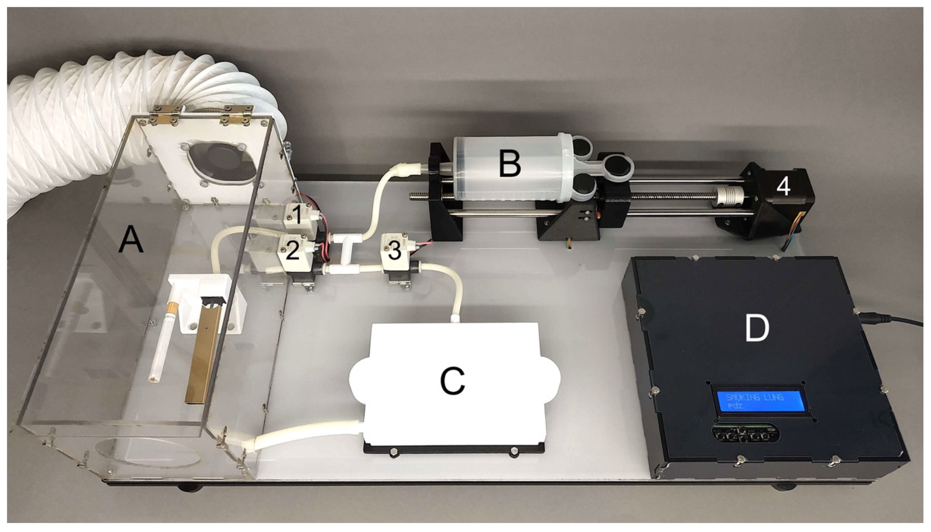
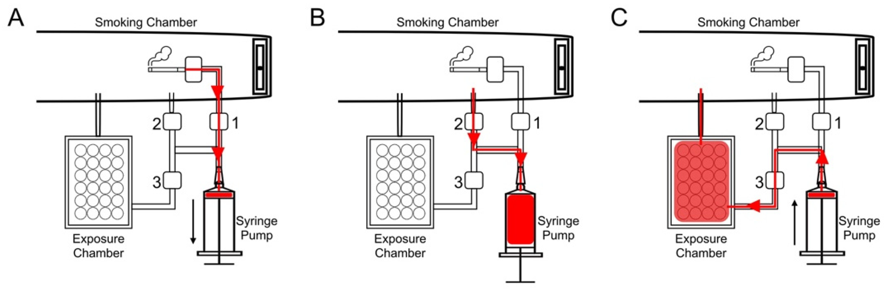
2.2. Automated Smoking Machine
2.3. Cigarette Smoke and Aerosol Cell Exposure on the Automated Generation System
2.4. Cell Viability Assay
2.5. Morphology and Cell Death Analysis
2.6. Inflammatory Gene Expression Analysis
2.7. Statistical Analysis
3. Results
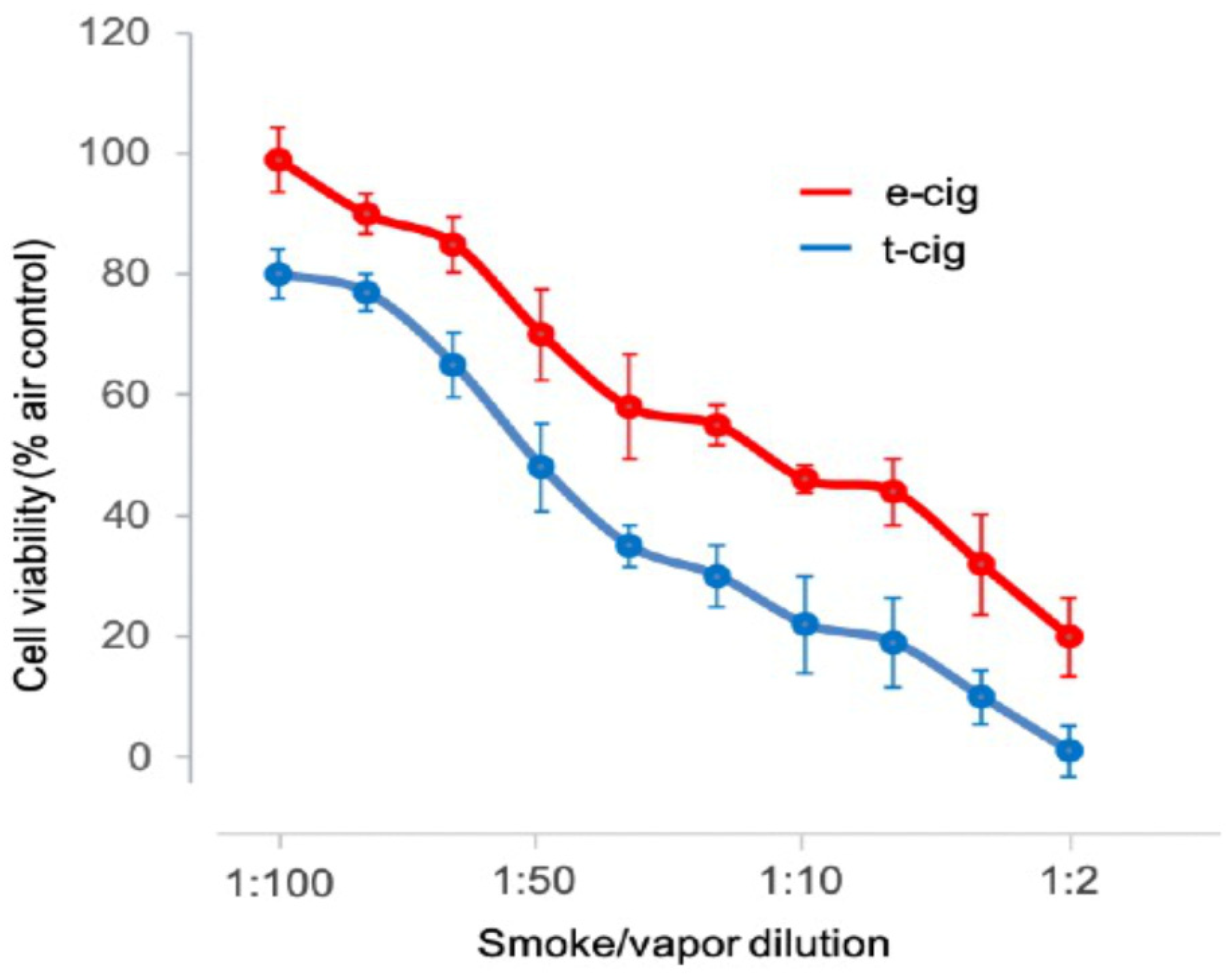
3.1. Cell Viability
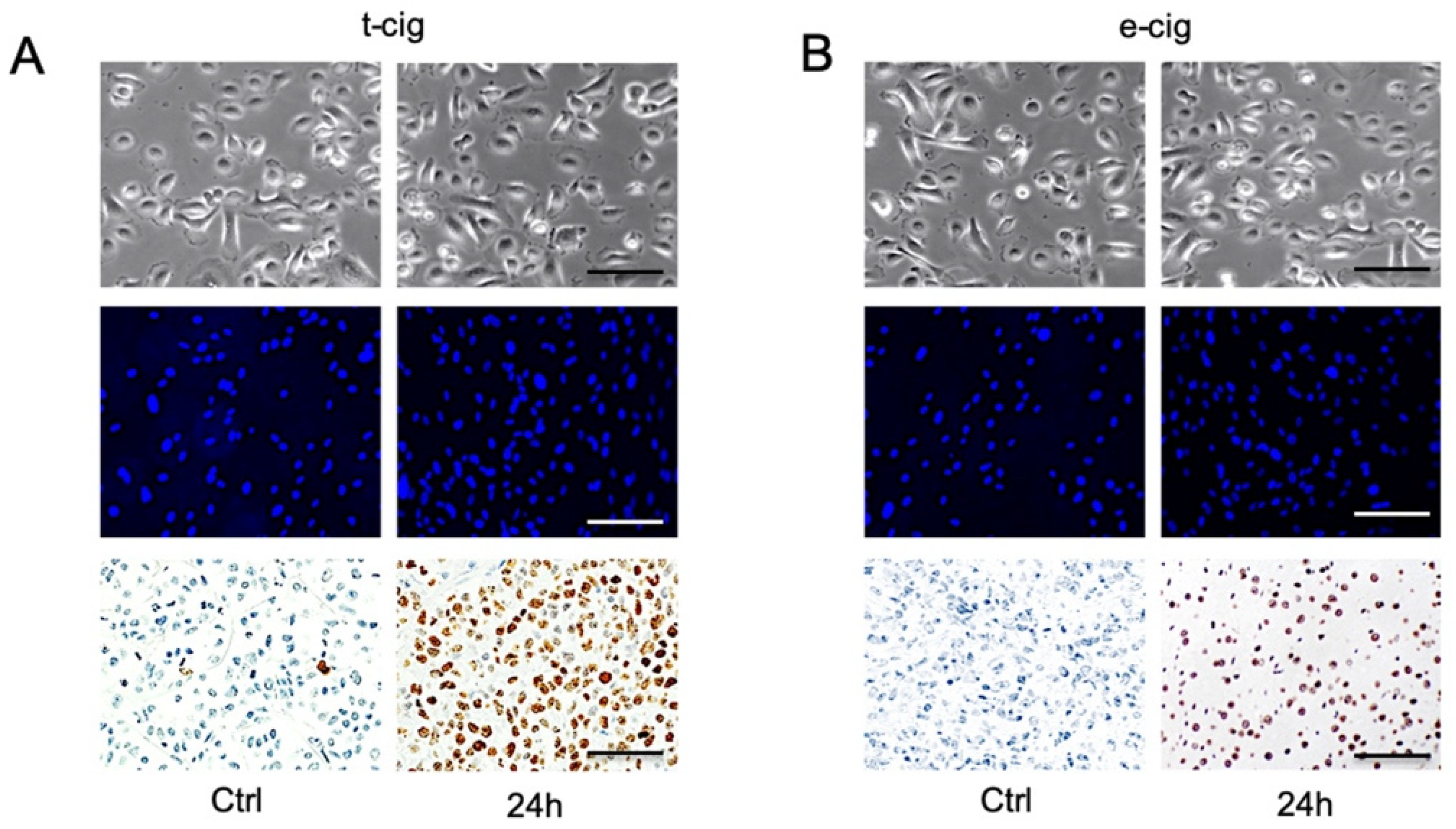
3.2. Cell Death
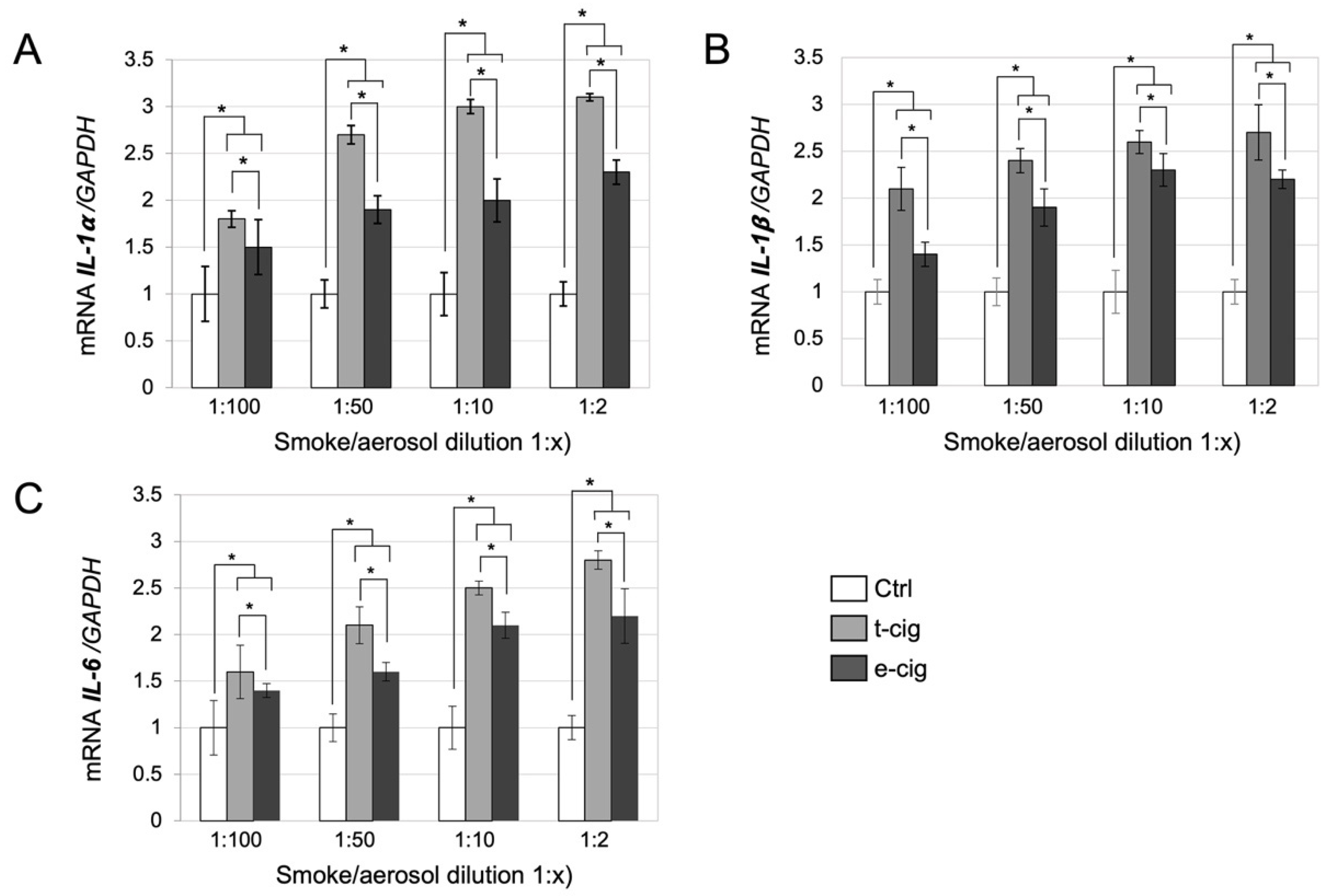
3.3. Gene Expression
4. Discussion
5. Conclusions
Author Contributions
Funding
Institutional Review Board Statement
Informed Consent Statement
Data Availability Statement
Conflicts of Interest
References
- Rahman, M.A.; Hann, N.; Wilson, A.; Worrall-Carter, L. Electronic cigarettes: Patterns of use, health effects, use in smoking cessation and regulatory issues. Tob. Induc. Dis. 2014, 12, 21. [Google Scholar] [CrossRef] [Green Version]
- Chun, L.F.; Moazed, F.; Calfee, C.S.; Matthay, M.A.; Gotts, J.E. Pulmonary toxicity of e-cigarettes. Am. J. Physiol. Lung Cell. Mol. Physiol. 2017, 313, 193–206. [Google Scholar] [CrossRef]
- Regan, A.K.; Promoff, G.; Dube, S.R.; Arrazola, R. Electronic nicotine delivery systems: Adult use and awareness of the ‘e-cigarette’ in the USA. Tob. Control. 2013, 22, 19–23. [Google Scholar] [CrossRef] [Green Version]
- Kalkhoran, S.; Glantz, S.A. E-cigarettes and smoking cessation in real-world and clinical settings: A systematic review and meta-analysis. Lancet Respir. Med. 2016, 4, 116–128. [Google Scholar] [CrossRef] [Green Version]
- Chatterjee, K.; Alzghoul, B.; Innabi, A.; Meena, N. Is vaping a gateway to smoking: A review of the longitudinal studies. Int. J. Adolesc. Med. Health 2016, 30, 3. [Google Scholar] [CrossRef]
- Fuoco, F.C.; Buonanno, G.; Stabile, L.; Vigo, P. Influential parameters on particle concentration and size distribution in the mainstream of e-cigarettes. Environ. Pollut. 2014, 184, 523–529. [Google Scholar] [CrossRef]
- Marini, S.; Buonanno, G.; Stabile, L.; Ficco, G. Short-term effects of electronic and tobacco cigarettes on exhaled nitric oxide. Toxicol. Appl. Pharmacol. 2014, 278, 9–15. [Google Scholar] [CrossRef]
- Ingebrethsen, B.J.; Cole, S.K.; Alderman, S.L. Electronic cigarette aerosol particle size distribution measurements. Inhal. Toxicol. 2012, 24, 976–984. [Google Scholar] [CrossRef]
- National Academies of Sciences, Engineering, and Medicine; Health and Medicine Division; Board on Population Health and Public Health Practice; Committee on the Review of the Health Effects of Electronic Nicotine Delivery Systems. Public Health Consequences of E-Cigarettes; Eaton, D.L., Kwan, L.Y., Stratton, K., Eds.; National Academies Press: Washington, DC, USA, 2018.
- Breland, A.; Soule, E.; Lopez, A.; Ramôa, C.; El-Hellani, A.; Eissenberg, T. Electronic cigarettes: What are they and what do they do? Ann. N. Y. Acad. Sci. 2017, 1394, 5–30. [Google Scholar] [CrossRef]
- Hiemstra, P.S.; Bals, R. Basic science of electronic cigarettes: Assessment in cell culture and in vivo models. Respir. Res. 2016, 17, 127. [Google Scholar] [CrossRef] [Green Version]
- Lerner, C.A.; Sundar, I.K.; Yao, H.; Gerloff, J.; Ossip, D.J.; McIntosh, S.; Robinson, R.; Rahman, I. Vapors produced by electronic cigarettes and e-juices with flavorings induce toxicity, oxidative stress, and inflammatory response in lung epithelial cells and in mouse lung. PLoS ONE 2015, 10, e0116732. [Google Scholar] [CrossRef] [PubMed]
- Schweitzer, K.S.; Chen, S.X.; Law, S.; Van Demark, M.; Poirier, C.; Justice, M.J.; Hubbard, W.C.; Kim, E.S.; Lai, X.; Wang, M.; et al. Endothelial disruptive proinflammatory effects of nicotine and e-cigarette vapor exposures. Am. J. Physiol. Lung Cell. Mol. Physiol. 2015, 309, 175–187. [Google Scholar] [CrossRef]
- Farsalinos, K.E.; Romagna, G.; Allifranchini, E.; Ripamonti, E.; Bocchietto, E.; Todeschi, S.; Tsiapras, D.; Kyrzopoulos, S.; Voudris, V. Comparison of the cytotoxic potential of cigarette smoke and electronic cigarette vapor extract on cultured myocardial cells. Int. J. Environ. Res. Public Health 2013, 10, 5146–5162. [Google Scholar] [CrossRef] [Green Version]
- Romagna, G.; Allifranchini, E.; Bocchietto, E.; Todeschi, S.; Esposito, M.; Farsalinos, K.E. Cytotoxicity evaluation of electronic cigarette vapor extract on cultured mammalian fibroblasts (ClearStream-LIFE): Comparison with tobacco cigarette smoke extract. Inhal. Toxicol. 2013, 25, 354–361. [Google Scholar] [CrossRef]
- Moses, E.; Wang, T.; Corbett, S.; Jackson, G.R.; Drizik, E.; Perdomo, C.; Perdomo, C.; Kleerup, E.; Brooks, D.; O’Connor, G.; et al. Molecular impact of electronic cigarette aerosol exposure in human bronchial epithelium. Toxicol. Sci. 2017, 155, 248–257. [Google Scholar] [CrossRef] [Green Version]
- Misra, M.; Leverette, R.D.; Cooper, B.T.; Bennett, M.B.; Brown, S.E. Comparative in vitro toxicity profile of electronic and tobacco cigarettes, smokeless tobacco and nicotine replacement therapy products: E-liquids, extracts and collected aerosols. Int. J. Environ. Res. Public Health 2014, 11, 11325–11347. [Google Scholar] [CrossRef]
- Scheffler, S.; Dieken, H.; Krischenowski, O.; Forster, C.; Branscheid, D.; Aufderheide, M. Evaluation of E-cigarette liquid vapor and mainstream cigarette smoke after direct exposure of primary human bronchial epithelial cells. Int. J. Environ. Res. Public Health 2015, 12, 3915–3925. [Google Scholar] [CrossRef] [Green Version]
- Cervellati, F.; Muresan, X.M.; Sticozzi, C.; Gambari, R.; Montagner, G.; Forman, H.J.; Torricelli, C.; Maioli, E.; Valacchi, G. Comparative effects between electronic and cigarette smoke in human keratinocytes and epithelial lung cells. Toxicol. In Vitro 2014, 28, 999–1005. [Google Scholar] [CrossRef] [Green Version]
- Husari, A.; Shihadeh, A.; Talih, S.; Hashem, Y.; El Sabban, M.; Zaatari, G. Acute exposure to electronic and combustible cigarette aerosols: Effects in an animal model and in human alveolar cells. Nicotine Tob. Res. 2016, 18, 613–619. [Google Scholar] [CrossRef] [Green Version]
- Yu, V.; Rahimy, M.; Korrapati, A.; Xuan, Y.; Zou, A.E.; Krishnan, A.R.; Tsui, T.; Aguilera, J.A.; Advani, S.; Crotty Alexander, L.E.; et al. Electronic cigarettes induce DNA strand breaks and cell death independently of nicotine in cell lines. Oral Oncol. 2016, 52, 58–65. [Google Scholar] [CrossRef] [Green Version]
- Sussan, T.E.; Gajghate, S.; Thimmulappa, R.K.; Ma, J.; Kim, J.H.; Sudini, K.; Consolini, N.; Cormier, S.A.; Lomnicki, S.; Hasan, F.; et al. Exposure to electronic cigarettes impairs pulmonary anti-bacterial and anti-viral defenses in a mouse model. PLoS ONE 2015, 10, e0116861. [Google Scholar] [CrossRef] [PubMed]
- Shen, Y.; Wolkowicz, M.J.; Kotova, T.; Fan, L.; Timko, M.P. Transcriptome sequencing reveals e-cigarette vapor and mainstream-smoke from tobacco cigarettes activate different gene expression profiles in human bronchial epithelial cells. Sci. Rep. 2016, 6, 23984. [Google Scholar] [CrossRef] [Green Version]
- Bao, K.; Akguel, B.; Bostanci, N. Establishment and characterization of immortalized gingival epithelial and fibroblastic cell lines for the development of organotypic cultures. Cells Tissues Organs 2014, 199, 228–237. [Google Scholar] [CrossRef] [Green Version]
- Schlage, W.K.; Iskandar, A.R.; Kostadinova, R.; Xiang, Y.; Sewer, A.; Majeed, S.; Kuehn, D.; Frentzel, S.; Talikka, M.; Geertz, M.; et al. In Vitro systems toxicology approach to investigate the effects of repeated cigarette smoke exposure on human buccal and gingival organotypic epithelial tissue cultures. Toxicol. Mech. Methods 2014, 24, 470–487. [Google Scholar] [CrossRef] [Green Version]
- Azzopardi, D.; Patel, K.; Jaunky, T.; Santopietro, S.; Camacho, O.M.; Mcaughey, J.; Gaca, M. Electronic cigarette aerosol induces significantly less cytotoxicity than tobacco smoke. Toxicol. Mech. Methods 2016, 26, 477–491. [Google Scholar] [CrossRef] [PubMed] [Green Version]
- Delaval, M.; Egli, D.; Schupfer, P.; Benarafa, C.; Geiser, M.; Burtscher, H. Novel instrument to generate representative e-cigarette vapors for physicochemical particle characterization and in-vitro toxicity. J. Aerosol Sci. 2019, 129, 40–52. [Google Scholar] [CrossRef]
- Ramenzoni, L.L.; Weber, F.E.; Attin, T.; Schmidlin, P.R. Cerium chloride application promotes wound healing and cell proliferation in human foreskin fibroblasts. Materials 2017, 10, 573. [Google Scholar] [CrossRef] [Green Version]
- Jin, M.; Earla, R.; Shah, A.; Earla, R.L.; Gupte, R.; Mitra, A.K.; Kumar, A.; Kumar, S. A LC-MS/MS method for concurrent determination of nicotine metabolites and role of CYP2A6 in nicotine metabolism in U937 macrophages: Implications in oxidative stress in HIV + smokers. J. Neuroimmune Pharmacol. 2012, 7, 289–299. [Google Scholar] [CrossRef] [Green Version]
- Yan, X.S.; D’Ruiz, C. Effects of using electronic cigarettes on nicotine delivery and cardiovascular function in comparison with regular cigarettes. Regul. Toxicol. Pharmacol. 2015, 71, 24–34. [Google Scholar] [CrossRef]
- Herr, C.; Beisswenger, C.; Hess, C.; Kandler, K.; Suttorp, N.; Welte, T.; Schroeder, J.M.; Vogelmeier, C.; R Bals for the CAPNETZ Study Group. Suppression of pulmonary innate host defense in smokers. Thorax 2009, 64, 144–149. [Google Scholar] [CrossRef] [Green Version]
- Jungnickel, C.; Wonnenberg, B.; Karabiber, O.; Wolf, A.; Voss, M.; Wolf, L.; Honecker, A.; Kamyschnikow, A.; Herr, C.; Bals, R.; et al. Cigarette smoke-induced disruption of pulmonary barrier and bacterial translocation drive tumor-associated inflammation and growth. Am. J. Physiol. Lung Cell. Mol. Physiol. 2015, 309, 605–613. [Google Scholar] [CrossRef] [Green Version]
- Voss, M.; Wonnenberg, B.; Honecker, A.; Kamyschnikow, A.; Herr, C.; Bischoff, M.; Tschernig, T.; Bals, R.; Beisswenger, C. Cigarette smoke-promoted acquisition of bacterial pathogens in the upper respiratory tract leads to enhanced inflammation in mice. Respir. Res. 2015, 16, 41. [Google Scholar] [CrossRef] [Green Version]
- Martin, E.M.; Clapp, P.W.; Rebuli, M.E.; Pawlak, E.A.; Glista-Baker, E.; Benowitz, N.L.; Fry, R.C.; Jaspers, I. E-cigarette use results in suppression of immune and inflammatory-response genes in nasal epithelial cells similar to cigarette smoke. Am. J. Physiol. Lung Cell. Mol. Physiol. 2016, 311, 135–144. [Google Scholar] [CrossRef]
- Wu, Q.; Jiang, D.; Minor, M.; Chu, H.W. Electronic cigarette liquid increases inflammation and virus infection in primary human airway epithelial cells. PLoS ONE 2014, 9, e108342. [Google Scholar] [CrossRef] [Green Version]
- Ganesan, S.; Comstock, A.T.; Sajjan, U.S. Barrier function of airway tract epithelium. Tissue Barriers 2013, 1, e24997. [Google Scholar] [CrossRef]
- Alexander, L.E.C.; Shin, S.; Hwang, J.H. Inflammatory diseases of the lung induced by conventional cigarette smoke a review. Chest 2015, 148, 1307–1322. [Google Scholar] [CrossRef]
- Rastrick, J.M.; Stevenson, C.S.; Eltom, S.; Grace, M.; Davies, M.; Kilty, I.; Evans, S.M.; Pasparakis, M.; Catley, M.C.; Lawrence, T.; et al. Cigarette smoke induced airway inflammation is independent of NF-kappaB signalling. PLoS ONE 2013, 8, e54128. [Google Scholar] [CrossRef]
- Heijink, I.H.; de Bruin, H.G.; van den Berge, M.; Bennink, L.J.C.; Brandenburg, S.M.; Gosens, R.; van Oosterhout, A.J.; Postma, D.S. Role of aberrant WNT signalling in the airway epithelial response to cigarette smoke in chronic obstructive pulmonary disease. Thorax 2013, 68, 709–716. [Google Scholar] [CrossRef] [Green Version]
- Aug, A.; Altraja, S.; Kilk, K.; Porosk, R.; Soomets, U.; Altraja, A. E-cigarette affects the Metabolome of primary Normal human bronchial epithelial cells. PLoS ONE 2015, 10, e0142053. [Google Scholar] [CrossRef]
- Johnson, N.; Bain, C. Tobacco and oral disease. Br. Dent. J. 2000, 189, 200–206. [Google Scholar] [CrossRef] [Green Version]
- Taybos, G. Oral changes associated with tobacco use. Am. J. Med. Sci. 2003, 326, 179–182. [Google Scholar] [CrossRef] [PubMed]
- Borojevic, T. Smoking and periodontal disease. Mater. Sociomed. 2012, 24, 274–276. [Google Scholar] [CrossRef] [PubMed] [Green Version]
- Baltacioglu, E.; Akalin, F.A.; Alver, A.; Deger, O.; Karabulut, E. Protein carbonyl levels in serum and gingival crevicular fluid in patients with chronic periodontitis. Arch. Oral Biol. 2008, 53, 716–722. [Google Scholar] [CrossRef]
- Sundar, I.K.; Javed, F.; Romanos, G.E.; Rahman, I. E-cigarettes and flavorings induce inflammatory and pro-senescence responses in oral epithelial cells and periodontal fibroblasts. Oncotarget 2016, 7, 77196–77204. [Google Scholar] [CrossRef] [Green Version]
- Javed, F.; Kellesarian, S.V.; Sundar, I.K.; Romanos, G.E.; Rahman, I. Recent updates on electronic cigarette aerosol and inhaled nicotine effects on periodontal and pulmonary tissues. Oral Dis. 2017, 23, 1052–1057. [Google Scholar] [CrossRef]
- Benowitz, N.L.; Jacob, P., 3rd; Bernert, J.T.; Wilson, M.; Wang, L.; Allen, F.; Dempsey, D. Carcinogen exposure during short-term switching from regular to “light” cigarettes. Cancer Epidemiol. Biomark. Prev. 2005, 14, 1376–1383. [Google Scholar] [CrossRef] [Green Version]


| Gene | Orientation | Sequence (5′→3′) | Tm (°C) | Annealing Temperature (°C) | Product Size (bp) |
|---|---|---|---|---|---|
| IL-1α | Forward | TGCCTATGTCTCAGCCTCTT | 58.13 | 64.3 | 642 |
| Reverse | GAGGCCATTTGGGAACTTCT | 57.78 | |||
| IL-β1 | Forward | TAGAGCTGCTGGCCTTGTTA | 58.72 | 64.8 | 210 |
| Reverse | ACCTGTAAAGGCTTCTCGGA | 58.36 | |||
| IL-6 | Forward | ATGAACTCCTTCTCCACAAGC | 57.94 | 64.1 | 264 |
| Reverse | GTTTTCTGCCAGTGCCTCTTTG | 60.54 | |||
| GAPDH | Forward | GCTCTCTGCTCCTCCCTGTT | 61.26 | 65.9 | 374 |
| Reverse | CACACCGACCTTCACCATCT | 59.68 |
Publisher’s Note: MDPI stays neutral with regard to jurisdictional claims in published maps and institutional affiliations. |
© 2022 by the authors. Licensee MDPI, Basel, Switzerland. This article is an open access article distributed under the terms and conditions of the Creative Commons Attribution (CC BY) license (https://creativecommons.org/licenses/by/4.0/).
Share and Cite
Ramenzoni, L.L.; Schneider, A.; Fox, S.C.; Meyer, M.; Meboldt, M.; Attin, T.; Schmidlin, P.R. Cytotoxic and Inflammatory Effects of Electronic and Traditional Cigarettes on Oral Gingival Cells Using a Novel Automated Smoking Instrument: An In Vitro Study. Toxics 2022, 10, 179. https://0-doi-org.brum.beds.ac.uk/10.3390/toxics10040179
Ramenzoni LL, Schneider A, Fox SC, Meyer M, Meboldt M, Attin T, Schmidlin PR. Cytotoxic and Inflammatory Effects of Electronic and Traditional Cigarettes on Oral Gingival Cells Using a Novel Automated Smoking Instrument: An In Vitro Study. Toxics. 2022; 10(4):179. https://0-doi-org.brum.beds.ac.uk/10.3390/toxics10040179
Chicago/Turabian StyleRamenzoni, Liza L., Andreas Schneider, Stephan C. Fox, Michael Meyer, Mirko Meboldt, Thomas Attin, and Patrick R. Schmidlin. 2022. "Cytotoxic and Inflammatory Effects of Electronic and Traditional Cigarettes on Oral Gingival Cells Using a Novel Automated Smoking Instrument: An In Vitro Study" Toxics 10, no. 4: 179. https://0-doi-org.brum.beds.ac.uk/10.3390/toxics10040179