Heavy Metal Mixture Exposure and Effects in Developing Nations: An Update
by

Brilliance Onyinyechi Anyanwu
1, 
Anthonet Ndidiamaka Ezejiofor
2,
Zelinjo Nkeiruka Igweze
3 and
Orish Ebere Orisakwe
2,*
1
World Bank Africa Centre of Excellence in Oilfield Chemicals Research, University of Port Harcourt, PMB, 5323 Port Harcourt, Rivers State, Nigeria
2
Department of Experimental Pharmacology & Toxicology, Faculty of Pharmacy, University of Port Harcourt, PMB, 5323 Port Harcourt, Rivers State, Nigeria
3
Department of Experimental Pharmacology & Toxicology, Faculty of Pharmacy, Madonna University Elele, PMB, 5001 Elele, Rivers State, Nigeria
*
Author to whom correspondence should be addressed.
Toxics 2018, 6(4), 65; https://0-doi-org.brum.beds.ac.uk/10.3390/toxics6040065
Submission received: 21 September 2018
/
Revised: 17 October 2018
/
Accepted: 23 October 2018
/
Published: 2 November 2018
(This article belongs to the Special Issue Toxicity of Chemical Mixtures)
Abstract
:The drive for development and modernization has come at great cost. Various human activities in developed and developing countries, particularly in sub-Saharan Africa (SSA) have given rise to environmental safety concerns. Increased artisanal mining activities, illegal refining, use of leaded petrol, airborne dust, arbitrary discarding and burning of toxic waste, absorption of production industries in inhabited areas, inadequate environmental legislation, and weak implementation of policies, have given rise to the incomparable contamination and pollution associated with heavy metals in recent decades. This review evaluates the public health effects of heavy metals and their mixtures in SSA. This shows the extent and size of the problem posed by exposure to heavy metal mixtures in regard to public health.
1. Introduction
Pollution in the environment is the price we have paid for growth in industrialization and urbanization. While advancement in technology has improved the standard of living, it has also released unwanted substances into the environment, thereby raising issues with public health. Ineffective regulations on pollution and emission controls due to increasing urbanization and industrialization have put humans at risk. Sub-Saharan Africa (SSA) has become heavily polluted with heavy metals and other chemicals [1].
Heavy metals are persistent environmental pollutants and humans are exposed to them through water, air, food, or industrial settings [2]. Natural and anthropogenic activities are the two sources of heavy metal pollution. Biological buildup in the food chain allows multi-heavy metal pollutants to increase [3]. Heavy metals are extensively used to uphold the standard of living in developed nations and they enter the environment through natural and anthropogenic sources, including artisanal mining, illegal refining, inadequate disposal of waste, and the constant increase in industrialization and urbanization. Thus, the risk of human exposure continues to increase as a result of the prevalence of heavy metals in the environment. Insufficient control of reclaim plans has led to unplanned exposure in the past [2]. Metal poisoning from various sources is a significant problem, from evolutionary, natural, and dietary perspectives [4,5].
The ubiquity of heavy metals poses major public health threats to adults and children. While toxicity from industrial exposure usually affects several organ systems, the severity of the health outcomes is dependent on the nature of the metal, the method of exposure, the age of the individual, and finally, the person’s individual susceptibility [6]. Humans are exposed to heavy metals, either voluntarily or involuntarily, from various sources resulting from an increase in industrial pollution, manmade or natural activities. According to the authors of Martinez-Finley et al. [7], there is a constant increase in heavy metal contamination around the globe and this has posed serious health concerns. Metals exist as mixtures in the environment [8]. Given that Pb, Hg and Cd are widely distributed in the environment through various sources, human exposure is inevitable because these metals are non-degradable, environmentally persistent, and can accumulate in ecosystems at very low levels.
The increase in population, urbanization, and industrialization, coupled with the rapid growth of buildings as a result of inadequate planning, have caused an increase in the production of waste without proper disposal systems [1]. Increasing artisanal mining activities, illegal refining, use of leaded petrol, airborne dust, arbitrary dumping, and burning of toxic waste, absorption of production industries in inhabited areas, as well as weak and insufficient environmental legislation, have all given rise to the unparalleled heavy metal pollution in past years [9]. According to UNEP [10], which is a report on the environmental assessment of Ogoniland, Rivers State, Nigeria, the levels of hydrocarbon and heavy metals in the soil, drinking water, and air in 10 communities are almost 1000 times higher than the permissible levels.
There were higher levels than the World Health Organization WHO recommended limits for blood Pb [11] Ni, Cr, and Mn [12] in pregnant women and children in Nigeria. Similarly, the concentration of heavy metal pollution in South Africa in maternal and umbilical cord blood from inhabitants of preferred areas showed intolerably high levels of Hg, Pb, Cd, and Se [13]. Heavy metals were found in the umbilical cord whole blood samples indicating high risks of heavy metal pollution to both adults and fetuses. Given the prevalent heavy metal exposure from both environmental and occupational settings in developing countries, an intimate knowledge of the effect of these noxious metals on public health is important for drafting robust policies for preventative medicine in Africa.
The present review provides an overview of the mechanisms of organ toxicity and public health effects of heavy metal mixtures arising from both occupational and environmental exposures in developing countries and SSA.
2. Materials and Methods
2.1. Database Searching and Search Strategy
To identify the papers focusing on heavy metal mixture exposure and public health effects in developing nations, we systematically reviewed Google scholar, Research Gate, Springer, Medline and PubMed databases by using the following key search words: ‘heavy metal mixtures’, ‘heavy metal risk assessment’, ‘heavy metal pollution in developing countries’, and ‘heavy metal public health effects’. The search was done independently in each database and the literature was pooled together afterwards. The collected research was scrutinized and double citations were excluded. Results were collated and studied by extracting relevant information.
2.2. Inclusion and Exclusion Criteria
The addition and omission yardstick was adopted when evaluating the title and abstract of each journal article. Articles were included if they detailed a relationship between heavy metal mixture exposure and public health effects, or heavy metal pollution in SSA. Additionally, mechanisms of action of these metal mixtures and their risk assessment in the environment were included. If two or more reports were published from the same study, then only the most recent study or the study with the best assessment of the metal mixture was included. Articles that detailed epidemiological studies were added to the review. Studies reporting metals as being beneficial to the body were excluded from the study. Additionally, the review excluded articles that were not written in the English language.
Initially, a total of 159 articles were kept for this review. After collecting the results and removing 4 duplicates, a total of 155 articles were kept for data retrieval. In the course of extracting relevant data for the study, 12 articles were omitted as they were not applicable to the focus of this review; three articles were removed because they were written in foreign languages; leaving a total of 140 articles for this review. In order to ascertain the suitability of the articles, six more articles were removed, as shown in Figure 1 below. After applying the selection criteria, 134 articles were retained and used in this systematic review. These articles explored the association between low dose metal mixture exposure and health effects in developing countries.
3. Exposure to Heavy Metal Mixture
Cd, Hg, Pb, Cr, and Ni are all toxic metals that are persistent in our environment [14] and they cause toxicity in different environmental media such as the soil, plants, air, wildlife, water, and domestic animals. In toxicology, the method of uptake of metals is through ingestion, inhalation, and skin contact. The absorption by an organism occurs either by diffusion or through conveyers [3]. The health risks posed by these metals are determined by several factors including age, gender, individual susceptibility, route of exposure, and duration of exposure.
In different environmental media, the joint toxicity of metals is linked with several processes caused by the interaction of the toxicant outside the organism, which introduces the issue of metal speciation, binding, and transport of toxicants. Processes such as absorption, distribution, metabolism, and excretion take part in the uptake and removal of metals. According to Lokke et al. [15], estimating the possible interaction that may occur improves on what is known about the kind of mechanism involved in the toxicity of mixtures. Figure 2 illustrates how humans become exposed to heavy metals from both natural and manmade activities and the possible adverse health effects that may arise through various mechanisms.
4. Effects of Heavy Metal Mixture to Tissues and Organs
4.1. Cytotoxicity
Healthy living cells can either be provoked to undergo accidental cell death (necrosis) or programmed cell death (apoptosis) by using a cytotoxic compound including heavy metals. Cytotoxicity becomes the capacity of some chemicals or mediator cells to destroy living cells. The co-exposure of Pb and Cd to secluded red blood cells of the common buzzard (Buteo buteo) showed that apoptosis is the main type of cell death resulting from this exposure [16]. A study by Jadhav et al. [17] examined the genotoxic effects of subchronic exposure through drinking water to a mixture of eight metals (Pb, Hg, Cd, Cr, Mn, As, Fe, and Ni) and found that exposure to this mixture produced genotoxicity in rat bone marrow and spleen cells in relatively high doses (10× and 100×). The results also showed that the cytogenetic effects were associated with a dose-dependent increase in lipid peroxidation (LPO) and decrease in the enzymatic and non-enzymatic antioxidative systems in the spleen. Cytogenetic effects induced by the mixture in bone marrow cells indicate its toxic consequences on bone marrow. The mixture components were reported to produce cytogenetic damage, including chromosomal aberrations, micronuclei induction, and sister chromatid exchange (SCE) in bone marrow and other cells [17]. Their study, therefore, concluded that the observed cytogenetic effects of the metal mixture might relate to oxidative stress-induced damage to DNA, interference with the DNA repair process and substitution of cellular essential metal ions [17].
When the body is sick, it is typified by behavioral, autonomic and endocrine changes that are activated by soluble mediators known as pro-inflammatory cytokines that are produced at the site of infection by activated accessory immune cells [18]. These mediators include interleukin-1alpha and beta (IL-1a and IL-1b), tumor necrosis factor-alpha (TNF-a), and interleukin-6 (IL-6). They are significant in coordinating the local and systemic inflammatory response to microbial pathogens. They also act on the brain to cause behavioral symptoms of sickness [18].
The intensity and duration of sickness behavior are regulated by anti-inflammatory cytokines possibly by inhibiting pro-inflammatory cytokine production and decreasing pro-inflammatory cytokine signaling [19]. Studies have shown that administration of IL-10 or insulin-like growth factor 1 (IGF-1) decreases behavioral signs of sickness induced by centrally injected lipopolysaccharide (LPS) [20,21]. Ageing is typified by an elevated activity of the innate immune system, which at the brain level translates into an improved manufacture of pro-inflammatory cytokines like IL-6, and a reduced production of anti-inflammatory cytokines such as IL-10 [22,23].
4.2. Oxidative Stress
According to Fowler et al. [24], oxidative stress may possibly be a key factor of the mechanism of toxicity of the metal mixture. Many definitions have been given to oxidative stress but the more useful one is described as a state where oxidative force becomes higher than the antioxidant systems due to loss of the balance between them [25]. This definition is generally accepted because oxidative stress is actually useful in some cases which include inducing programmed cell death to prepare the birth canal for delivery and also strengthening of biological defense mechanisms during physical exercise and ischemia [26].
Jadhav et al. [27] concluded that oxidative stress and lipid peroxidation were induced in several visceral tissues of rats on sub-chronic exposure to a mixture of metals. Several studies have shown that oxidative damage and lipid peroxidation in the liver, brain, and kidney of rats by continuous exposure to Pb [28], As [29], and Cd [30] through water was activated by the production of reactive oxygen species (ROS). Studies have also revealed that mercury, chromium, nickel, iron, and manganese that also induce lipid peroxidation (LPO) and show their toxicity through the generation of reactive oxygen species [31,32,33]. At high concentrations, ROS may cause structural damage to cells, proteins, nucleic acid, membranes, and lipids, leading to a stressed situation at the cellular level [34]. This is illustrated in Figure 3 below.
The production of reactive oxygen species and reactive nitrogen species were suggested as the underlying factor in estimating the toxicity of these metals [35]. Therefore, oxidative damage to tissues through enhanced lipid peroxidation can possibly be the result of a joint effect of the buildup of reactive oxygen species, ensuing from dysfunction GSH and antioxidases, and overproduction of free radicals during sub-chronic exposure to metal mixture [27].
According to Jadhav et al. [27], the inverse association between the levels of GSH and LPO in brain, kidney, and liver suggest that reduction of GSH was a significant cause of LPO. On the contrary, the overproduction of ROS can possibly cause a reduction of GSH. The superoxide dismutase (SOD) can be up-regulated by overproduction of ROS, and its inadequate expression in SOD knock-out results in oxidative stress [36].
Another study by Bhattacharyya et al. [37] revealed a negative correlation of LPO with SOD or GPX or catalase (CAT) in the brain, liver, and kidney and suggested that these antioxidant markers contribute to the oxidative stress-dependent toxicity caused by the metal mixture. The reduction in the activities of antioxidant enzymes may be because of their depletion in response to metal mixture-induced oxidative stress. A study by Jadhav et al. [27] suggested that the oxidative stress induced by subchronic exposure to metal mixture might be linked to the Fenton reaction mechanism and linked with the attenuation in the antioxidative capacity of the rats.
4.3. Immunotoxicity
The immune system is a multifaceted system of cells with many vital roles, which are controlled by soluble glycoproteins, the lymphokines [38]. Lymphokines are produced by immunocompetent cells, lymphocytes, and monocytes, but are also secreted by endothelial and epithelial cells [39,40,41]. The immune system functions to protect the body against bacteria, fungi, parasites, and viruses, and also destroy malignant cells or virus-producing cells.
Immunotoxicity is referred to as the deleterious effects of foreign compounds from occupational or environmental exposure characterized by either a suppression or enhancement of the immune system. Observations in epidemiological and experimental studies have shown that a number of environmental and industrial chemicals including heavy metals can adversely affect the immune system [42].
Immunotoxicity is a significant health risk of exposure to heavy metal [27]. As the effect of exposure to individual heavy metals is quite different from the combined effect of the metal mixtures, several studies have demonstrated whether subchronic or chronic exposure to metal mixtures can cause immunotoxicity to humans and experimental animals. A study [27] examined whether subchronic exposure to a mixture of eight metals (Pb, Hg, Cd, Cr, Mn, As, Fe, and Ni) can induce immunotoxicity in male albino rats. Their findings suggested that hematopoietic and immune systems are toxicologically susceptible to the mixture, which could lead to anaemia and suppression of humoural and cell-mediated immune responses in male rats.
Rajeshkumar et al. [43] studied the effects of exposure to a mixture of Cd, Cr, and Pb on biochemical, immunotoxicity level, and morphological features of different tissues of a biomarker freshwater fish at low concentrations and revealed that exposure of aquatic life to this metal mixture (Cd, Cr, and Pb) can change the cytokine alterations leading to immune suppression as well as immune dysfunction. Jung et al. [44] examined a group of metal workers exposed to a variety of metals including Pb, Cd, and Cu in their workplace and concluded that exposure to Pb and Cu in levels below or approximate the current threshold limit value (TLV) lead to clear immunotoxic effects.
4.4. Hepatotoxicity
The liver plays a significant role in the metabolism and removal of foreign compounds, which makes it vulnerable to their deleterious effects [45]. Hepatotoxicity refers to damage to the liver resulting from various chemicals and xenobiotics including heavy metals and their metabolites [46]. Symptoms associated with hepatotoxicity include jaundice leading to yellowing of the skin, eyes, and mucous membranes due to high level of bilirubin in the extracellular fluid, severe abdominal pain, generalized itching, nausea, fatigue, weakness, skin rashes, edema, and increase in weight in a short period of time, dark urine and light colored stool [47]. The liver performs more than 500 metabolic functions in the body [48]. The major role played by the liver in the clearance and transformation of chemicals exposes it to harmful effects [49].
Due to the ubiquity of metals in the environment, Bhattacharjee et al. [50] evaluated the effects of long-term exposure at a low dose to a mixture of Cd, As, and Pb and concluded that chronic exposure to a mixture of heavy metals at a very low environmentally relevant dose produced hepatotoxic effects in albino rats. Hepatoxicity has also been evidenced by Yuan et al. [51] to be among the toxicities resulting from the mixture of Pb and Cd on Sprague Dawley rats.
The liver could be exposed to high levels of foreign compounds and their intermediates. Metabolism of these foreign compounds can change the properties of hepatotoxicants by either elevating its toxicity (metabolic activation or toxication) or depleting its toxicity (detoxification) [52]. Figure 4 shows the biotransformation of hepatotoxicants.
Reactions associated with phase I produce toxic metabolites which become innocuous by phase II reactions. Phase II reactions entail the linking of chemicals with water-soluble moieties leading to a more water-soluble metabolite [52]. Lee [53] reported glutathione’s capacity to covalently bind to toxic metabolites by glutathione-S-transferase as another phase II reaction. Thus, these reactions are considered as detoxification pathways. Yet, phase II reactions can result in the production of unbalanced precursors to reactive species that can cause hepatotoxicity [54].
An epidemiological study on the joint toxicity of heavy metal mixtures in human liver cells has been conducted by Lin et al. [55]. The study exposed the liver cells to a mixture of eight metals, which included Hg, Cr, Pb, Cd, Cu, Zn, Mn, and nickel, and suggested the need to consider the assessment of the risk of co-exposure heavy metal contamination after the side effects of these metals on the liver cells.
4.5. Nephrotoxicity
Today, chronic kidney disease (CKD) has become one of the most prevalent diseases in the world. It is characterized by a permanent loss of nephrons and finally ends with a reduction in glomerular filtration rate (GFR) [56]. It is estimated that eight-16% of the world’s populace is affected by some form of CKD [57]. Metals such as As, Pb, Hg, and Cd are persistent environmental toxicants and are known nephrotoxicants [58]. A decrease in total glomerular filtration rate (GFR) causes foreign compounds and toxicants to accumulate in the blood and result in metabolic distortion and/or organ intoxication [59].
Owing to the ubiquity of heavy metals in the environment, individuals are exposed continuously overtime to pollutants that have the capacity to negatively affect various organs in the body system including the kidney leading to enlargement, histological changes, nuclear damage, mitochondria damage, decreased antioxidant capacity, and increased metal content and malondialdehyde (MDA). This is shown in Figure 5 below.
Yuan et al. [51] established that damage to the kidney was seen when rats were exposed to a mixture of Pb and Cd and further showed that their interactions were additive. Similar findings were observed by Hambach et al. [60], revealing that co-exposure to Pb and Cd increases the relationship between cadmium and renal biomarkers. Exposure to heavy metals has been reported to negatively affect the function of the remaining functional nephrons [61]. These side effects could result in an elevation in cell death and glomerulosclerosis, which would constantly reduce the functional renal mass of the individual. As the urinary excretion of foreign compounds and toxicants reduces, the functional renal mass of the individual is reduced affecting the overall health of the individual [61].
4.6. Neurotoxicity
Neurotoxicity refers to any deleterious effect on the structure or function of the nervous system (central and/or peripheral) produced by an agent whether physical, chemical, or biological that reduces the potentiality of an organism to live or acclimatize to its environment [62]. The effects of acute exposure to neurotoxicants may be compensated by the nervous system, but a chronic exposure even to the least concentration may result in delayed brain damage [63]. These effects can, therefore, be seen in later stages of life even when the events that led to them occurred decades earlier. This implies that chronic exposure to low concentrations can possibly give rise to a nation with a lifetime loss of intelligence and motor capacities and permanent psychological disturbances [64]. According to Landrigan et al. [65], these effects can lead to a reduction in economic productivity, and when this is inherent in a nation, the resulting economic effects could be higher than the costs of controlling metal pollution. In the environment, mixtures of neurotoxic metals naturally occur, and metals exist in the environment as mixtures [8]. Pb, Cd, Hg, and As are thought to exhibit their neurotoxic effects [66,67] through common mechanisms, such as the production of reactive oxygen species (ROS) [68] and interaction with micronutrients [66,69,70].
Most metals are known to elevate the vulnerability to cognitive dysfunction and neuro-degenerative outcomes [71]. Cecil et al. [72] proved that the brain volume of children exposed to Pb was modified. Exposure to Pb during early postnatal life generates a higher discrepancy in learning performance than in older animals [73]. It has been evidenced that As induces hippocampal-dependent behavioral deficits in rodents [74] and report has shown that elevated levels of As alters growth and development in children resulting in neurological deficits [75]. Some Hg compounds have been shown to cause neurotoxicity, affecting the usual maturity of the central nervous system [76]. In vitro studies with animals have shown that methylmercury can affect the biological processes thought to be involved in Alzheimer’s disease [77]. Cadmium has also been established to generate free radicals in the brain [78]. Long-term occupational exposure to cadmium slows the psychomotor functions of the brain [79].
When metals are mixed, they show competitive interactions with macromolecule due to the similarity in their functions. The toxic interactions associated with metal mixture could be dose additive, interactive (synergistic or antagonistic), or sovereign to each other, which can produce elevated biochemical changes in several parts of the brain. It has been proven that sub-chronic exposure to a mixture of Pb, Cd, and As in albino rats caused neuronal developmental disorder by synergistic action [80]. The result revealed a major proof of the metal mixture’s neurotoxic activity and their possible relations. The possible relations of a metal mixture for passing the blood-brain barrier (BBB) gives internal exposure, critical for estimating the effective concentration of individual metal in the mixture responsible for potential risk of cognitive dysfunction [81]. Figure 6 shows the conceptual framework of exposure to a metal mixture, toxicology, and disease scenarios in the brain.
The effects of heavy metals may alter neurotransmission and cause neurodegeneration, which can show cognitive problems, disorders in movement, learning, and memory dysfunction. Epidemiological studies have shown that heavy metals induced neurotoxicity has been associated with several neurological diseases including Alzheimer’s disease (AD), amyotrophic lateral sclerosis (ALS), autism spectrum disorders (ASDs), gulf war syndrome (GWS), Huntington’s disease (HD), Parkinson’s disease (PD), multiple sclerosis, manganism, and Wilson’s disease [82,83,84,85,86,87]. While there is a scarcity of information on the combined effect of metal mixture exposure, few studies have established that neurotoxicity is an effect from exposure to Pb and other metals (Hg, As, Mn, and Cd) [88]. McDermott et al. [89] reported that prenatal exposure to Pb and As increases the chance of intellectual disability when combined with exposure to an individual metal. Co-exposure to high levels of Pb and Cd tends to affect mental and psychomotor development in children [88,90]. Co-exposure to Mn and Pb in the prenatal stage has been shown to distort the cognitive and language development in children at their second year compared with single metal exposure [68]. These studies have as well established that if symptoms are earlier recognized and adequate treatment and elimination from exposure is supplied, neurologic and or psychological function can remain stable or actually improve regardless of the initial exposure.
Metal mixture exposure occurs in several stages (embryo, fetus, newborn, child, adult, and old age), which is termed as windows of exposure by Karri et al. [91]. The level of internal dose of metal in a brain may have high inter-individual variability and high dependability on the anatomical and physiological development in the brain barrier system [92]. Additionally, exposure of Pb and Cd to pregnant rat has been reported to have an additive effect on decreasing sodium-potassium adenosine triphosphatase (Na+/K+ -ATP ase) function, in which Cd activity is potentiated by Pb for causing failure of the Na+/K+ -ATP ase pump. The Na+/K+ -ATP ase pump depletion forms the inhibition of intracellular K+, accumulation of intracellular Na+, and elevation in intracellular free Ca+2 leading to intensified cognitive dysfunction [93].
Karri et al. [91] established the common links between As, Pb, MeHg, and Cd to cause cognitive dysfunction. Pb2+ has been reported to obstruct with the glutamate (Glu) transmission and may distort the N-methyl-D-aspartate (NMDA) expression in the synaptic region. Studies with rats showed that arsenic affects the synaptic activity of neurons found in the hippocampus by hindering the NMDA function similar to Pb and upregulating the AchE function. MeHg inhibits the Glutamic acid decarboxylase (GAD) Glu transporter affecting the Glu uptake and NMDA over-expression [94]. MeHg also distorts the microtubules in the brain due to high affinity to binding the sulphur containing amino acids –SH [95].
4.7. Development of Cancer
The World Health Organization in 2018 reported that cancer is the second leading cause of death in the universe today and was accountable for 8.8 million deaths in 2015. Globally, every one in six deaths results from cancer and approximately 70% of these deaths occur in low and middle-income countries. The frequency of individuals in many hospitals today with hormonal disorders could be as a result of heavy metal contamination in the environment, which has contributed to the disruption in the endocrine system [96].
The incidence and occurrence of cancer have been on the increase around the globe. In 2008, there were 12.7 million new cases and 7.6 million cancer-related deaths [97]. The newly reported cancer cases with 56% occurrence were in the developing countries and it is estimated that by 2030, 70% of all new cases of cancer will be found in developing countries [98]. Most of this increase in incidence is a consequence of population growth and increased life expectancy [99]. This risk may also result from overexposure to different heavy metals in the environment and their bioaccumulation for a long period of time, which is due to the increase in industrialization and urbanization in the developing countries. This effect can only be reduced when individuals start adopting a healthy way of life, when there is control in the occupational hazards and when there is a reduction in the exposure to heavy metals.
In Nigeria, the University of Port Harcourt Teaching Hospital (UPTH) Cancer registry, 2009-2013 reports the five most common cancers in both male and female living in Rivers State. The report shows that prostate cancer is the most occurring cancer in males, while breast cancer is the frequently occurring cancer in females. This is illustrated in Figure 7 below.
It has been estimated by Sylla and Wild [100] that one million Africans will die yearly from cancer-related causes by the year 2030. According to Sassman [101], cancer is the fourth leading cause of mortality and the eighth primary contributor to disability-adjusted life years (DALY) in South Africa. Despite the paucity of information on the cause-effect association between low dose metal mixture exposure and cancer, researchers have continued to find a relationship between the two.
Sassman [101] evaluated the dose-response connection between As and bladder cancer and reported an increased death from bladder cancer in areas exposed to As in their drinking water. Hopenhayn-Rich et al. [102] investigated the possible relationship between chronic environmental exposure to Pb and Cd and cancer incidence and reported higher incidences of gastrointestinal and lung cancers among individuals exposed to polluted rivers over 30 years. Wang et al. [103] established a strong relationship between pancreatic cancer and Cr, Se, and Mo by comparing the heavy metal composition of pancreatic juice collected from patients with pancreatic cancer exposed occupationally with others that were not exposed occupationally.
5. Public Health Effects of Heavy Metal Exposure in Sub Saharan Africa
The prevalence of mineral resources in SSA has resulted in threats in relation to environmental safety. Products of artisanal mining including heavy metals have caused environmental pollution as a result of poor regulation. In Zamfara State, Nigeria, an epidemic of lead poisoning from artisanal mining led to the deaths of about 163 people between March and June 2010, including 111 children under five years of age [104].
TerraGraphics Environmental Engineering (TG), World Health Organization (WHO), and Centers for Disease Control and Prevention (CDC), reported that approximately 400 children <five years old have been killed from the outbreak and thousands of people affected, including >2000 children left with permanent disabilities [105,106,107]. Dooyema et al. [107] measured the concentrations of soil Pb and soil Hg which showed >100,000 ppm and about 4600 ppm for Pb and Hg, respectively. The study found that surviving children < five years of age had blood Pb levels (BLL) of about 370 ug/dL which is above the CDC recommended BLL of 5 ug/dL [108].
While most studies in relation to the Zamfara Pb outbreak centred on clinical intervention and reduction of blood Pb level (BLL) in children, a study by Lo et al. [105] drew attention to some drawbacks like the non-assessment of Pb poisoning in livestock and other foods including dairy products. Since the populace may be exposed through a secondary pathway (i.e., through consumption of leaded foods), Lo et al. [105] suggested the importance of characterizing the magnitude of Pb distribution in livestock. Additionally, it is a known fact that lead does not exist as a single metal in the earth crust but occur as a combination with other heavy metal mixture on most cases of lead intoxication. These limitations formed the bedrock towards a research carried out Orisakwe [109] in Dareta and Abare, Zamfara State, Northern Nigeria.
The importance of various toxicant exposures is critical in global public health. Orisakwe et al. [109] reported that Pb may not be the only toxic metal of concern in the contaminated mining communities of Dareta, Abare, and Gasau of Zamfara State given the high levels of cadmium in meats and vegetables from these villages. The public health effects of exposure to lead either through ingestion or inhalation can cause damage to the brain, kidneys, bone marrow, and other body systems in young children. Blood Pb levels (BLLs) below 5 ug/dL have been shown to cause developmental problems including impaired cognitive function, behavioral difficulties, impaired hearing, and reduced stature in infants and children, while BLLs above 75 ug/dL has been implicated to cause coma, convulsions, and death [110]. Cd is an endocrine disruptor that crosses the placental barrier and accumulates in the foetus leading to neurodevelopmental toxicity [111].
Extraction of petroleum is one of the main causes of pollution in West Africa. Chindah et al. [112] and Oloruntegbe et al. [113] have reported the factors that result in the discharge of crude oil into the environment and they include oil pipeline corrosion, effluents from oil and gas industries and the recurrent act of damage to oil facilities in the South-South region of Nigeria resulting in contamination by heavy metals such as Pb, Zn, Cu, Cr, V, and Cd [113]. In a study assessing the link between industrial activities and pollution, Adekola and Eletta [114] attributed the high levels of Fe, Zn, Cu, Cr, and Mn in Asa River sediments in Nigeria to bottling, tannery, detergent, and other industries that discharge effluents into the river. In Ghana, contamination of water in the Iture estuary with Pb and Cd has been ascribed to waste carried by the Sorowie and Kakum River, which flow through a swiftly urbanized and industrialized central region [115].
Farming of food crops and vegetables in contaminated environments is also common in West Africa. This is done by small-scale farmers to maximize yields due to the seemingly high organic contents of waste dumpsite soils. Based on environmental studies over the past decade, it is clear that there is a steady accumulation of heavy metals in the African environment. The levels of pollution in many African countries are at dangerous points, as the present levels of many metals in water, soils, fish, vegetables, and food animals are above international limits [116].
Studies have recorded several effects of heavy metals in drinking water [117,118]. Smith et al. [119] detailed that drinking one liter per day water with As of 50 ug/L over an individual’s lifespan can possibly result to cancer of the liver, lung, kidney, or bladder in 13 per 1000 persons. Ahsan et al. [120] reported an improved incidence of skin lesions from As dose of 0.0012 mg/kg/day through drinking water. As has been reported to have a side effect on the central nervous system and cognitive development in children [121]. The central nervous system, renal, reproductive, neurological, cardiovascular, musculoskeletal, developmental and immunological systems have been reported to be affected by Pb [118]. Drinking water contaminated with Cd may lead to chronic renal failure [118,122]. Long-term exposure to low concentrations of Cd can possibly lead to deposition in the kidney, causing kidney disease, fragile bones, and lung damage [123].
Cancer risks have also been evaluated in Ghanian residents who eat foods produced from mining communities with soil showing increased levels of heavy metals [124]. Breast cancer risk has also been evaluated in a Nigerian population with volumes of tumors and body levels of Pb and other heavy metals [125]. The study also established evidence for interactions between Pb and Se. High Pb levels were directly proportional to tumor volumes in agreement with the identified tumour-inducing Pb effects, and selenium levels were inversely proportional to tumor volume, which is in conformity with its anti-proliferative effects. Hnizdo and Sluis-Cremern [126] reported the relationship between lung cancer and gold mining dust exposure to miners in South Africa. In a similar way in Southern Africa, McGlashen et al. [127] reported the correlation between lung, liver, oesophagal, and lymphatic system cancer with exposure to mining dust. According to Hnidzo et al. [128], individuals exposed to high mining dust stand a greater chance of having lung cancer. Similarly, in a Zimbabwean study, persons exposed to nickel during mining were found to be more vulnerable to having lung cancer risk [129].
Pb, Cd, Hg, and As are termed endocrine disrupting compounds [130] and exposure to them during pregnancy may have deleterious effects on the mother and unborn child [131]. It has been recorded by various studies that some adverse effects [132,133,134]. These heavy metals have been shown to influence the delicate maternal-fetal balance, hence causing long-term damage to the newborns [132,134].
Ajayi et al. [135] conducted an epidemiological research using 69 pregnant women including those who had previous spontaneous abortion history and control group without a record of recurrent spontaneous abortion. Blood samples were analyzed for heavy metals and results found indicated high levels of serum metals (Cd, Cr and Pb), which could cause recurrent spontaneous abortion. Additionally, Otebhi and Osadolor [136] reported a considerable increase in the blood toxic metals (Pb, Hg, Cd, and As) levels in pregnant women with a history of pregnancy complications compared with women who are also pregnant but without any record of pregnancy complications. Their findings were in conformity with other studies [135,137] where similar reports were closely related with spontaneous abortion. These findings show that increased serum heavy metals (Cd and Pb) can possibly lead to recurrent spontaneous abortion.
While there is a scarcity of literature on cases of heavy metal exposure in Nigeria, Orisakwe et al. [138] reported high concentrations of Pb, Cd, and Ni in some selected Nigerian fruits and vegetables. Results from this study concluded that from foods alone, the burden of Pb in an average Nigerian exceeds the values obtained in America and Europe. In the same vein, results gathered from the analysis of heavy metal content of some herbal remedies sold in Nigeria by Amadi et al. [139] showed high concentrations of Hg, Sn, and Sb in the products. These herbal remedies have been implicated to cause liver damage with a high incidence of mortalities and morbidities as reviewed by Amadi and Orisakwe [140].
Ideriah et al. [141] conducted research on the distribution of heavy metals in water and sediment along Abonnema shoreline, Nigeria. Their results showed that the shoreline was heavily contaminated as the concentrations of Cr, Zn, and Cu exceeded permissible limits set by the World Health Organization and therefore pose a serious health concern. Similarly, Owamah [142] assessed the heavy metals in a petroleum impacted river in the Niger Delta Region of Nigeria and reported that the levels of heavy metals, Cd, Cr, Cu, Fe, Ni, and Pb in River Ijana were generally above W.H.O. standards recommended for surface waters and concluded that this is an indication of pollution. Heavy metals discharged into the aquatic ecosystem are possible to be scavenged by particles leading to their buildup in sediments [143].
Some metals in trace amounts are biologically beneficial to the body such as Zn and Cu, while toxic metals build up in large quantities and cause deleterious health effects. Heavy metals occur as mixtures in the environment and may enter the body simultaneously through the air, water, or food. Once found in the human body, they accumulate rapidly and bio-accumulate leading to a rise in their concentration because they are not easily metabolized or excreted [144].
Nigeria crude oils have been studied by Akporido and Onianwa [144] and were reported to contain relatively appreciable concentrations of some heavy metals including Pb, Hg, Cu, Fe, Zn, and V. In Niger Delta, particularly in Port Harcourt, the arbitrary release of effluents by the petroleum companies into the environment constitutes a major factor to the degradation of the water and land ecosystem within its environs and contributes to the rise of the levels of heavy metals in this environment [145,146]. Toxicity arising from oil pollution can lead to many adverse effects in humans including respiratory illness, neurological, and kidney diseases [147]. Several studies have shown that heavy metal pollution has become a major characteristic trend in sub-Saharan Africa.
Table 1 describes the heavy metal pollution from different sources such as soils, sediments, fish, vegetables, and water from various regions in sub-Saharan Africa, showing their associated public health effects including but not limited to brain damage, nephrotoxicity, hepatotoxicity, bone diseases, carcinogenicity, and others. Heavy metals such as Zn, Fe, Cr, and Cu are beneficial to the body, while Pb, Cd, Hg, and As have no known beneficial roles in the body [148].
In Niger Delta, Nigeria, Oze et al. [149] showed that high levels of heavy metals such as Pb and Cr in fish were above the WHO/UNEP/FAO standards of 0.29 ppm and 0.1 ppm, respectively, while Cd was found to be below the standard of 0.05 ppm. Farombi et al. [150] reported high metal concentrations of 3.4 ppm of Pb, 2.1 ppm of Cd, 5.0 ppm of Cu, 20.35 ppm of Zn, and 2.3 ppm of As. These concentrations were far above the WHO/UNEP/FAO limits of 0.29 ppm, 0.05 ppm, 0.5 ppm, 5.0 ppm, and 0.01 ppm for Pb, Cd, Cu, Zn, and As in fish.
Many heavy metals are naturally occurring elements in the environment and affect almost all the organs and tissues of the human system. Pb causes nephrotoxicity and neurotoxicity and also affects heme synthesis [151]. Cd can distort calcium metabolism, renal tubular dysfunction, bone diseases, and also lung cancer [152]. Mercury has deleterious effects on the immune and digestive systems. It also causes neurotoxicity [153].
Studies have shown that heavy metal mixtures may have joint effects that are significantly different from their individual effects [154,155]. Many studies have revealed the individual toxicity of these metals [156,157,158], but only a few studies have shown the mixture effects, which actually represent the real-life situation in the world [159,160,161]. In the need to mimic the real-life situation using multiple heavy metal exposure, Kentson et al. [159] observed the effects of one dose of heavy metal mixture oral administration on rats after four weeks of exposure. Their study showed that exposure to heavy metal mixtures induced toxic effects in the form of loss of body weight, disturbance in the hepatic injury and renal insufficiency, haematological system, abnormal neurological disorders, and animal death [159].
A study by Whittaker et al. [162] revealed that lowest observed effects levels of Pb, Cd, and As mixtures resulted in the improved incidence of mediators of oxidative stress such as delta-aminolevulinic acid (ALA), Cu, and Fe. Studies have established that the toxicity arising from exposure to metal mixtures on various organs and tissues in the body system including cytogenicity [17,163,164], oxidative stress [27], neurotoxicity [165,166], bladder cancer [167], toxicity on embryogenesis [168], immunotoxicity [27], and mortality [169].
High concentrations of heavy metals such as Pb, Cd, Cr, Zn, and Cu in vegetables have been reported by several studies in SSA. In Ethiopia, [170] reported 0.345 mg/kg of Cd, 130 mg/kg of Cu, 130 mg/kg of Zn and 24.11 mg/kg of Cr in vegetables. In Kano Nigeria, Abdullahi et al. [171] reported 13.19 mg/kg of Pb, 0.735 mg/kg of Cd, and 12.89 mg/kg of Cr in vegetables. A Zimbabwean study by [172] reported 6.77 mg/kg of Pb, 3.68 mg/kg of Cd, 0.05 mg/kg of Hg, 111 mg/kg of Cu, 221 mg/kg of Zn and 16.1 mg/kg of Cr in vegetables, while a Ugandan study by [173] recorded 18.7 mg/kg of Pb and 1.87 mg/kg of Cd. The concentrations of these heavy metals reported were higher than the WHO/FAO recommended limits for Pb 0.3 mg/kg; Cd 0.2 mg/kg; Cu 40 mg/kg; Zn 99.40 mg/kg; and Cr 1.30 mg/kg.
Similarly, high concentrations of heavy metals have also been recorded in the agricultural soils of many countries in SSA. In Kumasi, Ghana, Odai et al. [116] reported 54.6 mg/kg, 2.87 mg/kg, 2606 mg/kg, and 2606 mg/kg of Pb, Cd, Cu, and Zn, respectively, in soil. According to UNEP [174], a Kenyan study reported 264 mg/kg of Pb, 40 mg/kg of Cd, 18.6 mg/kg of Hg, 105 mg/kg of Cu, 462 mg/kg of Zn, and 157 mg/kg of Cr in soil. Fakayode and Olu-Owolabi [175] reported 92.07 mg/kg of Pb, 3.6 mg/kg of Cd, 37.9 mg/kg of Cu, 71.9 mg/kg of Zn, and 17.3 mg/kg of Ni in a Nigerian soil. Most of the heavy metals found in these agricultural soils were found to be higher than the WHO/FAO/EU permissible limits of Pb 10–70 mg/kg; Cd 0.07–1.1 mg/kg; Cu 6–60 mg/kg; Zn 50–100 mg/kg; and Cr 65 mg/kg for agricultural soils.
Several studies have also highlighted high concentrations of heavy metals in SSA waters. In Ghana, Fianko et al. [115] reported 0.075 mg/L of Pb, 0.041 mg/L of Cd, 2.45 mg/L of Cu, and 2.45 mg/L of Zn in water. A Kenyan study by Mireji et al. [176] recorded 0.496 mg/L, 0.01 mg/L and 1.95 mg/L of Pb, Cd and Cr respectively. In Niger Delta, Nigeria, the water has been found to be contaminated with heavy metal concentrations of 0.025–0.064 mg/L of Pb, 0.01–0.11 mg/L of Cd, 0.03–0.081 mg/L of Cr, and 0.03–0.09 mg/L of Ni [177]. A Zimbabwean study by [178] reported 1.02 mg/L, 0.12 mg/L, 2.48 mg/L and 2.37 mg/L of Pb, Cd, Cr, and Ni, respectively, in water, while Fatoki and Mathabatha [179] recorded 16.3 mg/L of Pb, 72 mg/L of Cd, 42.6 mg/L of Cu, and 27.6 mg/L of Zn in South Africa. These concentrations were higher when compared with the WHO permissible limits of 0.01 mg/L Pb, 0.003 mg/L Cd, 2.0 mg/L Cu, 3.0 mg/L Zn, and 0.07 mg/L of Ni for drinking water.
There are several factors that could attribute to the high concentrations of heavy metals found in agricultural soils, water, vegetables, and fishes around SSA which include traffic emissions, biomass burning and domestic combustion, waste disposal, illegal refining, and artisanal mining. Waste disposal is also a contributor to the high concentrations of heavy metals in SSA [180]. The open burning of waste at both the residential level and at dumpsites have been reported to release harmful air pollutants including heavy metals, dioxins, and polyaromatic hydrocarbons [181].
Heavy metal exposure may contribute to metabolic syndrome; though available data seem to be conflicting [182] because epidemiological data are largely cross-sectional; and variation in the study design, including samples used for heavy metal measurements, the age of individuals at which metabolic syndrome effects are measured. Metabolic syndrome defines the co-occurrence of factors that increase one’s risk for heart disease and other disorders such as diabetes and stroke 182]. A review by Planchart et al. [182] suggested that future studies, standardization, or increased consistency across study designs and reporting, and molecular mechanisms informed by model system studies are important to better evaluate potential causal links between heavy metal exposure and metabolic syndrome.
Pb is known to cause toxicity by replacing Zn in heme synthesis and depleting the role of heme synthesizing enzymes [183]. Individuals highly intoxicated by lead have been reported to show different forms of neurological syndrome including Pb palsy and encephalopathy, especially in children [184]. As leads to coagulation of proteins, the formation of complexes with coenzymes and inhibits the production of adenosine triphosphate (ATP) during respiration [185]. It is a probable carcinogen and high-level exposure can cause death [186]. Exposure to Cr has been reported by [187] to cause adverse effects to the skin including ulcerations, dermatitis, and allergic skin reactions.
Cd and its compounds can interfere in calcium metabolism, renal tubular dysfunction, or osteoporosis [152]. The correlation between Cd exposure and certain cancers have been evidenced in some epidemiological studies [188]. Cd has been reported to cause neurodegenerative disorders, breast cancer, diabetes, and prostate cancer [189,190,191]. Nickel has been evidenced to cause allergic contact dermatitis, oral cancer, asthma, reproductive toxicity, and carcinogenesis [192,193,194].
Pb is a non-beneficial element multi-organ toxicant [216]. Pb exposure resulting from both environmental and occupational activities has been identified as among the public health problems affecting the globe [217]. Young children are at high risk to lead because of their propensity to pick up particles from the ground and put into their mouths, and due to high levels of absorption of ingested Pb compared to adults [110].
Studies have reported that both long-term and short-term exposures to moderate levels of Pb are followed with some deleterious effects [218,219]. Blood Pb levels ≤40 ug/dL of US occupational exposure limit guidelines have been implicated to cause both systolic and diastolic hypertension among women aged between 40–59 years old [220]. According to the National Research Council report [221] 1993, children, pregnant women and breastfeeding mothers were grouped to be more susceptible to Pb exposure due to high bone turnover associated with these physiological states. It has been evidenced by some researchers that bone Pb stores add to the circulating levels of Pb in blood particularly, in pregnant women [222,223]. This is as a result of the mobilization of Pb stored in the bones, especially, in individuals with low Ca intake [222]. Studies have reported that maternal BLLs ≤ 10 ug/dL may result in problems during pregnancy, such as increased risk of high blood pressure, miscarriage, reduced length of gestation, spontaneous abortion, and premature delivery [224,225].
Children living in SSA have an obvious risk for Pb poisoning [226]. Table 2 describes the blood lead levels (BLLs) and public health effects in SSA. Njoku and Orisakwe [11] reported that 78.9% of expectant mothers in Nigeria had BLLs ≥10 ug/dL (range: 0.5–448 ug/dL), while Adekunle et al. [227] and Ugwuja et al. [228] reported that the geometric mean of Nigerian pregnant women (15–40 years old) ranges between 2.7 and 73.8 ug/dL. Pb levels in cord blood varied from 2–17 ug/dL in South Africa [229]. The values obtained from these countries in SSA are higher than those reported in North Carolina women (USA) (0.07–0.13 ug/dL) [230] or from the Duke cohort (>75% of pregnant women with BLLs <1.00 ug/dL) [231] or from NHANES 2003–2004 (1.78 ug/dL) or in Quebec (1.50 ug/dL) [230]. It is seen from these facts that young children resident in SSA are more probably exposed to lead in utero than US children. The high mean BLLs shown in SSA children may be indicative of elevated levels of Pb in their environments [231].
Despite the report by Adeniyi and Anetor [232] that the general Nigerian population has high blood levels, there is still a scarcity of data on the BLLs in Nigerian pregnant women. The data from Table 2 shows that the BLLs of the Nigerian population including children and pregnant women are high. The high incidence of increased blood Pb among children and pregnant women may be indicative of high Pb content in Nigeria’s gasoline [233], which was estimated at 0.66 g/L [175]. It could also be attributed to increased use of petrol-powered generating set, causing lead pollution [228]. Furthermore, the high incidence of elevated BLLs in Nigerian pregnant women could result from the consumption of contaminated water and foods, inhalation of poor indoor air, and unregulated use of cosmetics [234].
6. Risk Assessment of Exposure to Mixtures of Heavy Metal
The mechanism of action of the metal is important while assessing the risk of heavy metal mixtures [244,245]. The mechanism of action refers to the methods by the interaction of the toxicant with the receptor and the progress through changes in the organism that leads to sub-lethal and deadly effects. According to Borgert et al. [246], it is the response shown by an organism exposed to a pollutant or the key features of the mechanism needed for the production of a biological response. The means of action is needed in the estimation of the toxicity of mixtures of toxicants in the assessment of risk.
The difference in the concentration of single metals in mixtures is the underlying factor causing the non-feasibility in assessing every metal mixture combination [247]. Many models have been used to mimic the toxicity of metal mixtures to organisms. Most of these models are dependent on the concepts of independent action (IA) and concentration additions (CAs) [248]. These models are based on several theories linked to the modes of action of a compound. The CA is used when two or more chemicals have a connected mode of action, for example, when they aim at the same enzyme. CA is based on the theory of dilution and assumes that any constituent of a mixture can be replaced by an effective concentration of another constituent, without changing the total effect of the mixture [249]. Independent action (IA) is used when two or more chemicals have dissimilar modes of action [250]. It is based on the principle of independent random events. It is assumed that the susceptibilities of an organism to each of the chemicals in the mixture are statistically independent.
Both CA and IA depart from the idea that substances do not interact at target sites. However, this prediction is not always satisfied because substances can increase or decrease each other’s toxicity, i.e., substances may interact when combined in a mixture. If the observed mixture effect is more or less than additive than the expected based on the reference model, the mixture acts either synergistically or antagonistically respectively [251,252].
Studies on metal mixture toxicity have revealed that mixture effects are hard to predict as all potential outcomes have been observed [253,254]. Studies have revealed that interactions can be conflicting across various experiments [253], or that interactions can be dependent on concentration [255,256].
Mixture effects and mixture interactions from chronic tests vary from those of acute tests because acute tests do not account for metal interactions taking place during longer-term detoxification. Some studies have concluded that there is a paucity of information on the validity of the mixture reference models for chronic metal mixture toxicity at low concentration doses to permit the addition of metal mixture toxicity in risk assessment frameworks [257,258].
A study by Spurgeon et al. [259] has explained the mechanisms underlying chemical mixtures by proposing a biologically based framework that shows the idea of external exposure. This idea explains the interaction of mixtures in the environment, its exposure and uptake by the host organism (toxicokinetic), to the expression of toxicity in the host organism (toxicodynamics), and finally, to the combined toxic effect known as toxicogenomics. Toxicogenomics is a recent scientific field, which shows how genomes respond to environmental pollutants. It explains the molecular mechanisms looking at both toxicity and biomarkers that reveal genetic vulnerability to toxicants [260]. Toxicogenomics is important because environmental pollutants such as heavy metals contain more than one mechanism of action and may interact with more than one specific site along an adverse outcome pathway [261]. The adverse outcome contains aspects of molecular interactions, followed by issues of responses to stress due to exposure to the toxicant, and finally, to deleterious effects resulting from exposure to the joint mixture [3].
7. Conclusions
It is clear from this review that evaluating exposures on an individual chemical basis does not adequately account for the wide array of mixtures encountered in the environment [262,263]. While there is paucity of information on epidemiological evidence to heavy metal mixture exposures and associated health effects in SSA, it is non-indicative of the increased level of heavy metal pollution in SSA due to several manmade activities in the region which include artisanal mining, illegal refining, and others. The prevalence of mineral resources in SSA has resulted in threats in relation to environmental safety. Products of artisanal mining including heavy metals have caused environmental pollution as a result of poor regulation. This is because health regulations concerning these exploratory activities are inadequate and feebly enforced. Additionally, urbanization and industrialization contribute largely to heavy metal pollution as seen from the reviewed literature above. With significant evidence from both occupational and environmental exposure to metal mixtures, it is imperative to note that low dose metal mixtures can cause deleterious effects to man.
In order to reduce the environmental and public health effects of heavy metal pollution, government and health agencies need to give additional attention to the environment and anthropogenic activities. There is need for regulatory authorities in SSA to be stricter in enhancing and enforcing existing rules in order to protect humans from heavy metal exposure resulting from the environment. Furthermore, since heavy metals occur as heterogeneous mixtures in the environment and are ubiquitous, there is need for researchers to start looking for an alternative medicare since heavy metals have been shown to ward off treatment with modern medicine due to their toxicity.
Author Contributions
B.O.A.: Writing-Original Draft Preparation and Writing-Review & Editing; A.N.E.: Supervision and Writing-Review & Editing; Z.N.I.: Writing-Review & Editing; O.E.O.: Conceptualization, Writing-Original Draft Preparation and Writing-Review & Editing.
Funding
This research received no external funding.
Conflicts of Interest
The authors declare no conflict of interest.
References
- Fasinu, P.S.; Orisakwe, O.E. Heavy metal pollution in sub-Saharan Africa and possible implications in cancer epidemiology. Asian Pac. J. Cancer Prev. 2013, 14, 3393–3402. [Google Scholar] [CrossRef] [PubMed]
- Jan, A.T.; Azam, M.; Siddiqui, K.; Ali, A.; Choi, I.; Haq, Q.M.R. Heavy metals and human health: Mechanistic insight into toxicity and counter defense system of antioxidants. Int. J. Mol. Sci. 2015, 16, 29592–29630. [Google Scholar] [CrossRef] [PubMed]
- Wu, X.; Cobbina, S.J.; Mao, G.; Xu, H.; Zhang, Z.; Yang, L. A review of toxicity and mechanisms of individual and mixtures of heavy metals in the environment. Environ. Sci. Pollut. Res. 2016, 23, 8244–8259. [Google Scholar] [CrossRef] [PubMed]
- Jaishankar, M.; Mathew, B.B.; Shah, M.S.; Murthy, K.T.P.; Gowda, K.R.S. Biosorption of few heavy metal ions using agricultural wastes. J. Environ. Pollut. Hum. Health 2014, 2, 1–6. [Google Scholar]
- Nagajyoti, P.C.; Lee, K.D.; Sreekanth, T.V.M. Heavy metals, occurrence andtoxicity for plants: A review. Environ. Chem. Lett. 2010, 8, 199–216. [Google Scholar] [CrossRef]
- Jan, A.T.; Ali, A.; Haq, Q.M.R. Glutathione as an Antioxidant in Inorganic Mercury Induced Nephrotoxicity. J. Postgrad. Med. 2011, 57, 72–77. [Google Scholar] [PubMed]
- Martinez-Finley, E.J.; Chakraborty, S.; Fretham, S.J.; Aschner, M. Cellular transport and homeostasis of essential and nonessential metals. Metallomics 2012, 4, 593–605. [Google Scholar] [CrossRef] [PubMed]
- Fairbrother, A.; Wenstel, R.; Sappington, K.; Wood, W. Framework for metals risk assessment. Ecotoxicol. Environ. Saf. 2007, 68, 145–227. [Google Scholar] [CrossRef] [PubMed]
- Yabe, J.; Ishizuka, M.; Umemura, T. Current levels of heavy metal pollution in Africa. J. Vet. Med. Sci. 2010, 72, 1257–1263. [Google Scholar] [CrossRef] [PubMed]
- United Nation Environment Programme (UNEP). Environmental Assessment of Ogoniland. 2011. Available online: http://postconflict.unep.ch/publications/OEA/UNEP_OEA.pdf (accessed on 4 August 2018).
- Njoku, C.O.; Orisakwe, O.E. Higher blood lead levels in rural than urban pregnant women in eastern Nigeria. Occup. Environ. Med. 2012, 69, 850–851. [Google Scholar] [CrossRef]
- Ibeto, C.N.; Okoye, C.O.B. High levels of Heavy metals in Blood of Urban population in Nigeria. Res. J. Environ. Sci. 2010, 4, 371–382. [Google Scholar] [CrossRef]
- Röllin, H.B.; Rudge, C.V.; Thomassen, Y.; Mathee, A.; Odland, J.Ø. Levels of toxicand essential metals in maternal and umbilical cord blood from selected areas of South Africa—Results of a pilot study. J. Environ. Monit. 2009, 11, 618–627. [Google Scholar] [CrossRef] [PubMed]
- Cheremisinoff, N.P. Agency for Toxic Substances and Disease Registry (Atsdr); John Wiley & Sons, Inc.: Hoboken, NJ, USA, 2016; pp. 83–93. [Google Scholar]
- Løkke, H.; Ragas, A.M.; Holmstrup, M. Tools and perspectives for assessing chemical mixture sand multiple stressors. Toxicology 2013, 313, 73–82. [Google Scholar] [CrossRef] [PubMed]
- Hernández-García, A.; Romero, D.; Gómez-Ramírez, P.; María-Mojica, P.; Martínez-López, E.; García-Fernández, A.J. In vitro evaluation of cell death induced by cadmium, lead and their binary mixtures on erythrocytes of Common buzzard (Buteo buteo). Toxicol. Vitro 2014, 28, 300–306. [Google Scholar] [CrossRef] [PubMed]
- Jadhav, S.H.; Sarkar, S.N.; Tripathi, H.C. Cytogenetic Effects of a Mixture of Selected Metals Following Subchronic Exposure through Drinking Water in Male rats. Indian J. Exp. Biol. 2006, 44, 997–1005. [Google Scholar] [PubMed]
- Dantzer, R.; O’Connor, J.C.; Freund, G.G.; Johnson, R.W.; Kelley, K.W. From inflammation to sickness and depression: When the immune system subjugates the brain. Nat. Rev. Neurosci. 2008, 9, 46–56. [Google Scholar] [CrossRef] [PubMed]
- Heyen, J.R.; Ye, S.M.; Finck, B.N.; Johnson, R.W. Interleukin (IL)-10 inhibits IL-6 production in microglia by preventing activation of NF-κB. Mol. Brain Res. 2000, 77, 138–147. [Google Scholar] [CrossRef]
- Bluthé, R.M.; Castanon, N.; Pousset, F.; Bristow, A.; Ball, C.; Lestage, J.; Dantzer, R. Central injection of IL-10 antagonizes the behavioural effects of lipopolysaccharide in rats. Psychoneuroendocrinology 1999, 24, 301–311. [Google Scholar] [CrossRef]
- Dantzer, R.; Gheusi, G.; Johnson, R.W.; Kelley, K.W. Central administration of insulin-like growth factor-1 inhibits lipopolysaccharide-induced sickness behavior in mice. NeuroReport 1999, 10, 289–292. [Google Scholar] [CrossRef] [PubMed]
- Ye, S.M.; Johnson, R.W. Increased interleukin-6 expression by microglia from brain of aged mice. J. Neuroimmunol. 1999, 93, 139–148. [Google Scholar] [CrossRef]
- Ye, S.M.; Johnson, R.W. An age-related decline in interleukin-10 may contribute to the increased expression of interleukin-6 in brain of aged mice. Neuroimmunomodulation 2001, 9, 183–192. [Google Scholar] [CrossRef] [PubMed]
- Fowler, B.A.; Whittaker, M.H.; Lipsky, M.; Wang, G.; Chen, X.Q. Oxidative stress induced by lead, cadmium and arsenic mixtures: 30-day, 90-day, and 180-day drinking water studies in rats: An overview. Biometals 2004, 17, 567–568. [Google Scholar] [CrossRef] [PubMed]
- Yoshikawa, T.; Naito, Y. What is oxidative stress? Jpn. Med. Assoc. J. 2002, 45, 271–276. [Google Scholar]
- Tan, S.; Sagara, Y.; Liu, Y.; Maher, P.; Schubert, D. The regulation of reactive oxygen species production during programmed cell death. J. Cell Boil. 1998, 141, 1423–1432. [Google Scholar] [CrossRef]
- Jadhav, S.H.; Sarkar, S.N.; Patil, R.D.; Tripathi, H.C. Effects of subchronic exposure via drinking water to a mixture of eight water-contaminating metals: A biochemical and histopathological study in male rats. Arch. Environ. Contam. Toxicol. 2007, 53, 667–677. [Google Scholar] [CrossRef] [PubMed]
- Aykin-Burns, N.; Laegeler, A.; Kellogg, G.; Ercal, N. Oxidative effects of lead in young and adult Fisher 344 rats. Arch. Environ. Contam. Toxicol. 2003, 44, 417–420. [Google Scholar] [CrossRef] [PubMed]
- Santra, A.; Maiti, A.; Das, S.; Lahiri, S.; Charkaborty, S.K.; Guha Mazumder, D.N.; GuhaMazumder, D. Hepatic damage caused by chronic arsenic toxicity inexperimentalanimals. J. Toxicol. Clin. Toxicol. 2000, 38, 395–405. [Google Scholar] [CrossRef] [PubMed]
- Bagchi, D.; Vuchetich, P.J.; Bagchi, M.; Hassoun, E.A.; Tran, M.X.; Tang, L.; Stohs, S.J. Induction of oxidative stress by chronic administration of sodium dichromate [chromium VI] and cadmium chloride [cadmium II] to rats. Free Radic. Biol. Med. 1997, 22, 471–478. [Google Scholar] [CrossRef]
- Bagchi, D.; Stohs, S.J.; Downs, B.W.; Bagchi, M.; Preuss, H.G. Cytotoxicity and oxidative mechanisms of different forms of chromium. Toxicology 2002, 180, 5–22. [Google Scholar] [CrossRef]
- Chen, C.Y.; Wang, Y.F.; Huang, W.R.; Huang, Y.T. Nickel induces oxidative stress and genotoxicity in human lymphocytes. Toxicol. Appl. Pharmacol. 2003, 189, 153–159. [Google Scholar] [CrossRef]
- Zhang, Z.; Wei, T.; Hou, J.; Li, G.; Yu, S.; Xin, W. Iron-induced oxidative damage and apoptosis in cerebellar granule cells: Attenuation by tetramethylpyrazine and ferulic acid. Eur. J. Pharmacol. 2003, 467, 41–47. [Google Scholar] [CrossRef]
- Mathew, B.B.; Tiwari, A.; Jatawa, S.K. Free radicals and antioxidants: A review. J. Pharm. Res. 2011, 4, 4340–4343. [Google Scholar]
- Valko, M.; Morris, H.; Cronin, M.T.D. Metals, toxicity and oxidative stress. Curr. Med. Chem. 2005, 12, 1161–1208. [Google Scholar] [CrossRef] [PubMed]
- Mates, J.M. Effects of antioxidant enzymes in the molecular control of reactive oxygen species toxicology. Toxicology 2000, 153, 83–104. [Google Scholar] [CrossRef]
- Bhattacharyya, A.; Chattopadhyay, R.; Mitra, S.; Crowe, S.E. Oxidative stress: An essential factor in the pathogenesis of gastrointestinal mucosal diseases. Physiol. Rev. 2014, 94, 329–354. [Google Scholar] [CrossRef] [PubMed]
- Carter, J.D.; Ghio, A.J.; Samet, J.M.; Devlin, R.B. Cytokine production by human airway epithelial cells after exposure to an air pollution particle is metal-dependent. Toxicol. Appl. Pharmacol. 1997, 146, 180–188. [Google Scholar] [CrossRef] [PubMed]
- Quay, J.L.; Reed, W.; Samet, J.; Devlin, R.B. Air pollution particles induce IL-6 gene expression in human airway epithelial cells via NF-κ B activation. Am. J. Respir. Cell Mol. Biol. 1998, 19, 98–106. [Google Scholar] [CrossRef] [PubMed]
- Gilliland, F.D.; McConnell, R.; Peters, J.; Gong, H., Jr. A theoretical basis for investigating ambient air pollution and children’s respiratory health. Environ. Health Perspect. 1999, 107, 403. [Google Scholar] [CrossRef] [PubMed]
- Samet, J.M.; Graves, L.M.; Quay, J.; Dailey, L.A.; Devlin, R.B.; Ghio, A.J.; Reed, W. Activation of MAPKs in human bronchial epithelial cells exposed to metals. Am. J. Physiol. Lung Cell. Mol. Physiol. 1998, 275, L551–L558. [Google Scholar] [CrossRef]
- Gulati, K.; Ray, A. Immunotoxicity. In Handbook of Toxicology of Chemical Warfare Agents; Elsevier Science: Amsterdam, The Netherlands, 2009; pp. 595–609. [Google Scholar]
- Rajeshkumar, S.; Liu, Y.; Ma, J.; Duan, H.Y.; Li, X. Effects of exposure to multipleheavy metals on biochemical and histopathological alterations in common carp, Cyprinus carpio L. Fish Shellfish Immunol. 2017, 70, 461–472. [Google Scholar] [CrossRef] [PubMed]
- Jung, D.; Bolm-Audorff, U.; Faldum, A.; Hengstler, J.G.; Attia, D.I.; Janssen, K.; Konietzko, J. Immunotoxicity of co-exposures to heavy metals: In vitro studies and results from occupational exposure to cadmium, cobalt and lead. EXCLI J. 2003, 2, 31–44. [Google Scholar]
- Miyai, K.; Meeks, R.G.; Harrison, S.D.; Bull, R.J. Structural organization of the liver. In Hepatotoxicology; CRC Press: London, UK, 1991; Volume 1. [Google Scholar]
- Navarro, V.J.; Senior, J.R. Drug-related hepatotoxicity. N. Engl. J. Med. 2006, 354, 731–739. [Google Scholar] [CrossRef] [PubMed]
- Bleibel, W.; Kim, S.; D’Silva, K.; Lemmer, E.R. Drug-induced liver injury. Dig. Dis. Sci. 2007, 52, 2463–2471. [Google Scholar] [CrossRef] [PubMed]
- Naruse, K.; Tang, W.; Makuuchi, M. Artificial and bioartificial liver support: A review of perfusion treatment for hepatic failure patients. World J. Gastroenterol. 2007, 13, 1516. [Google Scholar] [CrossRef] [PubMed] [Green Version]
- Saukkonen, J.J.; Cohn, D.L.; Jasmer, R.M.; Schenker, S.; Jereb, J.A.; Nolan, C.M.; Bernardo, J. An official ATS statement: Hepatotoxicity of antituberculosis therapy. Am. J. Respir. Crit. Care Med. 2006, 174, 935–952. [Google Scholar] [CrossRef] [PubMed]
- Bhattacharjee, T.; Bhattacharjee, S.; Choudhuri, D. Hepatotoxic and nephrotoxic effects of chronic low dose exposure to a mixture of heavy metals-lead, cadmium and arsenic. Int. J. Pharm. Chem. Biol. Sci. 2016, 6, 39. [Google Scholar]
- Yuan, G.; Dai, S.; Yin, Z.; Lu, H.; Jia, R.; Xu, J.; Zhao, X. Toxicological assessment of combined lead and cadmium: Acute and sub-chronic toxicity study in rats. Food Chem. Toxicol. 2014, 65, 260–268. [Google Scholar] [CrossRef] [PubMed]
- Kedderis, G.L. Biochemical basis of hepatocellular injury. Toxicol. Pathol. 1996, 24, 77–83. [Google Scholar] [CrossRef] [PubMed]
- Lee, W.M. Drug-induced hepatotoxicity. N. Engl. J. Med. 1995, 333, 1118–1127. [Google Scholar] [CrossRef] [PubMed]
- Anders, M.W.; Lash, L.; Dekant, W.; Elfarra, A.A.; Dohn, D.R.; Reed, D.J. Biosynthesis and biotransformation of glutathione S-conjugates to toxic metabolites. CRC Crit. Rev. Toxicol. 1988, 18, 311–341. [Google Scholar] [CrossRef] [PubMed]
- Lin, X.; Gu, Y.; Zhou, Q.; Mao, G.; Zou, B.; Zhao, J. Combined toxicity of heavy metal mixtures in liver cells. J. Appl. Toxicol. 2016, 36, 1163–1172. [Google Scholar] [CrossRef] [PubMed]
- Zalups, R.K.; Klotzbach, J.M.; Diamond, G.L. Enhanced accumulation of injected inorganic mercury in renal outer medulla after unilateral nephrectomy. Toxicol. Appl. Pharmacol. 1987, 89, 226–236. [Google Scholar] [CrossRef]
- Price, R.G. Urinary enzymes, nephrotoxicity and renal disease. Toxicology 1982, 23, 99–134. [Google Scholar] [CrossRef]
- Orr, S.E.; Bridges, C.C. Chronic kidney disease and exposure to nephrotoxic metals. Int. J. Mol. Sci. 2017, 18, 1039. [Google Scholar] [CrossRef]
- Hall, R.L.; Wilke, W.L.; Fettman, M.J. Renal resistance to mercuric chloride toxicity during prolonged exposure in rats. Vet. Hum. Toxicol. 1986, 28, 305–307. [Google Scholar] [PubMed]
- Hambach, R.; Lison, D.; D’haese, P.C.; Weyler, J.; De Graef, E.; De Schryver, A.; Van Sprundel, M. Co-exposure to lead increases the renal response to low levels of cadmium in metallurgy workers. Toxicol. Lett. 2013, 222, 233–238. [Google Scholar] [CrossRef] [PubMed]
- Houser, M.T.; Berndt, W.O. Unilateral nephrectomy in the rat: Effects on mercury handling and renal cortical subcellular distribution. Toxicol. Appl. Pharmacol. 1988, 93, 187–194. [Google Scholar] [CrossRef]
- Costa, L.G. Biochemical and molecular neurotoxicology: Relevance to biomarker development, neurotoxicity testing and risk assessment. Toxicol. Lett. 1998, 102, 417–421. [Google Scholar] [CrossRef]
- Lucchini, R.; Zimmerman, N. Lifetime cumulative exposure as a threat forneurodegeneration: Need for prevention strategies on a globalscale. NeuroToxicology 2009, 30, 1144–1148. [Google Scholar] [CrossRef] [PubMed]
- Kakkar, P.; Jaffery, F.N. Biological markers for metal toxicity. Environ. Toxicol. Pharmacol. 2005, 19, 335–349. [Google Scholar] [CrossRef] [PubMed]
- Landrigan, P.; Nordberg, M.; Lucchini, R.; Nordberg, G.; Grandjean, P.; Iregren, A.; Alessio, L. The declaration of Brescia on prevention of the neurotoxicity of metals. Am. J. Ind. Med. 2007, 50, 709–711. [Google Scholar] [CrossRef] [PubMed]
- Pohl, H.R.; Roney, N.; Abadin, H.G. Metal ions affecting the neurological system. Met. Ions Life Sci. 2011, 8, 247–262. [Google Scholar] [PubMed]
- Kaur, A.; Joshi, K.; Minz, R.W.; Gill, K.D. Neurofilament phosphorylation and disruption: A possible mechanism of chronic aluminium toxicity in Wistarrats. Toxicology 2006, 219, 1–10. [Google Scholar] [CrossRef] [PubMed]
- Flora, S.J. Arsenic-induced oxidative stress and its reversibility. Free Radic. Biol. Medic. 2011, 51, 257–281. [Google Scholar] [CrossRef] [PubMed]
- Lin, C.Y.; Hsiao, W.C.; Huang, C.J.; Kao, C.F.; Hsu, G.S.W. Heme oxygenase-1induction by the ROS–JNK pathway plays a role in aluminum-induced anemia. J. Inorg. Biochem. 2013, 128, 221–228. [Google Scholar] [CrossRef] [PubMed]
- Freitas Fonseca, M.; De Souza Hacon, S.; Grandjean, P.; Choi, A.; Rodrigues Bastos, W. Iron status as a covariate in methylmercury-associated neurotoxicity risk. Chemosphere 2014, 100, 89–96. [Google Scholar] [CrossRef] [PubMed]
- Clarkson, T.W. Metal toxicity in the central nervous system. Environ. Health Perspect. 1987, 75, 59. [Google Scholar] [CrossRef] [PubMed]
- Cecil, K.M.; Brubaker, C.J.; Adler, C.M.; Dietrich, K.N.; Altaye, M.; Egelhoff, J.C.; Lanphear, B.P. Decreased brain volume in adults with childhood lead exposure. PLoS Med. 2008, 5, 112. [Google Scholar] [CrossRef] [PubMed]
- Kuhlmann, A.C.; McGlothan, J.L.; Guilarte, T.R. Developmental lead exposurecauses spatial learning deficits in adult rats. Neurosci. Lett. 1997, 233, 101–104. [Google Scholar] [CrossRef]
- Martinez-Finley, E.J.; Goggin, S.L.; Labrecque, M.T.; Allan, A.M. Reduced expression of MAPK/ERK genes in perinatal arsenic-exposed offspring induced by glucocorticoid receptor deficits. Neurotoxicol. Teratol. 2011, 33, 530–537. [Google Scholar] [CrossRef] [PubMed] [Green Version]
- Bellinger, D.C. Inorganic arsenic exposure and children’s neurodevelopment: A review of the evidence. Toxics 2013, 1, 2–17. [Google Scholar] [CrossRef]
- Crespo-López, M.E.; Macêdo, G.L.; Pereira, S.I.; Arrifano, G.P.; Picanço-Diniz, D.L.; do Nascimento, J.L.M.; Herculano, A.M. Mercury and human genotoxicity: Critical considerations and possible molecular mechanisms. Pharmacol. Res. 2009, 60, 212–220. [Google Scholar] [CrossRef] [PubMed]
- Leong, C.C.; Syed, N.I.; Lorscheider, F.L. Retrograde degeneration of neurite membrane structural integrity of nerve growth cones following in vitro exposure tomercury. NeuroReport 2001, 12, 733–737. [Google Scholar] [CrossRef] [PubMed]
- Czarnecki, L.A.; Moberly, A.H.; Turkel, D.J.; Rubinstein, T.; Pottackal, J.; Rosenthal, M.C.; McGann, J.P. Functional rehabilitation of cadmium-induced neurotoxicity despite persistent peripheral pathophysiology in the olfactory system. Toxicol. Sci. 2012, 126, 534–544. [Google Scholar] [CrossRef] [PubMed]
- Viaene, M.K.; Masschelein, R.; Leenders, J.; De Groof, M.; Swerts, L.J.V.C.; Roels, H.A. Neurobehavioural effects of occupational exposure to cadmium: A cross sectionalepidemiological study. Occup. Environ. Med. 2000, 57, 19–27. [Google Scholar] [CrossRef] [PubMed]
- Rai, A.; Maurya, S.K.; Khare, P.; Srivastava, A.; Bandyopadhyay, S. Characterizationof developmental neurotoxicity of As, Cd, and Pb mixture: Synergistic action of metal mixture in glial and neuronal functions. Toxicolo. Sci. 2010, 118, 586–601. [Google Scholar] [CrossRef] [PubMed]
- Ankley, G.T.; Bennett, R.S.; Erickson, R.J.; Hoff, D.J.; Hornung, M.W.; Johnson, R.D.; Serrrano, J.A. Adverse outcome pathways: A conceptual framework to support ecotoxicology research and risk assessment. Environ. Toxicol. Chem. 2010, 29, 730–741. [Google Scholar] [CrossRef] [PubMed]
- Desai, V.; Kaler, S.G. Role of copper in human neurological disorders. Am. J. Clin. Nutr. 2008, 88, 855S–858S. [Google Scholar] [CrossRef] [PubMed] [Green Version]
- Shaw, C.A.; Tomljenovic, L. Aluminum in the central nervous system (CNS): Toxicity in humans and animals, vaccine adjuvants, and autoimmunity. Immunol. Res. 2013, 56, 304–316. [Google Scholar] [CrossRef] [PubMed]
- Authier, F.J.; Cherin, P.; Creange, A.; Bonnotte, B.; Ferrer, X.; Abdelmoumni, A.; Maisonobe, T. Central nervous system disease in patients with macrophagic myofasciitis. Brain 2001, 124, 974–983. [Google Scholar] [CrossRef] [PubMed] [Green Version]
- Strausak, D.; Mercer, J.F.; Dieter, H.H.; Stremmel, W.; Multhaup, G. Copper indisorders with neurological symptoms: Alzheimer’s, Menkes, and Wilson diseases. Brain Res. Bull. 2001, 55, 175–185. [Google Scholar] [CrossRef]
- Okuda, B.; Iwamoto, Y.; Tachibana, H.; Sugita, M. Parkinsonism after acute cadmium poisoning. Occup. Health Ind. Med. 1998, 5, 232–243. [Google Scholar] [CrossRef]
- Chen, P.; Chakraborty, S.; Peres, T.V.; Bowman, A.B.; Aschner, M. Manganese-induced neurotoxicity: From C. elegans to humans. Toxicol. Res. 2015, 4, 191–202. [Google Scholar] [CrossRef] [PubMed]
- Sanders, A.P.; Henn, B.C.; Wright, R.O. Perinatal and childhood exposure tocadmium, manganese, and metal mixtures and effects on cognition and behavior: A review of recent literature. Curr. Environ. Health Rep. 2015, 2, 284–294. [Google Scholar] [CrossRef] [PubMed]
- McDermott, S.; Wu, J.; Cai, B.; Lawson, A.; Aelion, C.M. Probability of intellectual disability is associated with soil concentrations of arsenic and lead. Chemosphere 2011, 84, 31–38. [Google Scholar] [CrossRef] [PubMed] [Green Version]
- Kim, Y.; Ha, E.H.; Park, H.; Ha, M.; Kim, Y.; Hong, Y.C.; Kim, B.N. Prenatal lead and cadmium co-exposure and infant neurodevelopment at 6 months of age: Mothers and Children’s Environ Health (MOCEH) study. Neurotoxicology 2013, 35, 15–22. [Google Scholar] [CrossRef] [PubMed]
- Karri, V.; Schuhmacher, M.; Kumar, V. Heavy metals (Pb, Cd, As and MeHg) as risk factors for cognitive dysfunction: A general review of metal mixture mechanism in brain. Environ. Toxicol. Pharmacol. 2016, 48, 203–213. [Google Scholar] [CrossRef] [PubMed]
- Yu, X.D.; Yan, C.H.; Shen, X.M.; Tian, Y.; Cao, L.L.; Yu, X.G.; Liu, J.X. Prenatal exposure to multiple toxic heavy metals and neonatal neurobehavioral development in Shanghai, China. Neurotoxicol. Teratol. 2011, 33, 437–443. [Google Scholar] [CrossRef] [PubMed]
- Antonio, M.T.; Corredor, L.; Leret, M.L. Study of the activity of several brain enzymes like markers of the neurotoxicity induced by perinatal exposure to lead and/or cadmium. Toxicol. Lett. 2003, 143, 331–340. [Google Scholar] [CrossRef]
- Farina, M.; Rocha, J.B.; Aschner, M. Mechanisms of methylmercury-induced neurotoxicity: Evidence from experimental studies. Life Sci. 2011, 89, 555–563. [Google Scholar] [CrossRef] [PubMed] [Green Version]
- Wang, B.; Du, Y. Cadmium and its neurotoxic effects. Oxid. Med. Cell. Longev. 2013, 2013, 898034. [Google Scholar] [CrossRef] [PubMed]
- Orisakwe, O.E. Lead and cadmium in public health in Nigeria: Physicians neglect and pitfall in patient management. N. Am. J. Med. Sci. 2014, 6, 61–70. [Google Scholar] [CrossRef] [PubMed]
- Ferlay, J.; Shin, H.R.; Bray, F.; Forman, D.; Mathers, C.; Parkin, D.M. Estimates of worldwide burden of cancer in 2008: GLOBOCAN 2008. Int. J. Cancer 2010, 127, 2893–2917. [Google Scholar] [CrossRef] [PubMed] [Green Version]
- Boyle, P.; Levin, B. World Cancer Report 2008; IARC Press, International Agency for Research Cancer: Lyon, France, 2008; Available online: https://www.cabdirect.org/cabdirect/search/?q=bn%3a%229789283204237%22 (accessed on 15 August 2018).
- Lyerly, H.K.; Abernethy, A.P.; Stockler, M.R.; Koczwara, B.; Aziz, Z.; Nair, R.; Seymour, L. Need for global partnership in cancer care: Perceptions of cancer care researchers attending the 2010 Australia and Asia Pacific Clinical Oncology Research Development Workshop. J. Oncol. Pract. 2011, 7, 324–329. [Google Scholar] [CrossRef] [PubMed]
- Sylla, B.S.; Wild, C.P. A million Africans a year dying from cancer by 2030: What can cancer research and control offer to the continent? Int. J. Cancer 2012, 130, 245–250. [Google Scholar] [CrossRef] [PubMed]
- Sassman, E. Types of cancer prevalent in South Africa: Cancer Association of South Africa. Health-e News. 2011. Available online: http://www.health-e.org.za/uploaded/00ceee132303b08c2e8a6f73622517e6.pdf (accessed on 3 August 2018).
- Hopenhayn-Rich, C.; Biggs, M.L.; Fuchs, A.; Bergoglio, R.; Tello, E.E.; Nicolli, H.; Smith, A.H. Bladder cancer mortality associated with arsenic in drinking water in Argentina. Epidemiology 1996, 7, 117–124. [Google Scholar] [CrossRef] [PubMed]
- Wang, M.; Song, H.; Chen, W.Q.; Lu, C.; Hu, Q.; Ren, Z.; Ling, W. Cancer mortality in a Chinese population surrounding a multi-metal sulphide mine in Guangdong province: An ecologic study. BMC Public Health 2011, 11, 319. [Google Scholar] [CrossRef] [PubMed]
- Carrigan, P.E.; Hentz, J.G.; Gordon, G.; Morgan, J.L.; Raimondo, M.; Anbar, A.D.; Miller, L.J. Distinctive heavy metal composition of pancreatic juice in patients with pancreatic carcinoma. Cancer Epidemiol. Biomark. Prev. 2007, 16, 2656–2663. [Google Scholar] [CrossRef] [PubMed]
- Lo, Y.C.; Dooyema, C.A.; Neri, A.; Durant, J.; Jefferies, T.; Medina-Marino, A.; Samson, M.Y. Childhood lead poisoning associated with gold ore processing: A village-level investigation—Zamfara State, Nigeria, October–November 2010. Environ. Health Perspect. 2012, 120, 1450–1455. [Google Scholar] [CrossRef] [PubMed]
- Von Lindern, I.H.; Von Braun, M.C.; Tirima, S.; Bartrem, C. Zamfara, Nigeria Lead Poisoning Epidemic Emergency Environmental Response, May 2010–March 2011, Final Report to the United Nations Childrens Fund (UNICEF). UNICEF Programme cooperation Agreement: YW-303 (01). 2011. Available online: http://www. blacksmithinstitute.org/files/FileUpload/files/Additional% 20Reports/Zamfara-Nigeria-Report. pdf (accessed on 7 August 2018).
- Dooyema, C.A.; Neri, A.; Lo, Y.C.; Durant, J.; Dargan, P.I.; Swarthout, T.; Biya, O.; Gidado, S.O.; Haladu, S.; Sani-Gwarzo, N.; et al. Outbreak of fatal childhood lead poisoning related to artisanal gold mining in northwestern Nigeria, 2010. Environ. Health Perspect. 2012, 120, 601–607. [Google Scholar] [CrossRef] [PubMed] [Green Version]
- CDC (Centers for Disease Control and Prevention). Response to the Advisory Committee on Childhood Lead Poisoning Prevention Report, Low Level Lead Exposure Harms Children: A Renewed Call for Primary Prevention; Morb Mortal Wkly Rep MMWR 61:383; CDC: Atlanta, GA, USA, 2012.
- Orisakwe, O.E.; Oladipo, O.O.; Ajaezi, G.C.; Udowelle, N.A. Horizontal and Vertical Distribution of Heavy Metals in Farm Produce and Livestock around Lead-Contaminated Goldmine in Dareta and Abare, Zamfara State, Northern Nigeria. J. Environ. Public Health 2017, 2017, 3506949. [Google Scholar] [CrossRef] [PubMed]
- Agency for Toxic Substances and Disease Registry. Toxicological Profile for Lead; US Department of Health and Human Services, Agency for Toxic Substances and Disease Registry: Atlanta, GA, USA, 2007.
- ATSDR Agency for Toxic Substances and Disease Registry. Toxicological Profile for Cadmium; Agency for Toxic Substances Disease Registry: Atlanta, GA, USA, 2008.
- Chindah, A.C.; Braide, A.S.; Sibeudu, O.C. Distribution of hydrocarbons and heavy metals in sediment and a crustacean(shrimps- Penaeus notialis) from the Bonny/New Calabar River Estuary, Niger Delta. Afr. J. Environ. Assess. Manag. 2004, 9, 1–17. [Google Scholar]
- Oloruntegbe, K.O.; Akinsete, M.A.; Odutuyi, M.O. Fifty years of oil exploration in Nigeria: Physico-chemical impacts and implication for environmental accounting and development. J. Appl. Sci. Res. 2009, 5, 2131–2137. [Google Scholar]
- Adekola, F.A.; Eletta, O.A.A. A study of heavy metal pollution of ASA River, Ilorin. Nigeria; trace metal monitoring and geochemistry. Environ. Monit. Assess. 2007, 125, 157–163. [Google Scholar] [CrossRef] [PubMed]
- Fianko, J.R.; Osae, S.; Adomako, D.; Adotey, D.K.; Serfor-Armah, Y. Assessment of heavy metal pollution of the Iture Estuary in the central region of Ghana. Environ. Monitor. Assess. 2007, 131, 467–473. [Google Scholar] [CrossRef] [PubMed]
- Odai, S.N.; Mensah, E.; Sipitey, D.; Ryo, S.; Awuah, E. Heavy metals uptake by vegetables cultivated on urban waste dumpsites: Case study of Kumasi, Ghana. Res. J. Environ. Toxicol. 2008, 2, 92–99. [Google Scholar] [CrossRef]
- USEPA (US Environmental Protection Agency). Regulated Drinking Water Contaminants; Online database; USEPA: Washington, DC, USA, 2015.
- ATSDR (Agency for Toxic Substances and Disease Registry). Toxicologial Profiles, Toxic Substances Portal; ATSDR: Atlanta, GA, USA, 2015.
- Smith, A.H.; Lingas, E.O.; Rahman, M. Contamination of drinking- water by arsenic in Bangladesh: A public health emergency. Bull. World Health Organ. 2000, 78, 1093–1103. [Google Scholar] [PubMed]
- Ahsan, H.; Chen, Y.; Parvez, F.; Zablotska, L.; Argos, M.; Hussain, I.; Van Geen, A. Arsenic exposure from drinking water and risk of premalignant skin lesions in Bangladesh: Baseline results from the Health Effects of Arsenic Longitudinal Study. Am. J. Epidemiol. 2006, 163, 1138–1148. [Google Scholar] [CrossRef] [PubMed]
- Rosado, J.L.; Ronquillo, D.; Kordas, K.; Rojas, O.; Alatorre, J.; Lopez, P.; Stoltzfus, R.J. Arsenic exposure and cognitive performance in Mexican schoolchildren. Environ. Health Perspect. 2007, 115, 1371–1375. [Google Scholar] [CrossRef] [PubMed]
- Bawaskar, H.S.; Himmatrao Bawaskar, P.; Himmatrao Bawaskar, P. Chronic renal failure associated with heavy metal contamination of drinking water: A clinical report from a small village in Maharashtra. Clin. Toxicol. 2010, 48, 768. [Google Scholar] [CrossRef] [PubMed]
- Bernard, A. Cadmium & its adverse effects on human health. Indian J. Med. Res. 2008, 128, 557–564. [Google Scholar] [PubMed]
- Armah, F.A.; Quansah, R.; Luginaah, I. A systematic review of heavy metals of anthropogenic origin in environmental media and biota in the context of gold mining in Ghana. Int. Sch. Res. Not. 2014, 2014, 252148. [Google Scholar] [CrossRef] [PubMed]
- Alatise, O.I.; Schrauzer, G.N. Lead exposure: A contributing cause of the current breast cancer epidemic in Nigerian women. Biol. Trace Elem. Res. 2010, 136, 127–139. [Google Scholar] [CrossRef] [PubMed]
- Hnizdo, E.; Sluis-Cremer, G.K. Silica exposure, silicosis, and lung cancer: A mortality study of South African gold miners. Occup. Environ. Med. 1991, 48, 53–60. [Google Scholar] [CrossRef]
- McGlashan, N.D.; Harington, J.S.; Chelkowska, E. Changes in the geographical and temporal patterns of cancer incidence among black gold miners working in South Africa, 1964–1996. Br. J. Cancer 2003, 88, 1361–1369. [Google Scholar] [CrossRef] [PubMed]
- Hnizdo, E.; Murray, J.; Klempman, S. Lung cancer in relation to exposure to silica dust, silicosis and uranium production in South African gold miners. Thorax 1997, 52, 271–275. [Google Scholar] [CrossRef] [PubMed] [Green Version]
- Parkin, D.M.; Vizcaino, A.P.; Skinner, M.E.; Ndhlovu, A. Cancer patterns and risk factors in the African population of southwestern Zimbabwe, 1963–1977. Cancer Epidemiol. Prev. Biomark. 1994, 3, 537–547. [Google Scholar]
- Georgescu, B.; Georgescu, C.; Dărăban, S.; Bouaru, A.; Paşcalău, S. Heavy metals acting as endocrine disrupters. Sci. Pap. Anim. Sci. Biotechnol. 2011, 44, 89–93. [Google Scholar]
- Rahman, A.; Kumarathasan, P.; Gomes, J. Infant and mother related outcomes from exposure to metals with endocrine disrupting properties during pregnancy. Sci. Total Environ. 2016, 569, 1022–1031. [Google Scholar] [CrossRef] [PubMed]
- Gundacker, C.; Hengstschläger, M. The role of the placenta in fetal exposure to heavy metals. Wien. Med. Wochenschr. 2012, 162, 201–206. [Google Scholar] [CrossRef] [PubMed]
- Needham, L.L.; Grandjean, P.; Heinzow, B.; Jørgensen, P.J.; Nielsen, F.; Patterson, D.G., Jr.; Weihe, P. Partition of environmental chemicals between maternal and fetal blood and tissues. Environ. Sci. Technol. 2010, 45, 1121–1126. [Google Scholar] [CrossRef] [PubMed] [Green Version]
- McDermott, S.; Bao, W.; Aelion, C.M.; Cai, B.; Lawson, A. When are fetuses and young children most susceptible to soil metal concentrations of arsenic, lead and mercury? Spat. Spatio-Temporal Epidemiol. 2012, 3, 265–272. [Google Scholar] [CrossRef] [PubMed] [Green Version]
- Ajayi, O.O.; Charles-Davies, M.A.; Arinola, O.G. Progesterone, selected heavy metals and micronutrients in pregnant Nigerian women with a history of recurrent spontaneous abortion. Afr. Health Sci. 2012, 12, 153–159. [Google Scholar] [CrossRef] [PubMed]
- Otebhi, O.E.; Osadolor, H.B. Select toxic metals status of pregnant women with history of pregnancy complications in Benin City, South-South Nigeria. J. Appl. Sci. Environ. Manag. 2016, 20, 5–10. [Google Scholar] [CrossRef]
- Coulam, C.B.; Stephenson, M.; Stern, J.J.; Clark, D.A. Immunotherapy for recurrent pregnancy loss: Analysis of results from clinical trials. Am. J. Reprod. Immunol. 1996, 35, 352–359. [Google Scholar] [CrossRef] [PubMed]
- Orisakwe, O.E.; Asomugha, R.; Obi, E.; Afonne, O.J.; Anisi, C.N.; Dioka, C.E. Impact of effluents from a car battery manufacturing plant in Nnewi, Nigeria onwater, soil and food qualities. Arch. Environ. Health Int. J. 2004, 59, 31–36. [Google Scholar] [CrossRef] [PubMed]
- Amadi, C.N.; Orisakwe, O.E.; Roberts, I.I. Elemental impurities in registered herbal supplements in Nigeria. A. look at Mercury, Antimony and Tin. Rasayan J. Chem. 2012, 5, 220–228. [Google Scholar]
- Amadi, C.N.; Orisakwe, O.E. Herb-induced liver injuries in developing nations: An update. Toxics 2018, 6, 24. [Google Scholar] [CrossRef] [PubMed]
- Ideriah, T.J.K.; David-Omiema, S.; Ogbonna, D.N. Distribution of heavy metals in water and sediment along Abonnema Shoreline, Nigeria. Res. Environ. 2012, 2, 33–40. [Google Scholar]
- Owamah, H.I. Heavy metals determination and assessment in a petroleum impacted River in the Niger Delta Region of Nigeria. J. Pet. Environ. Biotechnol. 2013, 4, 135–138. [Google Scholar] [CrossRef]
- Ansari, T.M.; Marr, I.L.; Tariq, N. Heavy metals in marine pollution perspective—Amini review. J. Appl. Sci. 2004, 4, 1–20. [Google Scholar] [CrossRef]
- Akporido, S.O.; Onianwa, P.C. Heavy Metals and Total Petroleum Hydrocarbon Concentrations in Surface Water of ESI River, Western Niger Delta. Res. J. Environ. Sci. 2015, 9, 88–100. [Google Scholar] [CrossRef]
- Cooper, Z.; Bringolf, R.; Cooper, R.; Loftis, K.; Bryan, A.L.; Martin, J.A. Heavy metal bioaccumulation in two passerines with differing migration strategies. Sci. Total Environ. 2017, 592, 25–32. [Google Scholar] [CrossRef] [PubMed]
- Egboh, S.H.O.; Nwajei, G.E.; Adaikpoh, E.O. Selected Heavy metals concentration in sediments from major roads and gutters in Warri, Delta State, Nigeria. Niger. J. Sci. Environ. 2000, 2, 105–111. [Google Scholar]
- Osakwe, S.A.; Otuya, B.O.; Adaikpoh, E.O. Determination of Pb, Cu, Ni, Fe and Hgin the Soils of Okpai Delta State, Nigeria. Niger. J. Sci. Environ. 2003, 3, 45–49. [Google Scholar]
- Ndubuisi, O.L.; Asia, I.O. Environmental pollution in oil producing areas of the Niger Delta Basin, Nigeria: Empirical assessment of trends and people’s perception. Environ. Res. J. 2007, 1, 18–26. [Google Scholar]
- Oze, G.; Oze, R.; Anunuso, C.; Ogukwe, C.; Nwanjo, H.; Okorie, K. Heavy metal pollution of fish of Qua-Iboe river estuary: Possible implications for neurotoxicity. Int. J. Toxicol. 2006, 3, 56–59. [Google Scholar]
- Farombi, E.O.; Adelowo, O.A.; Ajimoko, Y.R. Biomarkers of oxidative stress and heavy metal levels as indicators of environmental pollution in African cat fish (Clarias gariepinus) from Nigeria Ogun River. Int. J. Environ. Res. Public Health 2007, 4, 158–165. [Google Scholar] [CrossRef] [PubMed]
- Malik, R.N.; Zeb, N. Assessment of environmental contamination using feathers of Bubulcus ibis L., as a biomonitor of heavy metal pollution, Pakistan. Ecotoxicology 2009, 18, 522–536. [Google Scholar] [CrossRef] [PubMed]
- Avenant-Oldewage, A.; Marx, H.M. Bioaccumulation of chromium, copper and iron in the organs and tissues of Clarias gariepinus in the Olifants River, Kruger National Park. Water SA 2000, 26, 569–582. [Google Scholar]
- Abadin, H.; Ashizawa, A.; Stevens, Y.W.; Llados, F.; Diamond, G.; Sage, G.; Swarts, S.G. Health Effects in Toxicological Profile for Lead; ATSDR: Atlanta, GA, USA, 2007.
- Faroon, O.; Ashizawa, A.; Wright, S.; Tucker, P.; Jenkins, K.; Ingerman, L.; Rudisill, C. Toxicological Profile for Cadmium; Agency for Toxic Substances and Disease Registry: Atlanta, GA, USA, 2012.
- Kortenkamp, A. Ten years of mixing cocktails: A review of combination effects of endocrine-disrupting chemicals. Environ. Health Perspect. 2007, 115, 98–105. [Google Scholar] [CrossRef] [PubMed]
- Dopp, E.; Von Recklinghausen, U.; Diaz-Bone, R.; Hirner, A.V.; Rettenmeier, A.W. Cellular uptake, subcellular distribution and toxicity of arsenic compounds in methylating and non-methylating cells. Environ. Res. 2010, 110, 435–442. [Google Scholar] [CrossRef] [PubMed]
- Marsano, F.; Boatti, L.; Ranzato, E.; Cavaletto, M.; Magnelli, V.; Dondero, F.; Viarengo, A. Effects of mercury on Dictyostelium discoideum: Proteomics reveals the molecular mechanisms of physiological adaptation and toxicity. J. Proteome Res. 2010, 9, 2839–2854. [Google Scholar] [CrossRef] [PubMed]
- Naujokas, M.F.; Anderson, B.; Ahsan, H.; Aposhian, H.V.; Graziano, J.H.; Thompson, C.; Suk, W.A. The broad scope of health effects from chronic arsenic exposure: Update on a worldwide public health problem. Environ. Health Perspect. 2013, 121, 295–302. [Google Scholar] [CrossRef] [PubMed]
- Kenston, S.S.F.; Su, H.; Li, Z.; Kong, L.; Wang, Y.; Song, X.; Li, Z. The systemic toxicity of heavy metal mixtures in rats. Toxicol. Res. 2018, 7, 396–407. [Google Scholar] [CrossRef] [PubMed] [Green Version]
- Su, H.; Li, Z.; Kenston, S.S.F.; Shi, H.; Wang, Y.; Song, X.; Ding, M. Joint toxicity of different heavy metal mixtures after a short-term oral repeated-administration in rats. Int. J. Environ. Res. Public Health 2017, 14, 1164. [Google Scholar] [CrossRef] [PubMed]
- Silva, E.; Rajapakse, N.; Kortenkamp, A. Something from “nothing”—Eight weak estrogenic chemicals combined at concentrations below NOECs produce significant mixture effects. Environ. Sci. Technol. 2002, 36, 1751–1756. [Google Scholar] [CrossRef] [PubMed]
- Whittaker, M.H.; Wang, G.; Chen, X.Q.; Lipsky, M.; Smith, D.; Gwiazda, R.; Fowler, B.A. Exposure to Pb, Cd, and As mixtures potentiates the production of oxidative stress precursors: 30-day, 90-day, and 180-day drinking water studies in rats. Toxicol. Appl. Pharmacol. 2011, 254, 154–166. [Google Scholar] [CrossRef] [PubMed]
- Le, T.H.; Lim, E.S.; Hong, N.H.; Lee, S.K.; Shim, Y.S.; Hwang, J.R.; Min, J. Proteomic analysis in Daphnia magna exposed to As (III), As (V) and Cd heavy metals and their binary mixtures for screening potential biomarkers. Chemosphere 2013, 93, 2341–2348. [Google Scholar] [CrossRef] [PubMed]
- Varotto, L.; Domeneghetti, S.; Rosani, U.; Manfrin, C.; Cajaraville, M.P.; Raccanelli, S.; Venier, P. DNA damage and transcriptional changes in the gills of Mytilus galloprovincialis exposed to nanomolar doses of combined metal salts (Cd, Cu, Hg). PLoS ONE 2013, 8, e54602. [Google Scholar] [CrossRef] [PubMed]
- Vellinger, C.; Parant, M.; Rousselle, P.; Usseglio-Polatera, P. Antagonistic toxicity of arsenate and cadmium in a freshwater amphipod (Gammarus pulex). Ecotoxicology 2012, 21, 1817–1827. [Google Scholar] [CrossRef] [PubMed]
- Mohammed, S.A.; Folorunsho, J.O. Heavy metals concentration in soil and Amaranthus retroflexus grown on irrigated farmlands in the Makera Area, Kaduna, Nigeria. J. Geogr. Reg. Plan. 2015, 8, 210–217. [Google Scholar] [CrossRef]
- WHO/UNEP. Water Pollution Control—A Guide to the Use of Water Quality Management Principles; Case Study of Nigeria; WHO: Geneva, Switzerland; UNEP: Nairobi, Kenya, 1997; 14p. [Google Scholar]
- Asonye, C.C.; Okolie, N.P.; Okenwa, E.E.; Iwuanyanwu, U.G. Some physico-chemical characteristics and heavy metal profiles of Nigerian rivers, streams and waterways. Afr. J. Biotechnol. 2007, 6, 617–624. [Google Scholar]
- Ogedengbe, K.; Akinbile, C.O. Impact of industrial pollutants on quality of ground and surface waters at Oluyole Industrial Estate, Ibadan, Nigeria. Niger. J. Technol. Dev. 2004, 4, 139–144. [Google Scholar]
- Prabu, P.C. Impact of heavy metal contamination of Akaki river of Ethiopia on soil and metal toxicity on cultivated vegetable crops. Electron. J. Environ. Agric. Food Chem. 2009, 8, 818–827. [Google Scholar]
- Abdullahi, M.S.; Uzairu, A.; Harrison, G.F.S.; Balarabe, M.L. Trace metals screening of tomatoes and onions from irrigated farmlands on the bank of river Challawa, Kano, Nigeria. EJEA Che. 2007, 6, 1869–1878. [Google Scholar]
- Muchuweti, M.; Birkett, J.W.; Chinyanga, E.; Zvauya, R.; Scrimshaw, M.D.; Lester, J.N. Heavy metal content of vegetables irrigated with mixtures of wastewater and sewage sludge in Zimbabwe: Implications for human health. Agric. Ecosyst. Environ. 2006, 112, 41–48. [Google Scholar] [CrossRef] [Green Version]
- Nabulo, G.; Oryem-Origa, H.; Diamond, M. Assessment of lead, cadmium, and zinc contamination of roadside soils, surface films, and vegetables in Kampala City, Uganda. Environ. Res. 2006, 101, 42–52. [Google Scholar] [CrossRef] [PubMed]
- United Nations Environment Program, UNEP. Environmental Pollution and Impacts on Public Health: Implications of the Dandora Municipal Dumping Site in Nairobi, Kenya. 2007. Available online: http://www.unep.org/urban_environment/PDFs/DandoraWasteDump-ReportSummary.pdf (accessed on 4 August 2018).
- Fakayode, S.O.; Olu-Owolabi, B.I. Heavy metal contamination of roadside topsoil in Osogbo, Nigeria: Its relationship to traffic density and proximity to highways. Environ. Geol. 2003, 44, 150–157. [Google Scholar] [CrossRef]
- Mireji, P.O.; Keating, J.; Hassanali, A.; Mbogo, C.M.; Nyambaka, H.; Kahindi, S.; Beier, J.C. Heavy metals in mosquito larval habitats in urban Kisumu and Malindi, Kenya, and their impact. Ecotoxicol. Environ. Saf. 2008, 70, 147–153. [Google Scholar] [CrossRef] [PubMed] [Green Version]
- Emoyan, O.O.; Ogban, F.E.; Akarah, E. Evaluation of heavy metals loading of River Ijana in Ekpan–Warri, Nigeria. J. Appl. Sci. Environ. Manag. 2006, 10, 121–127. [Google Scholar] [CrossRef]
- Meck, M.; Love, D.; Mapani, B. Zimbabwean mine dumps and their impacts on river water quality—A reconnaissance study. Phys. Chem. Earth Parts A/B/C 2006, 31, 797–803. [Google Scholar] [CrossRef]
- Fatoki, O.S.; Mathabatha, S. An assessment of heavy metal pollution in the East London and Port Elizabeth harbours. Water SA 2001, 27, 233–240. [Google Scholar] [CrossRef]
- Naidja, L.; Ali-Khodja, H.; Khardi, S. Sources and levels of particulate matter in North African and Sub-Saharan cities: A literature review. Environ. Sci. Pollut. Res. 2018, 25, 12303–12328. [Google Scholar] [CrossRef] [PubMed]
- Park, S.S.; Sim, S.Y.; Bae, M.S.; Schauer, J.J. Size distribution of water-soluble components in particulate matter emitted from biomass burning. Atmos. Environ. 2013, 73, 62–72. [Google Scholar] [CrossRef]
- Planchart, A.; Green, A.; Hoyo, C.; Mattingly, C.J. Heavy metal exposure and metabolic syndrome: Evidence from human and model system studies. Curr. Environ. Health Rep. 2018, 5, 110–124. [Google Scholar] [CrossRef] [PubMed]
- Goyer, R.A.; Clarkson, T.M. Toxic effects of metals. In Casarett and Doull’s Toxicology; Klaassen, C.D., Ed.; McGraw-Hill: New York, NY, USA, 2001; pp. 811–868. [Google Scholar]
- Klaassen, C.D. Heavy metals. In Goodman and Gilman’s Pharmacological Basis of Therapeutics, 9th ed.; Hardman, J., Limbird, L., Eds.; McGraw Hill: New York, NY, USA, 1995; pp. 1049–1073. [Google Scholar]
- Institute of Environmental Conservation and Research, INECAR. Position Paper against Mining in Rapu-Rapu; INECAR, Ateneo de Naga University: Naga, Philippines, 2000; Available online: www.adnu.edu.ph/institutes/inecar/pospaper1.asp (accessed on 29 August 2018).
- Ogwuegbu, M.O.; Ijioma, M.A. Effects of certain heavy metals on the population due to mineral exploitation. In International Conference on Scientific and Environmental Issues in the Population, Environment and Sustainable Development in Nigeria; University of Ado Ekiti: Ado Ekiti, Nigerian, 2003; pp. 8–10. [Google Scholar]
- Velma, V.; Vutukuru, S.S.; Tchounwou, P.B. Ecotoxicology of hexavalent chromium in freshwater fish: A critical review. Rev. Environ. Health 2009, 24, 129–146. [Google Scholar] [CrossRef] [PubMed]
- Tchounwou, P.B.; Yedjou, C.G.; Patlolla, A.K.; Sutton, D.J. Heavy metal toxicity and the environment. In Molecular, Clinical and Environmental Toxicology; Springer: Basel, Switzerland, 2012; Volume 22, pp. 546–550. [Google Scholar] [CrossRef]
- Jiang, L.F.; Yao, T.M.; Zhu, Z.L.; Wang, C.; Ji, L.N. Impacts of Cd (II) on the conformation and self-aggregation of Alzheimer’s tau fragment corresponding to the hird repeat of microtubule-binding domain. Biochim. Biophys. Acta 2007, 1774, 1414–1421. [Google Scholar] [CrossRef] [PubMed]
- Nagata, C.; Nagao, Y.; Shibuya, C.; Kashiki, Y.; Shimizu, H. Urinary cadmium and serum levels of estrogens and androgens in postmenopausal Japanese women. Cancer Epidemiol. Biomark. Prev. 2005, 14, 705–708. [Google Scholar] [CrossRef] [PubMed]
- Schwartz, G.G.; Il’yasova, D.; Ivanova, A. Urinary cadmium, impaired fasting glucose, and diabetes in the NHANES III. Diabetes Care 2003, 26, 468–470. [Google Scholar] [CrossRef] [PubMed]
- Su, C.C.; Yang, H.F.; Huang, S.J.; Lian, I.B. Distinctive features of oral cancer in Changhua County: High incidence, buccal mucosa preponderance, and a close relation to betel quid chewing habit. J. Formos. Med. Assoc. 2007, 106, 225–233. [Google Scholar] [CrossRef]
- Salnikow, K.; Zhitkovich, A. Genetic and epigenetic mechanisms in metal carcinogenesis and cocarcinogenesis: Nickel, arsenic, and chromium. Chem. Res. Toxicol. 2007, 21, 28–44. [Google Scholar] [CrossRef] [PubMed]
- Chiou, Y.H.; Wong, R.H.; Chao, M.R.; Chen, C.Y.; Liou, S.H.; Lee, H. Nickel accumulation in lung tissues is associated with increased risk of p53 mutation in lung cancer patients. Environ. Mol. Mutagen. 2014, 55, 624–632. [Google Scholar] [CrossRef] [PubMed]
- Adewuyi, G.O.; Olowu, R.A. Assessment of oil and grease, total petroleum hydrocarbons and some heavy metals in surface and groundwater within the vicinity of NNPC oil depot in Apata, Ibadan metropolis, Nigeria. Int. J. Recent Res. Appl. Stud. 2012, 13, 166–174. [Google Scholar]
- Akporido, S.O.; Asagba, S.O. Quality characteristics of soil close to the Benin River in the vicinity of a lubricating oil producing factory, Koko, Nigeria. Int. J. Soil Sci. 2013, 8, 1–16. [Google Scholar] [CrossRef]
- Akpanyung, E.O.; Akanemesang, U.M.; Akpakpan, E.I.; Anodoze, N.O. Levels of heavy metals in fish obtained from two fishing sites in Akwa Ibom State, Nigeria. Afr. J. Environ. Sci. Technol. 2014, 8, 416–421. [Google Scholar] [CrossRef]
- Olowoyo, J.O.; Okedeyi, O.O.; Mkolo, N.M.; Lion, G.N.; Mdakane, S.T.R. Uptake and translocation of heavy metals by medicinal plants growing around a waste dump site in Pretoria, South Africa. S. Afr. J. Bot. 2012, 78, 116–121. [Google Scholar] [CrossRef]
- Mutune, A.N.; Makobe, M.A.; Abukutsa-Onyango, M.O.O. Heavy metal content of selected African leafy vegetables planted in urban and peri-urban Nairobi, Kenya. Afr. J. Environ. Sci. Technol. 2014, 8, 66–74. [Google Scholar] [CrossRef]
- Fosu-Mensah, B.Y.; Addae, E.; Yirenya-Tawiah, D.; Nyame, F. Heavy metals concentration and distribution in soils and vegetation at Korle Lagoon area in Accra, Ghana. Cogent Environ. Sci. 2017, 3, 1405887. [Google Scholar] [CrossRef]
- Malan, M.; Müller, F.; Cyster, L.; Raitt, L.; Aalbers, J. Heavy metals in the irrigation water, soils and vegetables in the Philippi horticultural area in the Western Cape Province of South Africa. Environ. Monit. Assess. 2015, 187, 4085. [Google Scholar] [CrossRef] [PubMed]
- Teta, C.; Ncube, M.; Naik, Y.S. Heavy metal contamination of water and fish in peri-urban dams around Bulawayo, Zimbabwe. Afr. J. Aquat. Sci. 2017, 42, 351–358. [Google Scholar] [CrossRef]
- Ekengele, N.L.; Myung, C.; Ombolo, A.; Ngatcha, N.; Georges, E.; Lape, M. Metal pollution in freshly deposited sediments from river Mingoa, main tributary to the Municipal lake of Yaounde, Cameroon. Geosci. J. 2008, 12, 337–347. [Google Scholar] [CrossRef]
- Essumang, D.K. Analysis and human health risk assessment of arsenic, cadmium, and mercury in Manta birostris (manta ray) caught along the Ghanaian coastline. Hum. Ecol. Risk Assess. 2009, 15, 985–998. [Google Scholar] [CrossRef]
- Kwadwo, A.A.; Tetsuro, A.; Annamalai, S.; Ansa-Asare, O.D.; Biney, C.A.; Tanabe, S. Contamination status of arsenic and other trace elements in drinking water and residents from Tarkwa, a historic mining township in Ghana. Chemosphere 2007, 66, 1513–1522. [Google Scholar]
- Taylor, M.P.; Kesterton, R.G. Heavy metal contamination of an arid river environment: Gruben River, Namibia. Geomorphology 2002, 42, 311–327. [Google Scholar] [CrossRef]
- Obi, E.; Akunyili, D.N.; Ekpo, B.; Orisakwe, O.E. Heavy metal hazards of Nigerianherbal remedies. Sci. Total Environ. 2006, 369, 35–41. [Google Scholar] [CrossRef] [PubMed]
- Ntekim, E.E.U.; Ekwere, S.J.; Ukpong, E.E. Heavy metal distribution in sediments from Calabar River, southeastern Nigeria. Environ. Geol. 1993, 21, 237–241. [Google Scholar] [CrossRef]
- Mombeshora, C.; Ajayi, S.O.; Osibanjo, O. Pollution studies on Nigerian rivers: Toxicheavy metal status of surface waters in Ibadan city. Environ. Int. 1981, 5, 49–53. [Google Scholar] [CrossRef]
- Fakayode, S.; Onianwa, P. Heavy metal contamination of soil, and bioaccumulation in Guinea grass (Panicum maximum) around Ikeja Industrial Estate, Lagos, Nigeria. Environ. Geol. 2002, 43, 145–150. [Google Scholar]
- Bahemuka, T.E.; Mubofu, E.B. Heavy metals in edible green vegetables grown along the sites of the Sinza and Msimbazi rivers in Dar es Salaam, Tanzania. Food Chem. 1999, 66, 63–66. [Google Scholar] [CrossRef]
- Kishe, M.A.; Machiwa, J.F. Distribution of heavy metals in sediments of Mwanza Gulf of Lake Victoria, Tanzania. Environ. Int. 2003, 28, 619–625. [Google Scholar] [CrossRef]
- Muwanga, A.; Barifaijo, E. Impact of industrial activities on heavy metal loading and their physico-chemical effects on wetlands of Lake Victoria basin (Uganda). Afr. J. Sci. Technol. 2006, 7. [Google Scholar] [CrossRef]
- Pettersson, U.T.; Ingri, J. The geochemistry of Co and Cu in the Kafue River as it drains the Copperbelt mining area, Zambia. Chem. Geol. 2001, 177, 399–414. [Google Scholar] [CrossRef]
- Von der Heyden, C.J.; New, M.G. Sediment chemistry: A history of mine contaminant remediation and an assessment of processes and pollution potential. J. Geochem. Explor. 2004, 82, 35–57. [Google Scholar] [CrossRef]
- Papanikolaou, N.C.; Hatzidaki, E.G.; Belivanis, S.; Tzanakakis, G.N.; Tsatsakis, A.M. Lead toxicity update. A brief review. Med. Sci. Monit. 2005, 11, RA329–RA336. [Google Scholar] [PubMed]
- Tong, S.; Schirnding, Y.E.V.; Prapamontol, T. Environmental lead exposure: A public health problem of global dimensions. Bull. World Health Organ. 2000, 78, 1068–1077. [Google Scholar] [PubMed]
- Sowers, M.; Jannausch, M.; Scholl, T.; Li, W.; Kemp, F.W.; Bogden, J.D. Blood lead concentrations and pregnancy outcomes. Arch. Environ. Health Int. J. 2002, 57, 489–495. [Google Scholar] [CrossRef] [PubMed]
- Rossi, E. Low level environmental lead exposure—A continuing challenge. Clin. Biochem. Rev. 2008, 29, 63–70. [Google Scholar] [PubMed]
- Nash, D.; Magder, L.; Lustberg, M.; Sherwin, R.W.; Rubin, R.J.; Kaufmann, R.B.; Silbergeld, E.K. Blood lead, blood pressure, and hypertension in perimenopausal and postmenopausal women. JAMA 2003, 289, 1523–1532. [Google Scholar] [CrossRef] [PubMed]
- National Research Council. Measuring Lead Exposure in Infants, Children, and Other Sensitive Populations; National Academies Press: Washington, DC, USA, 1993. [Google Scholar]
- Gulson, B.L.; Mizon, K.J.; Palmer, J.M.; Korsch, M.J.; Taylor, A.J.; Mahaffey, K.R. Blood lead changes during pregnancy and postpartum with calcium supplementation. Environ. Health Perspect. 2004, 112, 1499–1507. [Google Scholar] [CrossRef] [PubMed]
- Smith, D.R.; Osterloh, J.D.; Flegal, A.R. Use of endogenous, stable lead isotopes to determine release of lead from the skeleton. Environ. Health Perspect. 1996, 104, 60. [Google Scholar] [CrossRef] [PubMed]
- Falcon, M.; Vinas, P.; Luna, A. Placental lead and outcome of pregnancy. Toxicology 2003, 185, 59–66. [Google Scholar] [CrossRef]
- FaikoĞLu, R.; Savan, K.; Utku, Ç.; Takar, N.; Zebitay, A.G. Significance of maternal plasma lead level in early pregnancy loss. J. Environ. Sci. Health Part A 2006, 41, 501–506. [Google Scholar] [CrossRef] [PubMed]
- Ngueta, G.; Ndjaboue, R. Blood lead concentrations in sub-Saharan African children below 6 years: Systematic review. Trop. Med. Int. Health 2013, 18, 1283–1291. [Google Scholar] [CrossRef] [PubMed]
- Adekunle, I.M.; Olorundare, O.; Nwange, C. Assessments of lead levels and daily intakes from green leafy vegetables of southwest Nigeria. Nutr. Food Sci. 2009, 39, 413–422. [Google Scholar] [CrossRef]
- Ugwuja, E.I.; Ibiam, U.A.; Ejikeme, B.N.; Obuna, J.A.; Agbafor, K.N. Blood Pb levels in pregnant Nigerian women in Abakaliki, South-Eastern Nigeria. Environ. Monit. Assess. 2013, 185, 3795–3801. [Google Scholar] [CrossRef] [PubMed]
- Naicker, N.; Norris, S.A.; Mathee, A.; von Schirnding, Y.E.; Richter, L. Prenatal and adolescent blood lead levels in South Africa: Child, maternal and household risk factors in the Birth to Twenty cohort. Environ. Res. 2010, 110, 355–362. [Google Scholar] [CrossRef] [PubMed]
- Sanders, A.P.; Flood, K.; Chiang, S.; Herring, A.H.; Wolf, L.; Fry, R.C. Towards prenatal biomonitoring in North Carolina: Assessing arsenic, cadmium, mercury, and lead levels in pregnant women. PLoS ONE 2012, 7, e31354. [Google Scholar] [CrossRef] [PubMed]
- Miranda, M.L.; Edwards, S.E.; Swamy, G.K.; Paul, C.J.; Neelon, B. Blood lead levels among pregnant women: Historical versus contemporaneous exposures. Int. J. Environ. Res. Public Health 2010, 7, 1508–1519. [Google Scholar] [CrossRef] [PubMed]
- Rhainds, M.; Levallois, P.; Dewailly, É.; Ayotte, P. Lead, mercury, and organochlorine compound levels in cord blood in Quebec, Canada. Arch. Environ. Health Int. J. 1999, 54, 40–47. [Google Scholar] [CrossRef] [PubMed]
- Adeniyi, F.A.; Anetor, J.I. Lead-poisoning in two distant states of Nigeria: An indication of the real size of the problem. Afr. J. Med. Med. Sci. 1999, 28, 107–112. [Google Scholar] [PubMed]
- Ademuyiwa, O.; Arowolo, T.; Ojo, D.A.; Odukoya, O.O.; Yusuf, A.A.; Akinhanmi, T.F. Lead levels in blood and urine of some residents of Abeokuta, Nigeria. Trace Elem. Electrolytes 2002, 19, 63–69. [Google Scholar]
- Orisakwe, O.E.; Nwachukwu, E.; Osadolor, H.B.; Afonne, O.J.; Okocha, C.E. Liver and kidney function tests amongst paint factory workers in Nkpor, Nigeria. Toxicol. Ind. Health 2007, 23, 161–165. [Google Scholar] [CrossRef] [PubMed]
- Adekunle, I.M.; Ogundele, J.A.; Oguntoke, O.; Akinloye, O.A. Assessment of blood and urine lead levels of some pregnant women residing in Lagos, Nigeria. Environ. Monit. Assess. 2010, 170, 467–474. [Google Scholar] [CrossRef] [PubMed]
- Osuntogun, B.A.; Koku, C.A. Environmental impacts of urban road transportation in South-Western states of Nigeria. J. Appl. Sci. 2007, 7, 2356–2360. [Google Scholar] [CrossRef]
- Alasia, D.; Emem-chioma, P.; Wokoma, F.; Okojaja, R.; Bellgam, H.; Iyagba, A.; Akobo, D. NDT Plus. In Proceedings of the World Congress of Nephrology, Milan, Italy, 22–26 May 2009; Volume 2, p. Ii1733. [Google Scholar]
- Nriagu, J.; Afeiche, M.; Linder, A.; Arowolo, T.; Ana, G.; Sridhar, M.K.; Adesina, A. Lead poisoning associated with malaria in children of urban areas of Nigeria. Int. J. Hyg. Environ. Health 2008, 211, 591–605. [Google Scholar] [CrossRef] [PubMed]
- Babalola, O.O.; Ojo, L.O.; Aderemi, M.O. Lead levels in some biological samples of auto-mechanics in Abeokuta, Nigeria. 2005. Available online: http://nopr.niscair.res.in/handle/123456789/3547 (accessed on 4 August 2018).
- Arinola, O.G.; Nwozo, S.O.; Ajiboye, J.A.; Oniye, A.H. Evaluation of trace elements and total antioxidant status in Nigerian cassava processors. Pak. J. Nutr. 2008, 7, 770–772. [Google Scholar] [CrossRef]
- Bodeau-Livinec, F.; Glorennec, P.; Cot, M.; Dumas, P.; Durand, S.; Massougbodji, A.; LeBot, B. Elevated blood lead levels in infants and mothers in Benin and potential sources of exposure. Int. J. Environ. Res. Public Health 2016, 13, 316. [Google Scholar] [CrossRef] [PubMed]
- Tuakuila, J.; Mbuyi, F.; Kabamba, M.; Lantin, A.C.; Lison, D.; Hoet, P. Blood lead levels in the Kinshasa population: A pilot study. Arch. Public Health 2010, 68, 30–41. [Google Scholar] [CrossRef]
- Balistrieri, L.S.; Mebane, C.A. Predicting the toxicity of metal mixtures. Sci. Total Environ. 2014, 466, 788–799. [Google Scholar] [CrossRef] [PubMed]
- Charles, J.; Crini, G.; Degiorgi, F.; Sancey, B.; Morin-Crini, N.; Badot, P.M. Unexpected toxic interactions in the freshwater amphipod Gammarus pulex (L.) exposed to binary copper and nickel mixtures. Environ. Sci. Pollut. Res. 2014, 21, 1099–1111. [Google Scholar] [CrossRef] [PubMed]
- Borgert, C.J.; Quill, T.F.; McCarty, L.S.; Mason, A.M. Can mode of action predict mixture toxicity for risk assessment? Toxicol. Appl. Pharmacol. 2004, 201, 85–96. [Google Scholar] [CrossRef] [PubMed]
- Chen, J.; Jiang, Y.; Xu, C.; Yu, L.; Sun, D.; Xu, L.; Li, H. Comparison of two mathematical prediction models in assessing the toxicity of heavy metal mixtures to the feeding of the nematode Caenorhabditis elegans. Ecotoxicol. Environ. Saf. 2013, 94, 73–79. [Google Scholar] [CrossRef] [PubMed]
- Qin, L.T.; Liu, S.S.; Zhang, J.; Xiao, Q.F. A novel model integrated concentration addition with independent action for the prediction of toxicity of multi-component mixture. Toxicology 2011, 280, 164–172. [Google Scholar] [CrossRef] [PubMed]
- Berenbaum, M.C. The expected effect of a combination of agents: The general solution. J. Theor. Biol. 1985, 114, 413–431. [Google Scholar] [CrossRef]
- Backhaus, T.; Altenburger, R.; Boedeker, W.; Faust, M.; Scholze, M.; Grimme, L.H. Predictability of the toxicity of a multiple mixture of dissimilarly acting chemicals to Vibrio fischeri. Environ. Toxicol. Chem. 2000, 19, 2348–2356. [Google Scholar] [CrossRef]
- Jonker, M.J.; Svendsen, C.; Bedaux, J.J.; Bongers, M.; Kammenga, J.E. Significance testing of synergistic/antagonistic, dose level-dependent, or dose ratio-dependent effects in mixture dose-response analysis. Environ. Toxicol. Chem. 2005, 24, 2701–2713. [Google Scholar] [CrossRef] [PubMed]
- Jonker, M.J.; Gerhardt, A.; Backhaus, T.; van Gestel, C.A. Test design, mixture characterization, and data evaluation. In Mixture Toxicity: Linking Approaches from Ecological and Human Toxicology; CRC Press: Boca Raton, FL, USA, 2011; pp. 121–156. [Google Scholar]
- Norwood, W.P.; Borgmann, U.; Dixon, D.G.; Wallace, A. Effects of metal mixtures on aquatic biota: A review of observations and methods. Hum. Ecol. Risk Assess. 2003, 9, 795–811. [Google Scholar] [CrossRef]
- Vijver, M.G.; Elliott, E.G.; Peijnenburg, W.J.; De Snoo, G.R. Response predictions for organisms water-exposed to metal mixtures: A meta-analysis. Environ. Toxicol. Chem. 2011, 30, 1482–1487. [Google Scholar] [CrossRef] [PubMed]
- Liu, Y.; Vijver, M.G.; Qiu, H.; Baas, J.; Peijnenburg, W.J. Statistically significant deviations from additivity: What do they mean in assessing toxicity of mixtures? Ecotoxicol. Environ. Saf. 2015, 122, 37–44. [Google Scholar] [CrossRef] [PubMed]
- Sharma, S.S.; Schat, H.; Vooijs, R.; Van Heerwaarden, L.M. Combination toxicology of copper, zinc, and cadmium in binary mixtures: Concentration-dependent antagonistic, nonadditive, and synergistic effects on root growth in Silene vulgaris. Environ. Toxicol. Chem. Int. J. 1999, 18, 348–355. [Google Scholar] [CrossRef]
- Van Genderen, E.; Adams, W.; Dwyer, R.; Garman, E.; Gorsuch, J. Modeling and interpreting biological effects of mixtures in the environment: Introduction to the metal mixture modeling evaluation project. Environ. Toxicol. Chem. 2015, 34, 721–725. [Google Scholar] [CrossRef] [PubMed]
- Meyer, J.S.; Farley, K.J.; Garman, E.R. Metal mixtures modeling evaluation project: Background. Environ. Toxicol. Chem. 2015, 34, 726–740. [Google Scholar] [CrossRef] [PubMed]
- Spurgeon, D.J.; Jones, O.A.; Dorne, J.L.C.; Svendsen, C.; Swain, S.; Stürzenbaum, S.R. Systems toxicology approaches for understanding the joint effects of environmental chemical mixtures. Sci. Total Environ. 2010, 408, 3725–3734. [Google Scholar] [CrossRef] [PubMed]
- Koedrith, P.; Kim, H.; Weon, J.I.; Seo, Y.R. Toxicogenomic approaches forunderstanding molecular mechanisms of heavy metal mutagenicity and carcinogenicity. Int. J. Hyg. Environ. Health 2013, 216, 587–598. [Google Scholar] [CrossRef] [PubMed]
- Beyer, J.; Petersen, K.; Song, Y.; Ruus, A.; Grung, M.; Bakke, T.; Tollefsen, K.E. Environmental risk assessment of combined effects in aquatic ecotoxicology: A discussion paper. Mar. Environ. Res. 2014, 96, 81–91. [Google Scholar] [CrossRef] [PubMed]
- Suk, W.A.; Olden, K.; Yang, R.S. Chemical mixtures research: Significance and future perspectives. Environ. Health Perspect. 2002, 110, 891–892. [Google Scholar] [CrossRef] [PubMed]
- Sarigiannis, D.A.; Hansen, U. Considering the cumulative risk of mixtures of chemicals—A challenge for policy makers. Environ. Health 2012, 11, S18. [Google Scholar] [CrossRef] [PubMed]
Figure 1.
Study selection flow diagram.

Figure 2.
Conceptual framework of human exposure to heavy metal mixture from the environment.

Figure 3.
Attack of heavy metals on a cell resulting in the production of reactive oxygen species (ROS).
Figure 3.
Attack of heavy metals on a cell resulting in the production of reactive oxygen species (ROS).

Figure 4.
Biotransformation of hepatotoxicants.

Figure 5.
A conceptual framework of metal mixture effects on the kidney.

Figure 6.
A conceptual framework of metal mixture exposure, mode of action, and disease outcome in the brain, the - sign indicates inhibition of cellular elements and the + sign rising apoptotic factors.
Figure 6.
A conceptual framework of metal mixture exposure, mode of action, and disease outcome in the brain, the - sign indicates inhibition of cellular elements and the + sign rising apoptotic factors.

Figure 7.
Five predominant cancers in both males (A) and females (B) living in Rivers State (Cancer in Nigeria, 2009–2013).
Figure 7.
Five predominant cancers in both males (A) and females (B) living in Rivers State (Cancer in Nigeria, 2009–2013).


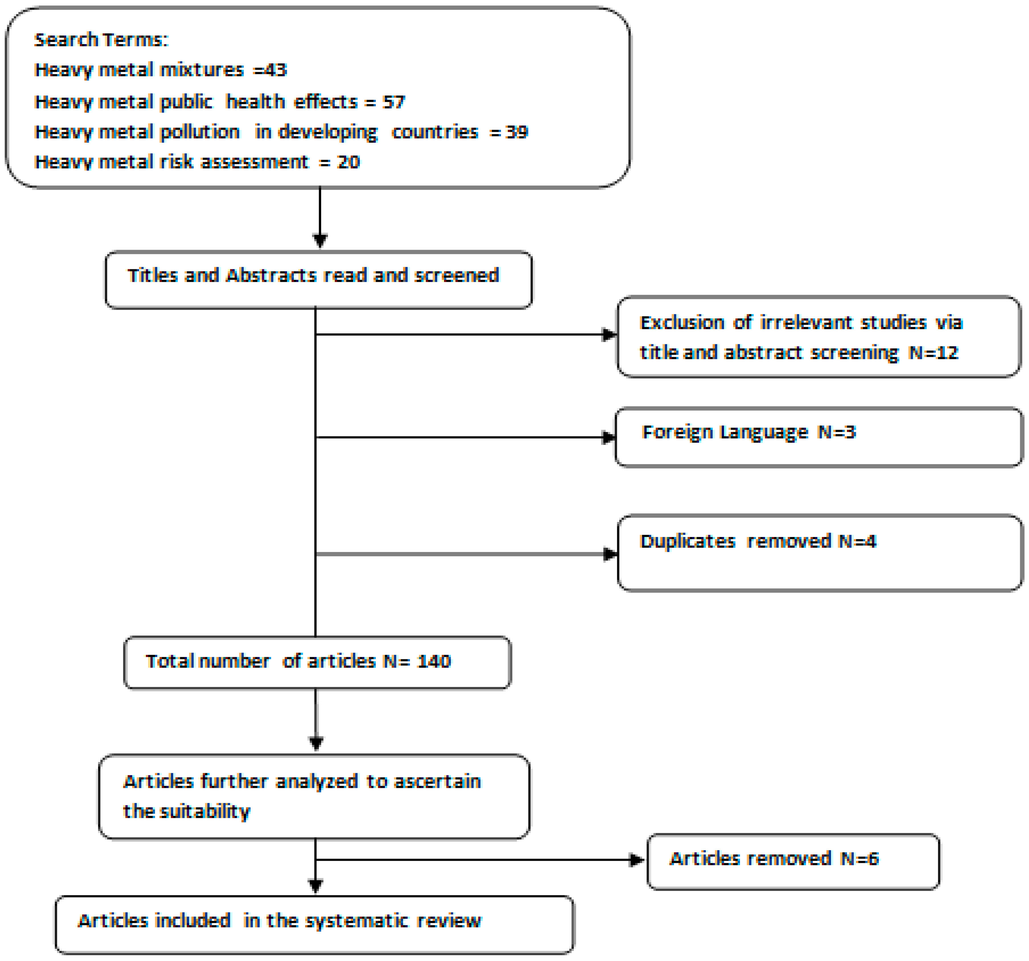
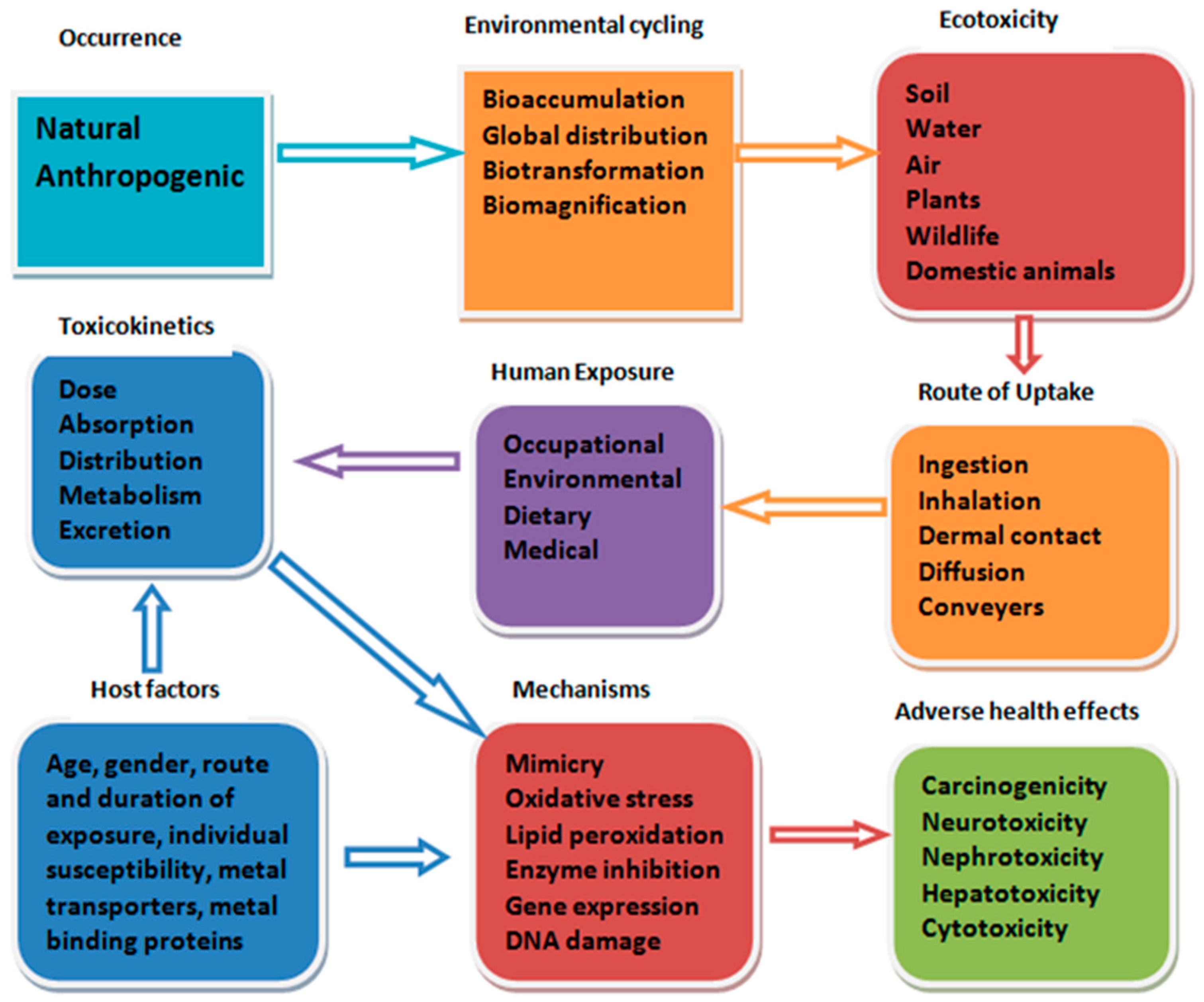
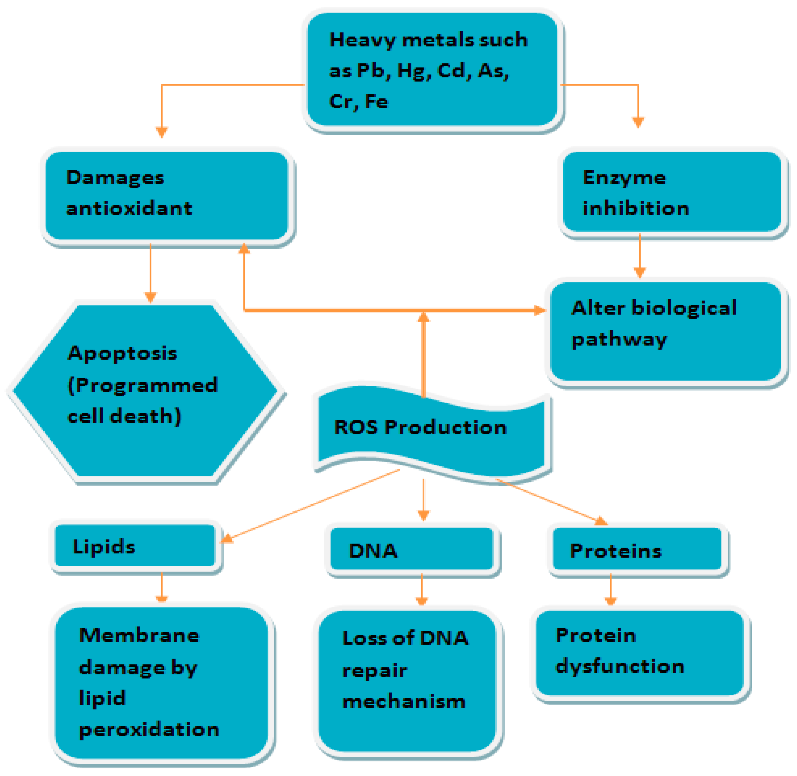
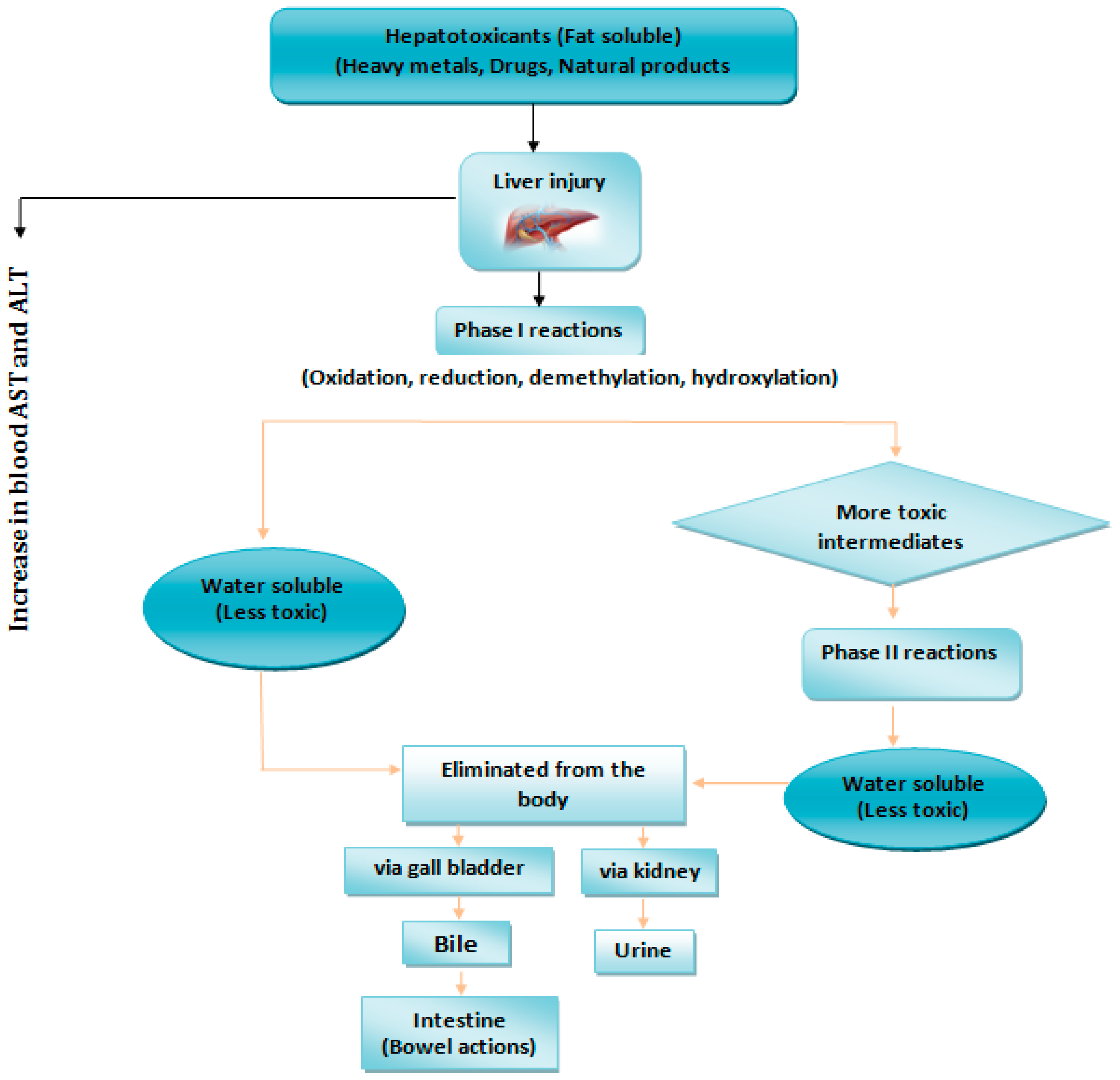
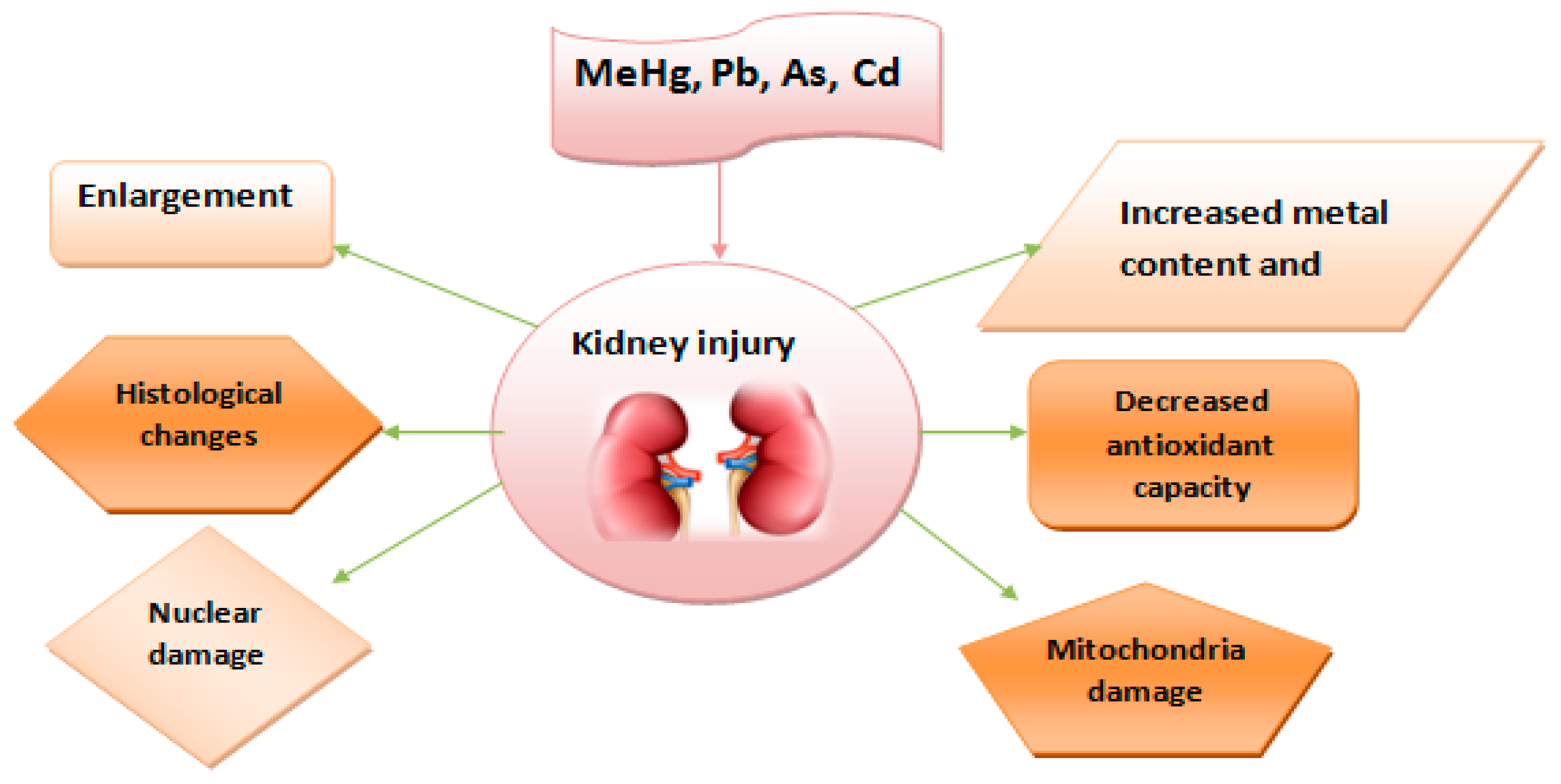
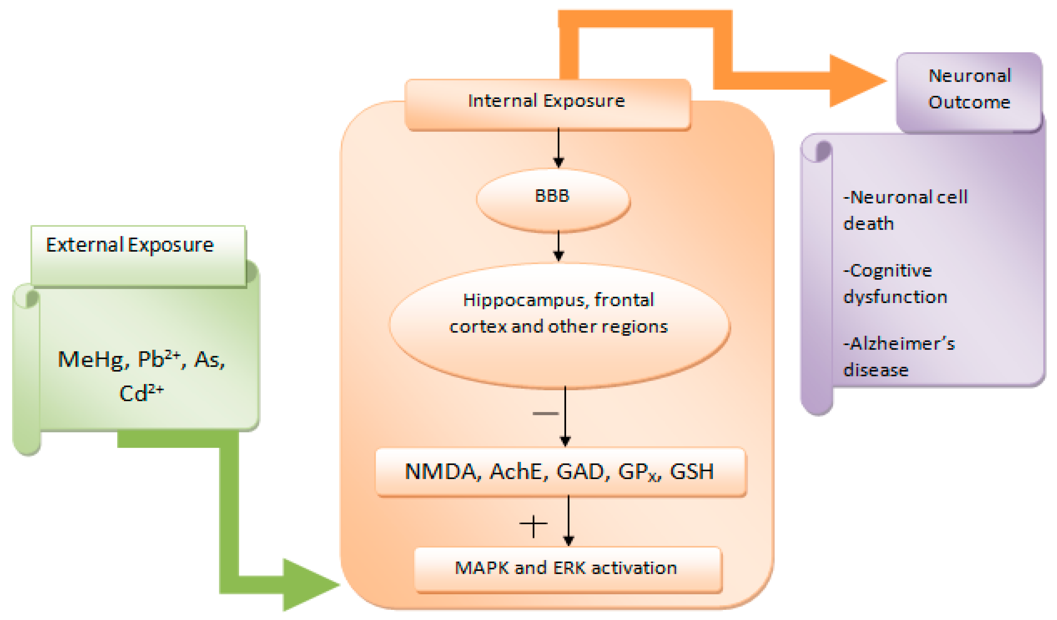
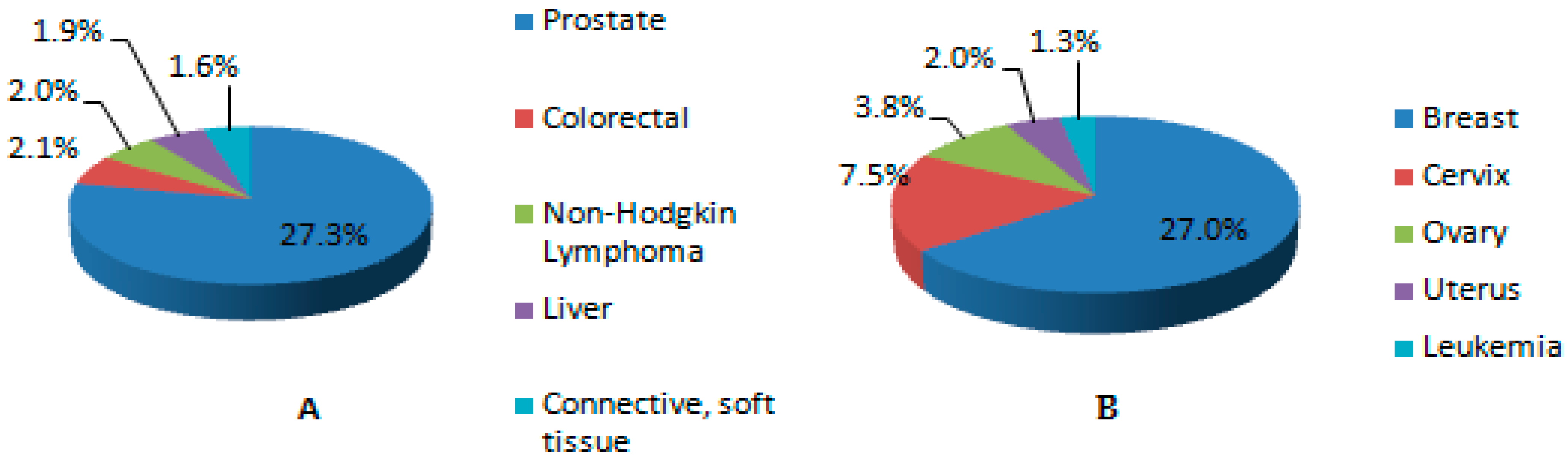
Table 1.
Heavy metal pollution in Sub-Saharan Africa.
| Country/Region | Pollution Source | Pb | Cd | Mn | Cu | Fe | Zn | Cr | Ni | As | Ref. |
|---|---|---|---|---|---|---|---|---|---|---|---|
| Nigeria/Kaduna | Soil | 0.87–1.41 | 0.0014–8.02 | 13.21–42.15 | 1425.0–1981.6 | 10.10–112.04 | 21.35–358.00 | [166] | |||
| Nigeria/Kaduna | Vegetable | 0.0014–0.001 | 0.013–2.12 | 0.582–8.65 | 331.6–1252 | 14.19–69.07 | 0.058–2.80 | [166] | |||
| Nigeria/Niger Delta | Water | 39 ± 33 | 2.4 ± 3.1 | 16.1 ± 7.4 | 107 ± 7.9 | 42 ± 17 | [144] | ||||
| Nigeria/Ibadan | Water | 0.162–0.195 | 0.279–0.315 | 8.744–10.307 | 5.063–5.096 | 0.052–0.059 | 0.103–0.133 | [195] | |||
| Nigeria/Benin | Soil | 227 ± 160 | 2.0 ± 2.9 | 562 ± 510 | 94.5 ± 150 | [196] | |||||
| Nigeria/Akwa Ibom | Fish | 0.013 ± 0.003 | 0.011 ± 0.004 | 81.36 ± 5.06 | 223.0 ± 23.47 | 0.044 ± 0.05 | 0.017 ± 0.02 | [197] | |||
| South Africa/Pretoria | Soil | 12.8–145 | 0.09–0.98 | 33.9–140 | 39.3–97.6 | 43.6–101 | [198] | ||||
| Kenya/Nairobi | Vegetable | 0–2.4 | 0–3.02 | 0.52–21.34 | 20.13–89.85 | 0–1.24 | [199] | ||||
| Kenya/Nairobi | Soil | 0.57–20 | 0–2.6 | 3.59–75.37 | 14.62–198.3 | 0.03–1.4 | [199] | ||||
| Ghana/Accra | Soil | 184.44 | 103.66 | 202.99 | 72.00 | [200] | |||||
| South Africa/Philippi horticultural area | Water | 0.04 ± 0.006 | 0.01 ± 0.002 | 0.02 ± 0.002 | 0.02 ± 0.003 | 0.02 ± 0.003 | 0.06 ± 0.009 | 0.02 ± 0.002 | [201] | ||
| South Africa/Philippi horticultural area | Soil | 19.24 ± 2.91 | 0.74 ± 0.18 | 96.74 ± 12.29 | 14.53 ± 2.02 | 30.13 ± 3.93 | 1.71 ± 0.40 | [201] | |||
| South Africa/Philippi horticultural area | Vegetable | 2.32 ± 0.91 | 0.22 ± 0.09 | 41.64 ± 5.21 | 5.55 ± 0.57 | 54.12 ± 9.24 | 2.68 ± 0.52 | 0.34 ± 0.25 | [202] | ||
| Zimbabwe/Bulawayo | Water | 0.19 ± 0.03 | 0.06 ± 0.03 | [202] | |||||||
| Zimbabwe/Bulawayo | Sediment | 51.67 ± 2.36 | 7.33 ± 0.76 | 79.17 ± 7.64 | 108.33 ± 17.02 | [202] | |||||
| Zimbabwe/Bulawayo | Fish | 35 | 5 | 120 | 10 | [202] | |||||
| Cameroon/Yaounde | River sediment | 20.3–249 | 2.8–15.6 | 42.8–142 | 26.8–341 | 94.7–199 | 2.68–32.7 | [203] | |||
| Ethiopia | Vegetable | 0.345 | 130 | 130 | 24.11 | [170] | |||||
| Ghana | Fish | 0.028 | 2.31 | [204] | |||||||
| Ghana/Iture | Water | 0.075 | 0.041 | 2.45 | 2.45 | [115] | |||||
| Ghana/Kumasi | Soil | 54.6 | 2.87 | 2606 | 2606 | [116] | |||||
| Ghana/Tarkwa | Water | 1.3 | [205] | ||||||||
| Kenya | Water | 0.496 | 0.01 | 1.95 | [176] | ||||||
| Kenya/Nairobi | Soil | 264 | 40 | 105 | 462 | 157 | [174] | ||||
| Namibia | Sediment | 10500 | 205 | 1950 | [206] | ||||||
| Nigeria | Herbal medicines | 27 | 4.75 | 97.5 | 25.5 | 78 | [207] | ||||
| Nigeria/Calabar | River sediment | 20 | 0.2 | 64 | 15 | 184 | 65 | 67 | [208] | ||
| Nigeria/Ibadan | River surface water | 0.046 | 0.0044 | 0.0033 | 0.018 | 0.14 | 0.0031 | [209] | |||
| Nigeria/Kano | Vegetable | 13.19 | 0.735 | 12.89 | [171] | ||||||
| Nigeria/Lagos | Soil | 67.5–426 | 1.61–5.31 | [210] | |||||||
| Nigeria/Niger Delta | Water | 0.025–0.064 | 0.01–0.11 | 0.03–0.081 | 0.03–0.09 | [177] | |||||
| Nigeria/Niger Delta | Fish | 0.3 | 0.03 | 0.53 | 0.21 | [149] | |||||
| Nigeria/Ogun | Fish | 3.4 | 2.1 | 5 | 20.35 | 2.3 | [150] | ||||
| Nigeria/Osogbo | Soil | 92.07 | 3.6 | 37.9 | 71.9 | 17.3 | [175] | ||||
| South Africa | Water | 16.3 | 72 | 42.6 | 27.6 | [175] | |||||
| Tanzania | Vegetable | 4.9 | 0.3 | [211] | |||||||
| Tanzania, along Lake Victoria | Water sediment | 54.6 | 7 | 26.1 | 83.7 | 12.9 | [212] | ||||
| Uganda | Vegetable | 18.7 | 1.87 | [173] | |||||||
| Uganda, along Lake Victoria | Water | 1.44 | 0.02 | 0.16 | 0.02 | 0.13 | [213] | ||||
| Zambia | Sediment | 12,855 ± 1445 | 1030 ± 58 | [214] | |||||||
| Zambia | Sediment | 9.75 | 0.8 | 125 | 130 | 220 | [215] | ||||
| Zimbabwe | Water | 1.02 | 0.12 | 2.48 | 2.37 | [178] | |||||
| Zimbabwe Harare | Vegetable | 6.77 | 3.68 | 0.05 | 111 | 221 | 16.1 | [172] |
Table 2.
Blood lead levels (ug/dL) and public health effects in Sub-Saharan Africa.
| S/N | Age Groups | Sex | Place of Study (City/Country) | Condition | Reported Concentrations | Disease Scenarios | Exposure Scenarios | Ref. |
|---|---|---|---|---|---|---|---|---|
| 1 | Adults | F | Abeokuta, Southwest Nigeria | Pregnant | 54.50 ± 4.4 | Spontaneous abortion, premature delivery, pregnancy complications, still birth, hypertension, low birth weight | Non-occupationally exposed | [234] |
| 2 | Adults | M | Nkpor, Nigeria | N/A | 39.00 ± 4.00 | Increases risk of hypertension and liver damage | Occupationally exposed | [235] |
| 3 | Adults | M | Nkpor, Nigeria | N/A | 17.00 ± 4.00 | Increases risk of hypertension and liver damage | Non-occupationally exposed | [235] |
| 4 | Adults | F | Niger Delta, Nigeria | Pregnant | 40.00 ± 16.50 | Spontaneous abortion, premature delivery, pregnancy complications, still birth, hypertension, low birth weight | Non-occupationally exposed | [228] |
| 5 | Adults | F | Niger Delta, Nigeria | Non-pregnant | 27.7 ± 1.10 | Hypertension, increased risk of renal failure, cardiovascular attacks | Non-occupationally exposed | [236] |
| 6 | Adults | M/F | Oshodi, Dopemu, & Ojota in Southwest Nigeria | N/A | 155.42 148.56 122.6 | Increases risk of hypertension and liver damage, endocrine disorder, reproductive disorder | Occupationally exposed | [237] |
| 7 | Adults | M | Port Harcourt, Nigeria | N/A | 50.37 ± 24.58 | Increases risk of hypertension and liver damage | Occupationally exposed | [238] |
| 8 | Adults | M | Port Harcourt, Nigeria | N/A | 41.40 ± 26.85 | Increases risk of hypertension and liver damage | Non-exposed | [238] |
| 9 | Children | M/F | Nigeria | N/A | >10 or >20 | Increases risk of hypertension and liver damage | Non-exposed | [239] |
| 10 | Adults | M | Abeokuta, Nigeria | N/A | 48.50 ± 9.08 | Increases risk of hypertension and liver damage | Exposed | [240] |
| 11 | Adults | M | Southwest, Nigeria | N/A | 63.00 ± 9.00 | Increases risk of hypertension and liver damage | Exposed | [241] |
| 12 | Adults | M | Southwest, Nigeria | N/A | 61.00 ± 11.00 | Increases risk of hypertension and liver damage | Non-exposed | [241] |
| 13 | Children | M/F | Allada, Benin Republic | N/A | 46.6 | Lowers intelligent quotient scores, aggressive and violent behaviours | Environmentally exposed | [242] |
| 14 | Adults | F | Allada, Benin Republic | Non-pregnant | 55.1 | Increases risk of hypertension and liver damage | Environmentally exposed | [242] |
| 15 | Adults | M | Kinshasa (Democratic Republic of Congo) | N/A | 127 | Increases risk of hypertension and liver damage | Environmentally exposed | [243] |
| 16 | Children | M/F | Kinshasa(Democratic Republic of Congo) | N/A | 11.5 | Lowers intelligent quotient scores, aggressive and violent behaviours | Environmentally exposed | [243] |
| 17 | Children | M/F | Northwest, Nigeria | N/A | 143.8 | Lowers intelligent quotient scores, aggressive and violent behaviours | Environmentally exposed | [107] |
| 18 | Adults | F | Abakaliki, Nigeria | Pregnant | 40.0 ± 16.5 | Spontaneous abortion, premature delivery, pregnancy complications | Environmentally exposed | [228] |
© 2018 by the authors. Licensee MDPI, Basel, Switzerland. This article is an open access article distributed under the terms and conditions of the Creative Commons Attribution (CC BY) license (http://creativecommons.org/licenses/by/4.0/).
Share and Cite
MDPI and ACS Style
Anyanwu, B.O.; Ezejiofor, A.N.; Igweze, Z.N.; Orisakwe, O.E. Heavy Metal Mixture Exposure and Effects in Developing Nations: An Update. Toxics 2018, 6, 65. https://0-doi-org.brum.beds.ac.uk/10.3390/toxics6040065
AMA Style
Anyanwu BO, Ezejiofor AN, Igweze ZN, Orisakwe OE. Heavy Metal Mixture Exposure and Effects in Developing Nations: An Update. Toxics. 2018; 6(4):65. https://0-doi-org.brum.beds.ac.uk/10.3390/toxics6040065
Chicago/Turabian StyleAnyanwu, Brilliance Onyinyechi, Anthonet Ndidiamaka Ezejiofor, Zelinjo Nkeiruka Igweze, and Orish Ebere Orisakwe. 2018. "Heavy Metal Mixture Exposure and Effects in Developing Nations: An Update" Toxics 6, no. 4: 65. https://0-doi-org.brum.beds.ac.uk/10.3390/toxics6040065
Note that from the first issue of 2016, this journal uses article numbers instead of page numbers. See further details here.