Review

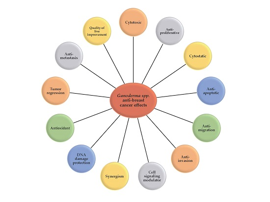
Ganoderma spp.: A Promising Adjuvant Treatment for Breast Cancer

by
Ivette J. Suárez-Arroyo
1,
Yaliz Loperena-Alvarez
2,
Raysa Rosario-Acevedo
3 and
Michelle M. Martínez-Montemayor
1,*
1
Department of Biochemistry, Universidad Central del Caribe, School of Medicine, P.O. Box 60327, Bayamón, PR 00960-6032, USA
2
Department of Science, Pontificia Universidad Católica-Mayagüez, P.O. Box 1326, Mayagüez, PR 00681, USA
3
Department of Neuroscience and Regenerative Medicine, Augusta University, 1120 15th St, Augusta, GA 30912, USA
*
Author to whom correspondence should be addressed.
Submission received: 29 December 2016 / Revised: 24 February 2017 / Accepted: 3 March 2017 / Published: 15 March 2017
(This article belongs to the Special Issue Complementary and Alternative Medicine for Cancer Patients)

Abstract

:
For the past several decades, cancer patients in the U.S. have chosen the use of natural products as an alternative or complimentary medicine approach to treat or improve their quality of life via reduction or prevention of the side effects during or after cancer treatment. The genus Ganoderma includes about 80 species of mushrooms, of which several have been used for centuries in traditional Asian medicine for their medicinal properties, including anticancer and immunoregulatory effects. Numerous bioactive compounds seem to be responsible for their healing effects. Among the approximately 400 compounds produced by Ganoderma spp., triterpenes, peptidoglycans and polysaccharides are the major physiologically-active constituents. Ganoderma anticancer effects are attributed to its efficacy in reducing cancer cell survival and growth, as well as by its chemosensitizing role. In vitro and in vivo studies have been conducted in various cancer cells and animal models; however, in this review, we focus on Ganoderma’s efficacy on breast cancers. Evidence shows that some species of Ganoderma have great potential as a natural therapeutic for breast cancer. Nevertheless, further studies are needed to investigate their potential in the clinical setting and to translate our basic scientific findings into therapeutic interventions for cancer patients.

Graphical Abstract

1. Introduction

Research proposes that different Ganoderma species (Ganoderma spp.), including G. lucidum, G. sinense, G. atrum, G. tsugae, G. neo-japonicum and, most recently, G. hainanense, carry promising anticancer properties. Bioactive substances isolated, characterized and identified from Ganoderma spp. include triterpenoids, polysaccharides, nucleosides, sterols, proteins and alkaloids. However, two main active ingredients, triterpenes and polysaccharides, have been shown to have significant anticancer effects in vitro and in vivo.
Triterpenes are compounds composed of one or more isoprene units and having anti-inflammatory and antitumorigenic activity [1]. G. lucidum possesses over 140 species of triterpenes and triterpenoids [1,2]. Triterpenes are originally isolated from Ganoderma spp. spores, and studies demonstrate outstanding therapeutic and pharmacological activities on various diseases, including cancer [3,4]. Studies confirm that subtypes of triterpenes extracted from G. lucidum may affect the viability of human cancer cell lines [4].
Ganoderma contains a significant number of polysaccharides (i.e., β-d-glucans) and peptidoglycans. Polysaccharides extracted from fruiting body, mycelia and spores usually consist of arabinose, galactose, glucose, xylose and mannose [5]. Studies demonstrate that Ganoderma polysaccharides exert anticancer effects in tumor therapy by enhancing the immune system [5]. Antitumor and antiglycemic effects, immunomodulation, antiviral activity, protective effects from free radicals and the reduction of cell damage by radiation are attributed to triterpenes and polysaccharides.
Breast cancer (BC) is the leading cause of cancer death in women in the U.S. Cancer is a disease distinguished by clinical behaviors, risk factors, molecular subtypes and response to treatment. BC molecular subtypes identified via gene expression profiles have provided information that has led to the biomarker identification that may facilitate prognosis and treatment [6,7,8]. For example, the presence or absence of estrogen receptor (ER), progesterone receptors (PR) and human epidermal growth factor receptor 2 (HER2) serve as molecular and pathological markers for treatment [8]. About 40% of BC are luminal A, thus making this the most common type of breast cancer [9]. These tumors are less aggressive and tend to have hormonal receptors present, thus having a favorable response to therapy [10]. About 10%–20% are luminal B and tend to be highly proliferative tumors [9,10,11,12]. Basal-like breast tumors account for ~10%–20% and are common in women positive for BRCA1 gene mutation, as well as African American and premenopausal women [9]. Because most of the basal-like cancers tend to be triple negative (no hormonal or HER2 receptor), their prognosis tends to be grim since for these tumor subtypes, no targeted therapies exist. Finally, ~10% of BC express HER2, while not expressing hormone receptors [9]. Similar to basal-like cancers, these tumors grow more aggressively and are associated with poorer prognosis [10]. Importantly, the availability of targeted anti-HER2 therapies has somewhat reversed the adverse prognosis.
Conventional therapy targeted to commonly deregulated signaling pathways in BC is a highly effective remedial strategy. Nevertheless, its usefulness is somewhat limited by intrinsic and acquired mechanisms of resistance, as well as by the fact that BC survivors who have received therapeutic interventions may develop conditions that affect their quality of life (QOL) and their overall survival (OS). Alterations in cell cycle and cell survival pathways and the evasion of apoptotic processes provide tumors with alternative proliferative and growth stimuli. Among pathways associated with therapy resistance are the phosphoinositide 3-kinase/AKT/mammalian target of rapamycin (PI3K/AKT/mTOR), mitogen-activated protein kinases (MAPK), the epidermal growth factor receptor family (ErbB1, also known as EGFR; ErbB2, also known as HER2; ErbB3; and ErbB4) and nuclear factor kappa B (NF-κB). Therefore, research has led investigators to assess the efficacy of alternative methods that sensitize resistant cells to therapy and that enhance QOL. Ganoderma spp. has been used in multiple in vitro and in vivo models because of its antiproliferative and growth inhibitory efficacy. In this review, we discuss the use of various Ganoderma spp. in BC models, patients and survivors to show the potential this medicinal mushroom has for its use in the clinical setting as a therapeutic for this deadly disease.

2. In Vitro Studies

2.1. Cytotoxic, Antiproliferative, Cytostatic and Antiapoptotic Effects of Ganoderma spp.

Cancer is characterized by a cascade of critical events that end in uncontrolled cell expansion and invasion. Deregulated cell proliferation together with the obliged compensatory suppression of apoptosis are necessary to support further tumor progression. Most of the chemotherapeutics are cytotoxic or cytostatic, causing toxicity or stopping the cancer cells from multiplying. Cell division is time regulated by the cyclin-dependent kinases (CDKs). Once the cyclins (the CDKs’ regulatory subunits) are bound to the CDKs, they form heterodimeric complexes. Cyclins may also be bound by CDK inhibitors that are responsible for cell cycle deceleration. The cell cycle progresses from the G1 to synthesis (S) to G2 to the mitosis (M) phase, while cells in G0 remain in a non-dividing state [13]. Cell proliferation and death are processes that are regulated to maintain homeostasis. Manipulation of the cell cycle may result in apoptosis prevention or induction; an association has been recognized for tumor suppressor genes, such as p53 and retinoblastoma (RB), c-Myc and various CDKs [14]. Apoptosis is a programmed cell-death process that occurs in normal development and turnover. It is characterized by the activation of endonucleases following the cleavage of chromatin DNA, followed by nuclear shrinkage, condensation of chromatin, membrane blebbing and DNA fragmentation [14,15]. However, improper regulation of apoptosis occurs in a variety of pathological disorders, such as cancer [14].
Whole mushroom extract or individual bioactive compounds of Ganoderma have been associated with cell death induction or cell cycle arrest of several human BC cells. In a study where aqueous extracts of G. lucidum, G. sinense and G. tsugae were used to assess their effectiveness against BC cells, data showed significant antiproliferative activities in MCF-7 cells and MDA-MB-231 in a concentration-dependent manner. Among the species tested, G. tsugae extract was most potent against MCF-7 cells, whereas the potencies for MDA-MB-231 cell proliferation inhibition were similar among the Ganoderma species tested. The extracts did not cause any cytotoxic effect on human noncancerous mammary epithelial (HMEC) cells [16]. Because of the accessibility of the commercially available products of Ganoderma, they have also been evaluated to determine their effectiveness against BC. G. lucidum in the form of powdered extract (20:1) with spores, which contains 13.5% polysaccharides and 6% triterpenes (ReishiMax GLp®, GLE), or the spore-only powder suppressed the proliferation of MDA-MB-231 cells in a dose- and time-dependent manner [17,18]. Treatment with GLE induced cell-cycle arrest at the G0/G1 phase, which was the result of the downregulation of cyclin D1 and CDK4 [17]. GLE also demonstrated a potent cytotoxic effect against noninvasive, estrogen-dependent MCF-7 than the highly invasive, estrogen-independent MDA-MB-231 BC cells [19]. GLE also showed strong cytotoxic effects in the triple negative BC cell lines, SUM-102 and MDA-MB-468, and also on the HER2 overexpressing MDA-MB-435. However, the noncancerous mammary epithelial cells, MCF-10A, were not affected by treatment [20,21]. Furthermore, the effect of GLE has been evaluated in inflammatory breast cancer (IBC), which is the most lethal type of advanced BC and which presents unique characteristics that differentiate it from non-IBCs [22]. In these studies, GLE significantly affected the viability or proliferation of SUM-149 and KPL-4 IBC cells by proapoptotic effects [20,21]. The authors demonstrated that the regulatory gene expression of cell-cycle progression was affected in SUM-149 IBC cells. Treatment with GLE decreased the expression of (cyclin D1) CCND1 and WEE, while it significantly decreased the abundance of (cyclin A2; B2) CCNA2 and CCNB2 cell cycle gene expression [23]. Gurunathan et al. showed that biologically-synthesized silver nanoparticles using G. neo-japonicum Imazeki mycelia aqueous extracts induced cell death through the generation of reactive oxygen species (ROS), caspase 3 activation and DNA fragmentation in MDA-MB-231 BC cells [24]. BreastDefend™ (BD) is a dietary supplement composed of various botanicals, including medicinal mushroom extracts (Coriolus versicolor, G. lucidum, Phellinus linteus), medicinal herbs (Scutellaria barbata, Astragalus membranaceus, Curcuma longa) and purified biologically-active nutritional compounds (diindolylmethane and quercetin). Studies show that BD suppressed proliferation of BC cells (MDA-MB-231) in a dose- and time-dependent manner predominantly through cytostatic effects. In an effort to elucidate which genes are responsible for BD’s cytostatic effect, a cDNA microarray analysis validated that BD increased the expression of GADD45A, which affects cell cycle growth arrest and downregulates the expression of cyclin A1 [25].
Alcohol extracts from Ganoderma spp. have been evaluated in BC, showing inhibitory results. Moreover, a dose- and time-dependent inhibition mediated through p21/Waf1 upregulation and cyclin D1 downregulation was obtained with a G. lucidum ethanolic extract. In this study, the extract significantly increased the expression of Bax (proapoptotic protein), while there was no change in the expression of Bcl-2 (antiapoptotic protein). The researchers demonstrated caspase-7 cleavage along with cleaved PARP expression after 36 h of treatment [26]. Proliferation assays in four tumor-cell lines, including MCF-7 and MDA-MB-231 BC cells, evaluated the effects of the ethanol extracts of G. lucidum and G. sinense. Human normal fibroblastic cells, Hs68, were used as the control. The study revealed that the G. lucidum antiproliferative effect was stronger than G. sinense extract and was more effective on the MDA-MB-231 BC cells. Moreover, G. lucidum significantly decreased G1/S phase transition, while G. sinense induced a cell cycle arrest at G2. As determined by terminal deoxynucleotidyl transferase dUTP nick and labeling (TUNEL) assay, both extracts induced apoptosis even at the lower dose of 40 µg/mL. Triterpenoids, sterols and nucleosides may contribute to the apoptotic induction [27]. The effect of alcohol extract of G. tsugae (GTE) was assessed in HER2-overexpressing cancer cells. The study evidenced the antiproliferative effects of the extract on BT-474 and SKBR-3 BC cell lines. Antiproliferative effects in SKBR-3 cells resulted in an increase in G1 with a decrease in S and G2/M phases via regulation of cyclins D1 and E [28]. Wu et al. assessed the anticancer effects of water or ethanol extracts of five different fungal species, including G. sinense. The results showed that water extracts exert moderate anticancer activities compared to that of ethanol extract in MDA-MB-231 cells [29]. Recently, a group of researchers from Turkey examined the cytotoxic effects of G. lucidum extracts obtained with five different solvents (ethanol-water, methanol, ethanol, ethyl acetate and ether) on MCF-7 cells at 24, 48 and 72 h. Based on the cytotoxicity results, they determined that G. lucidum ether extract (G.Ether) had greater potency (IC50 = 100 μg/mL at 72 h) against BC cells than the others [30,31]. To study the mechanism behind the extracts’ anticancer effects, they did telomerase activity assays and found a 32% decrease in telomerase activity in treated cells [31].
Many scientists have attributed the anticancer effects of Ganoderma spp. to the triterpenes. Ganodermanontriol (GDNT) is a biologically-active triterpene alcohol isolated from G. lucidum. To evaluate the antiproliferative effects of GDNT, MDA-MB-231 cells were treated with increasing concentrations of the triterpene for 24, 48 and 72 h, and proliferation was determined. GDNT significantly inhibited the proliferation of cells with an IC50 of 42.0, 15.7 and 11.6 µM at 24, 48 and 72 h, respectively, accompanied by a reduction in colony formation. In addition, GDNT suppressed MCF-7 cell proliferation while slightly affecting the proliferation of MCF-10A noncancerous mammary epithelial cells [32]. Two related complexes closely related to E3 ubiquitin ligase, the anaphase-promoting complex (APC) and the Skp1-Cullin1-F-box complex (SCF), are major driving forces controlling cell cycle progression. APC, the most complex E3 ubiquitin ligase, consists of at least 14 subunits, one of them being the coactivator, CDC20 (cell division cycle 20 homologue), which has been associated with oncogenesis [33]. Jiang et al. found the overexpression of CDC20 in BC cells and tissues. Interestingly, GDNT treatment resulted in suppressed CDC20 expression in MDA-MB-231 BC cells [32]. The anti-BC activity of the ethanol-soluble and acidic component (ESAC), which mainly contains triterpenes extracted from G. lucidum, was determined after 48 h of treatment on MCF-7 and MDA-MB-231 cells. The results showed that ESAC notably decreased the viability of BC cells in a concentration-dependent manner, mediated by G1 cell cycle arrest and apoptosis, as demonstrated by the condensation of nuclear chromatin, DNA fragmentation and increased expression of a PARP cleavage [34]. Moreover, a study testing ganoderic acid DM (GADM), a G. lucidum triterpenoid, showed a dose- and time-dependent decrease in cancer cell viability [35]. Wu et al. showed that a low concentration of the GADM triterpene extract effectively induces a G1 cell cycle arrest in MCF-7 cells [35]. Interestingly, this study revealed that MDA-MB-231 cells need higher concentrations of the triterpenoid extract to mediate G1 cell cycle arrest. This effect may be attributable to the greater proliferative and metastatic potential of these cells. The researchers confirmed their results by assessing the expression of the catalytic subunits of the CDK complex, CDK2 and CDK6, which are essential for the G1/S transition, as well as the expression of cyclin D1 and retinoblastoma (Rb) phosphorylation. GADM downregulated the abundance and p-Rb in a time-dependent manner [35]. Similarly, ganoderic acid ME (GA-Me) purified from G. lucidum inhibits cell proliferation and induces apoptosis of BC cells by decreasing the prosurvival proteins BCL-2 and c-Myc, as well as the cell-cycle regulator cyclin D1 [36]. The effects on BC cell viability and morphology of G. lucidum extract mainly containing ganoderiol A (GA), dihydrogenated GA, and GA isomer (GAEE) was studied in highly metastatic BC cells. GAEE exhibited no toxicity in MDA-MB-231 BC cells at concentrations between 5 µg/mL and 20 µg/mL. Moreover, GAEE did not induce cell cycle arrest or apoptosis [37]. Recently, Peng et al. isolated 19 lanostane triterpenoids (15 unknown and 5 known compounds) from G. hainanense, a rare species of Ganoderma. The group evaluated the cytotoxicities of 16 compounds, including ganoderone A, lucidadiol, ganodermanontriol and 4,4,14a-trimethyl-3,7-dioxo-5a-chol-8-en-24-oic acid. Compounds 10, 12 and 17 (lucidadiol) showed inhibitory activities against MCF-7 cells [38]. Polysaccharides do not have direct cytotoxic activity against tumor cells, but do stimulate the immune response. Shang et al. purified a G. lucidum polysaccharide extracted from the selenium (Se)-enriched mycelia (SeGLP-2B-1) of the mushroom [39]. The cytotoxic effect of SeGLP-2B-1 was assessed in BC using increasing concentrations of polysaccharides for 24–72 h. The results showed that at 24 h, MCF-7 BC cell viability was not significantly decreased. MCF-7 BC cell viability was significantly reduced in a dose-dependent manner mediated by apoptosis after 48 h of treatment. The mechanisms behind this process included the intrinsic and extrinsic apoptotic pathways. Apoptosis results showed the formation of sub-G1 apoptotic bodies mediated by an increase of caspases-8, -9 and -3 and cleavage of PARP. Moreover, the loss of mitochondrial action potential accompanied by the release of cytochrome c into the cytosol paralleled the apoptosis data, suggesting that SeGLP-2B-1 induced mitochondria-mediated cell death [40]. Proteins of Ganoderma spp. are also identified as bioactive compounds with immunomodulatory and anticancer activities. One of the most important immunomodulatory proteins isolated from G. lucidum is Lhing-Zhi-8 (LZ-8) [41]. LZ-8 is a potent mitogen of T cells and peripheral blood mononuclear cells (PBMCs), an activator of macrophages and of human monocyte-derived dendritic cells and an inducer of cytokines [42,43,44,45]. LZ-8 has also immunosuppressive effects in vivo, showing a reduction in antibody production [46]. The antitumor effects of LZ-8 were also demonstrated in human lung cancer. Results from this study showed that recombinant LZ-8 (rLZ-8) reduced the proliferation of A549 human lung cancer cells, inducing a G1 cell-cycle arrest mediated by the overexpression of p53 and p21. Moreover, rLZ-8 decreased tumor growth in xenograft models of Lewis lung carcinoma [47]. In addition, the anticancer effects of LZ-8 were evidenced in human gastric cancer cells. rLZ-8 induced SGC-7901 gastric cancer cell death by autophagy mediated by the activation of the ER-associated degradation systems (ERAD) in a caspase-independent manner [48]. An active fucose-containing glycoprotein fraction isolated from a water-soluble Ling-Zhi (G. lucidum) extract (FFLZ), exerts immunomodulating activities by stimulating the expression of inflammatory cytokines and antibody-mediated cytotoxicity in cancer cells [49,50]. Tsao et al. evaluated the effects of FLZZ on the growth of mouse BC cells 4T1 and on MDA-MB-231 and showed that treatment decreased the viability of both cell lines in a dose-dependent fashion. Furthermore, FFLZ-treated 4T1 cells formed fewer colonies than untreated cells, indicating that FFLZ inhibits the colony formation of 4T1 cells [51]. Khz is a crude polysaccharide isolated from the fusion of G. lucidum and Polyporus umbellatus mycelia. This protein selectively induced apoptosis in cancer cells by increasing intracellular [Ca+2] to generate ROS [52]. Recently, the same group of investigators showed that Khz decreased proliferation and induced apoptosis on MCF-7 cells following similar mechanisms evidenced before. The results also demonstrated that Khz proapoptotic effects were mediated by caspase-7, -8 and -9 [53]. Fungal immunomodulatory proteins (FIPs), which are small-molecule proteins isolated from higher basidiomycetes, have a variety of biological functions, including hemagglutination, antianaphylaxis, antitumor and immune-regulating activity. Currently, eight FIPs have been isolated from Ganoderma species, such as G. lucidum, G. tsugae, G. japonicum, G. microsporum, G. sinensis and G. atrum [54,55]. A FIP isolated from G. atrum (FIP-gat), is a new member of the FIP family. Recently, the anticancer effect of recombinant FIP-gat was evaluated in BC. Results showed that rFIP-gat reduced cell viability of MDA-MB-231 cells in a dose-dependent manner after 48 h and has agglutinating activity. rFIP-gat induced cell cycle arrest in G1 accompanied by proapoptosis effects. To study the effects of rFIP-gat in gene expression, investigators performed a gene microarray and found 669 differentially-expressed genes with a minimum fold change of two upon treatment. They validated 10 genes that were increased by the protein and that play an important role in cell death and cell growth, such as TNFSF8, SQSTM1 and DUSP1 [55]. These data suggest that Ganoderma spp. and their bioactive compounds are capable of inducing cytotoxicity, antiproliferative effects, proapoptotic processes and cell cycle arrest (Table 1) as part of its anti-BC-promoting properties.

2.2. Antimigration and Anti-Invasion Potential of Ganoderma spp.

A cancer trait is the unique capacity of cells to evolve from a hyperplastic state to an increasingly disorganized and invasive tumor that can eventually propagate to distant organs, thus metastasizing. Additional traits include motile and invasive properties, which are the first steps that a malignant cell utilizes to overcome the noncancerous tissue barriers [56]. Noncancerous tissue requires proper adhesion to the basement membrane or neighboring cells and a signaling network to create a homeostatic environment that is maintained. Cancer cells overcome these barriers via deregulation of the signaling network, resulting in modulation in the expression of proteins involved in basement membrane formation, motility and cytoskeletal remodeling. Cancer cells may detach from the primary tumor either as individual cells or collectively as cell sheets, strands and clusters to travel through surrounding extracellular stroma and to gain entry into blood and lymphatic vessels [56].
Anti-invasive properties of G. lucidum extracts have been shown in various cancer cell lines, including BC. Contrary to the antiproliferative and chemoresistance roles for which many Ganoderma spp. have been tested, anti-invasive or migratory properties in BC models have been studied mainly with G. lucidum and are described herein. GAEE was used to examine the anti-invasive effects in the human invasive BC cells MDA-MB-231. Wound-healing assays showed that increasing concentrations of GAEE extract inhibited cell migration as much as 73% when used in concentrations of 20 µg/mL. This effect was attributed to the ability of GAEE to attenuate the binding affinity between focal adhesion kinase (FAK) and the cytoskeletal protein paxillin, which might affect cell migration and adhesion. Moreover, GAEE downregulated RhoA, Rac1 and Cdc42 expression and decreased the interaction between N-WASP and Cdc42 [37]. Thyagarajan et al. evaluated a green tea extract (GTE) containing 97% polyphenols and 38% epigallocatechin gallate (EGCG), individually and in combination with GLE in MDA-MB-231 BC cells [57]. Although the sole GLE (0–500 μg/mL) or GTE (0–125 μg/mL) suppressed migration of MDA-MB-231 cells in a dose-response manner, the combination of GLE and GTE synergistically inhibited cell migration. Furthermore, GLE markedly inhibited invasion through the artificial basement membrane Matrigel®, and the addition of GTE increased invasion inhibition further, demonstrating a synergistic anti-invasive effect, as well [57]. To evaluate the anti-invasive effects of G. lucidum, Sliva et al. used different sources of commercially available extracts composed of whole spores, broken spores, ground-fruiting body particles/mushroom powder or GLE. In this study, they found that all extracts except for the ones composed of just broken spores or mushroom powder were effective in decreasing the adhesion, migration and invasion of MDA-MB-231 by more than 50%. Remarkably, GLE inhibited 99% of MDA-MB-231 migration [58], while aqueous extracts of G. lucidum spores or fruiting body both inhibited cell motility [59]. A G. lucidum dietary supplement also inhibited cell migration (MCF-7 and MDA-MB-231), cell adhesion to fibronectin and vitronectin and invasion of the metastatic cell line MDA-MB-231 [60]. Studies in IBC also support the anti-invasive effect of GLE. Treatment with GLE inhibited migration and invasion of SUM-149 cells after 24 and 72 h [20,21]. Ganoderic acid, GDNT and GA-Me also reduced BC (MDA-MB-231) cell adhesion to the extracellular matrix protein vitronectin, as well as cell migration and cell invasion [32,36,61]. G. lucidum also inhibited oxidative stress-induced migration of MCF-7 BC cells [62]. Recently, a mix of medicinal mushrooms and plants was developed for cancer treatment. Studies using the MycoPhyto® Complex, a mix of six varieties of mushroom (Agaricus blazei, Cordyceps sinensis, Coriolus versicolor, G. lucidum, Grifola frondosa and Polyporus umbellatus) mycelia, plus β-1, 3-glucan isolated from the yeast Saccharomyces cerevisiae and BD decreased MDA-MB-231 BC cell migration and invasion [25,63]. The effect of the glycoprotein FFLZ was evaluated in vitro using wound closure and migration/invasion assays. Results indicated that FFLZ significantly reduced MDA-MB-231 cell invasion and mobility [51]. The epithelial mesenchymal transition (EMT) process is the differentiation of epithelial cells into motile mesenchymal cells. This switch in cell behavior is mediated by reprogramming gene expression and cell signaling. During EMT, epithelial cells lose their junctions and cell polarity and reorganize their cytoskeleton. These changes increase their migration and invasion capacity [64]. MDA-MB-231 cells incubated with FFLZ displayed a distinct cell-cell adhesion and cluster morphology, formation of protrusions and destruction of actin filaments characteristic of the EMT process. To examine the mechanisms behind the phenomenon of morphology alteration, the investigators assessed the effects of FFLZ on the expression of EMT markers. They found that FFLZ decreases mesenchymal cell markers (e.g., N-cadherin and vimentin) or increases epithelial cell markers (e.g., E-cadherin and γ-catenin). Moreover, FFLZ decreases the TGFβ-induced Smad2/3-Smad4-Snail/Slug-axis pathway and the expression of the EMT-related transcriptional factors in BC cells [51]. There is growing evidence that tumor cell aggregates or spheroids provide a more representative model of tumors in an in vitro setting than what can be achieved with monolayer cultures. Parameters such as stiffness, pH, oxygen, glucose and growth factors are intentionally misaligned in standard 2D cell culture, making it a poor physiological model for tumor studies [65]. Spheroids create cell-cell connections and have decreased proliferation and increased cell survival rates. They may also show tumor dormancy and a hypoxic core, which are features of the tumor microenvironment [66]. These characteristics give rise to a more stratified composition, with the spheroids consisting of a border of proliferating cells, followed by a layer of quiescent cells [67]. Pathologically, many types of BC form tumor emboli as a mechanism of lymphovascular invasion, which are visualized in three-dimensional (3D) culture as tumor spheroids [68]. SUM-149 IBC cells typically form spheroids in culture when utilizing 3D cell matrixes. Studies show that SUM-149 cells invade as tumor spheroids in the control treatment, while GLE treatment disintegrates tumor spheroids by disrupting cell-cell interactions typically formed by invading cells after 48 or 72 h of treatment [20]. All of the gathered data suggest that Ganoderma inhibits the migration and invasive behavior of human BC cells in vitro.

3. Signaling Studies

The joint interaction between tumor cells and the reactive stroma strongly contributes to the development and progression of cancer. Cells use signaling cascades to amplify and send messages, generally for regulatory processes, such as control of transcription to maintain homeostasis. In cancer cells, many of these signaling cascades are deregulated or reprogrammed to evade apoptosis, thus maintaining a proliferative potential and resisting therapy. Medicinal mushroom supplements have been effective in signaling cascade modulation and cancer cell sensitization for conventional therapies. In this section of the review, we discuss signaling pathways involved in the oncogenesis of BC cells and how Ganoderma spp. and their compounds modulate them (Table 2).

3.1. HER2 Signaling Pathways

HER2 is a transmembrane tyrosine kinase receptor belonging to the EGFR family (which also includes EGFR, HER3 and HER4). All members of the EGFR family share a common molecular structure: an extracellular ligand-binding domain with an amino terminal, a single transmembrane spanning region and an intracellular cytoplasmic domain with tyrosine kinase activity. Once the receptor-specific ligand binds to the extracellular domain, the receptor adopts a specific conformation, which permits the receptor to form homodimers or heterodimers between the family members. However, HER2 is an orphan receptor that exists constitutively active. The formation of heterodimers or homodimers thereafter activates the intracellular tyrosine kinases and triggers the autophosphorylation of specific tyrosine residues, ending in the activation of signaling cascades, such as PI3K/Akt/mTOR and MAPK [71]. Although HER2 is vital for normal cell processes, the overexpression of HER2 leads to tumorigenesis. HER2 gene amplification and HER2 protein overexpression accounts for about 25%–30% of all BC and is associated with aggressive behavior, chemotherapy resistance, poor prognosis, a low OS rate and metastasis [72]. Kuo et al. demonstrated that GTE inhibited p-HER2 and p-Akt in SKBR-3, BT-474 and MCF-7/HER2 HER2-BC overexpressing cells. To investigate the mechanisms that underline the GTE-mediated downregulation of HER2, the authors assessed the effects of GTE on HER2 mRNA expression and HER2 protein stability. They found that GTE decreases the expression of HER2 mRNA and shortened the half-life of HER2 [28].

3.2. PI3K/AKT/mTOR

PI3K is a key intermediate in cell responses induced by various agonistic signals that result from downstream target activation by proteins and lipids [73]. PI3K is composed of the catalytic subunit p110 (α, β and δ) and regulatory subunits of 55, 87 or 101 kDa [74]. Lipids at the plasma membrane serve as docking sites for proteins that have pleckstrin homology (PH) domains, such as AKT. AKT is a central serine/threonine kinase involved in survival mechanisms of the cell by inhibiting apoptosis [75]. Activation of AKT leads to the phosphorylation of mTOR, which in turn activates translation by cap-dependent and independent pathways. The serine/threonine protein kinase mTOR is a cell metabolism, growth and survival central regulator. mTOR is activated in response to mitogenic signals, such as hormones, growth factors, nutrients, energy and stress, which leads to cell growth, proliferation and survival [76]. mTOR effectors are the eukaryotic translation initiation factor 4e binding protein (EIF4EBP1 or 4E-BP1) and the ribosomal protein S6 kinase (p70S6K) [77]. mTOR regulates the eIF4F cap-dependent translation initiation complex, which consists of the eIF4E cap-binding protein, eIF4A RNA helicase and the eIF4G scaffold protein. [77]. In IBC tissues and cells, overexpression of the eIF4GI is observed [78]. Overexpression of eIF4G drives greater cap-dependent translation, plus elevated levels of eIF4G enhances internal ribosome entry site (IRES)-dependent translation. The latter is used as an alternative translation initiation mechanism by a subset of mRNAs. In IBC, eIF4G is accountable for the strong homotypic cell interaction that drives tumor emboli formation and promotes IBC cell invasion [78]. Studies demonstrate that GLE significantly modulates mTOR signaling in SUM-149 IBC cells. This is shown by reduced expression of p-mTOR at Ser2481 (a site that promotes intrinsic catalytic activity) [79]. GLE also reduced p70S6K, S6, p-S6 and p-4E-BP1 expression [23]. Moreover, GLE induced a ~50% reduction in protein synthesis in IBC cells, an effect not seen in GLE-treated MCF-10A noncancerous cells [23].
Deregulation of PI3K/AKT/mTOR is associated with increased transformation and oncogenesis [80]. Constitutive activation of PI3K causes migration of MDA-MB-231 by means of both catalytic and regulatory subunits of the PI3K complex [73], an effect that GLE reduced by modulation of the NF-κB pathway [58]. Furthermore, Jiang et al. demonstrated that GLE inhibits AKT expression in a time-dependent manner with no inhibition of p-AKT-Thr308, which corresponds to the residue in the PI3-K activation loop by the phosphoinositide-dependent kinase 1 (PDK1). GLE decreased p-AKT-Ser473, which is the residue activated by mTOR. This reduction in turn suppressed NF-κB activity in MDA-MB-231 BC cells [17]. The effect of GLE was also studied on the PI3K/AKT pathway in SUM-149 IBC cells. GLE downregulated the gene expression of AKT1, CCND1, EIF4GI, MAPK1 and HRAS, while the expression of JUN and FOS was upregulated after 3 h of treatment [23]. Moreover, in vivo studies investigating GLE effects on IBC tumor lysates showed that GLE reduced the expression of the IBC biomarker, E-cadherin, of p120-catenin and of c-Myc. mTOR signaling protein abundance was significantly reduced in tumor lysates from GLE-treated mice, where mTOR, p70S6K and eIF4G expression was reduced [23]. Because loss of mTOR function impacts MAPK activation [81], the authors evaluated GLE’s effect on molecules from this pathway. Data show that GLE reduced RAS and p-ERK1/2 expression without affecting total ERK1/2 in tumor cell lysates [23].

3.3. NFκB

Nuclear factor-κB (NF-κB)/Rel is a group of proteins that comprise NF-κB p52/p100, NF-κB1 p50/p105, c-Rel, RelA/p65 and RelB [82]. NF-κB function as transcription factors that control genes regulating multiple processes, including innate and adaptive immunity, inflammation, stress response, B cell development and oncogenesis [83]. In most cells, NF-κB complexes are inactive, residing predominantly in the cytoplasm sequestered by inhibitory IκB proteins [82]. When this signaling pathway is activated (i.e., upon activation of the PI3-Kinase [84], interleukins or the tumor necrosis factor pathways), the IκB kinase (IKK) complex phosphorylates IκB and tags it for proteasomal degradation, liberating NF-κB, which dimerizes and translocates to the nucleus to modulate target gene expression [85]. NF-κB promotes proliferation and invasion and blocks apoptosis in different cancer types, including human BC [82,86] and activated NF-κB is detected in estrogen receptor-negative human BC cells with overexpressed EGFR [86]. Studies by Sliva et al. show that GLE suppresses the motility of BC cells by inhibiting NF-κB [59]. In accordance with the migration results explained in Section 2.2, the same extracts were also effective in the inhibition of the NF-κB pathway, which was directly linked to the invasion inhibition of this BC cell line [58]. Sliva et al. demonstrated that GLE inhibits the activity of NF-κB in BC cells. The researchers investigated the mechanistic basis of the inhibitory effects of G. lucidum on MCF-7 (estrogen dependent) and MDA-MB-231 (estrogen independent) proliferation. They found that GLE downregulates ERα expression, inhibits estrogen-inducible ER transactivation and inhibits tumor necrosis factor-alpha (TNFα)-stimulated activation of NF-κB in MCF-7 BC cells. GLE also decreased the estrogen response element (ERE) and NF-κB constitutive activity in MDA-MB-231 BC cells. Moreover, the ER and NF-κB pathway inhibition led to c-Myc downregulation [19]. As established earlier, GA-Me inhibits cell proliferation, induces apoptosis of BC cells by decreasing prosurvival proteins and decreases cell migration and invasion. GA-Me also has anti-BC tumor and antiangiogenesis properties [36]. The same study indicated that GA-Me inhibited NF-κB activity in the absence or presence of TNF-α without affecting the phosphorylation and degradation of its inhibitor (IkB-α). GA-Me downregulated c-Myc, cyclin D1, Bcl-2, MMP-9 VEGF, interleukin (IL)-6 and IL-8 expression, all of which are NF-κB-regulated genes [31].

3.4. AP-1

Activator protein 1 (AP-1) is a transcription factor composed of a dimeric complex that contains members of the JUN and FOS (c-Fos, Fra-1, Fra-2 and Fos-B) protein families. Members of the FOS family heterodimerize with members of the JUN family, creating complexes that are transcriptionally active [87]. Once dimerized, AP-1 binds to DNA response elements [tissue plasminogen activator (TPA) response elements and cAMP response elements (CREs)] in the promoter and enhancer regions of specific genes [88]. In vitro studies show that FOS-JUN heterodimers create more stable complexes and display stronger DNA-binding activity versus JUN homodimers [87]. Studies in human BC cells show that GLE suppresses the activity of constitutively-active AP-1, followed by urokinase-type plasminogen activator (uPA) and its receptor (uPAR) downregulation [59].

3.5. Proteases

The urokinase-type plasminogen activator specifically cleaves the Arg-X-Val bond in the zymogen form to activate the enzyme plasmin [89]. Plasmin mediates invasion directly by degrading collagen IV, fibronectin and laminin or indirectly by MMP 2, 3 and 9 and uPA activation. Moreover, uPA also regulates cell adhesion and chemotaxis [89,90] by stimulation of migration. First, it can act directly through proteolytic activity by transforming growth factor-β (TGF-β) activation [91]. An alternative mechanism involves uPA as a nonproteolytic protein, which stimulates cell migration directly through interaction with its receptor (uPAR) [92]. uPA plays a crucial role in tumor metastasis, and its overexpression in BC is a marker of poor prognosis [93,94]. GLE studies show that the expression of uPA and its receptor uPAR is downregulated upon GLE treatment in BC cells [59], while additional studies with the Ganoderma spp. triterpene, ganoderic acid, show uPA/uPAR signaling downregulation via AP-1 and NF-kB activity modulation in MDA-MB-231 BC cells [61,69]. Jiang et al. demonstrated that the antitumor and antimetastases effects of the BD dietary supplement were mediated via downregulation of the plasminogen activator urokinase (PLAU) and chemokine (C-X-C Motif) receptor 4 (CXCR4) gene expression [70].
Another group of proteases important in oncogenesis are the MMPs. These are zinc-dependent endopeptidases that degrade the extracellular matrix. MMPs are key mediators of invasion and metastasis and are involved in cell proliferation, survival, angiogenesis and migration [95]. The MMPs regulate various physiological and signaling events and play a key role in tumor and stroma communication [96]. Studies have evaluated the diagnostic and prognostic value of circulating MMP2 and MMP9 in BC patients because elevated levels of both metalloproteinases have been observed in BC patients’ blood. MMP levels are also correlated with stage and lymph node metastasis [97,98,99,100]. Studies demonstrated that GLE and GA-Me downregulates MMP9 gene expression in SUM-149 and MDA-MB-231 cells [20,36]. Moreover, gelatinase activity in response to 48 h of GLE treatment shows that the activity of MMP2 and MMP9 was significantly inhibited by almost 50% after normalizing for total cell number [20].

4. Synergistic Effects between Ganoderma spp. and Antineoplastic Drugs

Cancer patients frequently relapse after chemotherapy because of the acquisition of resistance to antitumor drugs. Resistance to cancer cytotoxic agents may occur by several mechanisms, such as poor absorption, inactivating metabolism, reduced drug availability or defective immune-system-mediated functions [101]. As part of the search for agents that could overcome this chemoresistant property, Ganoderma spp. plus conventional cancer therapies have been evaluated. Additive and synergistic effects between Ganoderma spp. and antineoplastic drugs have been shown in different cancer cells and tumors, such as in Lewis lung carcinoma [102], urothelial carcinoma [103], non-small [104] and small-cell lung cancer [105], chronic myelogenous leukemia [106], colon cancer [107], hepatocellular carcinoma [108], ovarian cancer [28,109], epidermoid carcinoma [110] and sarcoma [111].
Drug resistance is also characteristically found in advanced-stage BC. The inability of some BC patients to respond to targeted therapies starting at the beginning of treatment is called de novo drug resistance. In contrast, significant numbers of BC patients initially respond to chemotherapy and then turn refractory in a process of acquired resistance, resulting in poor prognosis [112]. The antitumor effects of bioactive Ganoderma spp. compounds present in a variety of BC models, as reviewed herein, suggest its use together with chemotherapeutic agents to overcome the chemoresistant phenotype. In studies using the methanolic extract of G. tsugae, the growth-inhibitory effect of taxol and cisplatin in the HER2+ BC cell line MBA-MD-435 was enhanced with 250 µg/mL of the extract [28]. Another study shows that the biologically-active compound ergosterol peroxide, isolated from G. lucidum, could overcome drug resistance conferred by miR-378 in MDA-MB-231 cells [113]. In vivo studies using MM46 mammary carcinoma C3H/HeN mice fed with a control or an AIN-93M diet containing 2.5% G. lucidum antler form extract and injected with 50 or 150 mg/kg of cyclophosphamide show that combining the cyclophosphamide plus 2.5% G. lucidum significantly inhibits tumor growth. Moreover, the respective final tumor weight was decreased to about 50% compared to tumors from mice fed control diets [114]. Recently, the interaction between G. lucidum and tamoxifen or doxorubicin was examined in MCF-7 cells, showing a synergistic interaction between the G.Ether and tamoxifen. Interestingly, G.Ether decreased the cytotoxic effects of doxorubicin in ER+ BC cells, showing an antagonistic effect between therapies [30]. Because of the high incidence of tyrosine kinase inhibitors (TKIs) targeted against EGFR resistance, a new study investigated the therapeutic potential of GLE in combination with the EGFR TKI Erlotinib in vitro and in vivo. In these studies, GLE synergizes with erlotinib to sensitize SUM-149 IBC cells to the conventional therapy. Moreover, GLE overcomes intrinsic (MDA-MB-231 BC cells) and developed (rSUM-149 IBC cells) erlotinib resistance. Furthermore, erlotinib/GLE decreased SUM-149 IBC cell viability, proliferation, migration and invasion. GLE increased erlotinib sensitivity via EGFR, AKT and ERK signaling inactivation [21]. These studies evidence that a combinatorial therapeutic approach using Ganoderma spp. and traditional therapies may be the best way to increase prognosis in BC patients.

5. Ganoderma spp. and DNA Damage Protection

In human cells, intrinsic and extrinsic factors, such as UV light, X-radiation, gamma irradiation and ionization, can cause DNA damage. Unrepaired or misrepaired DNA damage can result in the accumulation of genetic insults, leading to neoplastic transformation. Several studies have shown the radioprotective properties of Ganoderma spp. Studies have shown that using polysaccharides isolated from G. lucidum enhanced the repair process after gamma irradiation treatment using in vitro and in vivo models [115,116]. Lanostanoids isolated from G. tsugae protected human cells against damage induced by UVB light [117]. Moreover, a water-soluble extract of G. lucidum increased small intestinal crypt survival and enhanced body weight and complete blood counts of irradiated mice from radiation damage after X-irradiation [118,119].

Antioxidant Potential of Ganoderma spp.

The production of reactive compounds is the result of the oxidative metabolism in aerobic respiration. These normally low concentrations are necessary for enzyme activation, gene expression, disulfide bond formation during the folding of new proteins in the endoplasmic reticulum, signal transduction and caspase activity control. However, <5% of them may cause cell toxicity if their concentration increases by internal or external sources [120]. Reactive species can be classified into four groups based on the main atom involved. ROS are the most abundantly produced. When the pro-/anti-oxidant equilibrium is lost, oxidative stress occurs, which alters and damages DNA, RNA, lipids and proteins. ROS cause malfunctions in DNA repair and mutations in the DNA, enhancing aging and carcinogenesis. Antioxidants scavenge reactive species through the enzymatic superoxide dismutase (SOD), catalase (CAT) and glutathione peroxidases (GPXs) [120]. Deepalakshmi et al. evaluated the in vitro and in vivo antioxidant potential of G. lucidum fruiting-body ethanolic extract on 7,12-dimethylbenz(a)anthracene (DMBA)-induced mammary carcinogenesis in Sprague–Dawley rats. In vitro antioxidant and radical scavenging assays show that the extract had good scavenging activity. In vivo antioxidant enzymatic levels of SOD, CAT and GPx decreased in DMBA-induced animals. Furthermore, extract pretreatment in DMBA-induced animals significantly increased SOD, CAT and GPx levels in plasma, mammary and liver tissues [121]. These data evidence the potential of G. lucidum to be a natural source of antioxidants and a chemopreventive agent against BC.

6. In vivo Studies

6.1. Antitumor and Antimetastasis Effects of Ganoderma spp.

The invasion-to-metastasis transition is a multistep process that consists of several cell-biological changes. This transition starts by local invasion, followed by cancer cell intravasation, transit through the lymphatic and circulatory systems, extravasation, the formation of small colonies of cancer cells at the distant site (micrometastases) and colonization of the new organ [122].
We have already discussed that BD inhibits MDA-MB-231 cell proliferation, migration and invasion. However, to evaluate whether BD suppresses tumor growth and breast-to-lung metastasis, the authors used a human BC orthotopic model. Female immunocompromised mice were injected with 1 × 10⁶ MDA-MB-231 cells into the mammary fat pad. After 1–2 weeks of implantation, mice were orally gavaged with 0, 100, 200 and 400 mg BD per kg/BW for 33 d. After treatment, mice treated with the highest concentration of BD demonstrated a decrease in body weight in comparison with the control group. However, the necropsy did not show signs of toxicity, and liver, spleen, kidney, lung and heart weights were not different between the treatments. Furthermore, no abnormalities were seen in those organs. The lowest concentration of BD (100 mg per kg/BW) significantly decreased the tumor volume over time and the incidence of breast-to-lung cancer metastasis by 70% [70]. Because the natural supplement ReishiMax GLp® selectively decreases viability, migration and invasion of IBC cells, a study of the antitumor effects of GLE in a SUM-149 severe combined immunodeficient (SCID) xenograft model was performed. After 13 weeks of 28 mg/kg BW GLE via oral gavage, GLE-treated mice showed a 45% reduction in tumor weight and a 50% reduction in tumor volume when compared with vehicle-treated mice [23]. The antitumor activity of the extract was assessed via tumor lysate PCR arrays, and results showed eukaryotic initiation factor 4B and ribosomal protein S6 kinase, 70 kDa, polypeptide 1 (EIF4B, RPS6KB1), gap junction protein alpha 1, 43 kDa (GJA1), p21 protein (cdc42/Rac)-activated kinase 1 (PAK1) and pyruvate dehydrogenase kinase, isozyme 1 (PDK1) expression downregulation. Also in that study, the nuclear factor of kappa light polypeptide gene enhancer in B-cells inhibitor, alpha (NFKBIA) gene was upregulated [23]. Li et al. evaluated the antitumorigenic effect of GA-Me using xenograft models of BC. SCID mice were inoculated with MDA-MB-231 cells and received i.p. administration of increasing doses of GA-Me (4, 8, 16 and 32 mg/kg) three times weekly for eight weeks. Results showed that the highest dose of GA-Me significantly reduced tumor volume. Furthermore, tumor tissue samples were examined for angiogenesis and apoptosis by immunohistochemistry, staining with CD34 antibody, an angiogenesis marker and TUNEL analyses, respectively. Data demonstrated a gradual reduction in the number of CD34-positive vessels and an increase in the apoptosis index in the tumors of GA-Me-treated mice when compared to that of tumors in the controls [36]. Since FFLZ inhibits BC cell migration and alters the EMT phenotype, Tsao et al. investigated the antitumor activity of FFLZ. Using 4T1-bearing BALB/c mice model, they demonstrated that FFLZ reduces tumor weight and tumor volume [51]. All of these findings provide evidence of the potential of G. lucidum as an anti-BC therapy and provide other researchers with tools to further investigate the antitumor properties of other Ganoderma species.

6.2. Chemoprotective and Chemopreventive Effects

6.2.1. Animal Models

The potential of Ganoderma spp. as a preventive agent against the adverse effects of chemotherapy has been evaluated in recent years. A study was done to evaluate the protective property of a water-soluble extract from the culture medium of G. lucidum mycelia. Researchers administered various doses of the extract to B6C3F1/Crlj mice one week before treatment with 5-fluorouracil (5-FU), tegafur with uracil (UFT), cisplatin, cyclophosphamide or gefitinib, and then, they evaluated damages to the small intestine. G. lucidum protected against 5-FU-induced small intestinal injury and attenuated the extent of UFT or cisplatin-induced small intestinal injury. Moreover, cyclophosphamide or gefitinib plus G. lucidum mycelia extract promoted crypt regeneration [123]. Additionally, the polysaccharide (PSG-1) from G. atrum was used to study its chemoprotective effects in cyclophosphamide-treated mice. This study revealed that PSG-1 treatment accelerated recovery dose-dependently of hemopoietic function, serum cytokines and lymphocyte activity and also significantly increased the total antioxidant capacity [124]. Another chemotherapeutic agent used to treat a broad type of malignancies, including some forms of BC, is the platinum agent cisplatin. However, long-term cisplatin use may cause nephrotoxicity [125,126]. Interestingly, an in vivo study showed that oral administration of G. lucidum fruiting body terpenes prevents increases in urea and creatinine levels in cisplatin treated mice. Moreover, decreases in alkaline phosphatase (ALP) activity and enhanced renal antioxidant defense were also observed [127]. Additionally, G. lucidum has been evaluated to assist with common secondary effects of chemotherapy and radiation. An extract of G. lucidum attenuated cisplatin-induced nausea and vomiting and significantly increased the food intake of rats that was originally decreased after cisplatin treatment [128]. The effectiveness of DNA vaccines has been demonstrated in several animal models and is a promising therapy against several human diseases, including cancer. Although there are advantages to DNA immunization, a reduced level of immunogenicity has been identified as an impediment for their efficacy [129]. Thus, it is necessary to obtain an adequate adjuvant to overcome this limitation. In 2011, Lin et al. studied the potential of rLZ-8 as an adjuvant for the HER2 DNA vaccine against p185neu in MBT-2 cells in a mouse model of murine bladder carcinoma. The results showed that rLZ-8 increased the antitumor activity of the vaccine, promoting its Th1 response via the activation of DCs, evidencing the adjuvant potential of rLZ-8 [130]. Based on this evidence, the authors anticipate that further clinical trials administering anticancer DNA vaccines along with rLZ-8 as an adjuvant agent are possible [131].

6.2.2. BC Patients

Because of its promising anticancer properties, Ganoderma spp. is gaining popularity among cancer patients who use it as alternative medicine. Researchers from Australia and China evaluated the clinical effects of G. lucidum on long-term survival, tumor response, host immune functions, adverse effects and QOL of cancer patients. The results show that patients who take G. lucidum alongside chemo- or radio-therapy were more likely to respond positively versus chemo- or radio-therapy alone. G. lucidum treatment alone did not demonstrate the same regression rate as that seen in combined therapy [132]. Another physical impairment that normally occurs during cancer therapy is a vulnerable immune system. A study of 105 cancer patients receiving chemotherapy or radiotherapy treated with a mixture of citronellol and extracts of G. lucidum and Chinese medicinal herbs showed an improvement in the patients’ immune function [133]. However, in a study conducted with BC patients who received chemotherapy or radiotherapy and were treated with a mixture of Chinese medicinal herbs and G. tsugae, improvements in the immune system markers were not statistically significant when compared with patients in the control group [134]. Therefore, the Ganoderma spp. used may influence therapy response.

7. Ganoderma spp. and BC Patient Behavioral Comorbidities

BC survivors who have gone through radiation, chemotherapy or surgery develop symptoms that affect not only their QOL, but also their OS [135,136,137]. These conditions include behavioral comorbidities, such as fatigue, pain, anxiety and depression. Cancer-related fatigue is a stressful and persistent sense of physical, emotional and cognitive tiredness related to cancer or cancer treatment and is not proportional to physical activity [138]. Studies show that fatigue in BC patients undergoing hormone therapy could be a result of endocrine therapy or BC treatment-induced amenorrhea (premature menopause) attributable to ovarian toxicity caused by chemo- or radio-therapy and adjuvant endocrine treatment side effects [139,140,141]. Other factors include anemia, heart disease, metabolic abnormalities and emotional symptoms (anxiety and depression) [142,143]. A study details the effects of Ganoderma spp. on behavioral comorbidities in BC patients. A total of 48 BC patients with CRF undergoing hormone therapy were randomized into the experimental or control groups. The experimental group was administered 1000 mg of G. lucidum spore powder three times a day for four weeks, while the control group received placebo for four weeks. Patients were administered the Functional Assessment of Cancer Therapy–Fatigue (FACT-F) and QLQ-30 QOL questionnaires and the Hospital Anxiety and Depression Scale (HANDS). The patients in the experimental group showed statistically-significant improvements in physical well-being and fatigue after the intervention. They also reported reduced anxiety and depression and improved QOL. Therefore, the results of this study suggest that G. lucidum spore powder may have beneficial effects on cancer-related behavioral comorbidities in BC patients undergoing hormone therapy without significant adverse effects [144]. In a large population-based BC cohort study in Shanghai, researchers evaluated the associations of the regular use of ginseng and G. lucidum as complementary therapy with the QOL of BC survivors during the first 36 months after diagnosis. Of the 4149 participants, 58.8% and 36.2% reported that they used G. lucidum at the six- and 36-month surveys, respectively. Survivors using G. lucidum after their BC diagnosis reported a higher social well-being score, but a lower physical well-being score, versus nonusers [145].

8. Conclusions

The need for a definitive cure for BC has led investigators to search for innovative ideas to eradicate this disease using natural alternatives with minimal side effects. Ganoderma spp. and their bioactive compounds represent a viable alternative to combat BC either alone or in combination with conventional therapies. This review summarized areas of research performed on Ganoderma spp. and BC models. There is evidence that the various compounds found within Ganoderma spp. have an inhibitory anticancer effect manifested by reduced tumor growth, induced apoptosis, cell cycle arrest, inhibitory activity against invasive behavior, gene expression modulation, DNA damage protection and concomitant inhibition of various signaling pathways. The multiple studies discussed in this review demonstrate the relevant therapeutic implications of Ganoderma spp. in various subtypes of BC. The bioactive compounds most studied are polysaccharides, triterpenes and immunomodulatory proteins, such as LZ-8, as well as fruiting-body or cracked-spore extracts. In the case of polysaccharides, their effects have been mainly described as immunomodulatory agents, decreasing the immunosuppression of conventional therapy. However, the complete mechanism of action of Ganoderma spp. is not entirely understood and deserves further study. It is known that in BC, Ganoderma spp. modulate ERK1/2, PI3-K, AKT and mTOR pathways, which in turn modulate AP-1, NF-kB, MMPs, IL-8 and uPA in cell and animal models. The chemosensitizing effects of Ganoderma spp. is a new field of study. Combining conventional therapies with alternative approaches may be an alternative to reduce tumor growth and increase OS and shows promising results for BC patients. Ganoderma spp. effects occur via potentiating conventional therapy actions or by reducing the adverse effects caused by conventional therapy. We also describe the use of Ganoderma spp. on fatigue and QOL in BC patients. Results from these studies could provide evidence for efficacy and thus may be used to design more comprehensive studies in the future. Thus, Ganoderma spp. may be used as an effective, complementary anticancer approach for chemoprevention and as an adjuvant treatment for BC. Future research includes further detailed characterization of Ganoderma spp. compounds, as well as clinical trials to further assess their clinical efficacy and chemopreventive effect in BC patients and survivors.

Acknowledgments

Part of the work reported herein was supported by NIH grants SC3GM111171 (Martínez-Montemayor M.M.), G12MD007583 (Martínez-Montemayor M.M.), P20GM103475 (Martínez-Montemayor M.M.), U54MD008149 (Martínez-Montemayor M.M.) and U54MD007587 (Martínez-Montemayor M.M.).

Author Contributions

I.J.S.-A., Y.L.-A., R.R.-A. and M.M.M.-M. wrote and revised the manuscript. I.J.S.-A. created all tables and figures.

Conflicts of Interest

The authors declare no conflict of interest.

References

  1. Yue, Q.X.; Song, X.Y.; Ma, C.; Feng, L.X.; Guan, S.H.; Wu, W.Y.; Yang, M.; Jiang, B.H.; Liu, X.; Cui, Y.J.; et al. Effects of triterpenes from Ganoderma lucidum on protein expression profile of hela cells. Phytomedicine 2010, 17, 606–613. [Google Scholar] [CrossRef] [PubMed]
  2. Xing, Z.; Yu, Q.; Zhang, J.; Pan, Y. Comparative study on triterpenes in different Ganoderma species. Zhong Yao Cai 2004, 27, 575–576. [Google Scholar] [PubMed]
  3. Shi, L.; Ren, A.; Mu, D.; Zhao, M. Current progress in the study on biosynthesis and regulation of ganoderic acids. Appl. Microbiol. Biotechnol. 2010, 88, 1243–1251. [Google Scholar] [CrossRef] [PubMed]
  4. Yuen, J.W.; Gohel, M.D. Anticancer effects of Ganoderma lucidum: A review of scientific evidence. Nutr. Cancer 2005, 53, 11–17. [Google Scholar] [CrossRef] [PubMed]
  5. Xu, Z.; Chen, X.; Zhong, Z.; Chen, L.; Wang, Y. Ganoderma lucidum polysaccharides: Immunomodulation and potential anti-tumor activities. Am. J. Chin. Med. 2011, 39, 15–27. [Google Scholar] [CrossRef] [PubMed]
  6. Goldhirsch, A.; Wood, W.C.; Coates, A.S.; Gelber, R.D.; Thurlimann, B.; Senn, H.J. Strategies for subtypes–Dealing with the diversity of breast cancer: Highlights of the St Gallen international expert consensus on the primary therapy of early breast cancer 2011. Ann. Oncol. 2011, 22, 1736–1747. [Google Scholar] [CrossRef] [PubMed]
  7. Perou, C.M.; Sorlie, T.; Eisen, M.B.; van de Rijn, M.; Jeffrey, S.S.; Rees, C.A.; Pollack, J.R.; Ross, D.T.; Johnsen, H.; Akslen, L.A.; et al. Molecular portraits of human breast tumours. Nature 2000, 406, 747–752. [Google Scholar] [CrossRef] [PubMed]
  8. Reis-Filho, J.S.; Pusztai, L. Gene expression profiling in breast cancer: Classification, prognostication, and prediction. Lancet 2011, 378, 1812–1823. [Google Scholar] [CrossRef]
  9. Perou, C.M.; Borresen-Dale, A.L. Systems biology and genomics of breast cancer. Cold Spring Harb. Perspect. Biol. 2011, 3. [Google Scholar] [CrossRef] [PubMed]
  10. Blows, F.M.; Driver, K.E.; Schmidt, M.K.; Broeks, A.; van Leeuwen, F.E.; Wesseling, J.; Cheang, M.C.; Gelmon, K.; Nielsen, T.O.; Blomqvist, C.; et al. Subtyping of breast cancer by immunohistochemistry to investigate a relationship between subtype and short and long term survival: A collaborative analysis of data for 10,159 cases from 12 studies. PLoS Med. 2010, 7, e1000279. [Google Scholar] [CrossRef] [PubMed] [Green Version]
  11. Cheang, M.C.; Chia, S.K.; Voduc, D.; Gao, D.; Leung, S.; Snider, J.; Watson, M.; Davies, S.; Bernard, P.S.; Parker, J.S.; et al. Ki67 index, HER2 status, and prognosis of patients with luminal b breast cancer. J. Natl. Cancer Inst. 2009, 101, 736–750. [Google Scholar] [CrossRef] [PubMed]
  12. Voduc, K.D.; Cheang, M.C.; Tyldesley, S.; Gelmon, K.; Nielsen, T.O.; Kennecke, H. Breast cancer subtypes and the risk of local and regional relapse. J. Clin. Oncol. 2010, 28, 1684–1691. [Google Scholar] [CrossRef] [PubMed]
  13. Lim, S.; Kaldis, P. Cdks, cyclins and CKIs: Roles beyond cell cycle regulation. Development 2013, 140, 3079–3093. [Google Scholar] [CrossRef] [PubMed]
  14. Pucci, B.; Kasten, M.; Giordano, A. Cell cycle and apoptosis. Neoplasia (New York, N.Y.) 2000, 2, 291–299. [Google Scholar] [CrossRef]
  15. Kerr, J.F.; Wyllie, A.H.; Currie, A.R. Apoptosis: A basic biological phenomenon with wide-ranging implications in tissue kinetics. Br. J. Cancer 1972, 26, 239–257. [Google Scholar] [CrossRef] [PubMed]
  16. Yue, G.G.; Fung, K.P.; Tse, G.M.; Leung, P.C.; Lau, C.B. Comparative studies of various Ganoderma species and their different parts with regard to their antitumor and immunomodulating activities in vitro. J. Altern. Complement. Med. 2006, 12, 777–789. [Google Scholar] [CrossRef] [PubMed]
  17. Jiang, J.; Slivova, V.; Harvey, K.; Valachovicova, T.; Sliva, D. Ganoderma lucidum suppresses growth of breast cancer cells through the inhibition of Akt/Nf-Kappab signaling. Nutr. Cancer 2004, 49, 209–216. [Google Scholar] [CrossRef] [PubMed]
  18. Lu, Q.Y.; Sartippour, M.R.; Brooks, M.N.; Zhang, Q.; Hardy, M.; Go, V.L.; Li, F.P.; Heber, D. Ganoderma lucidum spore extract inhibits endothelial and breast cancer cells in vitro. Oncol. Rep. 2004, 12, 659–662. [Google Scholar] [CrossRef] [PubMed]
  19. Jiang, J.; Slivova, V.; Sliva, D. Ganoderma lucidum inhibits proliferation of human breast cancer cells by down-regulation of estrogen receptor and nf-kappab signaling. Int. J. Oncol. 2006, 29, 695–703. [Google Scholar] [PubMed]
  20. Martinez-Montemayor, M.M.; Acevedo, R.R.; Otero-Franqui, E.; Cubano, L.A.; Dharmawardhane, S.F. Ganoderma lucidum (Reishi) inhibits cancer cell growth and expression of key molecules in inflammatory breast cancer. Nutr. Cancer 2011, 63, 1085–1094. [Google Scholar] [CrossRef] [PubMed]
  21. Suárez-Arroyo, I.J.; Riosfuller, T.J.; Feliz-Mosquea, Y.R.; Lacourt-Ventura, M.; Leal-Alviarez, D.J.; Maldonado-Martinez, G.; Cubano, L.A.; Martínez-Montemayor, M.M. Ganoderma lucidum combined with the EGFR tyrosine kinase inhibitor, erlotinib synergize to reduce inflammatory breast cancer progression. J. Cancer 2016, 7, 500–511. [Google Scholar] [CrossRef] [PubMed]
  22. Robertson, F.M.; Bondy, M.; Yang, W.; Yamauchi, H.; Wiggins, S.; Kamrudin, S.; Krishnamurthy, S.; Le-Petross, H.; Bidaut, L.; Player, A.N.; et al. Inflammatory breast cancer: The disease, the biology, the treatment. CA Cancer J. Clin. 2010, 60, 351–375. [Google Scholar] [CrossRef] [PubMed]
  23. Suarez-Arroyo, I.J.; Rosario-Acevedo, R.; Aguilar-Perez, A.; Clemente, P.L.; Cubano, L.A.; Serrano, J.; Schneider, R.J.; Martínez-Montemayor, M.M. Anti-tumor effects of Ganoderma lucidum (Reishi) in inflammatory breast cancer in in vivo and in vitro models. PLoS ONE 2013, 8, e57431. [Google Scholar] [CrossRef] [PubMed]
  24. Gurunathan, S.; Raman, J.; Abd Malek, S.N.; John, P.A.; Vikineswary, S. Green synthesis of silver nanoparticles using ganoderma neo-japonicum imazeki: A potential cytotoxic agent against breast cancer cells. Int. J. Nanomed. 2013, 8, 4399–4413. [Google Scholar]
  25. Jiang, J.; Wojnowski, R.; Jedinak, A.; Sliva, D. Suppression of proliferation and invasive behavior of human metastatic breast cancer cells by dietary supplement breastdefend. Integr. Cancer Ther. 2011, 10, 192–200. [Google Scholar] [CrossRef] [PubMed]
  26. Hu, H.; Ahn, N.S.; Yang, X.; Lee, Y.S.; Kang, K.S. Ganoderma lucidum extract induces cell cycle arrest and apoptosis in MCF-7 human breast cancer cell. Int. J. Cancer 2002, 102, 250–253. [Google Scholar] [CrossRef] [PubMed]
  27. Liu, Y.W.; Gao, J.L.; Guan, J.; Qian, Z.M.; Feng, K.; Li, S.P. Evaluation of antiproliferative activities and action mechanisms of extracts from two species of ganoderma on tumor cell lines. J. Agric. Food Chem. 2009, 57, 3087–3093. [Google Scholar] [CrossRef] [PubMed]
  28. Kuo, H.P.; Hsu, S.C.; Ou, C.C.; Li, J.W.; Tseng, H.H.; Chuang, T.C.; Liu, J.Y.; Chen, S.J.; Su, M.H.; Cheng, Y.C.; et al. Ganoderma tsugae extract inhibits growth of her2-overexpressing cancer cells via modulation of HER2/PI3K/Akt signaling pathway. Evid. Based Complement. Alternat. Med. 2013, 2013, 219472. [Google Scholar] [CrossRef] [PubMed]
  29. Wu, H.-T.; Lu, F.-H.; Su, Y.-C.; Ou, H.-Y.; Hung, H.-C.; Wu, J.-S.; Yang, Y.-C.; Chang, C.-J. In vivo and in vitro anti-tumor effects of fungal extracts. Molecules 2014, 19. [Google Scholar] [CrossRef] [PubMed]
  30. Atay, S.; Ak, H.; Kalmis, E.; Kayalar, H.; Aydin, H.H. Diverse effects of the lingzhi or reishi medicinal mushroom, Ganoderma lucidum (Agaricomycetes), in combination with tamoxifen citrate and doxorubicin in MCF-7 breast cancer cells. Int. J. Med. Mushrooms 2016, 18, 489–499. [Google Scholar] [CrossRef]
  31. Gonul, O.; Aydin, H.H.; Kalmis, E.; Kayalar, H.; Ozkaya, A.B.; Atay, S.; Ak, H. Effects of Ganoderma lucidum (higher basidiomycetes) extracts on the mirna profile and telomerase activity of the MCF-7 breast cancer cell line. Int. J. Med. Mushrooms 2015, 17, 231–239. [Google Scholar] [CrossRef] [PubMed]
  32. Jiang, J.; Jedinak, A.; Sliva, D. Ganodermanontriol (GDNT) exerts its effect on growth and invasiveness of breast cancer cells through the down-regulation of CDC20 and uPA. Biochem. Biophys. Res. Commun. 2011, 415, 325–329. [Google Scholar] [CrossRef] [PubMed]
  33. Wang, L.; Zhang, J.; Wan, L.; Zhou, X.; Wang, Z.; Wei, W. Targeting CDC20 as a novel cancer therapeutic strategy. Pharmacol. Ther. 2015, 151, 141–151. [Google Scholar] [CrossRef] [PubMed]
  34. Wu, G.; Qian, Z.; Guo, J.; Hu, D.; Bao, J.; Xie, J.; Xu, W.; Lu, J.; Chen, X.; Wang, Y. Ganoderma lucidum extract induces G1 cell cycle arrest, and apoptosis in human breast cancer cells. Am. J. Chin. Med. 2012, 40, 631–642. [Google Scholar] [CrossRef] [PubMed]
  35. Wu, G.S.; Lu, J.J.; Guo, J.J.; Li, Y.B.; Tan, W.; Dang, Y.Y.; Zhong, Z.F.; Xu, Z.T.; Chen, X.P.; Wang, Y.T. Ganoderic acid DM, a natural triterpenoid, induces DNA damage, G1 cell cycle arrest and apoptosis in human breast cancer cells. Fitoterapia 2012, 83, 408–414. [Google Scholar] [CrossRef] [PubMed]
  36. Li, F.; Wang, Y.; Wang, X.; Li, J.; Cui, H.; Niu, M. Ganoderic acids suppress growth and angiogenesis by modulating the Nf-kappab signaling pathway in breast cancer cells. Int. J. Clin. Pharmacol. Ther. 2012, 50, 712–721. [Google Scholar] [CrossRef] [PubMed]
  37. Wu, G.-S.; Song, Y.-L.; Yin, Z.-Q.; Guo, J.-J.; Wang, S.-P.; Zhao, W.-W.; Chen, X.-P.; Zhang, Q.-W.; Lu, J.-J.; Wang, Y.-T. Ganoderiol A-enriched extract suppresses migration and adhesion of MDA-MB-231 cells by inhibiting FAK-SRC-paxillin cascade pathway. PLoS ONE 2013, 8, e76620. [Google Scholar] [CrossRef] [PubMed]
  38. Peng, X.; Liu, J.; Xia, J.; Wang, C.; Li, X.; Deng, Y.; Bao, N.; Zhang, Z.; Qiu, M. Lanostane triterpenoids from Ganoderma hainanense J. D. Zhao. Phytochemistry 2015, 114, 137–145. [Google Scholar] [CrossRef] [PubMed]
  39. Shang, D.; Zhang, J.; Wen, L.; Li, Y.; Cui, Q. Preparation, characterization, and antiproliferative activities of the Se-containing polysaccharide SeGLP-2B-1 from Se-enriched Ganoderma lucidum. J. Agric. Food Chem. 2009, 57, 7737–7742. [Google Scholar] [CrossRef] [PubMed]
  40. Shang, D.; Li, Y.; Wang, C.; Wang, X.; Yu, Z.; Fu, X. A novel polysaccharide from Se-enriched Ganoderma lucidum induces apoptosis of human breast cancer cells. Oncol. Rep. 2011, 25, 267–272. [Google Scholar] [CrossRef] [PubMed]
  41. Wachtel-Galor, S.; Yuen, J.; Buswell, J.A.; Benzie, I.F.F. Ganoderma lucidum (Lingzhi or Reishi): A medicinal mushroom. In Herbal medicine: Biomolecular and clinical aspects, 2nd ed.; Benzie, I.F.F., Wachtel-Galor, S., Eds.; CRC Press/Taylor & Francis: Boca Raton, FL, USA, 2011; pp. 175–199. [Google Scholar]
  42. Yeh, C.H.; Chen, H.C.; Yang, J.J.; Chuang, W.I.; Sheu, F. Polysaccharides PS-G and protein LZ-8 from reishi (Ganoderma lucidum) exhibit diverse functions in regulating murine macrophages and T lymphocytes. J. Agric. Food Chem. 2010, 58, 8535–8544. [Google Scholar] [CrossRef] [PubMed]
  43. Haak-Frendscho, M.; Kino, K.; Sone, T.; Jardieu, P. Ling zhi-8: A novel T cell mitogen induces cytokine production and upregulation of ICAM-1 expression. Cell Immunol. 1993, 150, 101–113. [Google Scholar] [CrossRef] [PubMed]
  44. van der Hem, L.G.; van der Vliet, J.A.; Bocken, C.F.; Kino, K.; Hoitsma, A.J.; Tax, W.J. Ling zhi-8: Studies of a new immunomodulating agent. Transplantation 1995, 60, 438–443. [Google Scholar] [CrossRef] [PubMed]
  45. Lin, Y.L.; Liang, Y.C.; Tseng, Y.S.; Huang, H.Y.; Chou, S.Y.; Hseu, R.S.; Huang, C.T.; Chiang, B.L. An immunomodulatory protein, ling zhi-8, induced activation and maturation of human monocyte-derived dendritic cells by the Nf-kappab and mapk pathways. J. Leukoc. Biol. 2009, 86, 877–889. [Google Scholar] [CrossRef] [PubMed]
  46. Kino, K.; Yamashita, A.; Yamaoka, K.; Watanabe, J.; Tanaka, S.; Ko, K.; Shimizu, K.; Tsunoo, H. Isolation and characterization of a new immunomodulatory protein, ling zhi-8 (LZ-8), from Ganoderma lucidium. J. Biol. Chem. 1989, 264, 472–478. [Google Scholar] [PubMed]
  47. Wu, C.T.; Lin, T.Y.; Hsu, H.Y.; Sheu, F.; Ho, C.M.; Chen, E.I. Ling zhi-8 mediates p53-dependent growth arrest of lung cancer cells proliferation via the ribosomal protein S7-MDM2-p53 pathway. Carcinogenesis 2011, 32, 1890–1896. [Google Scholar] [CrossRef] [PubMed]
  48. Liang, C.; Li, H.; Zhou, H.; Zhang, S.; Liu, Z.; Zhou, Q.; Sun, F. Recombinant LZ-8 from Ganoderma lucidum induces endoplasmic reticulum stress-mediated autophagic cell death in SGC-7901 human gastric cancer cells. Oncol. Rep. 2012, 27, 1079–1089. [Google Scholar] [PubMed]
  49. Hsu, H.Y.; Hua, K.F.; Lin, C.C.; Lin, C.H.; Hsu, J.; Wong, C.H. Extract of reishi polysaccharides induces cytokine expression via TLR4-modulated protein kinase signaling pathways. J. Immunol. 2004, 173, 5989–5999. [Google Scholar] [CrossRef] [PubMed]
  50. Liao, S.F.; Liang, C.H.; Ho, M.Y.; Hsu, T.L.; Tsai, T.I.; Hsieh, Y.S.; Tsai, C.M.; Li, S.T.; Cheng, Y.Y.; Tsao, S.M.; et al. Immunization of fucose-containing polysaccharides from reishi mushroom induces antibodies to tumor-associated globo H-series epitopes. Proc. Natl. Acad. Sci. USA 2013, 110, 13809–13814. [Google Scholar] [CrossRef] [PubMed]
  51. Tsao, S.M.; Hsu, H.Y. Fucose-containing fraction of ling-zhi enhances lipid rafts-dependent ubiquitination of TGFβ receptor degradation and attenuates breast cancer tumorigenesis. Sci. Rep. 2016, 6. [Google Scholar] [CrossRef] [PubMed]
  52. Kim, T.H.; Kim, J.S.; Kim, Z.H.; Huang, R.B.; Chae, Y.L.; Wang, R.S. Khz-cp (crude polysaccharide extract obtained from the fusion of Ganoderma lucidum and Polyporus umbellatus mycelia) induces apoptosis by increasing intracellular calcium levels and activating P38 and NADPH oxidase-dependent generation of reactive oxygen species in SNU-1 cells. BMC Complement Altern. Med. 2014, 14. [Google Scholar] [CrossRef]
  53. Kim, T.H.; Kim, J.S.; Kim, Z.H.; Huang, R.B.; Chae, Y.L.; Wang, R.S. Induction of apoptosis in MCF7 human breast cancer cells by khz (fusion of Ganoderma lucidum and Polyporus umbellatus mycelium). Mol. Med. Rep. 2016, 13, 1243–1249. [Google Scholar] [PubMed]
  54. Li, Q.Z.; Wang, X.F.; Zhou, X.W. Recent status and prospects of the fungal immunomodulatory protein family. Crit. Rev. Biotechnol. 2011, 31, 365–375. [Google Scholar] [CrossRef] [PubMed]
  55. Xu, H.; Kong, Y.Y.; Chen, X.; Guo, M.Y.; Bai, X.H.; Lu, Y.J.; Li, W.; Zhou, X.W. Recombinant fip-gat, a fungal immunomodulatory protein from Ganoderma atrum, induces growth inhibition and cell death in breast cancer cells. J. Agric. Food Chem. 2016, 64, 2690–2698. [Google Scholar] [CrossRef] [PubMed]
  56. Friedl, P.; Wolf, K. Tumour-cell invasion and migration: Diversity and escape mechanisms. Nat. Rev. Cancer 2003, 3, 362–374. [Google Scholar] [CrossRef] [PubMed]
  57. Thyagarajan, A.; Zhu, J.; Sliva, D. Combined effect of green tea and Ganoderma lucidum on ivasive behavior of breast cancer cells. Int. J. Oncol. 2007, 30, 963–970. [Google Scholar] [PubMed]
  58. Sliva, D.; Sedlak, M.; Slivova, V.; Valachovicova, T.; Lloyd, F.P., Jr.; Ho, N.W. Biologic activity of spores and dried powder from Ganoderma lucidum for the inhibition of highly invasive human breast and prostate cancer cells. J. Altern. Complement Med. 2003, 9, 491–497. [Google Scholar] [CrossRef] [PubMed]
  59. Sliva, D.; Labarrere, C.; Slivova, V.; Sedlak, M.; Lloyd, F.P., Jr.; Ho, N.W. Ganoderma lucidum suppresses motility of highly invasive breast and prostate cancer cells. Biochem. Biophys. Res. Commun. 2002, 298, 603–612. [Google Scholar] [CrossRef]
  60. Slivova, V.; Valachovicova, T.; Jiang, J.; Sliva, D. Ganoderma lucidum inhibits invasiveness of breast cancer cell. J. Cancer Integr. Med. 2004, 2, 25–30. [Google Scholar]
  61. Jiang, J.; Grieb, B.; Thyagarajan, A.; Sliva, D. Ganoderic acids suppress growth and invasive behavior of breast cancer cells by modulating ap-1 and nf-kappab signaling. Int. J. Mol. Med. 2008, 21, 577–584. [Google Scholar] [PubMed]
  62. Thyagarajan, A.; Jiang, J.; Hopf, A.; Adamec, J.; Sliva, D. Inhibition of oxidative stress-induced invasiveness of cancer cells by Ganoderma lucidum is mediated through the suppression of interleukin-8 secretion. Int. J. Mol. Med. 2006, 18, 657–664. [Google Scholar] [CrossRef] [PubMed]
  63. Jiang, J.; Sliva, D. Novel medicinal mushroom blend suppresses growth and invasiveness of human breast cancer cells. Int. J. Oncol. 2010, 37, 1529–1536. [Google Scholar] [PubMed]
  64. Lamouille, S.; Xu, J.; Derynck, R. Molecular mechanisms of epithelial-mesenchymal transition. Nat. Rev. Mol. Cell Biol. 2014, 15, 178–196. [Google Scholar] [CrossRef] [PubMed]
  65. Krausz, E.; de Hoogt, R.; Gustin, E.; Cornelissen, F.; Grand-Perret, T.; Janssen, L.; Vloemans, N.; Wuyts, D.; Frans, S.; Axel, A.; et al. Translation of a tumor microenvironment mimicking 3d tumor growth co-culture assay platform to high-content screening. J. Biomol. Screen 2013, 18, 54–66. [Google Scholar] [CrossRef] [PubMed]
  66. Ivascu, A.; Kubbies, M. Rapid generation of single-tumor spheroids for high-throughput cell function and toxicity analysis. J. Biomol. Screen 2006, 11, 922–932. [Google Scholar] [CrossRef] [PubMed]
  67. Sutherland, R.M. Cell and environment interactions in tumor microregions: The multicell spheroid model. Science 1988, 240, 177–184. [Google Scholar] [CrossRef] [PubMed]
  68. Hoffmeyer, M.R.; Wall, K.M.; Dharmawardhane, S.F. In vitro analysis of the invasive phenotype of SUM 149, an inflammatory breast cancer cell line. Cancer Cell Int. 2005, 5. [Google Scholar] [CrossRef] [PubMed] [Green Version]
  69. Sliva, D.; English, D.; Lyons, D.; Lloyd, F.P., Jr. Protein kinase c induces motility of breast cancers by upregulating secretion of urokinase-type plasminogen activator through activation of ap-1 and nf-kappab. Biochem. Biophys. Res. Commun. 2002, 290, 552–557. [Google Scholar] [CrossRef] [PubMed]
  70. Jiang, J.; Thyagarajan-Sahu, A.; Loganathan, J.; Eliaz, I.; Terry, C.; Sandusky, G.E.; Sliva, D. Breastdefend prevents breast-to-lung cancer metastases in an orthotopic animal model of triple-negative human breast cancer. Oncol. Rep. 2012, 28, 1139–1145. [Google Scholar] [PubMed]
  71. Lv, Q.; Meng, Z.; Yu, Y.; Jiang, F.; Guan, D.; Liang, C.; Zhou, J.; Lu, A.; Zhang, G. Molecular mechanisms and translational therapies for human epidermal receptor 2 positive breast cancer. Int. J. Mol. Sci. 2016, 17. [Google Scholar] [CrossRef] [PubMed]
  72. Slamon, D.J. Studies of the HER-2/neu proto-oncogene in human breast cancer. Cancer Invest. 1990, 8, 253–254. [Google Scholar] [CrossRef] [PubMed]
  73. Sliva, D.; Rizzo, M.T.; English, D. Phosphatidylinositol 3-kinase and nf-kappab regulate motility of invasive mda-mb-231 human breast cancer cells by the secretion of urokinase-type plasminogen activator. J. Biol. Chem. 2002, 277, 3150–3157. [Google Scholar] [CrossRef] [PubMed]
  74. Cully, M.; You, H.; Levine, A.J.; Mak, T.W. Beyond pten mutations: The pi3k pathway as an integrator of multiple inputs during tumorigenesis. Nat. Rev. Cancer 2006, 6, 184–192. [Google Scholar] [CrossRef] [PubMed]
  75. Heerding, D.A.; Rhodes, N.; Leber, J.D.; Clark, T.J.; Keenan, R.M.; Lafrance, L.V.; Li, M.; Safonov, I.G.; Takata, D.T.; Venslavsky, J.W.; et al. Identification of 4-(2-(4-amino-1,2,5-oxadiazol-3-yl)-1-ethyl-7-{[(3s)-3-piperidinylmethyl]oxy}-1h- imidazo[4,5-c]pyridin-4-yl)-2-methyl-3-butyn-2-ol (gsk690693), a novel inhibitor of akt kinase. J. Med. Chem. 2008, 51, 5663–5679. [Google Scholar] [CrossRef] [PubMed]
  76. Kim, D.H.; Sarbassov, D.D.; Ali, S.M.; Latek, R.R.; Guntur, K.V.; Erdjument-Bromage, H.; Tempst, P.; Sabatini, D.M. Gbetal, a positive regulator of the rapamycin-sensitive pathway required for the nutrient-sensitive interaction between raptor and mtor. Mol. Cell 2003, 11, 895–904. [Google Scholar] [CrossRef]
  77. Laplante, M.; Sabatini, D.M. Mtor signaling in growth control and disease. Cell 2012, 149, 274–293. [Google Scholar] [CrossRef] [PubMed]
  78. Silvera, D.; Arju, R.; Darvishian, F.; Levine, P.H.; Zolfaghari, L.; Goldberg, J.; Hochman, T.; Formenti, S.C.; Schneider, R.J. Essential role for eif4gi overexpression in the pathogenesis of inflammatory breast cancer. Nat. Cell Biol. 2009, 11, 903–908. [Google Scholar] [CrossRef] [PubMed]
  79. Soliman, G.A.; Acosta-Jaquez, H.A.; Dunlop, E.A.; Ekim, B.; Maj, N.E.; Tee, A.R.; Fingar, D.C. Mtor ser-2481 autophosphorylation monitors mtorc-specific catalytic activity and clarifies rapamycin mechanism of action. J. Biol. Chem. 2010, 285, 7866–7879. [Google Scholar] [CrossRef] [PubMed]
  80. Lopez-Knowles, E.; O'Toole, S.A.; McNeil, C.M.; Millar, E.K.; Qiu, M.R.; Crea, P.; Daly, R.J.; Musgrove, E.A.; Sutherland, R.L. Pi3k pathway activation in breast cancer is associated with the basal-like phenotype and cancer-specific mortality. Int. J. Cancer 2010, 126, 1121–1131. [Google Scholar] [CrossRef] [PubMed]
  81. Carracedo, A.; Ma, L.; Teruya-Feldstein, J.; Rojo, F.; Salmena, L.; Alimonti, A.; Egia, A.; Sasaki, A.T.; Thomas, G.; Kozma, S.C.; et al. Inhibition of mtorc1 leads to mapk pathway activation through a pi3k-dependent feedback loop in human cancer. J. Clin. Invest. 2008, 118, 3065–3074. [Google Scholar] [CrossRef] [PubMed]
  82. Barkett, M.; Gilmore, T.D. Control of apoptosis by rel/nf-kappab transcription factors. Oncogene 1999, 18, 6910–6924. [Google Scholar] [CrossRef] [PubMed]
  83. Hayden, M.S.; Ghosh, S. Nf-kappab in immunobiology. Cell Res. 2011, 21, 223–244. [Google Scholar] [CrossRef] [PubMed]
  84. Beraud, C.; Henzel, W.J.; Baeuerle, P.A. Involvement of regulatory and catalytic subunits of phosphoinositide 3-kinase in nf-kappab activation. Proc. Natl. Acad. Sci. USA 1999, 96, 429–434. [Google Scholar] [CrossRef] [PubMed]
  85. Liu, S.; Chen, Z.J. Expanding role of ubiquitination in nf-kappab signaling. Cell Res. 2011, 21, 6–21. [Google Scholar] [CrossRef] [PubMed]
  86. Biswas, D.K.; Martin, K.J.; McAlister, C.; Cruz, A.P.; Graner, E.; Dai, S.C.; Pardee, A.B. Apoptosis caused by chemotherapeutic inhibition of nuclear factor-kappab activation. Cancer Res. 2003, 63, 290–295. [Google Scholar] [PubMed]
  87. Kharman-Biz, A.; Gao, H.; Ghiasvand, R.; Zhao, C.; Zendehdel, K.; Dahlman-Wright, K. Expression of activator protein-1 (AP-1) family members in breast cancer. BMC Cancer 2013, 13. [Google Scholar] [CrossRef] [PubMed] [Green Version]
  88. Milde-Langosch, K.; Roder, H.; Andritzky, B.; Aslan, B.; Hemminger, G.; Brinkmann, A.; Bamberger, C.M.; Loning, T.; Bamberger, A.M. The role of the ap-1 transcription factors c-fos, fosb, fra-1 and fra-2 in the invasion process of mammary carcinomas. Breast Cancer Res. Treat. 2004, 86, 139–152. [Google Scholar] [CrossRef] [PubMed]
  89. Harbeck, N.; Kates, R.E.; Gauger, K.; Willems, A.; Kiechle, M.; Magdolen, V.; Schmitt, M. Urokinase-type plasminogen activator (upa) and its inhibitor pai-i: Novel tumor-derived factors with a high prognostic and predictive impact in breast cancer. Thromb. Haemost. 2004, 91, 450–456. [Google Scholar] [CrossRef] [PubMed]
  90. De Petro, G.; Tavian, D.; Copeta, A.; Portolani, N.; Giulini, S.M.; Barlati, S. Expression of urokinase-type plasminogen activator (u-pa), u-pa receptor, and tissue-type pa messenger rnas in human hepatocellular carcinoma. Cancer Res. 1998, 58, 2234–2239. [Google Scholar] [PubMed]
  91. Santibanez, J.F. Transforming growth factor-beta and urokinase-type plasminogen activator: Dangerous partners in tumorigenesis-implications in skin cancer. ISRN Dermatol. 2013, 2013. [Google Scholar] [CrossRef] [PubMed]
  92. Waltz, D.A.; Fujita, R.M.; Yang, X.; Natkin, L.; Zhuo, S.; Gerard, C.J.; Rosenberg, S.; Chapman, H.A. Nonproteolytic role for the urokinase receptor in cellular migration in vivo. Am. J. Respir. Cell Mol. Biol. 2000, 22, 316–322. [Google Scholar] [CrossRef] [PubMed]
  93. Folgueira, M.A.; Maistro, S.; Katayama, M.L.; Roela, R.A.; Mundim, F.G.; Nanogaki, S.; de Bock, G.H.; Brentani, M.M. Markers of breast cancer stromal fibroblasts in the primary tumour site associated with lymph node metastasis: A systematic review including our case series. Biosci. Rep. 2013, 33. [Google Scholar] [CrossRef] [PubMed]
  94. Zhao, Y.; Gao, J.; Lu, F. Human adipose-derived stem cell adipogenesis induces paracrine regulation of the invasive ability of mcf-7 human breast cancer cells in vitro. Exp. Ther. Med. 2013, 6, 937–942. [Google Scholar] [PubMed]
  95. Deryugina, E.I.; Quigley, J.P. Matrix metalloproteinases and tumor metastasis. Cancer Metastasis Rev. 2006, 25, 9–34. [Google Scholar] [CrossRef] [PubMed]
  96. Radisky, E.S.; Radisky, D.C. Matrix metalloproteinase-induced epithelial-mesenchymal transition in breast cancer. J. Mammary Gland Biol. Neoplasia 2010, 15, 201–212. [Google Scholar] [CrossRef] [PubMed]
  97. Chaturvedi, A.K.; Kuntz, D.J.; Rao, N.G. Metabolic aspects of the toxicology of mixtures of parathion, toxaphene and/or 2,4-d in mice. J. Appl. Toxicol. 1991, 11, 245–251. [Google Scholar] [CrossRef] [PubMed]
  98. Coskun, U.; Yamac, D.; Gulbahar, O.; Sancak, B.; Karaman, N.; Ozkan, S. Locally advanced breast carcinoma treated with neoadjuvant chemotherapy: Are the changes in serum levels of ykl-40, mmp-2 and mmp-9 correlated with tumor response? Neoplasma 2007, 54, 348–352. [Google Scholar] [PubMed]
  99. La Rocca, G.; Pucci-Minafra, I.; Marrazzo, A.; Taormina, P.; Minafra, S. Zymographic detection and clinical correlations of mmp-2 and mmp-9 in breast cancer sera. Br. J. Cancer 2004, 90, 1414–1421. [Google Scholar] [CrossRef] [PubMed]
  100. Talvensaari-Mattila, A.; Turpeenniemi-Hujanen, T. Preoperative serum mmp-9 immunoreactive protein is a prognostic indicator for relapse-free survival in breast carcinoma. Cancer Lett. 2005, 217, 237–242. [Google Scholar] [CrossRef] [PubMed]
  101. Tortora, G.; Bianco, R.; Daniele, G.; Ciardiello, F.; McCubrey, J.A.; Ricciardi, M.R.; Ciuffreda, L.; Cognetti, F.; Tafuri, A.; Milella, M. Overcoming resistance to molecularly targeted anticancer therapies: Rational drug combinations based on egfr and mapk inhibition for solid tumours and haematologic malignancies. Drug Resist. Updat 2007, 10, 81–100. [Google Scholar] [CrossRef] [PubMed]
  102. Furusawa, E.; Chou, S.C.; Furusawa, S.; Hirazumi, A.; Dang, Y. Antitumour activity of Ganoderma lucidum, an edible mushroom, on intraperitoneally implanted lewis lung carcinoma in synergenic mice. Phytother. Res. 1992, 6, 300–304. [Google Scholar] [CrossRef]
  103. Huang, C.Y.; Chen, J.Y.; Wu, J.E.; Pu, Y.S.; Liu, G.Y.; Pan, M.H.; Huang, Y.T.; Huang, A.M.; Hwang, C.C.; Chung, S.J.; et al. Ling-zhi polysaccharides potentiate cytotoxic effects of anticancer drugs against drug-resistant urothelial carcinoma cells. J. Agric. Food Chem. 2010, 58, 8798–8805. [Google Scholar] [CrossRef] [PubMed]
  104. Yu, Y.H.; Kuo, H.P.; Hsieh, H.H.; Li, J.W.; Hsu, W.H.; Chen, S.J.; Su, M.H.; Liu, S.H.; Cheng, Y.C.; Chen, C.Y.; et al. Ganoderma tsugae induces s phase arrest and apoptosis in doxorubicin-resistant lung adenocarcinoma H23/0.3 cells via modulation of the PI3K/Akt signaling pathway. Evid. Based Complement. Alternat. Med. 2012, 2012. [Google Scholar] [CrossRef] [PubMed]
  105. Sadava, D.; Still, D.W.; Mudry, R.R.; Kane, S.E. Effect of ganoderma on drug-sensitive and multidrug-resistant small-cell lung carcinoma cells. Cancer Lett. 2009, 277, 182–189. [Google Scholar] [CrossRef] [PubMed]
  106. Li, W.D.; Zhang, B.D.; Wei, R.; Liu, J.H.; Lin, Z.B. Reversal effect of Ganoderma lucidum polysaccharide on multidrug resistance in K562/ADM cell line. Acta Pharmacol. Sin. 2008, 29, 620–627. [Google Scholar] [CrossRef] [PubMed]
  107. Hsu, S.C.; Ou, C.C.; Li, J.W.; Chuang, T.C.; Kuo, H.P.; Liu, J.Y.; Chen, C.S.; Lin, S.C.; Su, C.H.; Kao, M.C. Ganoderma tsugae extracts inhibit colorectal cancer cell growth via G2/M cell cycle arrest. J. Ethnopharmacol. 2008, 120, 394–401. [Google Scholar] [CrossRef] [PubMed]
  108. Yao, X.; Li, G.; Xu, H.; Lu, C. Inhibition of the JAK-STAT3 signaling pathway by ganoderic acid A enhances chemosensitivity of HepG2 cells to cisplatin. Planta Med. 2012, 78, 1740–1748. [Google Scholar] [CrossRef] [PubMed]
  109. Zhao, S.; Ye, G.; Fu, G.; Cheng, J.X.; Yang, B.B.; Peng, C. Ganoderma lucidum exerts anti-tumor effects on ovarian cancer cells and enhances their sensitivity to cisplatin. Int. J. Oncol. 2011, 38, 1319–1327. [Google Scholar] [PubMed]
  110. Hsu, S.C.; Ou, C.C.; Chuang, T.C.; Li, J.W.; Lee, Y.J.; Wang, V.; Liu, J.Y.; Chen, C.S.; Lin, S.C.; Kao, M.C. Ganoderma tsugae extract inhibits expression of epidermal growth factor receptor and angiogenesis in human epidermoid carcinoma cells: In vitro and in vivo. Cancer Lett. 2009, 281, 108–116. [Google Scholar] [CrossRef] [PubMed]
  111. Li, W.; Nie, S.; Chen, Y.; Wang, Y.; Li, C.; Xie, M. Enhancement of cyclophosphamide-induced antitumor effect by a novel polysaccharide from Ganoderma atrum in sarcoma 180-bearing mice. J. Agric. Food Chem. 2011, 59, 3707–3716. [Google Scholar] [CrossRef] [PubMed]
  112. Ahmad, A.; Sarkar, F. Current understanding of drug resistance mechanisms and therapeutic targets in her2 overexpressing breast cancers. In Breast Cancer Metastasis and Drug Resistance; Ahmad, A., Ed.; Springer: New York, NY, USA, 2013; pp. 261–274. [Google Scholar]
  113. Wu, Q.P.; Xie, Y.Z.; Deng, Z.; Li, X.M.; Yang, W.; Jiao, C.W.; Fang, L.; Li, S.Z.; Pan, H.H.; Yee, A.J.; et al. Ergosterol peroxide isolated from Ganoderma lucidum abolishes microrna mir-378-mediated tumor cells on chemoresistance. PLoS ONE 2012, 7, 30. [Google Scholar] [CrossRef] [PubMed]
  114. Nonaka, Y.; Ishibashi, H.; Nakai, M.; Shibata, H.; Kiso, Y.; Abe, S. Effects of the antlered form of Ganoderma lucidum on tumor growth and metastasis in cyclophosphamide-treated mice. Biosci. Biotechnol. Biochem. 2008, 72, 1399–1408. [Google Scholar] [CrossRef] [PubMed]
  115. Pillai, T.G.; Nair, C.K.; Janardhanan, K.K. Polysaccharides isolated from Ganoderma lucidum occurring in southern parts of india, protects radiation induced damages both in vitro and in vivo. Environ. Toxicol. Pharmacol. 2008, 26, 80–85. [Google Scholar] [CrossRef] [PubMed]
  116. Pillai, T.G.; Nair, C.K.K.; Janardhanan, K.K. Enhancement of repair of radiation induced DNA strand breaks in human cells by Ganoderma mushroom polysaccharides. Food Chem. 2010, 119, 1040–1043. [Google Scholar] [CrossRef]
  117. Lin, K.W.; Chen, Y.T.; Yang, S.C.; Wei, B.L.; Hung, C.F.; Lin, C.N. Xanthine oxidase inhibitory lanostanoids from Ganoderma tsugae. Fitoterapia 2013, 89, 231–238. [Google Scholar] [CrossRef] [PubMed]
  118. Hsu, H.Y.; Lian, S.L.; Lin, C.C. Radioprotective effect of Ganoderma lucidum (leyss. Ex. Fr.) karst after x-ray irradiation in mice. Am. J. Chin. Med. 1990, 18, 61–69. [Google Scholar] [CrossRef] [PubMed]
  119. Kubo, N.; Myojin, Y.; Shimamoto, F.; Kashimoto, N.; Kyo, E.; Kamiya, K.; Watanabe, H. Protective effects of a water-soluble extract from cultured medium of Ganoderma lucidum (Rei-shi) mycelia and Agaricus blazei murill against X-irradiation in B6C3F1 mice: Increased small intestinal crypt survival and prolongation of average time to animal death. Int. J. Mol. Med. 2005, 15, 401–406. [Google Scholar] [PubMed]
  120. Sosa, V.; Moline, T.; Somoza, R.; Paciucci, R.; Kondoh, H.; ME, L.L. Oxidative stress and cancer: An overview. Ageing Res. Rev. 2013, 12, 376–390. [Google Scholar] [CrossRef] [PubMed]
  121. Deepalakshmi, K.; Mirunalini, S.; Krishnaveni, M.; Arulmozhi, V. In vitro and in vivo antioxidant potentials of an ethanolic extract of Ganoderma lucidum in rat mammary carcinogenesis. Chin. J. Nat. Med. 2013, 11, 621–627. [Google Scholar] [CrossRef]
  122. Hanahan, D.; Weinberg, R.A. Hallmarks of cancer: The next generation. Cell 2011, 144, 646–674. [Google Scholar] [CrossRef] [PubMed]
  123. Kashimoto, N.; Ishii, S.; Myojin, Y.; Ushijima, M.; Hayama, M.; Watanabe, H. A water-soluble extract from cultured medium of Ganoderma lucidum (Reishi) mycelia attenuates the small intestinal injury induced by anti-cancer drugs. Oncol. Lett. 2010, 1, 63–68. [Google Scholar] [CrossRef] [PubMed]
  124. Yu, Q.; Nie, S.P.; Wang, J.Q.; Liu, X.Z.; Yin, P.F.; Huang, D.F.; Li, W.J.; Gong, D.M.; Xie, M.Y. Chemoprotective effects of Ganoderma atrum polysaccharide in cyclophosphamide-induced mice. Int. J. Biol. Macromol. 2013, 64C, 395–401. [Google Scholar] [CrossRef] [PubMed]
  125. Arany, I.; Safirstein, R.L. Cisplatin nephrotoxicity. Semin Nephrol. 2003, 23, 460–464. [Google Scholar] [CrossRef]
  126. Arunkumar, P.A.; Viswanatha, G.L.; Radheshyam, N.; Mukund, H.; Belliyappa, M.S. Science behind cisplatin-induced nephrotoxicity in humans: A clinical study. Asian Pac. J. Trop. Biomed. 2012, 2, 640–644. [Google Scholar] [CrossRef]
  127. Pillai, T.G.; John, M.; Sara Thomas, G. Prevention of cisplatin induced nephrotoxicity by terpenes isolated from Ganoderma lucidum occurring in southern parts of India. Exp. Toxicol. Pathol. 2011, 63, 157–160. [Google Scholar] [CrossRef] [PubMed]
  128. Yuan, C.-S.; McEntee, E.; Guan, X.; Chang, W.-T.; Mehendale, S.R.; Aung, H.H.; Basila, D.; Wang, C.-Z. Effects of Ganoderma lucidum extract on chemotherapy-induced nausea and vomiting in a rat model. Am. J. Chin. Med. 2005, 33, 807–815. [Google Scholar]
  129. Hasson, S.S.A.A.; Al-Busaidi, J.K.Z.; Sallam, T.A. The past, current and future trends in DNA vaccine immunisations. Asian Pac. J. Trop. Biomed. 2015, 5, 344–353. [Google Scholar] [CrossRef]
  130. Lin, C.-C.; Yu, Y.-L.; Shih, C.-C.; Liu, K.-J.; Ou, K.-L.; Hong, L.-Z.; Chen, J.D.C.; Chu, C.-L. A novel adjuvant ling zhi-8 enhances the efficacy of DNA cancer vaccine by activating dendritic cells. Cancer Immunol. Immunother. 2011, 60, 1019. [Google Scholar] [CrossRef] [PubMed]
  131. Chu, C.-L.; Chen, D.-C.; Lin, C.-C. A novel adjuvant ling zhi-8 for cancer DNA vaccines. Hum. Vaccines 2011, 7, 1161–1164. [Google Scholar] [CrossRef] [PubMed]
  132. Jin, X.; Ruiz Beguerie, J.; Sze, D.M.; Chan, G.C. Ganoderma lucidum (Reishi mushroom) for cancer treatment. Cochrane Database Syst. Rev. 2012, 6. [Google Scholar] [CrossRef]
  133. Zhuang, S.R.; Chen, S.L.; Tsai, J.H.; Huang, C.C.; Wu, T.C.; Liu, W.S.; Tseng, H.C.; Lee, H.S.; Huang, M.C.; Shane, G.T.; et al. Effect of citronellol and the Chinese medical herb complex on cellular immunity of cancer patients receiving chemotherapy/radiotherapy. Phytother. Res. 2009, 23, 785–790. [Google Scholar] [CrossRef] [PubMed]
  134. Zhuang, S.R.; Chiu, H.F.; Chen, S.L.; Tsai, J.H.; Lee, M.Y.; Lee, H.S.; Shen, Y.C.; Yan, Y.Y.; Shane, G.T.; Wang, C.K. Effects of a Chinese medical herbs complex on cellular immunity and toxicity-related conditions of breast cancer patients. Br. J. Nutr. 2012, 107, 712–718. [Google Scholar] [CrossRef] [PubMed]
  135. Bower, J.E.; Ganz, P.A.; Desmond, K.A.; Bernaards, C.; Rowland, J.H.; Meyerowitz, B.E.; Belin, T.R. Fatigue in long-term breast carcinoma survivors: A longitudinal investigation. Cancer 2006, 106, 751–758. [Google Scholar] [CrossRef] [PubMed]
  136. Dow, K.H.; Ferrell, B.R.; Leigh, S.; Ly, J.; Gulasekaram, P. An evaluation of the quality of life among long-term survivors of breast cancer. Breast Cancer Res. Treat. 1996, 39, 261–273. [Google Scholar] [CrossRef] [PubMed]
  137. Eaker, S.; Wigertz, A.; Lambert, P.C.; Bergkvist, L.; Ahlgren, J.; Lambe, M. Breast cancer, sickness absence, income and marital status. A study on life situation 1 year prior diagnosis compared to 3 and 5 years after diagnosis. PLoS ONE 2011, 6, e18040. [Google Scholar] [CrossRef] [PubMed]
  138. Borneman, T.; Piper, B.F.; Sun, V.C.; Koczywas, M.; Uman, G.; Ferrell, B. Implementing the fatigue guidelines at one NCCN member institution: Process and outcomes. J. Natl. Compr. Canc. Netw. 2007, 5, 1092–1101. [Google Scholar] [PubMed]
  139. Huang, X.; Zhang, Q.; Kang, X.; Song, Y.; Zhao, W. Factors associated with cancer-related fatigue in breast cancer patients undergoing endocrine therapy in an urban setting: A cross-sectional study. BMC Cancer 2010, 10. [Google Scholar] [CrossRef] [PubMed]
  140. Rotonda, C.; Guillemin, F.; Bonnetain, F.; Conroy, T. Factors correlated with fatigue in breast cancer patients before, during and after adjuvant chemotherapy: The fatsein study. Contemp. Clin. Trials 2011, 32, 244–249. [Google Scholar] [CrossRef] [PubMed]
  141. Taunk, N.K.; Haffty, B.G.; Chen, S.; Khan, A.J.; Nelson, C.; Pierce, D.; Goyal, S. Comparison of radiation-induced fatigue across 3 different radiotherapeutic methods for early stage breast cancer. Cancer 2011, 117, 4116–4124. [Google Scholar] [CrossRef] [PubMed]
  142. Campos, M.P.; Hassan, B.J.; Riechelmann, R.; Del Giglio, A. Cancer-related fatigue: A review. Rev. Assoc. Med. Bras. (1992) 2011, 57, 211–219. [Google Scholar] [CrossRef]
  143. Escalante, C.P.; Manzullo, E.F. Cancer-related fatigue: The approach and treatment. J. Gen. Intern. Med. 2009, 24 Suppl 2, S412–S416. [Google Scholar] [CrossRef] [PubMed]
  144. Zhao, H.; Zhang, Q.; Zhao, L.; Huang, X.; Wang, J.; Kang, X. Spore powder of Ganoderma lucidum improves cancer-related fatigue in breast cancer patients undergoing endocrine therapy: A pilot clinical trial. Evid. Based Complement. Alternat. Med. 2012, 2012. [Google Scholar] [CrossRef] [PubMed]
  145. Bao, P.P.; Lu, W.; Cui, Y.; Zheng, Y.; Gu, K.; Chen, Z.; Zheng, W.; Shu, X.O. Ginseng and Ganoderma lucidum use after breast cancer diagnosis and quality of life: A report from the shanghai breast cancer survival study. PLoS ONE 2012, 7, e39343. [Google Scholar] [CrossRef] [PubMed]
Table 1. Cytostatic effects of Ganoderma spp.
SourceBC Cell LineEffectReference
ReishiMax GLp®MDA-MB-231G0/G1 cell cycle arrest; downregulation of cyclin D1 and CDK4[17]
ReishiMax GLp®SUM-149Downregulation of CCND1 and WEE; downregulation of CCNA2, CCNB2[23]
BreastDefend™MDA-MB-231Upregulation of GADD45A; downregulation of CCNDA1[25]
G. lucidum ethanolic extractMCF-7Upregulation of p21/Waf1; downregulation of cyclin D1[26]
G. lucidum ethanolic extractMDA-MB 231Decreased G1/S phase transition[27]
G. sinense ethanolic extractMDA-MB 231G2 cell cycle arrest[27]
G. tsugae methanolic extractSKBR-3G1 cell cycle arrest; downregulation of cyclins D1 and E[28]
GanodermanontriolMDA-MB 231Downregulation of CDC20[32]
ethanol-soluble and acidic component from G. lucidumMCF-7
MDA-MB-231
G1 cell cycle arrest[34]
GADM from G. lucidumMCF-7
MDA-MB-231
G1 cell cycle arrest; downregulation of total and p-Rb[35]
GA-Me from G. lucidumMDA-MB-231Downregulation of cyclin D1[36]
FIP-gat from G. atrumMDA-MB-231G1 cell cycle arrest[55]
Table 2. Anti-breast-cancer mechanisms of Ganoderma spp. FIP, fungal immunomodulatory protein; ESAC, ethanol-soluble and acidic component.
CompoundTarget MoleculeEffectBiological FunctionReferences
G. tsugae; ReishiMax GLp®AKT, p-AKTDownregulatedCell survival, proliferation[17,21,23,28]
ReishiMax GLp®AP-1DownregulatedProliferation, migration[59,61,69]
G. lucidumBaxUpregulatedApoptosis[26]
GA-MeBCL-2DownregulatedSurvival[36]
G. neo-japonicum; SeGLP-2B-1Caspase 3UpregulatedApoptosis[24,40]
G. lucidum; KhzCaspase 7UpregulatedApoptosis[26,53]
SeGLP-2B-1; KhzCaspase 8UpregulatedApoptosis[40,53]
SeGLP-2B-1; KhzCaspase 9UpregulatedApoptosis[40,53]
GanodermanontriolCDC20DownregulatedCell cycle[32]
GAEECDC42DownregulatedMigration[37]
ReishiMax GLp®CDK4UpregulatedCell cycle[17]
GA-Me;
ReishiMax GLp®
c-MycDownregulatedCell survival, proliferation, oncogenesis[19,23,31,36]
BreastDefend™CXCR4DownregulatedInflammation, metastasis[70]
BreastDefend™Cyclin A1DownregulatedCell cycle[25]
ReishiMax GLp®Cyclin A2DownregulatedCell cycle[23]
ReishiMax GLp®Cyclin B2DownregulatedCell cycle[23]
ReishiMax GLp®; G. lucidum
G. tsugae; GA-Me
Cyclin D1DownregulatedCell cycle[17,23,26,28,31]
G. tsugaeCyclin EDownregulatedCell cycle[28]
FIP-gatDUSP1DownregulatedProliferation[55]
ReishiMax GLp®E-cadherinDownregulatedMigration, invasion[23]
ReishiMax GLp®EGFRDownregulatedCell survival, proliferation[21]
ReishiMax GLp®EIF4BDownregulatedProtein synthesis[23]
ReishiMax GLp®eIF4GDownregulatedProtein synthesis[23]
ReishiMax GLp®ERK2, p-ERK1/2DownregulatedCell survival, proliferation[21,23]
ReishiMax GLp®ERαDownregulatedOncogenesis[19]
GAEEFAKDownregulatedMigration[37]
ReishiMax GLp®FOSUpregulatedProliferation[23]
BreastDefend™GADD45AUpregulatedCell cycle[25]
ReishiMax GLp®GJA1DownregulatedCell signaling[23]
G. tsugaeHER2, p-HER2DownregulatedCell survival, proliferation[28]
GA-MeIL-8DownregulatedMigration, invasion[36]
GA-MeIL-6DownregulatedMigration, invasion[36]
ReishiMax GLp®JUNUpregulatedProliferation[23]
ReishiMax GLp®; GA-MeMMP-2DownregulatedInvasion, metastasis[20,36]
ReishiMax GLp®; GA-MeMMP-9DownregulatedInvasion, metastasis[20,36]
ReishiMax GLp®NFKBIAUpregulatedProliferation, invasion[23]
ReishiMax GLp®; GA-Me; BreastDefend™NF-κBDownregulatedProliferation, invasion[19,36,58,59,61,69,70]
ReishiMax GLp®p120-cateninDownregulatedCell survival, proliferation[23]
G. lucidump21/Waf1UpregulatedApoptosis[26]
ReishiMax GLp®p-4E-BP1DownregulatedProtein synthesis[23]
ReishiMax GLp®p70S6KDownregulatedProtein synthesis[23]
ReishiMax GLp®PAK1DownregulatedProliferation, migration[23]
G. lucidum; ESAC; SeGLP-2B-1PARPCleavedApoptosis[26,34,40]
GAEEpaxillinDownregulatedMigration[37]
ReishiMax GLp®PDK1DownregulatedCell survival, proliferation[23]
ReishiMax GLp®mTOR, p-mTORDownregulatedCell survival, proliferation[23]
GAEERac1DownregulatedMigration[37]
ReishiMax GLp®RASDownregulatedCell survival, proliferation[23]
GADMRb, p-RbDownregulatedCell cycle[35]
GAEERhoADownregulatedMigration[37]
ReishiMax GLp®S6, p-S6DownregulatedProtein synthesis[23]
FFLZTGFRβ/Smad2/3-Smad4-Snail/Slug-axisDownregulatedEMT, metastasis[51]
FIP-gatSQSTM1DownregulatedAutophagy, apoptosis[55]
FIP-gatTNFSF8DownregulatedProliferation[55]
ganoderic acid; ReishiMax GLp®; BreastDefend™uPA/uPARDownregulatedMigration, invasion, metastasis[59,61,69,70]
GA-MeVEGFDownregulatedAngiogenesis[36]
G. lucidumWEEDownregulatedCell cycle[23]

