Article

Screening of Crude Drugs Used in Japanese Kampo Formulas for Autophagy-Mediated Cell Survival of the Human Hepatocellular Carcinoma Cell Line

1
Department of Pharmacognosy, Graduate School of Pharmaceutical Sciences, Nagasaki International University, Nagasaki 859-3298, Japan
2
Department of Pharmacognosy, Faculty of Pharmaceutical Sciences, Nagasaki International University, Nagasaki 859-3298, Japan
*
Author to whom correspondence should be addressed.
Submission received: 5 May 2019 / Revised: 27 May 2019 / Accepted: 28 May 2019 / Published: 3 June 2019
(This article belongs to the Special Issue Plant-Derived Medicines and Biological Activity Evaluation)

Abstract

:
Background: Autophagy is a catabolic process through which dysfunctional proteins and organelles are degraded, and that is associated with the proliferation of cancer cells. The aim of this study was to screen approximately 130 kinds of crude drugs used in Japanese Kampo formulas to identify crude drugs that would regulate the proliferation through autophagy of human hepatocellular carcinoma HepG2 cells. Methods: Extracts of each crude drug were prepared using methanol. Protein levels were determined using Western blotting. Cell viability was measured by MTT assay. Results: Among the 130 crude extracts, 24 of them increased LC3-II expression. Among these, Goboshi (burdock fruit), Soboku (sappan wood), Mokko (saussurea root), Rengyo (forsythia fruit), and Hikai (dioscorea) notably suppressed the proliferation of HepG2 cells and increased p62 expression levels, which suggested that these five extracts downregulate the autophagic activity resulting in the accumulation of p62. On the other hand, Hishinomi (water chestnut), Biwayo (loquat leaf), and Binroji (areca) induced cell growth and decreased or were uninvolved with p62 expression levels, which implied that these three extracts might induce autophagy modulators for cell growth. Conclusions: The results suggest that the compounds contained in the crude drugs selected for this study could control cell viability by regulating autophagic activity in HepG2 cells. The isolation and identification of the active compounds in these drugs might lead to the development of agents for autophagy research and cancer chemoprevention.

Graphical Abstract

1. Introduction

Macroautophagy (hereafter, autophagy) is an evolutionarily conserved and self-consumption process that degrades cellular organelles and proteins and maintains cellular biosynthesis during nutrient deprivation or metabolic stress [1,2,3,4]. Accumulating evidence has indicated the importance of autophagy in various human diseases [2,3,4]. The steps in autophagy are initiation, phagophore elongation, autophagosome maturation, autophagosome fusion with the lysosome, and proteolytic degradation of the contents [1,2,3,4]. The products of degradation are recycled back into the cytosol and are reused to enhance cell survival during nutrient deprivation. In response to starvation, autophagy provides a nutrient source, promoting cell survival; however, autophagy is also induced by a broad range of other stressors and can degrade protein aggregates, oxidized lipids, and damaged organelles [1,2,3,4,5].
The entire autophagy process is complex and involves many critical proteins. Among these, LC3 and SQSTM1/p62 (p62) are responsible for autophagy’s membrane remodeling and trafficking events [6,7,8,9,10,11,12]. LC3 is an autophagosome membrane-bound protein first found in mammals [6]. Although there are several proteins that bind to the phagophore and autophagosome membranes, LC3 is widely used as a standard marker for these membranes because its binding is particularly stable [6]. After LC3 is synthesized as proLC3, it is immediately cleaved by cysteine protease to form LC3-I. Covalent binding of phosphatidylethanolamine to LC3-I results in LC3-II, which localizes to the phagophore and autophagosome membranes. The amount of LC3-II is proportional to the number of autophagosomes present within the cell [6,7]. p62 is a multifunctional signaling molecule involved in a variety of cellular pathways [8,9,10,11,12] that contains a ubiquitin-associated (UBA) domain for binding to ubiquitinated proteins. The presence of the UBA domain enables p62 to serve as an adaptor for selective autophagy of ubiquitinated substrates [8]. In addition, p62 itself can translocate not only to the phagophore membrane but also to the autophagosome formation site, even doing so independently of LC3 binding [12]. Therefore, p62 is a critical autophagic substrate and is widely used as an indicator of autophagic degradation [13].
In contrast to normal cells in tissues, tumors often locate within an environment deprived of nutrients, growth factors, and oxygen as a result of insufficient or abnormal vascularization. Thus, autophagy is important for supporting tumor growth [14,15,16,17]. In cancers, both the upregulation and downregulation of autophagic activity have been observed, which suggests its dual oncogenic and tumor-suppressing properties during malignant transformation [16,17]. Identification of the autophagy modulators is essential for the development of new therapeutic agents; however, no therapies are available that specifically focus on autophagy modulation. Although drugs such as chloroquine, hydroxychloroquine, and rapamycin act as autophagy modulators, they were not originally developed for this purpose. Thus, autophagy could represent a new and promising pharmacologic target in the development of future drugs and for therapeutic applications against human diseases, including cancer.
Recent studies have shown that natural products have beneficial effects on cancer therapy through autophagic activity [18,19,20,21,22]. Most of this research has reported that natural products induce autophagic activity in cancer cells. Berberine (BBR), an isoquinoline alkaloid isolated from Coptis chinensis, induces autophagic cell death by inhibiting the mammalian target of rapamycin complex 1 (mTORC1) through AMP-activated protein kinase (AMPK) activation in human hepatocellular carcinoma HepG2 cells [18]. BBR was also reported to induce autophagic cell death by enhancing glucose-regulated protein 78 (GRP78) levels in HepG2 cells and human colon carcinoma-116 and DLD1 cells [19]. Zanthoxylum fruit extracts from the Japanese pepper plant Zanthoxylum piperitum enhances autophagic cell death through the phosphorylation of c-Jun N-terminal kinase (JNK) in DLD1 cells [20]. On the other hand, it has recently been indicated that natural products have beneficial effects on cancer therapy by also downregulating autophagic activity. RA-XII, a natural cyclopeptide isolated from Rubia yunnanensis, suppresses protective autophagy to enhance apoptosis through AMPK/mTOR/70 kDa ribosomal protein S6 kinase (P70S6K) pathways in HepG2 cells [21]. Moreover, phyllanthusmins, which are isolated from various Phyllanthus species, inhibit late-stage autophagy by reducing lysosomal acidification, similar to the effects of bafilomycin A1 as a late-stage inhibitor of autophagy, and followed by apoptotic cell death in high-grade serous ovarian cancer (OVCAR3 and OVCAR8) cells [22]. These demonstrate the importance of identifying the modulators from natural products that both induce and inhibit autophagic activity to create new cancer therapy strategies.
Traditional Japanese Kampo medicine has been widely used in clinical practice in Japan. Although it was originally based on traditional Chinese medicine, Kampo medicine has developed more unique methods by which to diagnose and treat diseases. The Kampo formula is a combination of several crude drugs, most of which are derived from natural plants, but some of which are derived from animals and minerals. Parts of the crude drugs used in the Kampo formulas are also used in health foods and supplements. Kampo formulas have traditionally been prescribed for a number of health conditions, including chronic hepatitis, bronchial asthma, allergic rhinitis, anemia, and gastric cancer, and gradually reemerged in Japan as alternatives to Western medicine [23,24,25,26,27,28]. In a recent survey, approximately 90% of Japanese doctors prescribe Kampo formulas in their daily medical practice [29]. Kampo medicine enables physicians to deal with difficult-to-treat conditions by Western medicine alone. Moreover, each patient can grasp their own systemic state and improve their lifestyle. To extend healthy life expectancy, Kampo medicine plays an important role in health care in Japan [29]. Although several of the biological activities of the crude drugs used in the Kampo formulas were evaluated [30,31,32,33], there have been few reports that have investigated their effects on autophagic activity. In this study, we screened approximately 130 kinds of crude drugs used in the Kampo formulas in the comprehensive search for crude drugs exhibiting autophagy control in HepG2 cells. In addition, the effects of the selected crude extracts on cell viability were investigated to elucidate the relationship between modulation of autophagic activity and cancer cell viability.

2. Materials and Methods

2.1. Preparation of Crude Drug Extracts

All crude drugs were purchased from Yamaguchi Kampo Pharmacy (Nagasaki, Japan). Five grams were extracted from each crude drug overnight at room temperature using MeOH. The supernatant was evaporated under nitrogen gas to obtain the crude extract. The crude extracts were dissolved in dimethyl sulfoxide (DMSO) as a stock solution at the concentration of 100 mg/mL and stored at −20 °C.

2.2. Reagents

BBR chloride was purchased from Wako Pure Chemical Industries (Osaka, Japan). Antibodies against LC3B and SQSTM1/p62 were obtained from Cell Signaling Technology (Beverly, MA, USA). The antibody against β-actin and RIPA lysis buffer were purchased from Santa Cruz Biotechnology (Santa Cruz, CA, USA). Fetal bovine serum (FBS) was purchased from GIBCO (Gaithersburg, MD, USA). All other chemicals were obtained from Wako Pure Chemical Industries.

2.3. Cell Culture and Treatment

HepG2 cells were obtained from the RIKEN BioResource Center Cell Bank (Ibaraki, Japan). The cells were grown in Dulbecco’s Modified Eagle’s Medium supplemented with 10% FBS and 1% penicillin-streptomycin-L-glutamine and incubated at 37 °C with 5% CO2 under fully humidified conditions. During the cell treatments, the concentration of DMSO in the cell culture medium did not exceed 0.2% (v/v), and the controls were always treated with the same amount of DMSO as was used in the corresponding experiments.

2.4. Western Blot Analysis

Cells (1 × 10⁶ cells/dish) were plated on 6 cm dishes. After replacing with fresh medium, the cells were treated with each crude extract or BBR for various time periods, after which they were harvested and lysed in RIPA lysis buffer containing protease and phosphatase inhibitors. After centrifuging for 15 min at 12,000× g and 4 °C, the protein content of the samples was determined using a dye-binding protein assay kit according to the manufacturer’s instructions (Bio-Rad, Richmond, CA, USA). Equal amounts of lysate protein were subjected to SDS-PAGE. The proteins were electrotransferred to PVDF membranes and detected as previously described [30]. BBR was then used as a positive control [18,19]. The relative intensity of the indicated band was quantified using ImageJ software (1.50i; Java 1.6.0_24 (64-bit), National Institutes of Health, Bethesda, MD, USA), and the value was normalized to a corresponding loading control and expressed as the fold change in the control group.

2.5. Determination of Cell Viability

Cell viability was determined using a 3-(4,5-dimethylthiazol-2-yl)-2,5-diphenyltetrazolium bromide (MTT) assay and 0.9 × 10⁴ cells/well were cultured on 96-well plates. After replacing the original medium with fresh medium, the cells were treated with various concentrations of each crude extract for 24 h. At the end of treatment, 10 μL of 5 mg/mL MTT solution were added to each well, and the cells were incubated for another 4 h. The precipitated MTT-formazan was dissolved in 100 μL of 0.04 N HCl-isopropanol, and the amount of formazan was measured at 595 nm using an iMark microplate reader (Bio-Rad, Tokyo, Japan). Cell viability was expressed as a percentage of the control culture.

2.6. Statistical Analyses

All data were derived from at least three independent treatment repetitions. The results are expressed as the mean ± SD under each condition. Differences among the groups were analyzed using Student’s t-test. p < 0.005 was considered statistically significant.

3. Results

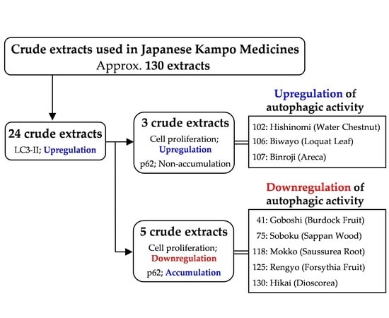
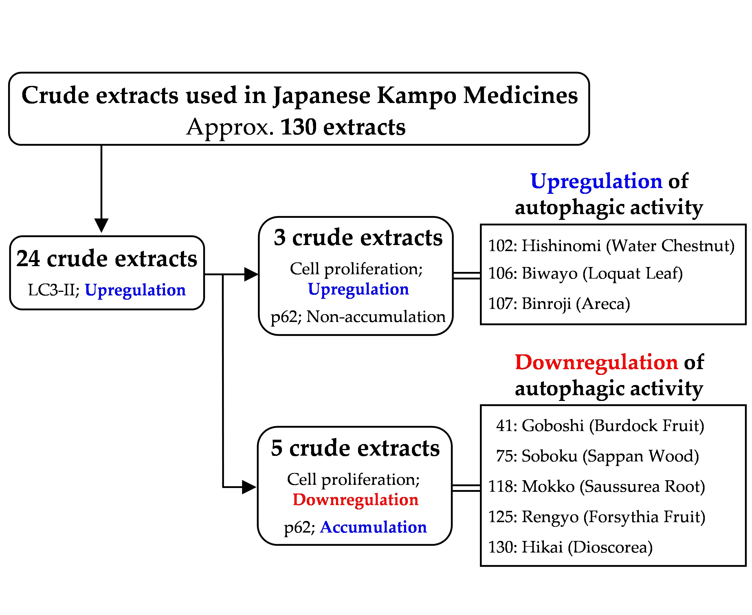
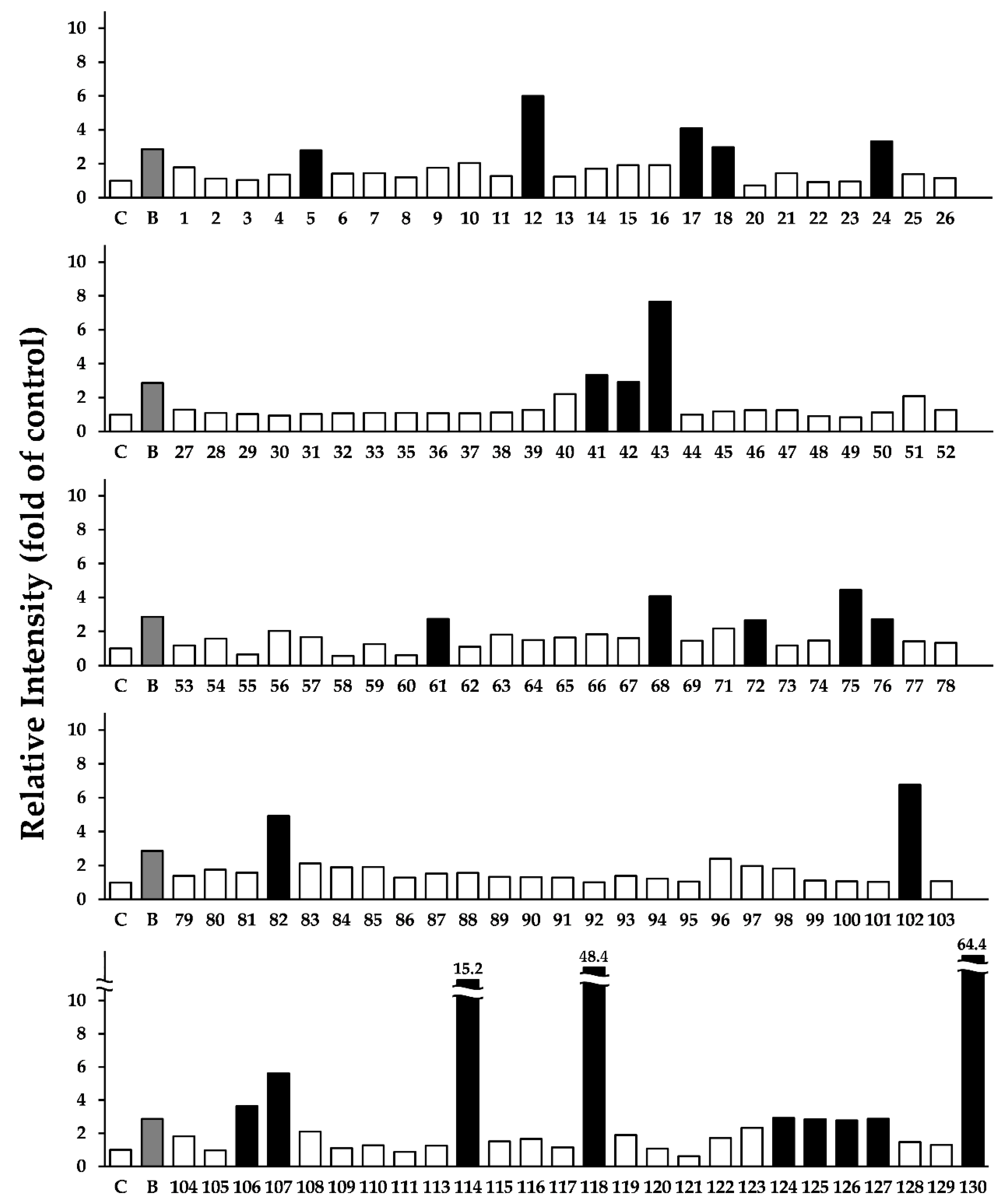
3.1. Screening the Crude Drugs for LC3-II Expression

Table S1 (Supplementary Materials) shows the crude drugs used in this study. The listed crude drugs cover not only those in Japanese Pharmacopoeia but also those commonly used in medical therapy in Japan. We first investigated the effects of MeOH extracts prepared from the crude drugs on the expression of LC3-II in HepG2 cells. The cells were treated with the approximately 130 kinds of crude extracts at 20 μg/mL for 24 h. LC3-II protein levels were examined using Western blotting, and the band intensities were quantified and expressed as the fold changes in the control. BBR, which induces autophagy and increases LC3-II expression in HepG2 cells, was used as a positive control [18,19]. As shown in Figure 1 and Figure S1, among the 130 crude extracts, 24 crude extracts, which are shown in black columns, increased LC3-II expression, which suggests that they might exhibit autophagic activity.

3.2. Effects of the Selected 24 Crude Drugs on Cell Viability

It has been suggested that autophagic activity is intimately related to the proliferation of cancer cells [14,15,16,17,18,19,20,21,22]; therefore, we next investigated the effects of the selected 24 crude drugs on the proliferation of HepG2 cells. The cells were treated with 5, 10, and 20 μg/mL of each of the 24 crude extracts for 24 h, and cell viability was measured using MTT assay. Table 1 indicates the percentage of cell viability after treatment with 5, 10, and 20 μg/mL of each extract. Among the 24 crude extracts, cell viability was significantly suppressed by >10% compared with that in the control using the following five kinds of crude extracts, namely, 41 (Goboshi; burdock fruit), 75 (Soboku; sappan wood), 118 (Mokko; saussurea root), 125 (Rengyo; forsythia fruit), and 130 (Hikai; dioscorea). On the other hand, three extracts, namely, 102 (Hishinomi; water chestnut), 106 (Biwayo; loquat leaf), and 107 (Binroji; areca), increased cell viability by >10% compared with that of the control. The remaining 16 crude extracts had no effect on cell viability. Figure 2 shows the cell viability of only eight crude extracts (five extracts that suppressed cell viability and three that increased cell viability). These results suggest that these eight crude drugs might influence cell viability by modulating autophagic activity.

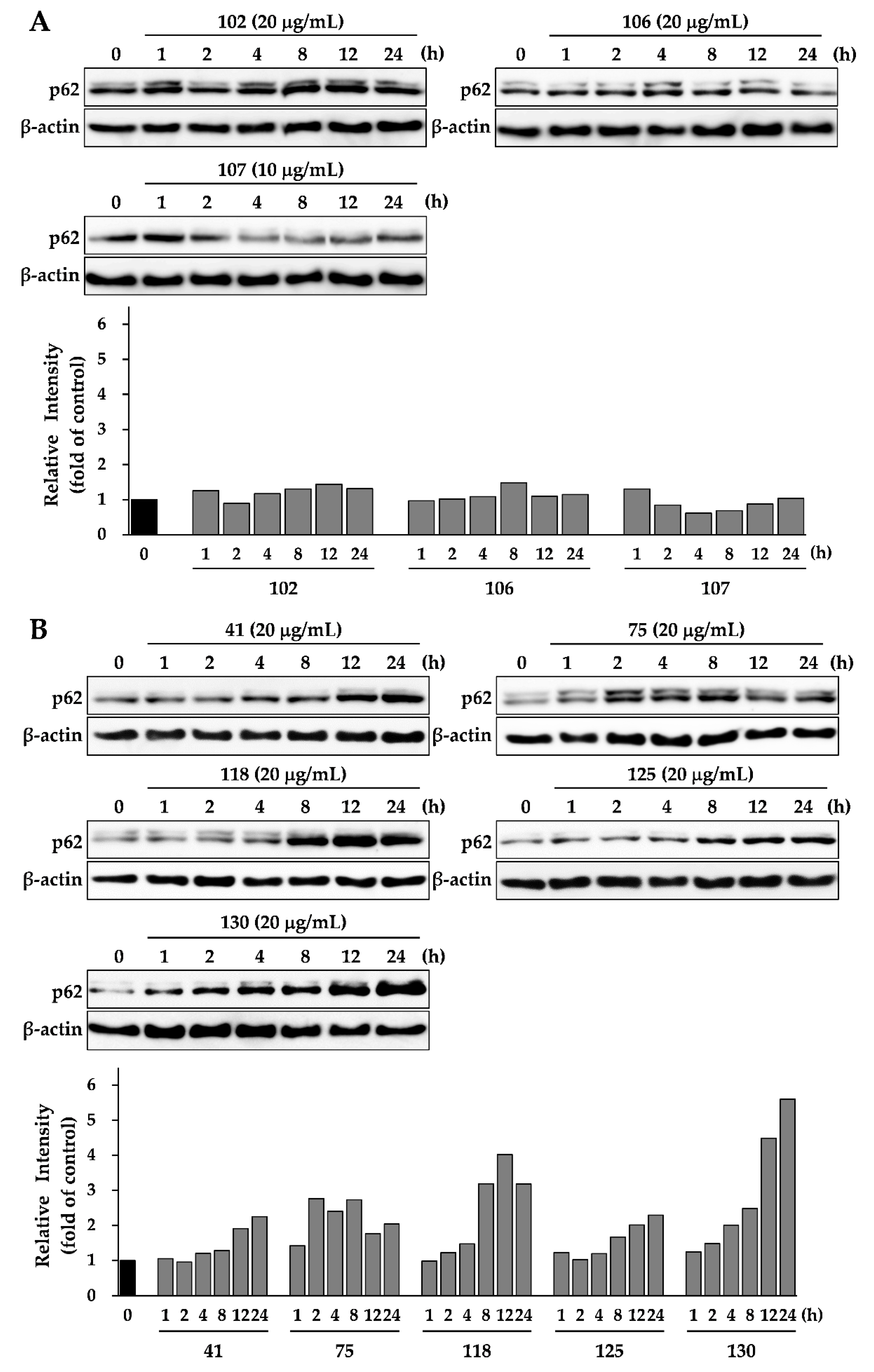
3.3. Effects of Eight Crude Drugs on P62 Expression

p62 is selectively incorporated into autophagosomes and is efficiently degraded by autophagy [8,9,10,11,12,13]; therefore, the p62 expression levels are inversely related to the autophagic activity. To identify whether the eight crude extracts in Figure 2 induce or inhibit autophagy, it was necessary to determine the autophagic flux assessed by monitoring p62 levels; therefore, we conducted a time-course experiment to determine the effect of the eight crude extracts on p62 expression levels. Figure 3A indicates the p62 expression levels regulated by the three crude extracts that increased cell viability. Crude drug numbers 102 and 106 had no effect on p62 expression levels during treatment. Crude drug number 107 decreased p62 expression level within a short time (approximately 4 h) after treatment and then p62 expression recovered to basal levels after 12 h. Previous studies have reported that the impairment of autophagy causes massive accumulation of p62 in HepG2 cells [13,21]. Conversely, when autophagy is activated, p62 rapidly degrades but then recovers to basal levels after several hours [34]. The results of these studies suggest that crude drug numbers 102, 106, and 107 did not block autophagic flux. Furthermore, the crude drug number 107 clearly induced autophagy. Figure 3B shows the p62 expression levels of treatment with five crude extracts that suppressed cell viability. All of these crude extracts increased p62 expression levels over time. In particular, crude drug number 130 strongly induced p62 expression levels by more than fourfold after treatments for 12 and 24 h. Crude drug number 118 also induced p62 expression by more than fourfold after treatment for 12 h. These results suggest that crude drug numbers 41, 75, 118, 125, and 130 deregulate the autophagic pathway by blocking autophagic flux, which results in p62 accumulation.

4. Discussion

Liver cancer is the second leading cause of cancer-related death and the sixth most diagnosed cancer worldwide [35]. Autophagy plays multiple roles in maintaining liver homeostasis. In the absence of Atg5 and Atg7, which are key autophagy genes involved in its initiation, nonfunctional proteins and organelles accumulate in liver cells [36]. It has been reported that conditional Atg7 knockout mice developed hepatomegaly and different metabolic liver disorders [37]; therefore, autophagy is important for suppressing tumorigenesis in the liver. Cancer cells use autophagy to ensure an alternative energy source for growth and survival in a stressful microenvironment with high hypoxia, scarce nutrients, and, very often, under stressful therapeutic conditions [14,15,16,17]. It has been reported that in advanced hepatocellular carcinomas, autophagy plays an oncogenic (pro-survival) role observed as an increased LC3-II expression that positively correlates with progression of malignancy and a poor prognosis [38]. However, one should be careful when coming to conclusions about the influence of LC3-II overexpression on autophagic activity. Hence, LC3-II overexpression cannot be solely used as a marker for increased autophagic activity because its increased accumulation might have resulted from autophagy inhibition at the post-lipidation stage.
In this study, we screened approximately 130 kinds of crude drugs used in Japanese Kampo formulas to identify those drugs that regulate HepG2 proliferation through autophagy. First, we screened 130 crude extracts to select those that increased LC3-II expression. Among them, 24 crude extracts did so, which suggests that these extracts might have autophagic activity. Among the 24 crude extracts that increased LC3-II expression, three induced cell growth but five suppressed cell viability. Interestingly, the remaining 16 crude extracts had no effect on cell viability despite LC3-II upregulation. These results suggest that the regulation of HepG2 proliferation is not indispensable for LC3-II induction. Because the results of screening for LC3-II expression cannot be directly applied to the selection of extracts that inhibit autophagy, we determined that autophagic flux could be assessed by monitoring p62 expression levels. Among the three crude extracts that increased cell viability, crude drug numbers 102 and 106 had no effect on p62 expression levels, and crude drug number 107 decreased p62 expression levels within a short duration after treatment, but then the levels finally recovered to their basal levels. It appears that crude drug number 107 induced cell growth by inducing autophagic activity, and we expect that this crude drug might contain compounds that are specific activators of autophagy. Although crude drug numbers 102 and 106 did not show obvious induction of autophagic activity, they showed that LC3-II expression can be increased without blocking autophagic flux. These results probably reflect the conclusion that crude drug numbers 102 and 106 induced cell growth by inducing autophagic activity, but further investigations are needed to confirm the findings. In fact, crude drug number 107 (Binroji; areca) has been reported to induce autophagy in several cell lines, such as leukemic Jurkat T cells [39] and oral carcinoma cells (OECM-1), Cal-27, and Scc-9 [40] by activating the AMPK/mTOR signaling pathway after accumulating reactive oxygen species [40]. However, the relationship between autophagy and crude drug numbers 102 (Hishinomi; water chestnut) or 106 (Biwayo; loquat leaf) has not been previously reported.
On the other hand, the five crude extracts that suppressed cell viability increased p62 expression levels, which suggests that these extracts downregulate the autophagic pathway by inhibiting autophagic flux, leading to p62 accumulation. Among these five extracts, crude drug numbers 41 (Goboshi; burdock fruit) and 125 (Rengyo; forsythia fruit) contain the same major compound, arctigenin (AG) of lignans. It has been reported that AG protects against drug-induced hepatitis by suppressing the immune system and regulating autophagy by inhibiting the IFN-γ/IL-6/Stat1 and IL-6/Bnip3 pathways in mice [41]. However, our results in this study indicate that crude drug numbers 75 (Soboku; sappan wood) and 118 (Mokko; saussurea root) reduced autophagic activity with p62 accumulation. The brazilin contained in crude drug number 75 was reported to induce autophagic cell death by disturbing calcium homeostasis in osteosarcoma MG-63 cells, thereby suppressing cell viability [42]. The costunolide contained in crude drug number 118 was reported to inhibit cell viability of multidrug-resistant human ovarian cancer OAW42-A cells by activating apoptotic and autophagic pathways via the decreased expression of B-cell lymphoma 2 (Bcl-2) [43]. This implies that the differences in the reactivity and signaling pathways depend on the cell type. Our results indicate, and those of previous reports have indicated, that these extracts contain compounds that modulate autophagic activity. In addition, our findings support the possibility that these crude extracts regulate autophagic activity to recover the medical conditions of patients, and provide new insights into pharmacological action underlying Japanese Kampo formulas in future investigations. We conclude that the crude drugs selected for this study could serve as sources of lead compounds in the development of agents for autophagy research and cancer therapy, and that, additional studies are needed to isolate and identify the active compounds in the drugs.

Supplementary Materials

The following are available online at https://0-www-mdpi-com.brum.beds.ac.uk/2305-6320/6/2/63/s1, Table S1: List of crude drugs, Figure S1: The effect of crude extracts including 24 selected crude drugs on LC3-II protein.

Author Contributions

Conceptualization, S.O. and T.U.; Data curation, S.O.; Formal analysis, S.O.; Funding acquisition, S.O.; Investigation, S.O., H.K., and A.K.; Methodology, S.O. and T.U.; Project administration, T.U.; Resources, S.O. and T.O.; Supervision, Y.S.; Writing—original draft, S.O., H.K., and T.O.; Writing—review and editing, T.U.

Funding

This research was funded by the Sasakawa Scientific Research Grant from The Japan Science Society, grant number 2018-4039.

Conflicts of Interest

The authors declare no conflict of interest.

References

  1. Rabinowitz, J.D.; White, E. Autophagy and metabolism. Science 2010, 330, 1344–1348. [Google Scholar] [CrossRef]
  2. Augustine, M.K.; Choi, M.D.; Stefan, W.; Ryter, P.H.D.; Beth Levine, M.D. Autophagy in human health and disease. N. Engl. J. Med. 2013, 368, 651–662. [Google Scholar] [CrossRef]
  3. Lassen, K.G.; Xavier, R.J. Mechanisms and function of autophagy in intestinal disease. Autophagy 2018, 14, 216–220. [Google Scholar] [CrossRef] [PubMed] [Green Version]
  4. Ueno, T.; Komatsu, M. Autophagy in the liver: Functions in health and disease. Nat. Rev. Gastroenterol. Hepatol. 2017, 14, 170–184. [Google Scholar] [CrossRef] [PubMed]
  5. Maiuri, M.C.; Kroemer, G. Autophagy in stress and disease. Cell Death Differ. 2015, 22, 365–366. [Google Scholar] [CrossRef] [PubMed] [Green Version]
  6. Kabeya, Y.; Mizushima, N.; Ueno, T.; Yamamoto, A.; Kirisako, T.; Noda, T.; Kominami, E.; Ohsumi, Y.; Yoshimori, T. LC3, a mammalian homologue of yeast Apg8p, is localized in autophagosome membranes after processing. EMBO J. 2000, 19, 5720–5728. [Google Scholar] [CrossRef]
  7. Kabeya, Y.; Mizushima, N.; Yamamoto, A.; Oshitani-Okamoto, S.; Ohsumi, Y.; Yoshimori, T. LC3, GABARAP and GATE16 localize to autophagosomal membrane depending on form-II formation. J. Cell Sci. 2004, 117, 2805–2812. [Google Scholar] [CrossRef] [Green Version]
  8. Geetha, T.; Wooten, M.W. Structure and functional properties of the ubiquitin binding protein p62. FEBS Lett. 2002, 512, 19–24. [Google Scholar] [CrossRef] [Green Version]
  9. Seibenhener, M.L.; Babu, J.R.; Geetha, T.; Wong, H.C.; Krishna, N.R.; Wooten, M.W. Sequestosome 1/p62 is a polyubiquitin chain binding protein involved in ubiquitin proteasome degradation. Mol. Cell. Biol. 2004, 24, 8055–8068. [Google Scholar] [CrossRef]
  10. Johansen, T.; Lamark, T. Selective autophagy mediated by autophagic adapter proteins. Autophagy 2011, 7, 279–296. [Google Scholar] [CrossRef]
  11. Pankiv, S.; Clausen, T.H.; Lamark, T.; Brech, A.; Bruun, J.A.; Outzen, H.; Øvervatn, A.; Bjørkøy, G.; Johansen, T. p62/SQSTM1 binds directly to Atg8/LC3 to facilitate degradation of ubiquitinated protein aggregates by autophagy. J. Biol. Chem. 2007, 282, 24131–24145. [Google Scholar] [CrossRef] [PubMed]
  12. Itakura, E.; Mizushima, N. p62 Targeting to the autophagosome formation site requires self-oligomerization but not LC3 binding. J. Cell. Biol. 2011, 192, 17–27. [Google Scholar] [CrossRef] [PubMed]
  13. Mizushima, N.; Yoshimori, T.; Levine, B. Methods in mammalian autophagy research. Cell 2010, 140, 313–326. [Google Scholar] [CrossRef] [PubMed]
  14. Degenhardt, K.; Mathew, R.; Beaudoin, B.; Bray, K.; Anderson, D.; Chen, G.; Mukherjee, C.; Shi, Y.; Gélinas, C.; Fan, Y.; et al. Autophagy promotes tumor cell survival and restricts necrosis, inflammation, and tumorigenesis. Cancer Cell 2006, 10, 51–64. [Google Scholar] [CrossRef] [PubMed] [Green Version]
  15. Turcotte, S.; Chan, D.A.; Sutphin, P.D.; Hay, M.P.; Denny, W.A.; Giaccia, A.J. A molecule targeting VHL-deficient renal cell carcinoma that induces autophagy. Cancer Cell 2008, 14, 90–102. [Google Scholar] [CrossRef] [PubMed]
  16. Yang, Z.J.; Chee, C.E.; Huang, S.; Sinicrope, F.A. The role of autophagy in cancer: Therapeutic implications. Mol. Cancer Ther. 2011, 10, 1533–1541. [Google Scholar] [CrossRef] [PubMed]
  17. Marinković, M.; Šprung, M.; Buljubašić, M.; Novak, I. Autophagy modulation in cancer: Current knowledge on action and therapy. Oxid. Med. Cell. Longev. 2018, 2018, 8023821. [Google Scholar] [CrossRef] [PubMed]
  18. Yu, R.; Zhang, Z.Q.; Wang, B.; Jiang, H.X.; Cheng, L.; Shen, L.M. Berberine-induced apoptotic and autophagic death of HepG2 cells requires AMPK activation. Cancer Cell Int. 2014, 14, 49. [Google Scholar] [CrossRef]
  19. La, X.; Zhang, L.; Li, Z.; Yang, P.; Wang, Y. Berberine-induced autophagic cell death by elevating GRP78 levels in cancer cells. Oncotarget 2017, 8, 20909–20924. [Google Scholar] [CrossRef] [Green Version]
  20. Nozaki, R.; Kono, T.; Bochimoto, H.; Watanabe, T.; Oketani, K.; Sakamaki, Y.; Okubo, N.; Nakagawa, K.; Takeda, H. Zanthoxylum fruit extract from Japanese pepper promotes autophagic cell death in cancer cells. Oncotarget 2016, 7, 70437–70446. [Google Scholar] [CrossRef] [Green Version]
  21. Song, L.; Wang, Z.; Wang, Y.; Guo, D.; Yang, J.; Chen, L.; Tan, N. Natural cyclopeptide RA-XII, a new autophagy inhibitor, suppresses protective autophagy for enhancing apoptosis through AMPK/mTOR/P70S6K pathways in HepG2 cells. Molecules 2017, 22, 1934. [Google Scholar] [CrossRef] [PubMed]
  22. Young, A.N.; Herrera, D.; Huntsman, A.C.; Korkmaz, M.A.; Lantvit, D.D.; Mazumder, S.; Kolli, S.; Coss, C.C.; King, S.; Wang, H.; et al. Phyllanthusmin derivatives induce apoptosis and reduce tumor burden in high-grade serous ovarian cancer by late-stage autophagy inhibition. Mol. Cancer Ther. 2018, 17, 2123–2135. [Google Scholar] [CrossRef] [PubMed]
  23. Watanabe, S.; Toyama, T.; Sato, T.; Suzuki, M.; Morozumi, A.; Sakagami, H.; Hamada, N. Kampo therapies and the use of herbal medicines in the dentistry in Japan. Medicines 2019, 6, 34. [Google Scholar] [CrossRef] [PubMed]
  24. Matsumoto, T.; Sekiguchi, K.; Kawakami, Z.; Watanabe, J.; Mizoguchi, K.; Ikarashi, Y.; Yamamoto, M. Basic study of drug-drug interaction between memantine and the traditional Japanese Kampo medicine yokukansan. Molecules 2018, 24, 115. [Google Scholar] [CrossRef] [PubMed]
  25. Sakagami, H.; Watanabe, T.; Hoshino, T.; Suda, N.; Mori, K.; Yasui, T.; Yamauchi, N.; Kashiwagi, H.; Gomi, T.; Oizumi, T.; et al. Recent progress of basic studies of natural products and their dental application. Medicines 2018, 6, 4. [Google Scholar] [CrossRef] [PubMed]
  26. Sunagawa, M.; Yamaguchi, K.; Tsukada, M.; Ebihara, N.; Ikemoto, H.; Hisamitsu, T. Kampo (traditional Japanese herbal) formulae for treatment of stomatitis and oral mucositis. Medicines 2018, 5, 130. [Google Scholar] [CrossRef] [PubMed]
  27. Amitani, M.; Amitani, H.; Sloan, R.A.; Suzuki, H.; Sameshima, N.; Asakawa, A.; Nerome, Y.; Owaki, T.; Inui, A.; Hoshino, E. The translational aspect of complementary and alternative medicine for cancer with particular emphasis on Kampo. Front. Pharmacol. 2015, 6, 150. [Google Scholar] [CrossRef] [PubMed]
  28. Sato, N.; Li, W.; Takemoto, H.; Takeuchi, M.; Nakamura, A.; Tokura, E.; Akahane, C.; Ueno, K.; Komatsu, K.; Kuriyama, N.; et al. Comprehensive evaluation ofantioxidant effects of Japanese Kampo medicines led to identification of Tsudosan formulation as a potent antioxidant agent. J. Nat. Med. 2019, 1, 163–172. [Google Scholar] [CrossRef]
  29. Kobayashi, Y. Kampo medicine in the new model core curriculum of pharmaceutical education. Yakugaku Zasshi 2016, 136, 423–432. [Google Scholar] [CrossRef]
  30. Uto, T.; Morinaga, O.; Tanaka, H.; Shoyama, Y. Analysis of the synergistic effect of glycyrrhizin and other constituents in licorice extract on lipopolysaccharide-induced nitric oxide production using knock-out extract. Biochem. Biophys. Res. Commun. 2012, 417, 473–478. [Google Scholar] [CrossRef] [PubMed]
  31. Uto, T.; Tung, N.H.; Taniyama, R.; Miyanowaki, T.; Morinaga, O.; Shoyama, Y. Anti-inflammatory activity of constituents isolated from aerial part of Angelica acutiloba Kitagawa. Phytother. Res. 2015, 12, 1956–1963. [Google Scholar] [CrossRef] [PubMed]
  32. Okubo, S.; Uto, T.; Goto, A.; Tanaka, H.; Nishioku, T.; Yamada, K.; Shoyama, Y. Berberine induces apoptotic cell death via activation of caspase-3 and -8 in HL-60 human leukemia cells: Nuclear localization and structure-activity relationships. Am. J. Chin. Med. 2017, 45, 1497–1511. [Google Scholar] [CrossRef] [PubMed]
  33. Uto, T.; Tung, N.H.; Ohta, T.; Juengsanguanpornsuk, W.; Hung, L.Q.; Hai, N.T.; Long, D.D.; Thuong, P.T.; Okubo, S.; Hirata, S.; et al. Antiproliferative activity and apoptosis induction by trijuganone C isolated from the root of Salvia miltiorrhiza Bunge (Danshen). Phytother. Res. 2018, 32, 657–666. [Google Scholar] [CrossRef] [PubMed]
  34. Mayurbhai, H.S.; Itakura, E.; Mizushima, N. Expression of the autophagy substrate SQSTM1/p62 is restored during prolonged starvation depending on transcriptional upregulation and autophagy-derived amino acids. Autophagy 2014, 10, 431–441. [Google Scholar] [CrossRef] [Green Version]
  35. Gravitz, L. Liver cancer. Nature 2014, 516, 7529. [Google Scholar] [CrossRef]
  36. Takamura, A.; Komatsu, M.; Hara, T.; Sakamoto, A.; Kishi, C.; Waguri, S.; Eishi, Y.; Hino, O.; Tanaka, K.; Mizushima, N. Autophagy-deficient mice develop multiple liver tumors. Genes. Dev. 2011, 25, 795–800. [Google Scholar] [CrossRef] [Green Version]
  37. Komatsu, M.; Waguri, S.; Ueno, T.; Iwata, J.; Murata, S.; Tanida, I.; Ezaki, J.; Mizushima, N.; Ohsumi, Y.; Uchiyama, Y.; et al. Impairment of starvation-induced and constitutive autophagy in Atg7-deficient mice. J. Cell Biol. 2005, 169, 425–434. [Google Scholar] [CrossRef]
  38. Wu, D.H.; Jia, C.C.; Chen, J.; Lin, Z.X.; Ruan, D.Y.; Li, X.; Lin, Q.; Min-Dong; Ma, X.K.; Wan, X.B.; Cheng, N.; et al. Autophagic LC3B overexpression correlates with malignant progression and predicts a poor prognosis in hepatocellular carcinoma. Tumour Biol. 2014, 12225–12233. [Google Scholar] [CrossRef]
  39. Yen, C.Y.; Chiang, W.F.; Liu, S.Y.; Lin, C.C.; Liao, K.A.; Lin, C.Y.; Hsieh, W.F.; Cheng, Y.C.; Hsu, KC.; Lin, P.Y.; Chen, T.C.; et al. Impacts of autophagy-inducing ingredient of areca nut on tumor cells. PLoS ONE 2015, 10, e0128011. [Google Scholar] [CrossRef]
  40. Xu, Z.; Huang, C.M.; Shao, Z.; Zhao, X.P.; Wang, M.; Yan, T.L.; Zhou, X.C.; Jiang, E.H.; Liu, K.; Shang, Z.J. Autophagy induced by areca nut extract contributes to decreasing cisplatin toxicity in oral squamous cell carcinoma cells: Roles of reactive oxygen species/AMPK signaling. Int. J. Mol. Sci. 2017, 18, 524. [Google Scholar] [CrossRef]
  41. Feng, Q.; Yao, J.; Zhou, G.; Xia, W.; Lyu, J.; Li, X.; Zhao, T.; Zhang, G.; Zhao, N.; Yang, J. Quantitative proteomic analysis reveals that arctigenin alleviates Concanavalin A-induced hepatitis through suppressing immune system and regulating autophagy. Front. Immunol. 2018, 9, 1881. [Google Scholar] [CrossRef] [PubMed]
  42. Kang, Y.; He, P.; Wang, H.; Ye, Y.; Li, X.; Xie, P.; Wu, B. Brazilin induces FOXO3A-dependent autophagic cell death by disturbing calcium homeostasis in osteosarcoma cells. Cancer Chemother. Pharmacol. 2018, 82, 479–491. [Google Scholar] [CrossRef] [PubMed]
  43. Fang, Y.; Li, J.; Wu, Y.; Gui, J.; Shen, Y. Costunolide inhibits the growth of OAW42-A multidrug-resistant human ovarian cancer cells by activating apoptotic and autophagic pathways, production of reactive oxygen species (ROS), cleaved caspase-3 and cleaved caspase-9. Med. Sci. Monit. 2019, 25, 3231–3237. [Google Scholar] [CrossRef] [PubMed]
Figure 1. Relative intensity of LC3-II protein levels by the approximately 130 kinds of crude extracts. The noticeable increase is shown in the black column. HepG2 cells were treated with 20 µg/mL of each crude extract for 24 h, and the LC3-II expression levels were determined using Western blotting. The data shown are representative of three independent treatments with similar results. Notes: C, control; B, positive control (BBR 50 μM); 1–130, crude drug number (see Table S1, Supplementary Materials).
Medicines 06 00063 g001
Figure 2. Effects of eight crude extracts on the proliferation of HepG2 cells. Cells were treated with each crude extract at various concentrations for 24 h, and cell viabilities were determined using MTT assay. The data represent the mean ± SD of three individual treatments using the same concentrations. ∗ p < 0.005 compared with the control group.
Medicines 06 00063 g002
Figure 3. Effects of eight crude extracts on p62 expression in HepG2 cells. Cells were treated with each crude extract at the indicated concentrations and for different durations. The expressions levels of p62 and β-actin were determined by Western blotting. (A) The effect of crude extracts on cell viability. (B) The effect of crude extracts that suppressed cell viability. The data shown are representative of three independent treatments using the same parameters with similar results.
Medicines 06 00063 g003
Table 1. Effects of selected 24 crude extracts on the proliferation.
Drug No.Japanese NameEnglish NameConcentration (µg/mL)
51020
5UzuAconite Root100.4100.3103.3
12OnjiPolygala Root100.497.396.4
17KakkoPogostemon Herb96.6100.2100.5
18KakkonPueraria Root101.6107.4109.5
24KikyoPlatycodon Root103.6106.9106.8
41 (b)GoboshiBurdock Fruit89.582.976.7
42GomishiSchisandra Fruit105.5107.3106.2
43SaikoBupleurum Root100.8100.396.1
61JashoshiCnidium Monnieri Fruit96.196.192.6
68ShomaCimicifuga Rhizome100.2104.3106.3
72SentaiCicada Slough105.6103.2101.4
75 (b)SobokuSappan Wood100.894.086.9
76SoyoPerilla Herb102.7103.8105.9
82ChimoAnemarrhena Rhizome99.4100.896.9
102 (a)HishinomiWater Chestnut105.1108.1116.3
106 (a)BiwayoLoquat Leaf106.9110.7113.9
107 (a)BinrojiAreca105.1114.6108.7
114MaoEphedra Herb101.595.097.7
118 (b)MokkoSaussurea Root94.282.367.3
124RyokyoAlpinia Officinarum Rhizome101.4103.2109.5
125 (b)RengyoForsythia Fruit90.489.588.7
126RennikuNelumbo Seed103.5105.6103.2
127TanjinSalvia Miltiorrhiza Root98.299.895.3
130 (b)HikaiDioscorea71.850.120.0
(a) Extracts that increased cell viability by >10% compared with the control. (b) Extracts that suppressed cell viability by >10% compared with the control.

