Contrast-Enhanced Ultrasonography for Screening and Diagnosis of Hepatocellular Carcinoma: A Case Series and Review of the Literature


Abstract
:1. Introduction
2. Materials and Methods
3. Case Presentation
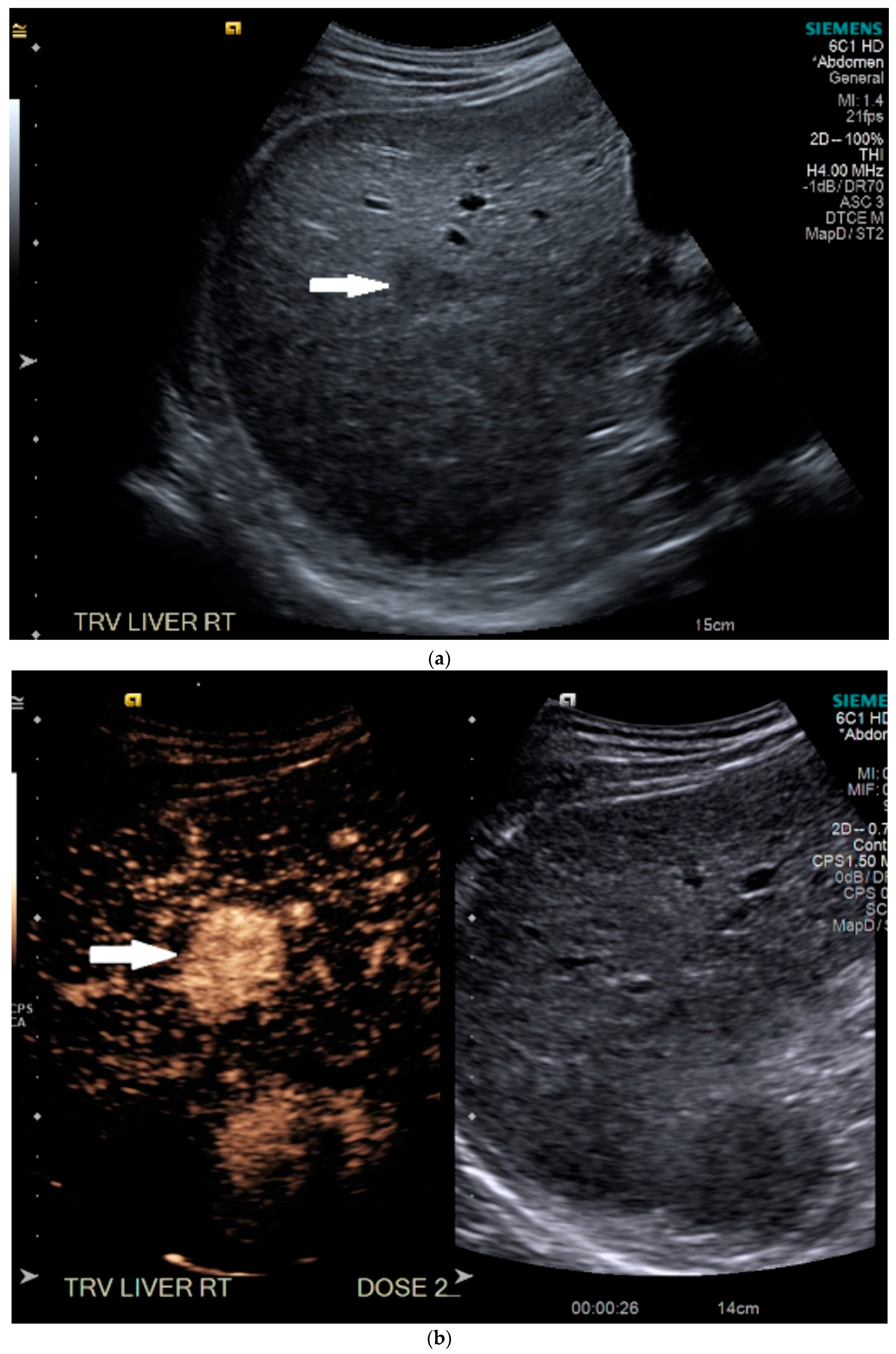
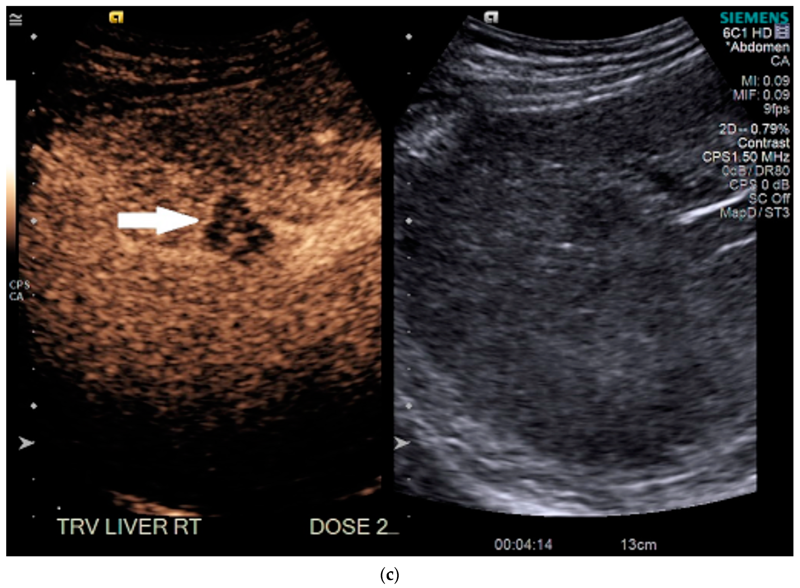
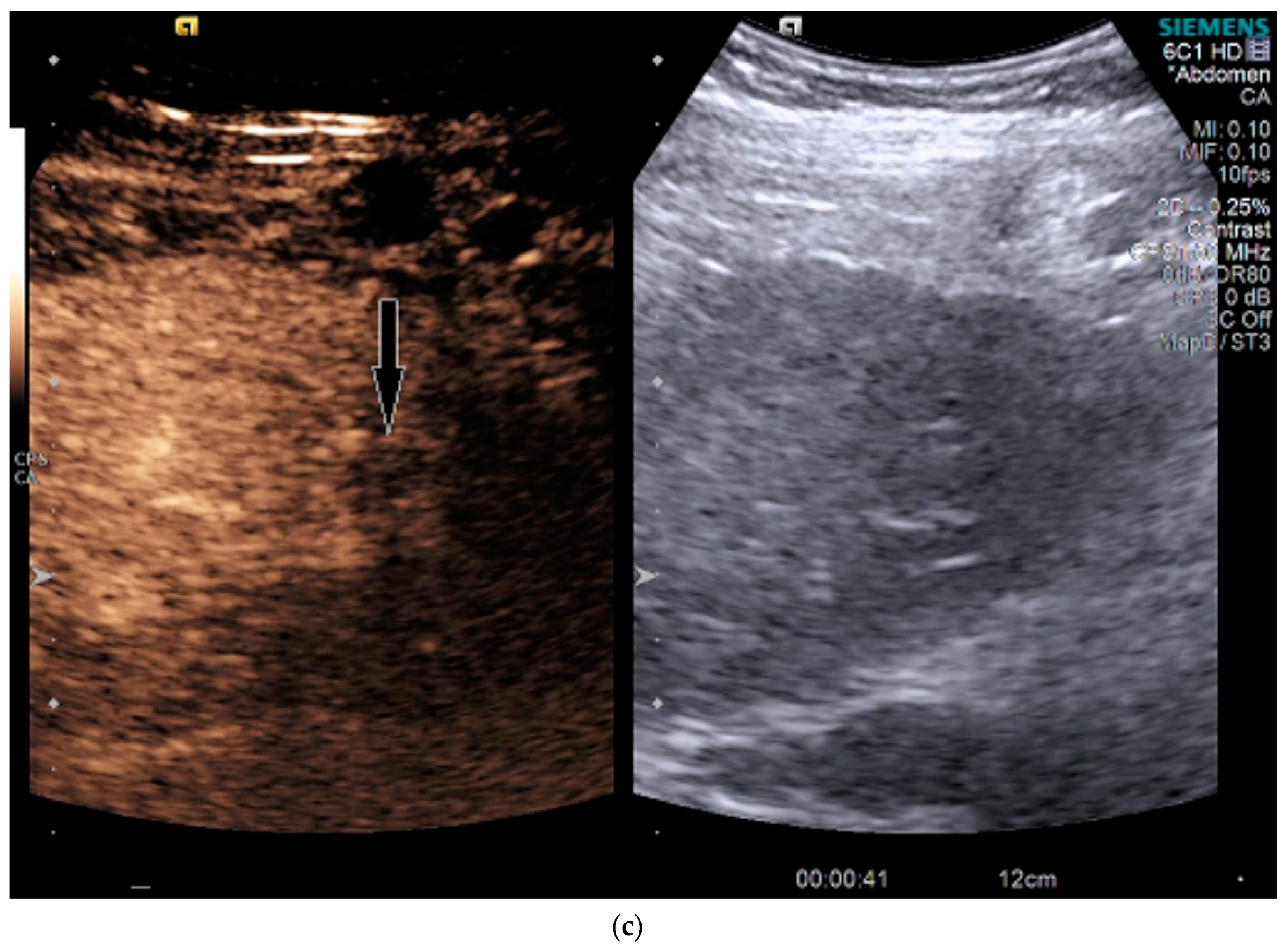
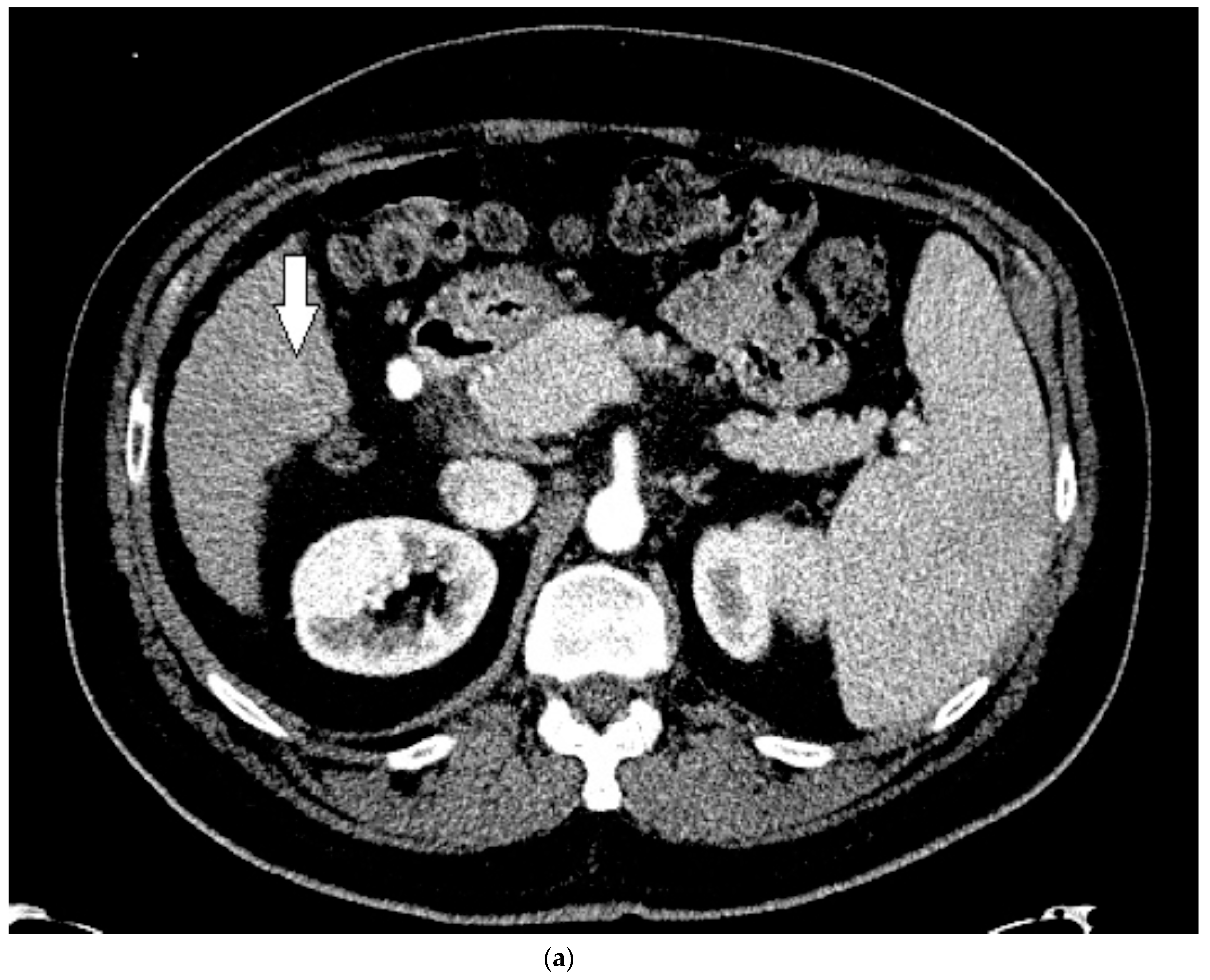
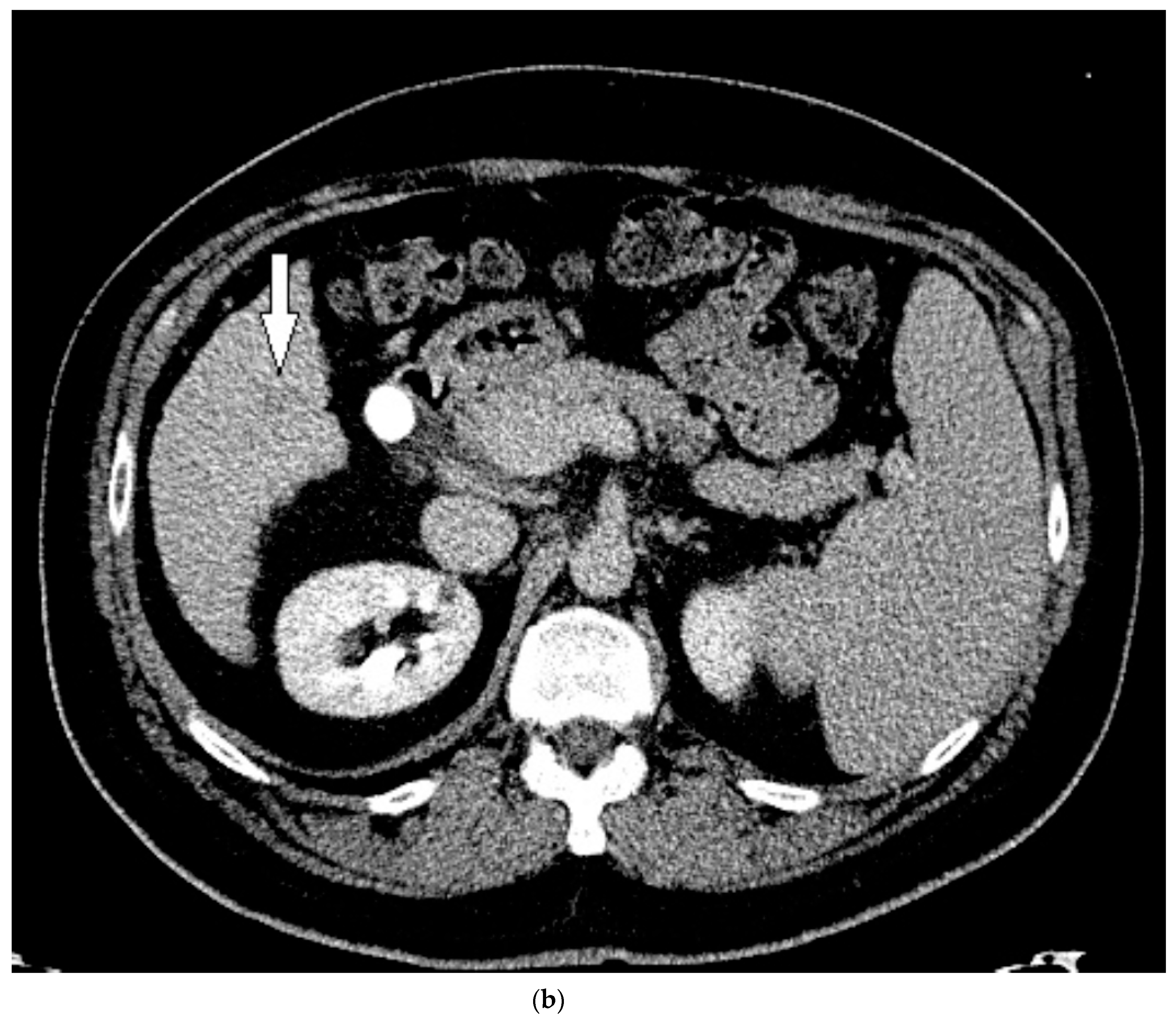
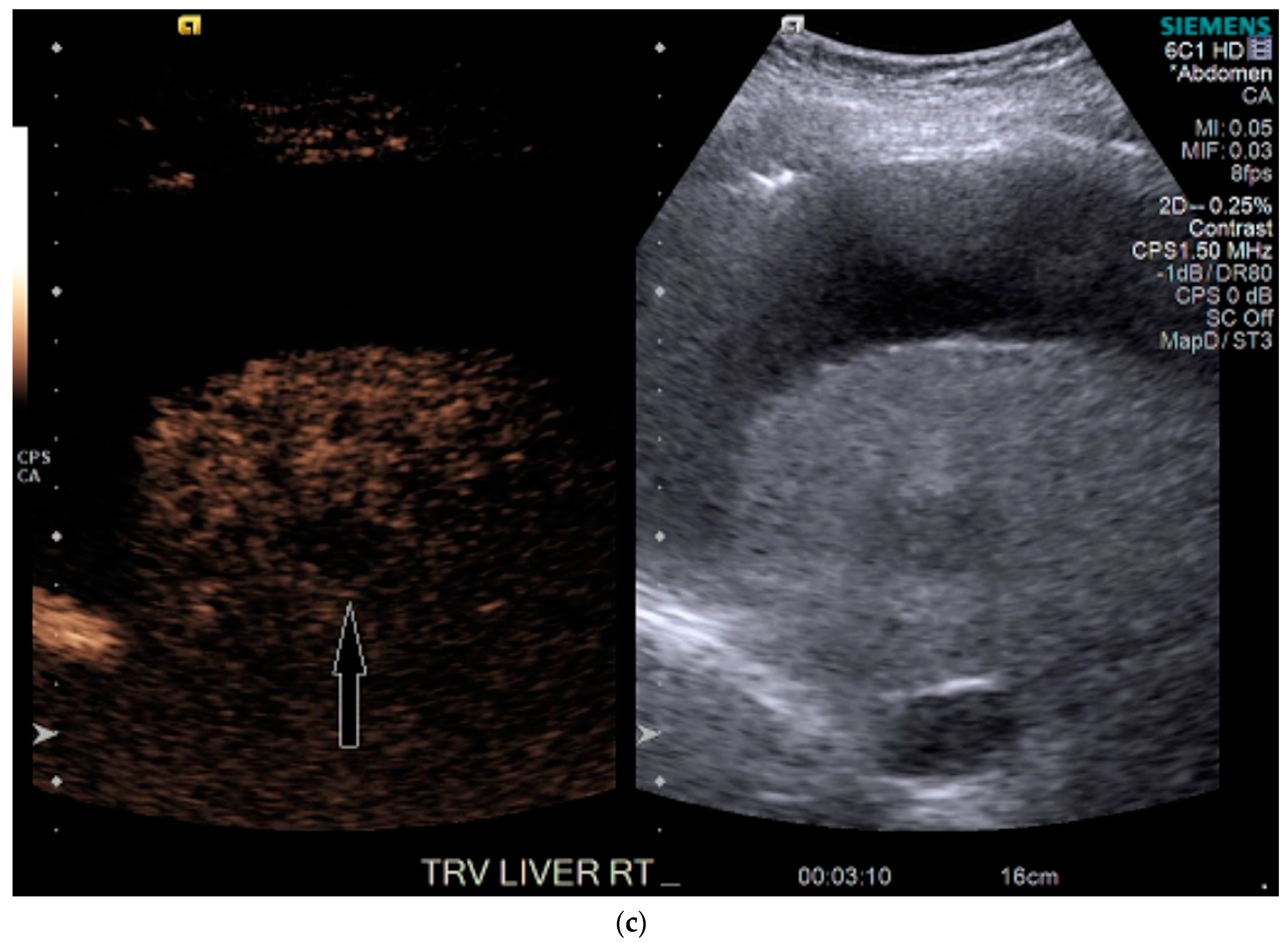
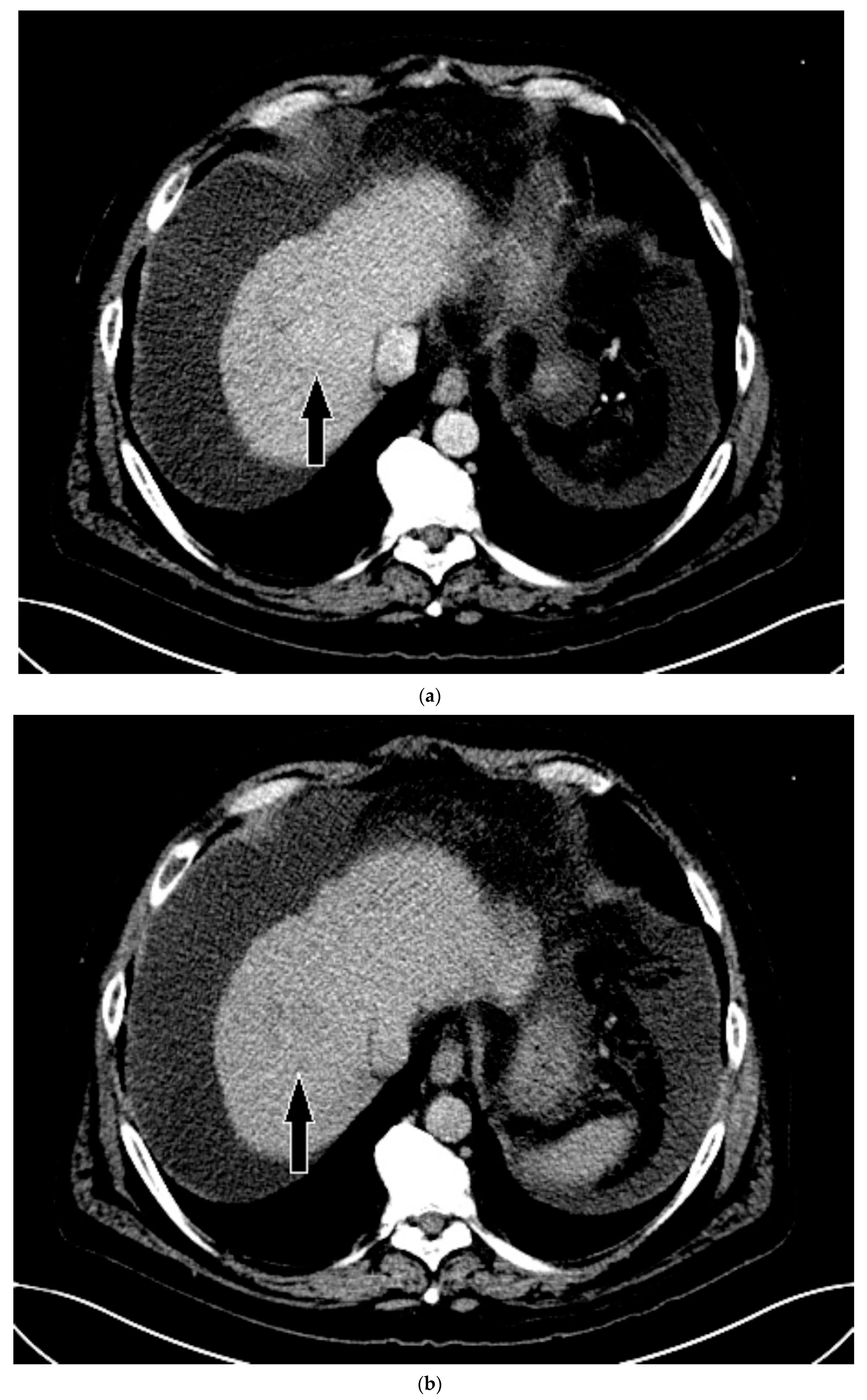
3.1. Case 1
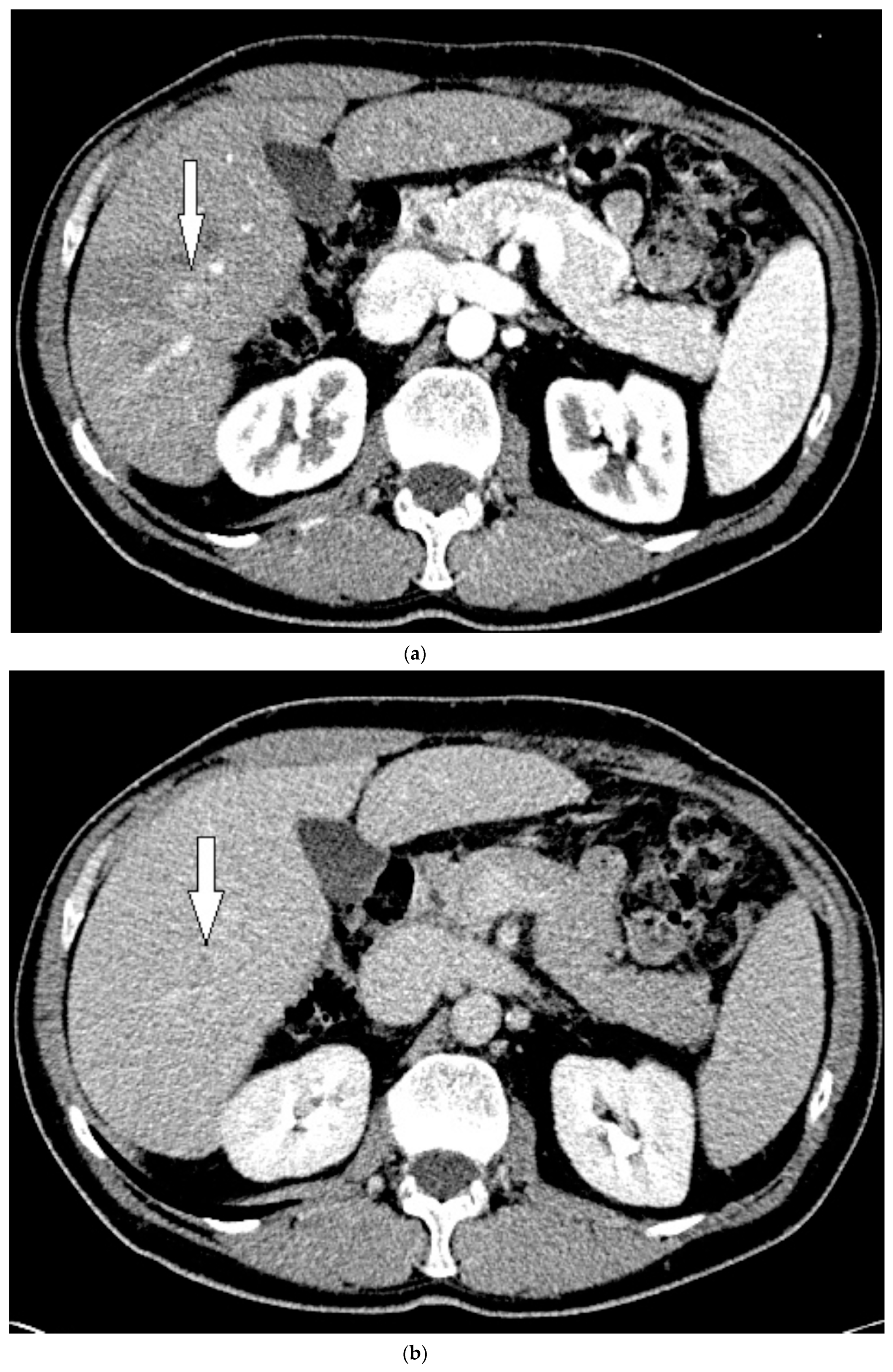
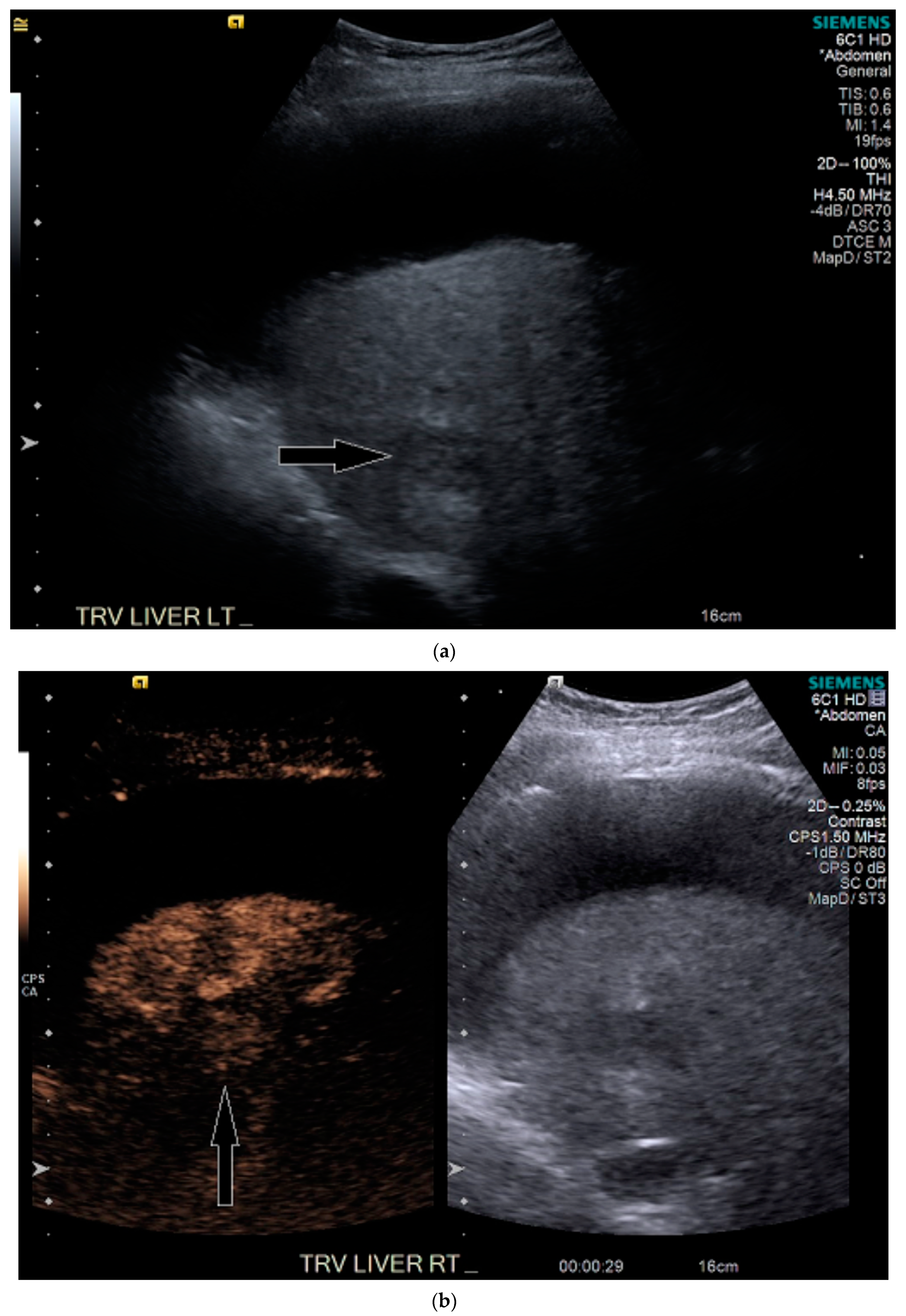
3.2. Case 2
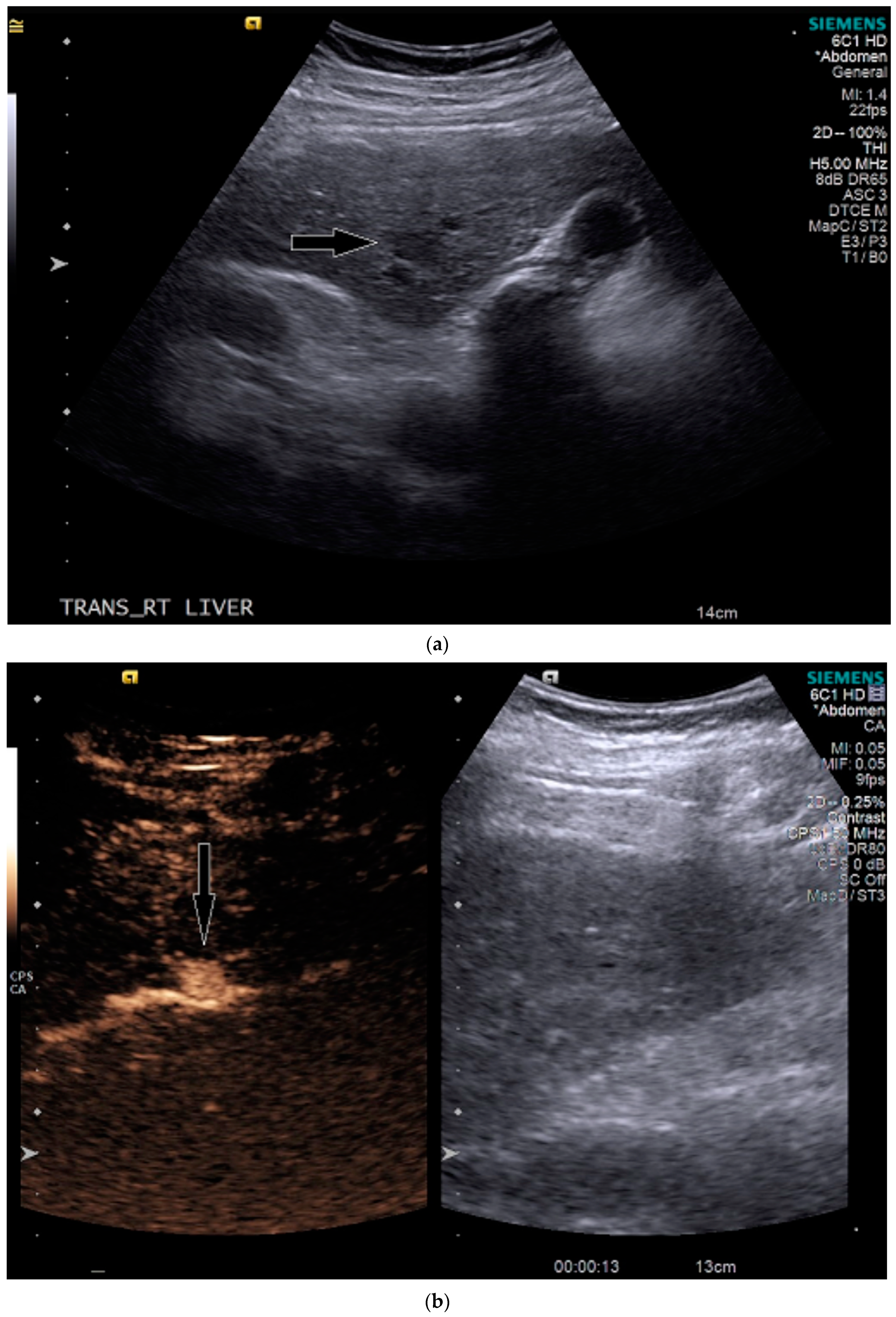
3.3. Case 3
4. Discussion
5. Conclusions
Supplementary Materials
Author Contributions
Funding
Conflicts of Interest
References
- El-Serag, H.B.; Rudolph, K.L. Hepatocellular carcinoma: Epidemiology and molecular carcinogenesis. Gastroenterology 2007, 132, 2557–2576. [Google Scholar] [CrossRef] [PubMed]
- Janevska, D.; Chaloska-Ivanova, V.; Janevski, V. Hepatocellular carcinoma: Risk factors, diagnosis and treatment. Open Access Maced. J. Med. Sci. 2015, 3, 732–736. [Google Scholar] [CrossRef] [PubMed] [Green Version]
- Heidelbaugh, J.J.; Bruderly, M. Cirrhosis and chronic liver failure: Part I. Diagnosis and evaluation. Am. Fam. Physician 2006, 74, 756–762. [Google Scholar] [PubMed]
- Singal, A.G.; Pillai, A.; Tiro, J. Early detection, curative treatment, and survival rates for hepatocellular carcinoma surveillance in patients with cirrhosis: A meta-analysis. PLoS Med. 2014, 11, e1001624. [Google Scholar] [CrossRef]
- Schaub, S.K.; Hartvigson, P.E.; Lock, M.I.; Høyer, M.; Brunner, T.B.; Cardenes, H.R.; Dawson, L.A.; Kim, E.Y.; Mayr, N.A.; Lo, S.S.; et al. Stereotactic body radiation therapy for hepatocellular carcinoma: Current trends and controversies. Technol. Cancer Res. Treat. 2018, 17, 1533033818790217. [Google Scholar] [CrossRef]
- Shetty, S.K.; Rosen, M.P.; Raptopoulos, V.; Goldberg, S.N. Cost-effectiveness of percutaneous radiofrequency ablation for malignant hepatic neoplasms. J Vasc. Interv. Radiol. 2001, 12, 823–833. [Google Scholar] [CrossRef]
- Vogl, T.J.; Naguib, N.N.; Nour-Eldin, N.E.; Rao, P.; Emami, A.H.; Zangos, S.; Nabil, M.; Abdelkader, A. Review on transarterial chemoembolization in hepatocellular carcinoma: Palliative, combined, neoadjuvant, bridging, and symptomatic indications. Eur. J. Radiol. 2009, 72, 505–516. [Google Scholar] [CrossRef]
- Marks, E.I.; Yee, N.S. Molecular genetics and targeted therapy in hepatocellular carcinoma. Curr. Cancer Drug Targets 2016, 16, 53–70. [Google Scholar] [CrossRef]
- Posadas, K.; Ankola, A.; Zang, Z.; Yee, N.S. Tumor molecular profiling for an individualized approach to the treatment of hepatocellular carcinoma: A patient case study. Biomedicines 2018, 6, 46. [Google Scholar] [CrossRef] [Green Version]
- Yee, N.S. Update in systemic and targeted therapies in gastrointestinal oncology. Biomedicines 2018, 6, 34. [Google Scholar] [CrossRef] [Green Version]
- Marrero, J.A.; Kulik, L.M.; Sirlin, C.B.; Zhu, A.X.; Finn, R.S.; Abecassis, M.M.; Roberts, L.R.; Heimbach, J.K. Diagnosis, staging, and management of hepatocellular carcinoma: 2018 practice guidance by the American association for the study of liver diseases. Hepatology 2018, 68, 723–750. [Google Scholar] [CrossRef] [PubMed] [Green Version]
- Zhang, J.; Yu, Y.; Li, Y.; Wei, L. Diagnostic value of contrast-enhanced ultrasound in hepatocellular carcinoma: A meta-analysis with evidence from 1998 to 2016. Oncotarget 2017, 8, 75418–75426. [Google Scholar] [CrossRef] [Green Version]
- Pang, E.H.T.; Chan, A.; Ho, S.G.; Harris, A.C. Contrast-Enhanced ultrasound of the liver: Optimizing technique and clinical applications. AJR Am. J. Roentgenol. 2018, 210, 320–332. [Google Scholar] [CrossRef] [PubMed]
- Quaia, E. Microbubble ultrasound contrast agents: An update. Eur. Radiol. 2007, 17, 1995–2008. [Google Scholar] [CrossRef] [PubMed]
- Burrowes, D.P.; Medellin, A.; Harris, A.C.; Milot, L.; Wilson, S.R. Contrast-enhanced US approach to the diagnosis of focal liver masses. Radiographics 2017, 37, 1388–1400. [Google Scholar] [CrossRef] [PubMed]
- American College of Radiology. Liver Reporting & Data System (LI-RADS). 2020. Available online: https://www.acr.org/Clinical-Resources/Reporting-and-Data-Systems/LI-RADS. (accessed on 30 June 2020).
- Kim, T.K.; Noh, S.Y.; Wilson, S.R.; Kono, Y.; Piscaglia, F.; Jang, H.J.; Lyshchik, A.; Dietrich, C.F.; Willmann, J.K.; Vezeridis, A.; et al. Contrast-enhanced ultrasound (CEUS) liver imaging reporting and data system (LI-RADS) 2017—A review of important differences compared to the CT/MRI system. Clin. Mol. Hepatol. 2017, 23, 280–289. [Google Scholar] [CrossRef] [Green Version]
- Van der Pol, C.B.; Lim, C.S.; Sirlin, C.B.; McGrath, T.A.; Salameh, J.P.; Bashir, M.R.; Tang, A.; Singal, A.G.; Costa, A.F.; Fowler, K.; et al. Accuracy of the liver imaging reporting and data system in computed tomography and magnetic resonance image analysis of hepatocellular carcinoma or overall Malignancy-A systematic review. Gastroenterology 2019, 156, 976–986. [Google Scholar] [CrossRef] [Green Version]
- Schima, W.; Heiken, J. LI-RADS v2017 for liver nodules: How we read and report. Cancer Imaging 2018, 18, 14. [Google Scholar] [CrossRef] [Green Version]
- American College of Radiology. CEUS LI-RADS® v2017 CORE. Available online: https://www.acr.org/-/media/ACR/Files/RADS/LI-RADS/CEUS-LI-RADS-2017-Core.pdf (accessed on 30 June 2020).
- Quaia, E.; Alaimo, V.; Baratella, E.; Medeot, A.; Midiri, M.; Cova, M.A. The added diagnostic value of 64-row multidetector CT combined with contrast-enhanced US in the evaluation of hepatocellular nodule vascularity: Implications in the diagnosis of malignancy in patients with liver cirrhosis. Eur. Radiol. 2009, 19, 651–663. [Google Scholar] [CrossRef]
- Burrowes, D.P.; Kono, Y.; Medellin, A.; Wilson, S.R. RadioGraphics Update: Contrast-enhanced US approach to the diagnosis of focal liver masses. Radiographics 2020, 40, E16–E20. [Google Scholar] [CrossRef]
- Wang, D.C.; Jang, H.J.; Kim, T.K. Characterization of indeterminate liver lesions on CT and MRI With contrast-enhanced ultrasound: What is the evidence? AJR Am. J. Roentgenol. 2020, 214, 1295–1304. [Google Scholar] [CrossRef] [PubMed]
- Schellhaas, B.; Bernatik, T.; Bohle, W.; Borowitzka, F.; Chang, J.; Dietrich, C.; Dirks, K.; Donoval, R.; Drube, K.; Friedrich-Rust, M.; et al. Contrast-Enhanced ultrasound algorithms (CEUS-LIRADS/ESCULAP) for the noninvasive diagnosis of hepatocellular carcinoma—A prospective multicenter DEGUM study. Ultraschall Med. 2020. [Google Scholar] [CrossRef]
- Wildner, D.; Bernatik, T.; Greis, C.; Seitz, K.; Neurath, M.; Strobel, D. CEUS in hepatocellular carcinoma and intrahepatic cholangiocellular carcinoma in 320 patients—Early or late washout matters: A subanalysis of the DEGUM multicenter trial. Ultraschall Med. 2015, 36, 132–139. [Google Scholar] [CrossRef] [PubMed]
- Huang, J.; Li, J.; Lu, Q.; Luo, Y.; Lin, L.; Shi, Y.; Li, T.; Liu, J.; Lyshchik, A. Diagnostic accuracy of CEUS LI-RADS for the characterization of liver nodules 20 mm or smaller in patients at risk for hepatocellular carcinoma. Radiology 2020, 294, 329–339. [Google Scholar] [CrossRef]
- Gordic, S.; Thung, S.N.; Roayaie, S.; Wagner, M.; Taouli, B. Hepatic adenomatosis in liver cirrhosis. Eur. J. Radiol. Open. 2017, 4, 115–117. [Google Scholar] [CrossRef] [Green Version]
- Katabathina, V.S.; Menias, C.O.; Shanbhogue, A.K.; Jagirdar, J.; Paspulati, R.M.; Prasad, S.R. Genetics and imaging of hepatocellular adenomas: 2011 update. Radiographics 2011, 31, 1529–1543. [Google Scholar] [CrossRef] [Green Version]
- Tarantino, L.; Ambrosino, P.; Di Minno, M.N. Contrast-enhanced ultrasound in differentiating malignant from benign portal vein thrombosis in hepatocellular carcinoma. World J. Gastroenterol. 2015, 21, 9457–9460. [Google Scholar] [CrossRef]
- Jang, J.Y.; Kim, M.Y.; Jeong, S.W.; Kim, T.; Kim, S.; Lee, S.; Suk, K.; Park, S.; Woo, H.; Kim, S.G.; et al. Current consensus and guidelines of contrast enhanced ultrasound for the characterization of focal liver lesions. Clin. Mol. Hepatol. 2013, 19, 1–16. [Google Scholar] [CrossRef]
- Schaible, J.; Stroszczynski, C.; Beyer, L.P.; Jung, E.M. Quantitative perfusion analysis of hepatocellular carcinoma using dynamic contrast enhanced ultrasound (CEUS) to determine tumor microvascularization. Clin. Hemorheol. Microcirc. 2019, 73, 95–104. [Google Scholar] [CrossRef]
- Hackl, C.; Schacherer, D.; Anders, M.; Wiedemann, L.M.; Mohr, A.; Schlitt, H.J.; Stroszczynski, C.; Tranquart, F.; Jung, E.M. Improved detection of preclinical colorectal liver metastases by high resolution ultrasound including molecular ultrasound imaging using the targeted contrast agent BR55. Ultraschall Med. 2016, 37, 290–296. [Google Scholar] [CrossRef]
- Unnikrishnan, S.; Klibanov, A.L. Microbubbles as ultrasound contrast agents for molecular imaging: Preparation and application. AJR Am. J. Roentgenol. 2012, 199, 292–299. [Google Scholar] [CrossRef] [PubMed]


© 2020 by the authors. Licensee MDPI, Basel, Switzerland. This article is an open access article distributed under the terms and conditions of the Creative Commons Attribution (CC BY) license (http://creativecommons.org/licenses/by/4.0/).
Share and Cite
McGillen, K.L.; Zaidi, S.; Ahmed, A.; Harter, S.; Yee, N.S. Contrast-Enhanced Ultrasonography for Screening and Diagnosis of Hepatocellular Carcinoma: A Case Series and Review of the Literature. Medicines 2020, 7, 51. https://0-doi-org.brum.beds.ac.uk/10.3390/medicines7090051
McGillen KL, Zaidi S, Ahmed A, Harter S, Yee NS. Contrast-Enhanced Ultrasonography for Screening and Diagnosis of Hepatocellular Carcinoma: A Case Series and Review of the Literature. Medicines. 2020; 7(9):51. https://0-doi-org.brum.beds.ac.uk/10.3390/medicines7090051
Chicago/Turabian StyleMcGillen, Kathryn L., Syeda Zaidi, Amer Ahmed, Shantell Harter, and Nelson S. Yee. 2020. "Contrast-Enhanced Ultrasonography for Screening and Diagnosis of Hepatocellular Carcinoma: A Case Series and Review of the Literature" Medicines 7, no. 9: 51. https://0-doi-org.brum.beds.ac.uk/10.3390/medicines7090051