Next Issue
Volume 6, September
Previous Issue
Volume 6, March


Bioengineering, Volume 6, Issue 2 (June 2019) – 28 articles

Cover Story (view full-size image): 
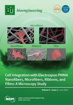
In tissue engineering, designs of scaffolds are crucial for cell attachment and proliferation. In this study, we explored electrospun PMMA scaffolds and spin-coated film in terms of initial osteoblast anchoring to produce geometries: nanofibers, microfibers, ribbons, and films. We showed high-resolution SEM images of cells shapes, attachments, and filopodia formation with respect to scaffold architectures. PMMA microfibers with the highest average fiber diameter in the range of 3.5 µm provided beneficial micro-environment for cell spreading and promoted the best integration. View this paper.
  • Issues are regarded as officially published after their release is announced to the table of contents alert mailing list.
  • You may sign up for e-mail alerts to receive table of contents of newly released issues.
  • PDF is the official format for papers published in both, html and pdf forms. To view the papers in pdf format, click on the "PDF Full-text" link, and use the free Adobe Reader to open them.
Order results
Result details
Section
Select all
Export citation of selected articles as:
12 pages, 35103 KiB  
Article
Influence of Pathogens and Mechanical Stimuli in Inflammation
by Israr B.M. Ibrahim and Ramana Pidaparti
Bioengineering 2019, 6(2), 55; https://0-doi-org.brum.beds.ac.uk/10.3390/bioengineering6020055 - 25 Jun 2019
Cited by 3 | Viewed by 5401
Abstract
Inflammation is a process driven by underlying cell-cell communication and many other factors. In this study, a model of cell-cell communications was proposed to study factors driving the inflammation time course. Analyses of inflammations that are driven by the combined effects of strain [...] Read more.
Inflammation is a process driven by underlying cell-cell communication and many other factors. In this study, a model of cell-cell communications was proposed to study factors driving the inflammation time course. Analyses of inflammations that are driven by the combined effects of strain (mechanical stimuli) and/or pathogens are considered in this paper. An agent-based model was employed to simulate inflammation where macrophages and fibroblasts influence each other through cell signaling cytokines that diffuse and spread in the tissue space. The communication network of macrophages and fibroblasts was then inferred and its network model (termed TE network) was generated and analyzed. The results suggest that factors driving inflammation time course can be discriminated by the characteristics of TE networks. Inflammation driven only by pathogens has certain TE network characteristics indicating slower and lower information exchange among cells. Multiple stimuli can help to maintain sufficient information exchange among cells, which is beneficial for inflammation resolution. The TE network captures the unfolding of the innate immune system over time, and the history of pathogens invasion. The resulting network leads to an improved understanding of the resilience of the system to future pathogen invasion. Full article
Show Figures

Graphical abstract

16 pages, 1216 KiB  
Article
Application of Plasmid Engineering to Enhance Yield and Quality of Plasmid for Vaccine and Gene Therapy
by Olusegun Folarin, Darren Nesbeth, John M. Ward and Eli Keshavarz-Moore
Bioengineering 2019, 6(2), 54; https://0-doi-org.brum.beds.ac.uk/10.3390/bioengineering6020054 - 19 Jun 2019
Cited by 5 | Viewed by 8204
Abstract
There is an increased interest in plasmid DNA as therapeutics. This is evident in the number of ongoing clinical trials involving the use of plasmid DNA. In order to be an effective therapeutic, high yield and high level of supercoiling are required. From [...] Read more.
There is an increased interest in plasmid DNA as therapeutics. This is evident in the number of ongoing clinical trials involving the use of plasmid DNA. In order to be an effective therapeutic, high yield and high level of supercoiling are required. From the bioprocessing point of view, the supercoiling level potentially has an impact on the ease of downstream processing. We approached meeting these requirements through plasmid engineering. A 7.2 kb plasmid was developed by the insertion of a bacteriophage Mu strong gyrase-binding sequence (Mu-SGS) to a 6.8 kb pSVβ-Gal and it was used to transform four different E. coli strains, and cultured in order to investigate the Mu-SGS effect and dependence on strain. There was an increase of over 20% in the total plasmid yield with pSVβ-Gal398 in two of the strains. The supercoiled topoisomer content was increased by 5% in both strains leading to a 27% increase in the overall yield. The extent of supercoiling was examined using superhelical density (σ) quantification with pSVβ-Gal398 maintaining a superhelical density of −0.022, and pSVβ-Gal −0.019, in both strains. This study has shown that plasmid modification with the Mu-phage SGS sequence has a beneficial effect on improving not only the yield of total plasmid but also the supercoiled topoisomer content of therapeutic plasmid DNA during bioprocessing. Full article
Show Figures

Figure 1

26 pages, 2752 KiB  
Review
Engineered Gold-Based Nanomaterials: Morphologies and Functionalities in Biomedical Applications. A Mini Review
by Iole Venditti
Bioengineering 2019, 6(2), 53; https://doi.org/10.3390/bioengineering6020053 - 10 Jun 2019
Cited by 68 | Viewed by 8904
Abstract
In the last decade, several engineered gold-based nanomaterials, such as spheres, rods, stars, cubes, hollow particles, and nanocapsules have been widely explored in biomedical fields, in particular in therapy and diagnostics. As well as different shapes and dimensions, these materials may, on their [...] Read more.
In the last decade, several engineered gold-based nanomaterials, such as spheres, rods, stars, cubes, hollow particles, and nanocapsules have been widely explored in biomedical fields, in particular in therapy and diagnostics. As well as different shapes and dimensions, these materials may, on their surfaces, have specific functionalizations to improve their capability as sensors or in drug loading and controlled release, and/or particular cell receptors ligands, in order to get a definite targeting. In this review, the up-to-date progress will be illustrated regarding morphologies, sizes and functionalizations, mostly used to obtain an improved performance of nanomaterials in biomedicine. Many suggestions are presented to organize and compare the numerous and heterogeneous experimental data, such as the most important chemical-physical parameters, which guide and control the interaction between the gold surface and biological environment. The purpose of all this is to offer the readers an overview of the most noteworthy progress and challenges in this research field. Full article
(This article belongs to the Special Issue Noble Metal Functionalized Nanoparticles for Biomedical Applications)
Show Figures

Graphical abstract

35 pages, 8464 KiB  
Article
A Multidisciplinary Approach toward High Throughput Label-Free Cytotoxicity Monitoring of Superparamagnetic Iron Oxide Nanoparticles
by Sonia Abad Tan, Georg Zoidl and Ebrahim Ghafar-Zadeh
Bioengineering 2019, 6(2), 52; https://0-doi-org.brum.beds.ac.uk/10.3390/bioengineering6020052 - 10 Jun 2019
Cited by 5 | Viewed by 7140
Abstract
This paper focuses on cytotoxicity examination of superparamagnetic iron oxide nanoparticles (SPIONs) using different methods, including impedance spectroscopy. Recent advances of SPIONs for clinical and research applications have triggered the need to understand their effects in cells. Despite the great advances in adapting [...] Read more.
This paper focuses on cytotoxicity examination of superparamagnetic iron oxide nanoparticles (SPIONs) using different methods, including impedance spectroscopy. Recent advances of SPIONs for clinical and research applications have triggered the need to understand their effects in cells. Despite the great advances in adapting various biological and chemical methods to assess in-vitro toxicity of SPIONs, less attention has been paid on the development of a high throughput label-free screening platform to study the interaction between the cells and nanoparticles including SPIONs. In this paper, we have taken the first step toward this goal by proposing a label-free impedimetric method for monitoring living cells treated with SPIONs. We demonstrate the effect of SPIONs on the adhesion, growth, proliferation, and viability of neuroblastoma 2A (N2a) cells using impedance spectroscopy as a label-free method, along with other standard microscopic and cell viability testing methods as control methods. Our results have shown a decreased viability of the cells as the concentration of SPIONs increases with percentages of 59%, 47%, and 40% for 100 µg/mL (C4), 200 µg/mL (C5), 300 µg/mL (C6), respectively. Although all SPIONs concentrations have allowed the growth of cells within 72 h, C4, C5, and C6 showed slower growth compared to the control (C1). The growth and proliferation of N2a cells are faster in the absence or low concentration of SPIONS. The percent coefficient of variation (% CV) was used to compare cell concentrations obtained by TBDE assay and a Scepter cell counter. Results also showed that the lower the SPIONs concentration, the lower the impedance is expected to be in the sensing electrodes without the cells. Meanwhile, the variation of surface area (∆S) was affected by the concentration of SPIONs. It was observed that the double layer capacitance was almost constant because of the higher attachment of cells, the lower surface area coated by SPIONs. In conclusion, impedance changes of electrodes exposed to the mixture of cells and SPIONs offer a wide dynamic range (>1 MΩ using Electric Cell-substrate Impedance electrodes) suitable for cytotoxicity studies. Based on impedance based, viability testing and microscopic methods’ results, SPIONs concentrations higher than 100 ug/mL and 300 ug/mL cause minor and major effects, respectively. We propose that a high throughput impedance-based label-free platform provides great advantages for studying SPIONs in a cell-based context, opening a window of opportunity to design and test the next generation of SPIONs with reduced toxicity for biomedical or medical applications. Full article
(This article belongs to the Collection Nanoparticles for Therapeutic and Diagnostic Applications)
Show Figures

Graphical abstract

2 pages, 142 KiB  
Editorial
Engineering Bone-Implant Materials
by Mohammad Elahinia, Hamdy Ibrahim, Mohammad Javad Mahtabi and Reza Mehrabi
Bioengineering 2019, 6(2), 51; https://0-doi-org.brum.beds.ac.uk/10.3390/bioengineering6020051 - 09 Jun 2019
Cited by 2 | Viewed by 5119
Abstract
This special issue is dedicated to the simulation as well as experimental studies of biomechanical behavior of biomaterials, especially those that are used for bone implant applications [...] Full article
(This article belongs to the Special Issue Engineering Bone-Implant Materials)
20 pages, 7549 KiB  
Article
A Microcavity Array-Based 4D Cell Culture Platform
by Cordula Nies, Tobias Rubner, Hanna Lorig, Vera Colditz, Helen Seelmann, Andreas Müller and Eric Gottwald
Bioengineering 2019, 6(2), 50; https://0-doi-org.brum.beds.ac.uk/10.3390/bioengineering6020050 - 31 May 2019
Cited by 5 | Viewed by 7129
Abstract
(1) Background: We describe a 4D cell culture platform with which we tried to detect and to characterize migration dynamics of single hematopoietic stem cells in polymer film microcavity arrays integrated into a microtiter plate. (2) Methods: The system was set up with [...] Read more.
(1) Background: We describe a 4D cell culture platform with which we tried to detect and to characterize migration dynamics of single hematopoietic stem cells in polymer film microcavity arrays integrated into a microtiter plate. (2) Methods: The system was set up with CD34-expressing KG-1a cells as a surrogate for hematopoietic stem cells. We then evaluated the system as an artificial hematopoietic stem cell niche model comprised of a co-culture of human hematopoietic stem cells from cord blood (cord blood CD34+ cells, hHSCs) and human mesenchymal stromal cells (hMSCs) from bone marrow over a period of 21 days. We used a software-based cell detection method to count single hematopoietic stem cells (HSCs) in microcavities. (3) Results: It was possible to detect single HSCs and their migration behavior within single microcavities. The HSCs displayed a pronounced migration behavior with one population of CD34-expressing cells located at the bottom of the microcavities and one population located in the middle of the microcavities at day 14. However, at day 21 the two populations seemed to unite again so that no clear distinction between the two was possible anymore. (4) Conclusions: Single cell migration detection was possible but microscopy and flow cytometry delivered non-uniform data sets. Further optimization is currently being developed. Full article
(This article belongs to the Special Issue Advanced Dynamic Cell and Tissue Culture, Volume 2)
Show Figures

Figure 1

16 pages, 4386 KiB  
Article
Mechanical Response of Porcine Liver Tissue under High Strain Rate Compression
by Joseph Chen, Sourav S. Patnaik, R. K. Prabhu, Lauren B. Priddy, Jean-Luc Bouvard, Esteban Marin, Mark F. Horstemeyer, Jun Liao and Lakiesha N. Williams
Bioengineering 2019, 6(2), 49; https://0-doi-org.brum.beds.ac.uk/10.3390/bioengineering6020049 - 30 May 2019
Cited by 8 | Viewed by 7595
Abstract
In automobile accidents, abdominal injuries are often life-threatening yet not apparent at the time of initial injury. The liver is the most commonly injured abdominal organ from this type of trauma. In contrast to current safety tests involving crash dummies, a more detailed, [...] Read more.
In automobile accidents, abdominal injuries are often life-threatening yet not apparent at the time of initial injury. The liver is the most commonly injured abdominal organ from this type of trauma. In contrast to current safety tests involving crash dummies, a more detailed, efficient approach to predict the risk of human injuries is computational modelling and simulations. Further, the development of accurate computational human models requires knowledge of the mechanical properties of tissues in various stress states, especially in high-impact scenarios. In this study, a polymeric split-Hopkinson pressure bar (PSHPB) was utilized to apply various high strain rates to porcine liver tissue to investigate its material behavior during high strain rate compression. Liver tissues were subjected to high strain rate impacts at 350, 550, 1000, and 1550 s−1. Tissue directional dependency was also explored by PSHPB testing along three orthogonal directions of liver at a strain rate of 350 s−1. Histology of samples from each of the three directions was performed to examine the structural properties of porcine liver. Porcine liver tissue showed an inelastic and strain rate-sensitive response at high strain rates. The liver tissue was found lacking directional dependency, which could be explained by the isotropic microstructure observed after staining and imaging. Furthermore, finite element analysis (FEA) of the PSHPB tests revealed the stress profile inside liver tissue and served as a validation of PSHPB methodology. The present findings can assist in the development of more accurate computational models of liver tissue at high-rate impact conditions allowing for understanding of subfailure and failure mechanisms. Full article
(This article belongs to the Special Issue Advances in Biological Tissue Biomechanics)
Show Figures

Figure 1

24 pages, 1820 KiB  
Review
Expansion Culture of Human Pluripotent Stem Cells and Production of Cardiomyocytes
by Minh Nguyen Tuyet Le and Kouichi Hasegawa
Bioengineering 2019, 6(2), 48; https://0-doi-org.brum.beds.ac.uk/10.3390/bioengineering6020048 - 24 May 2019
Cited by 18 | Viewed by 9605
Abstract
Transplantation of human pluripotent stem cell (hPSCs)-derived cardiomyocytes for the treatment of heart failure is a promising therapy. In order to implement this therapy requiring numerous cardiomyocytes, substantial production of hPSCs followed by cardiac differentiation seems practical. Conventional methods of culturing hPSCs involve [...] Read more.
Transplantation of human pluripotent stem cell (hPSCs)-derived cardiomyocytes for the treatment of heart failure is a promising therapy. In order to implement this therapy requiring numerous cardiomyocytes, substantial production of hPSCs followed by cardiac differentiation seems practical. Conventional methods of culturing hPSCs involve using a 2D culture monolayer that hinders the expansion of hPSCs, thereby limiting their productivity. Advanced culture of hPSCs in 3D aggregates in the suspension overcomes the limitations of 2D culture and attracts immense attention. Although the hPSC production needs to be suitable for subsequent cardiac differentiation, many studies have independently focused on either expansion of hPSCs or cardiac differentiation protocols. In this review, we summarize the recent approaches to expand hPSCs in combination with cardiomyocyte differentiation. A comparison of various suspension culture methods and future prospects for dynamic culture of hPSCs are discussed in this study. Understanding hPSC characteristics in different models of dynamic culture helps to produce numerous cells that are useful for further clinical applications. Full article
(This article belongs to the Special Issue Advanced Dynamic Cell and Tissue Culture, Volume 2)
Show Figures

Graphical abstract

40 pages, 7532 KiB  
Review
Mechanics of the Tricuspid Valve—From Clinical Diagnosis/Treatment, In-Vivo and In-Vitro Investigations, to Patient-Specific Biomechanical Modeling
by Chung-Hao Lee, Devin W. Laurence, Colton J. Ross, Katherine E. Kramer, Anju R. Babu, Emily L. Johnson, Ming-Chen Hsu, Ankush Aggarwal, Arshid Mir, Harold M. Burkhart, Rheal A. Towner, Ryan Baumwart and Yi Wu
Bioengineering 2019, 6(2), 47; https://0-doi-org.brum.beds.ac.uk/10.3390/bioengineering6020047 - 22 May 2019
Cited by 29 | Viewed by 10026
Abstract
Proper tricuspid valve (TV) function is essential to unidirectional blood flow through the right side of the heart. Alterations to the tricuspid valvular components, such as the TV annulus, may lead to functional tricuspid regurgitation (FTR), where the valve is unable to prevent [...] Read more.
Proper tricuspid valve (TV) function is essential to unidirectional blood flow through the right side of the heart. Alterations to the tricuspid valvular components, such as the TV annulus, may lead to functional tricuspid regurgitation (FTR), where the valve is unable to prevent undesired backflow of blood from the right ventricle into the right atrium during systole. Various treatment options are currently available for FTR; however, research for the tricuspid heart valve, functional tricuspid regurgitation, and the relevant treatment methodologies are limited due to the pervasive expectation among cardiac surgeons and cardiologists that FTR will naturally regress after repair of left-sided heart valve lesions. Recent studies have focused on (i) understanding the function of the TV and the initiation or progression of FTR using both in-vivo and in-vitro methods, (ii) quantifying the biomechanical properties of the tricuspid valve apparatus as well as its surrounding heart tissue, and (iii) performing computational modeling of the TV to provide new insight into its biomechanical and physiological function. This review paper focuses on these advances and summarizes recent research relevant to the TV within the scope of FTR. Moreover, this review also provides future perspectives and extensions critical to enhancing the current understanding of the functioning and remodeling tricuspid valve in both the healthy and pathophysiological states. Full article
(This article belongs to the Special Issue Advances in Biological Tissue Biomechanics)
Show Figures

Figure 1

18 pages, 4582 KiB  
Article
Controlling Electronic Devices with Brain Rhythms/Electrical Activity Using Artificial Neural Network (ANN)
by Yar Muhammad and Daniil Vaino
Bioengineering 2019, 6(2), 46; https://0-doi-org.brum.beds.ac.uk/10.3390/bioengineering6020046 - 17 May 2019
Cited by 8 | Viewed by 8486
Abstract
The purpose of this research study was to explore the possibility to develop a brain-computer interface (BCI). The main objective was that the BCI should be able to recognize brain activity. BCI is an emerging technology which focuses on communication between software and [...] Read more.
The purpose of this research study was to explore the possibility to develop a brain-computer interface (BCI). The main objective was that the BCI should be able to recognize brain activity. BCI is an emerging technology which focuses on communication between software and hardware and permitting the use of brain activity to control electronic devices, such as wheelchairs, computers and robots. The interface was developed, and consists of EEG Bitronics, Arduino and a computer; moreover, two versions of the BCIANNET software were developed to be used with this hardware. This BCI used artificial neural network (ANN) as a main processing method, with the Butterworth filter used as the data pre-processing algorithm for ANN. Twelve subjects were measured to collect the datasets. Tasks were given to subjects to stimulate brain activity. The purpose of the experiments was to test and confirm the performance of the developed software. The aim of the software was to separate important rhythms such as alpha, beta, gamma and delta from other EEG signals. As a result, this study showed that the Levenberg–Marquardt algorithm is the best compared with the backpropagation, resilient backpropagation, and error correction algorithms. The final developed version of the software is an effective tool for research in the field of BCI. The study showed that using the Levenberg–Marquardt learning algorithm gave an accuracy of prediction around 60% on the testing dataset. Full article
(This article belongs to the Special Issue Advances in Artificial Intelligence and Machine Learning for BCI/BMI)
Show Figures

Figure 1

15 pages, 4263 KiB  
Article
Tissue Level Mechanical Properties and Extracellular Matrix Investigation of the Bovine Jugular Venous Valve Tissue
by Adam A. Benson and Hsiao-Ying Shadow Huang
Bioengineering 2019, 6(2), 45; https://0-doi-org.brum.beds.ac.uk/10.3390/bioengineering6020045 - 14 May 2019
Cited by 8 | Viewed by 7001
Abstract
Jugular venous valve incompetence has no long-term remedy and symptoms of transient global amnesia and/or intracranial hypertension continue to discomfort patients. During this study, we interrogate the synergy of the collagen and elastin microstructure that compose the bi-layer extracellular matrix (ECM) of the [...] Read more.
Jugular venous valve incompetence has no long-term remedy and symptoms of transient global amnesia and/or intracranial hypertension continue to discomfort patients. During this study, we interrogate the synergy of the collagen and elastin microstructure that compose the bi-layer extracellular matrix (ECM) of the jugular venous valve. In this study, we investigate the jugular venous valve and relate it to tissue-level mechanical properties, fibril orientation and fibril composition to improve fundamental knowledge of the jugular venous valves toward the development of bioprosthetic venous valve replacements. Steps include: (1) multi loading biaxial mechanical tests; (2) isolation of the elastin microstructure; (3) imaging of the elastin microstructure; and (4) imaging of the collagen microstructure, including an experimental analysis of crimp. Results from this study show that, during a 3:1 loading ratio (circumferential direction: 900 mN and radial direction: 300 mN), elastin may have the ability to contribute to the circumferential mechanical properties at low strains, for example, shifting the inflection point toward lower strains in comparison to other loading ratios. After isolating the elastin microstructure, light microscopy revealed that the overall elastin orients in the radial direction while forming a crosslinked mesh. Collagen fibers were found undulated, aligning in parallel with neighboring fibers and orienting in the circumferential direction with an interquartile range of −10.38° to 7.58° from the circumferential axis (n = 20). Collagen crimp wavelength and amplitude was found to be 38.46 ± 8.06 µm and 4.51 ± 1.65 µm, respectively (n = 87). Analyzing collagen crimp shows that crimp permits about 12% true strain circumferentially, while straightening of the overall fibers accounts for more. To the best of the authors’ knowledge, this is the first study of the jugular venous valve linking the composition and orientation of the ECM to its mechanical properties and this study will aid in forming a structure-based constitutive model. Full article
(This article belongs to the Special Issue Advances in Biological Tissue Biomechanics)
Show Figures

Figure 1

6 pages, 2777 KiB  
Perspective
Biofabrication of Bacterial Constructs: New Three-Dimensional Biomaterials
by Amin Shavandi and Esmat Jalalvandi
Bioengineering 2019, 6(2), 44; https://0-doi-org.brum.beds.ac.uk/10.3390/bioengineering6020044 - 14 May 2019
Cited by 8 | Viewed by 7071
Abstract
An enormous number of bacteria live in almost every environment; from deep oceans to below the surface of the earth or in our gastrointestinal tract. Although biofabrication is growing and maturing very quickly, the involvement of bacteria in this process has not been [...] Read more.
An enormous number of bacteria live in almost every environment; from deep oceans to below the surface of the earth or in our gastrointestinal tract. Although biofabrication is growing and maturing very quickly, the involvement of bacteria in this process has not been developed at a similar pace. From the development of a new generation of biomaterials to green bioremediation for the removal of hazardous environmental pollutants or to develop innovative food products in a recent trend, researchers have used cutting-edge biofabrication techniques to reveal the great potential of 3D structured bacterial constructs. These 3D bacterial workhouses may fundamentally change our approach toward biomaterials. Full article
(This article belongs to the Special Issue Biocomposite Inks for 3D Printing)
Show Figures

Figure 1

28 pages, 5570 KiB  
Article
New Evaluation Procedure for Multi-Dimensional Mechanical Strains and Tangent Moduli of Breast Implants: IDEAL IMPLANT® Structured Breast Implant Compared to Silicone Gel Implants
by Harold J. Brandon, Larry S. Nichter and Dwight D. Back
Bioengineering 2019, 6(2), 43; https://0-doi-org.brum.beds.ac.uk/10.3390/bioengineering6020043 - 12 May 2019
Cited by 4 | Viewed by 7961
Abstract
The IDEAL IMPLANT® Structured Breast Implant is a dual lumen saline-filled implant with capsular contracture and deflation/rupture rates much lower than single-lumen silicone gel-filled implants. To better understand the implant’s mechanical properties and to provide a potential explanation for these eight-year clinical [...] Read more.
The IDEAL IMPLANT® Structured Breast Implant is a dual lumen saline-filled implant with capsular contracture and deflation/rupture rates much lower than single-lumen silicone gel-filled implants. To better understand the implant’s mechanical properties and to provide a potential explanation for these eight-year clinical results, a novel approach to compressive load testing was employed. Multi-dimensional strains and tangent moduli, metrics describing the shape stability of the total implant, were derived from the experimental load and platen spacing data. The IDEAL IMPLANT was found to have projection, diametric, and areal strains that were generally less than silicone gel implants, and tangent moduli that were generally greater than silicone gel implants. Despite having a relatively inviscid saline fill, the IDEAL IMPLANT was found to be more shape stable compared to gel implants, which implies potentially less interaction with the capsule wall when the implant is subjected to compressive loads. Under compressive loads, the shape stability of a higher cross-link density, cohesive gel implant was unexpectedly found to be similar to or the same as a gel implant. In localized diametric compression testing, the IDEAL IMPLANT was found to have a palpability similar to a gel implant, but softer than a cohesive gel implant. Full article
Show Figures

Figure 1

10 pages, 1135 KiB  
Article
Antimicrobial Photodynamic Therapy Protocols on Streptococcus mutans with Different Combinations of Wavelengths and Photosensitizing Dyes
by Elisabetta Merigo, Stefania Conti, Tecla Ciociola, Maddalena Manfredi, Paolo Vescovi and Carlo Fornaini
Bioengineering 2019, 6(2), 42; https://0-doi-org.brum.beds.ac.uk/10.3390/bioengineering6020042 - 10 May 2019
Cited by 11 | Viewed by 5828
Abstract
The aim of the study is to test the application of different laser wavelengths, with and without different photosensitizing dyes on different types of cultures. Laser irradiation was realized on Streptococcus mutans in both solid and liquid culture media in continuous mode at [...] Read more.
The aim of the study is to test the application of different laser wavelengths, with and without different photosensitizing dyes on different types of cultures. Laser irradiation was realized on Streptococcus mutans in both solid and liquid culture media in continuous mode at three different fluences (10, 20, and 30 J/cm2) with a red diode (650 nm) with toluidine blue dye, a blue-violet diode (405 nm) with curcumin dye, and a green diode (532 nm) with erythrosine dye. Without a photosensitizer, no growth inhibition was obtained with the red diode at any fluence value. Inhibition rates of 40.7% and 40.2% were obtained with the blue diode and green diode. The blue diode laser used with curcumin obtained results in terms of growth inhibition up to 99.26% at a fluence of 30 J/cm2. The red diode laser used with toluidine blue obtained results in terms of growth inhibition up to 100% at fluences of 20 and 30 J/cm2. The KTP (potassium-titanyl-phosphate) laser used with erythrosine was able to determine a complete growth inhibition (100%) at the different fluence values. The combination of a laser and its proper color may dramatically change the results in terms of bactericidal effect. It will be interesting to confirm these data by further in vivo studies. Full article
Show Figures

Figure 1

12 pages, 4788 KiB  
Article
Cell Integration with Electrospun PMMA Nanofibers, Microfibers, Ribbons, and Films: A Microscopy Study
by Daniel P. Ura, Joanna E. Karbowniczek, Piotr K. Szewczyk, Sara Metwally, Mateusz Kopyściański and Urszula Stachewicz
Bioengineering 2019, 6(2), 41; https://0-doi-org.brum.beds.ac.uk/10.3390/bioengineering6020041 - 09 May 2019
Cited by 32 | Viewed by 7627
Abstract
Tissue engineering requires properly selected geometry and surface properties of the scaffold, to promote in vitro tissue growth. In this study, we obtained three types of electrospun poly(methyl methacrylate) (PMMA) scaffolds—nanofibers, microfibers, and ribbons, as well as spin-coated films. Their morphology was imaged [...] Read more.
Tissue engineering requires properly selected geometry and surface properties of the scaffold, to promote in vitro tissue growth. In this study, we obtained three types of electrospun poly(methyl methacrylate) (PMMA) scaffolds—nanofibers, microfibers, and ribbons, as well as spin-coated films. Their morphology was imaged by scanning electron microscopy (SEM) and characterized by average surface roughness and water contact angle. PMMA films had a smooth surface with roughness, Ra below 0.3 µm and hydrophilic properties, whereas for the fibers and the ribbons, we observed increased hydrophobicity, with higher surface roughness and fiber diameter. For microfibers, we obtained the highest roughness of 7 µm, therefore, the contact angle was 140°. All PMMA samples were used for the in vitro cell culture study, to verify the cells integration with various designs of scaffolds. The detailed microscopy study revealed that higher surface roughness enhanced cells’ attachment and their filopodia length. The 3D structure of PMMA microfibers with an average fiber diameter above 3.5 µm, exhibited the most favorable geometry for cells’ ingrowth, whereas, for other structures we observed cells growth only on the surface. The study showed that electrospinning of various scaffolds geometry is able to control cells development that can be adjusted according to the tissue needs in the regeneration processes. Full article
(This article belongs to the Special Issue Designing Tissue Scaffolds with Electrospun Fibers)
Show Figures

Graphical abstract

24 pages, 10007 KiB  
Article
Compressive Mechanical Properties of Porcine Brain: Experimentation and Modeling of the Tissue Hydration Effects
by Raj K. Prabhu, Mark T. Begonia, Wilburn R. Whittington, Michael A. Murphy, Yuxiong Mao, Jun Liao, Lakiesha N. Williams, Mark F. Horstemeyer and Jianping Sheng
Bioengineering 2019, 6(2), 40; https://0-doi-org.brum.beds.ac.uk/10.3390/bioengineering6020040 - 07 May 2019
Cited by 12 | Viewed by 6845
Abstract
Designing protective systems for the human head—and, hence, the brain—requires understanding the brain’s microstructural response to mechanical insults. We present the behavior of wet and dry porcine brain undergoing quasi-static and high strain rate mechanical deformations to unravel the effect of hydration on [...] Read more.
Designing protective systems for the human head—and, hence, the brain—requires understanding the brain’s microstructural response to mechanical insults. We present the behavior of wet and dry porcine brain undergoing quasi-static and high strain rate mechanical deformations to unravel the effect of hydration on the brain’s biomechanics. Here, native ‘wet’ brain samples contained ~80% (mass/mass) water content and ‘dry’ brain samples contained ~0% (mass/mass) water content. First, the wet brain incurred a large initial peak stress that was not exhibited by the dry brain. Second, stress levels for the dry brain were greater than the wet brain. Third, the dry brain stress–strain behavior was characteristic of ductile materials with a yield point and work hardening; however, the wet brain showed a typical concave inflection that is often manifested by polymers. Finally, finite element analysis (FEA) of the brain’s high strain rate response for samples with various proportions of water and dry brain showed that water played a major role in the initial hardening trend. Therefore, hydration level plays a key role in brain tissue micromechanics, and the incorporation of this hydration effect on the brain’s mechanical response in simulated injury scenarios or virtual human-centric protective headgear design is essential. Full article
(This article belongs to the Special Issue Advances in Biological Tissue Biomechanics)
Show Figures

Graphical abstract

14 pages, 5801 KiB  
Article
Elastin-Dependent Aortic Heart Valve Leaflet Curvature Changes During Cyclic Flexure
by Melake D. Tesfamariam, Asad M. Mirza, Daniel Chaparro, Ahmed Z. Ali, Rachel Montalvan, Ilyas Saytashev, Brittany A. Gonzalez, Amanda Barreto, Jessica Ramella-Roman, Joshua D. Hutcheson and Sharan Ramaswamy
Bioengineering 2019, 6(2), 39; https://0-doi-org.brum.beds.ac.uk/10.3390/bioengineering6020039 - 07 May 2019
Cited by 7 | Viewed by 6585
Abstract
The progression of calcific aortic valve disease (CAVD) is characterized by extracellular matrix (ECM) remodeling, leading to structural abnormalities and improper valve function. The focus of the present study was to relate aortic valve leaflet axial curvature changes as a function of elastin [...] Read more.
The progression of calcific aortic valve disease (CAVD) is characterized by extracellular matrix (ECM) remodeling, leading to structural abnormalities and improper valve function. The focus of the present study was to relate aortic valve leaflet axial curvature changes as a function of elastin degradation, which has been associated with CAVD. Circumferential rectangular strips (L × W = 10 × 2.5 mm) of normal and elastin-degraded (via enzymatic digestion) porcine AV leaflets were subjected to cyclic flexure (1 Hz). A significant increase in mean curvature (p < 0.05) was found in elastin-degraded leaflet specimens in comparison to un-degraded controls at both the semi-constrained (50% of maximum flexed state during specimen bending and straightening events) and fully-constrained (maximally-flexed) states. This significance did not occur in all three flexed configurations when measurements were performed using either minimum or maximum curvature. Moreover, the mean curvature increase in the elastin-degraded leaflets was most pronounced at the instance of maximum flexure, compared to un-degraded controls. We conclude that the mean axial curvature metric can detect distinct spatial changes in aortic valve ECM arising from the loss in bulk content and/or structure of elastin, particularly when there is a high degree of tissue bending. Therefore, the instance of maximum leaflet flexure during the cardiac cycle could be targeted for mean curvature measurements and serve as a potential biomarker for elastin degradation in early CAVD remodeling. Full article
(This article belongs to the Special Issue Advances in Biological Tissue Biomechanics)
Show Figures

Figure 1

14 pages, 1639 KiB  
Perspective
Bioengineering an Artificial Human Blood–Brain Barrier in Rodents
by Kimia Kamal and Ben Waldau
Bioengineering 2019, 6(2), 38; https://0-doi-org.brum.beds.ac.uk/10.3390/bioengineering6020038 - 30 Apr 2019
Cited by 7 | Viewed by 8502
Abstract
Our group has recently created a novel in-vivo human brain organoid vascularized with human iPSC-derived endothelial cells. In this review article, we discuss the challenges of creating a perfused human brain organoid model in an immunosuppressed rodent host and discuss potential applications for [...] Read more.
Our group has recently created a novel in-vivo human brain organoid vascularized with human iPSC-derived endothelial cells. In this review article, we discuss the challenges of creating a perfused human brain organoid model in an immunosuppressed rodent host and discuss potential applications for neurosurgical disease modeling. Full article
(This article belongs to the Special Issue Organs-on-Chips)
Show Figures

Figure 1

33 pages, 14302 KiB  
Review
Application of NiTi in Assistive and Rehabilitation Devices: A Review
by Mohammadreza Nematollahi, Keyvan Safaei Baghbaderani, Amirhesam Amerinatanzi, Hashem Zamanian and Mohammad Elahinia
Bioengineering 2019, 6(2), 37; https://0-doi-org.brum.beds.ac.uk/10.3390/bioengineering6020037 - 29 Apr 2019
Cited by 69 | Viewed by 9190
Abstract
Shape memory alloys (SMAs) have found widespread applications as biomedical devices. Biocompatibility, corrosion resistance, and ductility make these alloys attractive for medical devices such as stents and filters. For these implants, the superelastic property is the primary function of SMAs. Additionally, these alloys, [...] Read more.
Shape memory alloys (SMAs) have found widespread applications as biomedical devices. Biocompatibility, corrosion resistance, and ductility make these alloys attractive for medical devices such as stents and filters. For these implants, the superelastic property is the primary function of SMAs. Additionally, these alloys, such as NiTi as the prime example, can be used for actuation. Several modes of actuation such as displacement control, force control, and compliance control have been used as harnesses with SMA devices. These two unique properties have opened another application in the form of neurosurgery and robot-assisted surgery devices, as well as controlled assistive and rehabilitation devices. This paper reviews the state of the art of application of SMAs in the latter category where control is applied to harness innovative medical devices. To this end, two major subsets of these devices: prosthesis and orthosis which take the advantage of SMAs in assistive and rehabilitation devices are studied. These devices are further categorized to hand prosthetics, elbow, knee and ankle orthotics. In most of these designs, SMA wires act as artificial muscles to mimic the motion of limbs in the target joints. The evolution of each category is explained, and the specific results of them are reported. The paper also reviews the SMA applications for neurological and neuromuscular rehabilitation. To this end, different categories of rehabilitation devices as a passive and aided exercise for the ankle, knee, and elbow are highlighted. The SMA actuator in these devices can be EMG-controlled to improved patient outcome. In addition to providing a comprehensive overview of the biomedical devices, this paper identifies several possible future directions of SMA related research in the area of assistive and rehabilitation devices. Full article
(This article belongs to the Special Issue Engineering Bone-Implant Materials)
Show Figures

Figure 1

5 pages, 2797 KiB  
Communication
Inhibition of Demineralization at Restoration Margins of Z100 and Tetric EvoCeram Bulk Fill in Dentin and Enamel
by Cristina Leon-Pineda and Kevin Donly
Bioengineering 2019, 6(2), 36; https://0-doi-org.brum.beds.ac.uk/10.3390/bioengineering6020036 - 28 Apr 2019
Cited by 1 | Viewed by 5580
Abstract
Recurrent caries is still considered the main reason restorations need to be replaced. There are different materials available now that promise to reduce the possibility of recurrent caries by releasing fluoride and inhibiting restoration marginal caries. The purpose of this in vitro study [...] Read more.
Recurrent caries is still considered the main reason restorations need to be replaced. There are different materials available now that promise to reduce the possibility of recurrent caries by releasing fluoride and inhibiting restoration marginal caries. The purpose of this in vitro study was to evaluate the demineralization inhibition potential of a non-fluoride-releasing resin (Z100TM 3M, St. Paul, MN, USA) and a glass containing resin-based composite (Tetric EvoCeram Bulk Fill, Ivoclar/Vivadent AG, Schaan, Liechtenstein), which contains fluoride. Class V preparations were placed on 22 premolars; the gingival margin was below the cementoenamel junction and the occlusal margin was placed above the cemento-enamel junction. Ten teeth were randomly selected to be restored with Z100 while the other 10 were restored with Tetric EvoCeram Bulk Fill. Both groups were restored following manufacturer’s instructions. All teeth had an acid resistant varnish placed within one millimeter of the preparation margins. Both groups were placed in artificial caries challenge solution (pH 4.4). At the end of the 4 days; 100 µm buccolingual sections were obtained for each tooth; these were photographed under polarized light microscopy and the demineralized areas adjacent to the restorations were measured and quantified. The mean (±S.D.) area (µm2) of demineralization from the occlusal margin (enamel) and dentin margin were: Z100 2781.889 ± 1045.213; 3960.455 ± 705.964 and for Tetric EvoCeram Bulk Fill 1541.545 ± 1167.027; 3027.600 ± 512.078. Student’s t-test indicated that there was significantly less enamel and dentin demineralization adjacent to Tetric EvoCeram Bulk Fill compared to Z100; there was significantly less demineralization in enamel compared to dentin in both Tetric EvoCeral Bulk Fill and Z100. Tetric EvoCeram Bulk Fill performed better inhibiting demineralization at restoration margins when compared to Z100 and provided better demineralization inhibition in enamel than cementum/dentin. Full article
Show Figures

Figure 1

14 pages, 1052 KiB  
Article
Oxygen Persufflation in Liver Transplantation Results of a Randomized Controlled Trial
by Anja Gallinat, Dieter Paul Hoyer, Georgios Sotiropoulos, Jürgen Treckmann, Tamas Benkoe, Jennifer Belker, Fuat Saner, Andreas Paul and Thomas Minor
Bioengineering 2019, 6(2), 35; https://0-doi-org.brum.beds.ac.uk/10.3390/bioengineering6020035 - 27 Apr 2019
Cited by 7 | Viewed by 6722
Abstract
Oxygen persufflation has shown experimentally to favorably influence hepatic energy dependent pathways and to improve survival after transplantation. The present trial evaluated oxygen persufflation as adjunct in clinical liver preservation. A total of n = 116 adult patients (age: 54 (23–68) years, M/F: [...] Read more.
Oxygen persufflation has shown experimentally to favorably influence hepatic energy dependent pathways and to improve survival after transplantation. The present trial evaluated oxygen persufflation as adjunct in clinical liver preservation. A total of n = 116 adult patients (age: 54 (23–68) years, M/F: 70/46), were enrolled in this prospective randomized study. Grafts were randomized to either oxygen persufflation for ≥2 h (O2) or mere cold storage (control). Only liver grafts from donors ≥55 years and/or marginal grafts after multiple rejections by other centers were included. Primary endpoint was peak-aspartate aminotransferase (AST) level until post-operative day 3. Standard parameters including graft- and patient survival were analyzed by uni- and multivariate analysis. Both study groups were comparable except for a longer ICU stay (4 versus 3 days) of the donors and a higher recipient age (57 versus 52 years) in the O2-group. Serum levels of TNF alpha were significantly reduced after oxygen persufflation (p < 0.05). Median peak-AST values did not differ between the groups (O2: 580 U/l, control: 699 U/l). Five year graft- and patient survival was similar. Subgroup analysis demonstrated a positive effect of oxygen persufflation concerning the development of early allograft dysfunction (EAD), in donors with a history of cardiopulmonary resuscitation and elevated ALT values, and concerning older or macrosteatotic livers. This study favors pre-implantation O2-persufflation in concrete subcategories of less than optimal liver grafts, for which oxygen persufflation can be considered a safe, cheap and easy applicable reconditioning method. Full article
(This article belongs to the Special Issue Bioengineering Liver Transplantation)
Show Figures

Figure 1

33 pages, 1494 KiB  
Review
Polyhydroxyalkanoate Biosynthesis at the Edge of Water Activity-Haloarchaea as Biopolyester Factories
by Martin Koller
Bioengineering 2019, 6(2), 34; https://0-doi-org.brum.beds.ac.uk/10.3390/bioengineering6020034 - 16 Apr 2019
Cited by 81 | Viewed by 9254
Abstract
Haloarchaea, the extremely halophilic branch of the Archaea domain, encompass a steadily increasing number of genera and associated species which accumulate polyhydroxyalkanoate biopolyesters in their cytoplasm. Such ancient organisms, which thrive in highly challenging, often hostile habitats characterized by salinities between 100 and [...] Read more.
Haloarchaea, the extremely halophilic branch of the Archaea domain, encompass a steadily increasing number of genera and associated species which accumulate polyhydroxyalkanoate biopolyesters in their cytoplasm. Such ancient organisms, which thrive in highly challenging, often hostile habitats characterized by salinities between 100 and 300 g/L NaCl, have the potential to outperform established polyhydroxyalkanoate production strains. As detailed in the review, this optimization presents due to multifarious reasons, including: cultivation setups at extreme salinities can be performed at minimized sterility precautions by excluding the growth of microbial contaminants; the high inner-osmotic pressure in haloarchaea cells facilitates the recovery of intracellular biopolyester granules by cell disintegration in hypo-osmotic media; many haloarchaea utilize carbon-rich waste streams as main substrates for growth and polyhydroxyalkanoate biosynthesis, which allows coupling polyhydroxyalkanoate production with bio-economic waste management; finally, in many cases, haloarchaea are reported to produce copolyesters from structurally unrelated inexpensive substrates, and polyhydroxyalkanoate biosynthesis often occurs in parallel to the production of additional marketable bio-products like pigments or polysaccharides. This review summarizes the current knowledge about polyhydroxyalkanoate production by diverse haloarchaea; this covers the detection of new haloarchaea producing polyhydroxyalkanoates, understanding the genetic and enzymatic particularities of such organisms, kinetic aspects, material characterization, upscaling and techno-economic and life cycle assessment. Full article
(This article belongs to the Special Issue Advances in Polyhydroxyalkanoate (PHA) Production, Volume 2)
Show Figures

Figure 1

16 pages, 2308 KiB  
Article
Acacia Holosericea: An Invasive Species for Bio-char, Bio-oil, and Biogas Production
by Md Sumon Reza, Ashfaq Ahmed, Wahyu Caesarendra, Muhammad S. Abu Bakar, Shahriar Shams, R. Saidur, Navid Aslfattahi and Abul K. Azad
Bioengineering 2019, 6(2), 33; https://0-doi-org.brum.beds.ac.uk/10.3390/bioengineering6020033 - 16 Apr 2019
Cited by 61 | Viewed by 10149
Abstract
To evaluate the possibilities for biofuel and bioenergy production Acacia Holosericea, which is an invasive plant available in Brunei Darussalam, was investigated. Proximate analysis of Acacia Holosericea shows that the moisture content, volatile matters, fixed carbon, and ash contents were 9.56%, 65.12%, [...] Read more.
To evaluate the possibilities for biofuel and bioenergy production Acacia Holosericea, which is an invasive plant available in Brunei Darussalam, was investigated. Proximate analysis of Acacia Holosericea shows that the moisture content, volatile matters, fixed carbon, and ash contents were 9.56%, 65.12%, 21.21%, and 3.91%, respectively. Ultimate analysis shows carbon, hydrogen, and nitrogen as 44.03%, 5.67%, and 0.25%, respectively. The thermogravimetric analysis (TGA) results have shown that maximum weight loss occurred for this biomass at 357 °C for pyrolysis and 287 °C for combustion conditions. Low moisture content (<10%), high hydrogen content, and higher heating value (about 18.13 MJ/kg) makes this species a potential biomass. The production of bio-char, bio-oil, and biogas from Acacia Holosericea was found 34.45%, 32.56%, 33.09% for 500 °C with a heating rate 5 °C/min and 25.81%, 37.61%, 36.58% with a heating rate 10 °C/min, respectively, in this research. From Fourier transform infrared (FTIR) spectroscopy it was shown that a strong C–H, C–O, and C=C bond exists in the bio-char of the sample. Full article
Show Figures

Figure 1

25 pages, 1623 KiB  
Review
Extracellular Vesicles in the Oviduct: Progress, Challenges and Implications for the Reproductive Success
by Carmen Almiñana and Stefan Bauersachs
Bioengineering 2019, 6(2), 32; https://0-doi-org.brum.beds.ac.uk/10.3390/bioengineering6020032 - 12 Apr 2019
Cited by 55 | Viewed by 7994
Abstract
The oviduct is the anatomical part of the female reproductive tract where the early reproductive events take place, from gamete transport, fertilization and early embryo development to the delivery of a competent embryo to the uterus, which can implant and develop to term. [...] Read more.
The oviduct is the anatomical part of the female reproductive tract where the early reproductive events take place, from gamete transport, fertilization and early embryo development to the delivery of a competent embryo to the uterus, which can implant and develop to term. The success of all these events rely upon a two-way dialogue between the oviduct (lining epithelium and secretions) and the gametes/embryo(s). Recently, extracellular vesicles (EVs) have been identified as major components of oviductal secretions and pointed to as mediators of the gamete/embryo-maternal interactions. EVs, comprising exosomes and microvesicles, have emerged as important agents of cell-to-cell communication by the transfer of biomolecules (i.e., mRNAs, miRNAs, proteins) that can modulate the activities of recipient cells. Here, we provide the current knowledge of EVs in the oviductal environment, from isolation to characterization, and a description of the EVs molecular content and associated functional aspects in different species. The potential role of oviductal EVs (oEVs) as modulators of gamete/embryo-oviduct interactions and their implications in the success of early reproductive events is addressed. Lastly, we discuss current challenges and future directions towards the potential application of oEVs as therapeutic vectors to improve pregnancy disorders, infertility problems and increase the success of assisted reproductive technologies. Full article
(This article belongs to the Special Issue Extracellular Vesicles: From Biology to Biomedical Applications)
Show Figures

Figure 1

8 pages, 2151 KiB  
Article
The Impact of a New “Inverted Arch” Prosthetic Annuloplasty Ring on the Mitral Valve’s 3-D Motion: An Experimental Ex-Vivo Study
by Philippe Caimmi, Emmanouil Kapetanakis, Carla Beggino, Giovanni Vacca, Elena Grossini, Florian Stratica, Roberto Sacco and Andrea Capponi
Bioengineering 2019, 6(2), 31; https://0-doi-org.brum.beds.ac.uk/10.3390/bioengineering6020031 - 08 Apr 2019
Viewed by 5857
Abstract
This experimental study aimed to evaluate the ex-vivo three-dimensional (3-D) motion of the Inverted Arch Ring (IAR), an innovative new design concept for a flexible incomplete annuloplasty prosthesis with an incorporated stabilizing rigid arch that can be used in correcting mitral valve regurgitation. [...] Read more.
This experimental study aimed to evaluate the ex-vivo three-dimensional (3-D) motion of the Inverted Arch Ring (IAR), an innovative new design concept for a flexible incomplete annuloplasty prosthesis with an incorporated stabilizing rigid arch that can be used in correcting mitral valve regurgitation. Twenty explanted porcine hearts were placed in a circulation simulation system. Ultrasonometry transducers implanted in the mitral annulus were used to measure the 3-D valvular motion during a simulated cardiac cycle. Annular distance measurements were recorded and compared in each heart before and after the implantation of the IAR prosthesis at pressures corresponding to mid-systole and mid-diastole. Distances measured in mid-systole and mid-diastole demonstrated no significant differences in annular motion or in valve areas either prior to or after IAR implantation. Therefore, the results of this study confirm the minimal effects exerted by the IAR prosthesis on the mitral valve’s 3-D motion during a simulated cardiac cycle. Full article
(This article belongs to the Special Issue Implantable Medical Devices)
Show Figures

Figure 1

13 pages, 2326 KiB  
Article
3D Encapsulation Made Easy: A Coaxial-Flow Circuit for the Fabrication of Hydrogel Microfibers Patches
by Chiara Emma Campiglio, Francesca Ceriani and Lorenza Draghi
Bioengineering 2019, 6(2), 30; https://0-doi-org.brum.beds.ac.uk/10.3390/bioengineering6020030 - 06 Apr 2019
Cited by 6 | Viewed by 6121
Abstract
To fully exploit the potential of hydrogel micro-fibers in the design of regenerative medicinal materials, we designed a simple, easy to replicate system for cell embedding in degradable fibrous scaffolds, and validated its effectiveness using alginate-based materials. For scaffold fabrication, cells are suspended [...] Read more.
To fully exploit the potential of hydrogel micro-fibers in the design of regenerative medicinal materials, we designed a simple, easy to replicate system for cell embedding in degradable fibrous scaffolds, and validated its effectiveness using alginate-based materials. For scaffold fabrication, cells are suspended in a hydrogel-precursor and injected in a closed-loop circuit, where a pump circulates the ionic cross-linking solution. The flow of the cross-linking solution stretches and solidifies a continuous micro-scaled, cell-loaded hydrogel fiber that whips, bends, and spontaneously assembles in a self-standing, spaghetti-like patch. After investigation and tuning of process- and solution-related parameters, homogeneous microfibers with controlled diameters and consistent scaffolds were obtained from different alginate concentrations and blends with biologically favorable macromolecules (i.e., gelatin or hyaluronic acid). Despite its simplicity, this coaxial-flow encapsulation system allows for the rapid and effortless fabrication of thick, well-defined scaffolds, with viable cells being homogeneously distributed within the fibers. The reduced fiber diameter and the inherent macro-porous structure that is created from the random winding of fibers can sustain mass transport, and support encapsulated cell survival. As different materials and formulations can be processed to easily create homogeneously cell-populated structures, this system appears as a valuable platform, not only for regenerative medicine, but also, more in general, for 3D cell culturing in vitro. Full article
(This article belongs to the Special Issue Applying Polymeric Biomaterials in 3D Tissue Constructs)
Show Figures

Graphical abstract

17 pages, 2966 KiB  
Article
QbD Based Media Development for the Production of Fab Fragments in E. coli
by Deepak Kumar, Jyoti Batra, Claire Komives and Anurag S. Rathore
Bioengineering 2019, 6(2), 29; https://0-doi-org.brum.beds.ac.uk/10.3390/bioengineering6020029 - 28 Mar 2019
Cited by 14 | Viewed by 6883
Abstract
Ranibizumab is a biotherapeutic Fab fragment used for the treatment of age-related macular degeneration and macular oedema. It is currently expressed in the gram-negative bacterium, Escherichia coli. However, low expression levels result in a high manufacturing cost. The protein expression can be [...] Read more.
Ranibizumab is a biotherapeutic Fab fragment used for the treatment of age-related macular degeneration and macular oedema. It is currently expressed in the gram-negative bacterium, Escherichia coli. However, low expression levels result in a high manufacturing cost. The protein expression can be increased by manipulating nutritional requirements (carbon source, nitrogen source, buffering agent), process parameters (pH, inducer concentration, agitation, temperature), and the genetic make-up of the producing strain. Further, understanding the impact of these factors on product quality is a requirement as per the principles of Quality by Design (QbD). In this paper, we examine the effect of various media components and process parameters on the expression level and quality of the biotherapeutic. First, risk analysis was performed to shortlist different media components based on the literature. Next, experiments were performed to screen these components. Eight components were identified for further investigation and were examined for their effect and interactions using a Fractional Factorial experimental design. Sucrose, biotin, and pantothenate were found to have the maximum effect during Fab production. Furthermore, cyanocobalamin glutathione and biotin-glutathione were the most significant interactions observed. Product identification was performed with Liquid Chromatography–Mass Spectrometry (LC-MS), the expression level was quantified using Bio-layer Interferometry, Reverse Phase-HPLC, and SDS-PAGE, and product quality were measured by RP-HPLC. Overall, a five-fold enhancement of the target protein titer was obtained (from 5 mg/L to 25 mg/L) using the screened medium components vis-a-vis the basal medium, thereby demonstrating the efficacy of the systematic approach purported by QbD. Full article
Show Figures

Graphical abstract

13 pages, 1203 KiB  
Article
Microwave Pretreatment and Enzymolysis Optimization of the Lotus Seed Protein
by Bi Foua Claude Alain Gohi, Jinze Du, Hong-Yan Zeng, Xiao-ju Cao and Kai min Zou
Bioengineering 2019, 6(2), 28; https://0-doi-org.brum.beds.ac.uk/10.3390/bioengineering6020028 - 27 Mar 2019
Cited by 23 | Viewed by 5521
Abstract
Pretreatment with a microwave was conducted before enzymolysis and shown to enhance the enzymolysis, which changed the secondary structure of the lotus seed protein. Under high-power microwave irradiation, sub bonds of the protein were broken, causing disaggregation and unfolding of the secondary structure, [...] Read more.
Pretreatment with a microwave was conducted before enzymolysis and shown to enhance the enzymolysis, which changed the secondary structure of the lotus seed protein. Under high-power microwave irradiation, sub bonds of the protein were broken, causing disaggregation and unfolding of the secondary structure, namely a decrease in the intermolecular aggregate structure and increase in the random coil structure, making the protein bonds susceptible to papain in the enzymolysis. On the other hand, a response surface methodology (RSM) was launched to investigate the influence of the enzymolysis process variables on the DH (degree of hydrolysis). The statistical analysis revealed that the optimized conditions were a protein substrate concentration of 15 g/L, pH of 5.5, enzymolysis temperature of 57 °C, papain amount of 0.5 g/L, and enzymolysis time of 45 min, for which the predicted value of the DH was 35.64%. The results indicated that a microwave also had better potential for applications in the enzymolysis of foods. Full article
Show Figures

Graphical abstract

Previous Issue
Next Issue
Back to TopTop