Repositioning Natural Antioxidants for Therapeutic Applications in Tissue Engineering


1
Department for Life Quality Studies, Alma Mater Studiorum, University of Bologna, Corso d’Augusto 237, 47921 Rimini (RN), Italy
2
School of Pharmacy and Biomolecular Sciences, Royal College of Surgeons in Ireland (RCSI), 123 St Stephen’s Green, 2 D02 Dublin, Ireland
3
Science Foundation Ireland Advanced Materials and Bioengineering (AMBER) Centre, RCSI, 2 D02 Dublin, Ireland
*
Author to whom correspondence should be addressed.
Bioengineering 2020, 7(3), 104; https://0-doi-org.brum.beds.ac.uk/10.3390/bioengineering7030104
Submission received: 29 June 2020
/
Revised: 29 August 2020
/
Accepted: 31 August 2020
/
Published: 2 September 2020
(This article belongs to the Special Issue Engineered Biomaterials: Design, Modeling, Synthesis, Characterization and Applications)
Abstract
:Although a large panel of natural antioxidants demonstrate a protective effect in preventing cellular oxidative stress, their low bioavailability limits therapeutic activity at the targeted injury site. The importance to deliver drug or cells into oxidative microenvironments can be realized with the development of biocompatible redox-modulating materials. The incorporation of antioxidant compounds within implanted biomaterials should be able to retain the antioxidant activity, while also allowing graft survival and tissue recovery. This review summarizes the recent literature reporting the combined role of natural antioxidants with biomaterials. Our review highlights how such functionalization is a promising strategy in tissue engineering to improve the engraftment and promote tissue healing or regeneration.
1. Oxidative Stress in Tissue Engineering: The Rationale for Antioxidant Integration
Today, translation of tissue engineering research from the laboratory into the clinic continues to grow at pace, with applications ranging from aesthetic medicine to in situ biomedical sensors. Typically, clinical applications seek to repair or stimulate regeneration of degenerating or aging organs by surgical implantation of biocompatible biomaterials or cell-loaded biomaterials with the appropriate cues, as outlined in the principle of the “tissue engineering triad” [1]. This discipline is considered to hold a critical place in the future of widespread clinical practice, potentially forming a central role in treating an ageing population suffering from cardiovascular [2,3], musculoskeletal, periodontal [4], and diabetic conditions [5,6]. However, although promising in concept, the reality is that, in implementation, implanted tissue engineered constructs face obstacles that extend beyond the triad, with particular exposure to a stressed oxidative environment that can disrupt successful cellular repopulation and tissue regeneration after transplantation [7].
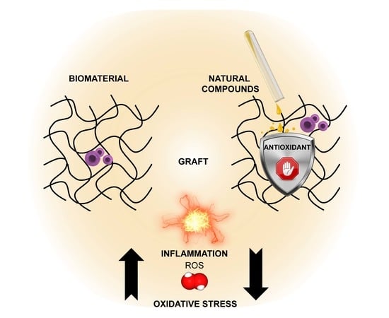
As a consequence of injury or pathology, damaged tissue is exposed to a slew of biomolecules that are produced as the body attempts to repair itself. Among these molecules, free radicals and pro-oxidant species are generated by surrounding connective tissue and by immune responses. Although oxidative stress performs many physiological roles at low concentrations [8,9], it can be harmful in activity, depending on spatiotemporal factors such as the site, the amount of reactive oxygen species (ROS) generated, and their persistence over time. Accordingly, there is an increasing body of literature suggesting that the production of ROS and subsequent cellular response to oxidative stress are important for engraftment success [10]. Graft-versus-host disease (GVHD), for example, has been correlated to oxidative stress [11]. Indeed, tissue-engineered grafts are vulnerable to rising ROS levels (Figure 1), with reperfusion injury following ischemia reported in organ and tissue engineering transplantation [12,13]. Moreover, tissue oxygenation during in vitro graft preservation [14], along with its need during surgery, can cause a production of local hyperoxia and exacerbate ROS-related organ damage [15]. Taken together, these issues underscore the potential benefit that combating ROS with antioxidants in the implantation microenvironment can bring to limit post-implantation failure and tissue regeneration.
Thus, a rationale has formed to consider the combination of anti-oxidant delivery with tissue engineering in order to improve outcomes in regeneration and restoration of organ function. Notably, an emerging trend is to reposition natural antioxidants to functionalize biomaterials with bioactive, ROS-reducing molecules (Figure 2). These biomolecules can regulate oxidation-reduction (redox)-mediated cell survival and homeostasis, whether for addressing redox balance in the in vivo microenvironment, or indeed for the more complex conditions of in vitro three-dimensional (3D) cell culture prior to surgical implantation. However, such functionalization is offset by the possible harm of total absence of ROS, which can stimulate initial healing processes. Moreover, the reactivity of antioxidants in their own right can pose stability concerns from a biomaterial perspective. Overall, in order to successfully reposition natural antioxidants into tissue engineering applications, it is critical to understand the essential concepts of antioxidants and how best to align them with tissue engineering principles. Accordingly, this review aims to provide such a context on core concepts and state-of-the-art. Firstly, an overview of antioxidants is outlined, followed by a summary of recent literature capturing the combined role of natural antioxidants with biomaterials, with a view to highlight how functionalization may be a promising strategy to improve the scaffold engraftment in tissue engineering.
2. Natural Antioxidants: An Overview
Antioxidants are defined as substances that, at low concentrations, could prevent the oxidation of a substrate [16]. They can be further divided in groups on the basis of their main mechanism of action (Figure 3) or on the basis of chemical structure. Direct antioxidants are chemicals that neutralize ROS through simple, direct reaction, and exert a classical scavenging activity. Indirect antioxidants are often activators of the Kelch-like ECH-associated protein 1 (Keap1)/nuclear factor erythroid 2-related factor 2 (Nrf2)/antioxidant response element (ARE) pathway, in which they induce phase 2 enzymes and augment cellular endogenous cytoprotective defences [17]. In comparison with a direct antioxidant that must be regenerated in order to continue its activity and can evoke a pro-oxidant effect, an indirect antioxidant may not be redox active and is unlikely to evoke a pro-oxidant effect [18]. Phenolic compounds are a large group of natural antioxidants. The polyphenols constitute a heterogenous sub-group of phenolic compounds, including flavonoids [19] and non-flavonoids. Flavonoids are sub-divided in flavan-3-ols (e.g., epigallocatechin gallate (EGCG)), flavonols (e.g., quercetin (Qu)), flavanones, flavones, isoflavones, and anthocyanins [20]. The non-flavonoid phenolic compounds are the proanthocyanidins (PA), the hydroxycinnamates [21] (e.g., caffeic acid, rosmarinic acid (RA), ferulic acid, and cinnamic acid), the lignans (e.g., sesamol), the stilbenes (e.g., resveratrol), benzoic acids (e.g., ellagic acid), coumarins (e.g., osthole), and curminoids (e.g., curcumin (Cur)) [22]. Terpenoids (e.g., carotenoids, thymol) are also common dietary antioxidants, usually occurring alongside polyphenols [20]. Other bioactive phytochemicals are nitrogen-containing compounds (e.g., isothioacyanates) or sulfur-containing ones [20]. Some vitamins, like vitamin C and E, are essential nutrients as well as molecules displaying an important antioxidant effect.
Conventionally, antioxidant sources have been largely investigated in terms of their scavenging potential of oxidant species; however, in the last decades, increasing attention has been directed to demonstrate a natural antioxidant protective effect on cells and organs. Principally, the polyphenols naturally biosynthesized by plants and marine organisms are well known as antioxidant and protective compounds. Several biochemical pathways, including regulating nuclear transcription factors, have been associated especially with polyphenols to promote health benefits [20]. Indeed, natural antioxidants raise attention because of their several capacities, that is, to induce the stress-response pathway activated by the Nrf-2 transcription factor [23] or inhibit nuclear factor kappa-B (NF-kB)-mediated inflammation pathway [24] or decrease the NADPH oxidase (NOX) family effect [25].
One disadvantage of dietary antioxidants is their bioavailability and stability. In fact, antioxidants, like their opposite ROS, are highly reactive [26], making it difficult to store them without preventing accidental oxidation. In the same manner, even after the in vivo delivery, their reactivity can result in low bioavailability. Two hypotheses are proposed for reduced bioavailability. First, in the case of oral administration, some natural antioxidants are activated only after enzymatic cleavage by human microbiota. The bioavailability of antioxidants following the dietary intake depends on digestion, then on gut absorption, metabolism, and clearance. Second, their active form is not feasible to be delivered after the implantation to the specific tissue target because of its intrinsic physicochemical properties. One potential solution to explore to overcome the instability related to a single antioxidant is to consider the use of multiple antioxidants together in a complex network. Indeed, recently in vitro results have suggested that a combination of antioxidants synergizes their effect in terms of cytoprotection [27], regeneration [28] and healing [29]. Such a consideration supports a similar co-delivery strategy for medical tissue engineering purposes. To date, the effects of the antioxidants in available human clinical studies were not significant and have some limitations [30], including the lack of clinical biomarkers and monitoring of oxidative load for effectiveness of the prescribed antioxidant [30]. Although the current antioxidants appear unsuccessful in the treatment of cardiovascular diseases [31], antioxidant strategies still represent an avenue of treatment for cardiovascular disease [31]. Similar disappointing clinical outcomes were reported for disease-modifying therapies for stroke [32] or in treating chronic neurodegenerative diseases [33]. The limitations for single antioxidant treatment in chronic neurodegenerative conditions can belong to insufficient knowledge about the biological equilibrium that can maintain a proper level of oxidative activity as well as a “chemical failure” due to poor blood–brain barrier permeability [32]. Unfortunately, most trials for vitamins were done in patients with established pathology, but preventing the disease could be a more realistic expectation than abolishing symptoms [30].
3. Biomaterials: Overview of Natural Polymers
When considering the design of a biomaterial scaffold (Figure 4), naturally-derived materials that resemble native tissue architecture can offer clear advantages [34]. Decellularized extracellular matrix (ECM) understandably [34] displays the closest approximation to native tissue. Decellularized matrix can be obtained both by animals and plants and protein matrix isolated can retain cell cues and growth factors (GFs). Animal-free matrices are very useful for 3D bioprinting of grafts [35] and decellularized matrices are ideal materials for generating a bioink [35]. As animal-derived matrices have to deal with xenogenic and ethical issues [36], plant-derived matrices are a worthy option. Matrices obtainable via plant tissue decellularization [37] are extremely challenging in the tissue engineering field and scaffolding for 3D cell cultures, however, they could need smart functionalization because of some lack of biochemical mammalian cues [38]. Strategically, plant-derived scaffolds and decellularized matrices [39] are biocompatible [40], bioprintable [41], and suitable to induce human stem cell differentiation [42] like osteogenesis from induced pluripotent stem cells (iPSCs). Polysaccharides, another class of natural biomaterial, are feasible polymers to develop ideal non-immunogenic scaffolds, usually in the form of hydrogels [43]. A critical review of all the possible interactions of polysaccharide biomaterial with the immune system [44] highlighted the need for standardization protocols for the natural polysaccharides and that the components of a composites could differentially produce unknown responses. Polysaccharides [45] often have good biocompatibility and are eco-compatible with a sustainable industry [39]. Polymers are attractive as future antioxidant materials, as many of them derive from plant sources [46]. Indeed, instead of biomaterial functionalization by antioxidants, there are hydrogels made of polyphenolic antioxidants in their own right. Aloe vera (AV) is the most known species of Aloe genus. AV is both a rich source of phytochemicals and an exciting natural scaffold for tissue engineering [47]. Its clinical efficacy was demonstrated [48]; indeed, it is used in topical dermatology applications; in cosmetics; or in wound healing as an anti-inflammatory, immunomodulatory [49] and antiseptic/antimicrobial agent [50]. Similar to cellulose, the lignins from forestry biomass can complex with synthetic polymers, for example, polylactic acid (PLA), to fabricate scavenging antioxidant material by [51] printing. Natural gums, namely marine alginate (ALG) [52], or plant-derived gum like arabic gum, can provide structural compatibility to the in vivo architecture, and thus are attractive biomaterials for innovative tissue engineering strategies [53]. Therefore, polymer scaffolds can derive from naturally agricultural products and byproducts [37], implementing the panel of techniques available for in 3D in vitro culture of mammalian cells. This could be a sustainable approach towards the next decade of biomedical needs. Other popular natural biomaterials include silk and chitosan.
Silk fibroin (SF) is a structural fibrous protein obtained from the cocoon of Bombyx Mori and it is also suitable for electrospinning [54]. Indeed, SF use is abundant to design porous scaffold, especially nanofibers [55] for bone [56] and cartilage [57] tissue engineering. Chitosan (CH) is a well-known, semisynthetic biomaterial [58] derivative of chitin [59,60] that has similarities to cellulose in its structure. Chitosan is an outstanding scaffold [61] because it is sustainable [62] and exerts antimicrobial [63] and immunomodulatory properties [64]. Not only does it possess intrinsic antioxidant capacity [65], but chitosan is also a good candidate for drug delivery [66], tissue engineering [67], and pharmaceutical engineering [67], and thus open to vast functionalization.
Thus far, biomaterials designed from metals, ceramics, and recently bioglasses [68] mimic particular biomechanical features (e.g., for bone and enamel). They are usually divided into inert or bioactive. The inert materials have been a spread use in the past for prosthetic implants where a mechanical support was the primary sufficient and requested activity for the biomaterial.
4. New Opportunities for Natural Antioxidant Combination with Tissue Engineering
4.1. Oxidative Stress Reduction Ex Vivo and In Vivo
Essentially, the antioxidant potential of bioactive compounds, together with their health protective effects, could represent a promising tool in regenerative medicine. The studies showing antioxidant activities mediated by different functionalized biomaterials are listed in Table 1. Antioxidant solutions have been tested or used to slow ex vivo organ degeneration [10] before transplantation, and research is investigating alternative antioxidant buffers for the preservation of ex vivo biological material viability. In a similar manner, antioxidant supply could preserve ex vivo graft survival, such as cornea and skin graft that, in the last decades, have been used at good manufacturing practices (GMP) level for clinical application [69,70]. As all tissues share many basic mechanisms to maintain a redox balance, the modulation of oxidant species levels by antioxidants can be widely applicable. Basically, the introduction of an antioxidant in tissue replacements will cause an effect depending on released concentration and ECM interactions. Finally, it could result in scavenging activity of radicals or in endogenous defence enhancement. Endogenous antioxidant molecules may cooperate with exogenous antioxidants to decrease the chance of failure of the graft, as well as they will promote a safe fate of both exogenous and endogenous cells connected to the implanted biomaterials. More appreciably, antioxidant supply can avoid biomaterial-mediated increase of oxidative stress after implantation. In respect to tissue oxygenation, the delivery of antioxidants could avoid excessive tissue oxygen levels that may lead to ROS production and subsequent dysfunction in the transplanted graft. “Tuneable” activation of antioxidant biomaterials is highly desirable [71]. Indeed, sensing before being responsive is a need during relevant increase in oxidative stress. Such a kind of activation is ideal for clinical application of antioxidant biomaterials [72]. With the goal of reducing oxidative stress, new biomaterials can be exploitable in order to control the release of redox sensitive molecules, that is, disulphide containing biomaterials [73]. Moreover, safeguarding redox equilibrium is also critical in tissue engineering for angiogenesis [74], an essential step promoting long-term survival and engraftment. In summary, antioxidants can preserve the viability of transplantable cells before and after transplantation, as well as control the oxidative stress in the microenvironment of implanted biomaterials (Figure 5).
4.2. Local Delivery
Locally targeted therapy is a must in a clinical setting to avoid organ toxicity and side-effects. For biomaterials, their direct association with specific growth factors can involve and activate many pathways, however, leading to specific different effects on different cell types via cell receptors. In contrast, physiological concentrations of antioxidants whose delivery is restricted to host tissue adjacent to the biomaterial will possibly have a broad effect on redox homeostasis, with a mechanism broadly shared among all cells within the body. Implants and grafts are often at the boundaries among different tissues, thus the functional molecules coupled to the biomaterials should not be detrimental to the close tissues. Furthermore, the effect of the antioxidant should be active specifically towards target tissue. The development of functionalised biomaterials [75] specifically retaining different natural bioactive compounds can contribute to a new rational delivery of antioxidants, with the aim of reducing oxidative stress and their downstream risk to aggravate a disease in the human body. Antioxidants can augment the potential of such biomaterials through local delivery. They can be introduced in biomaterials together with their biochemical limitations. Antioxidant nutraceutical compounds are hydrophilic or lipophilic molecules. In addition, active compounds also showed different cellular distribution. The inclusion of the antioxidant into the biomaterial via coating or during scaffold synthesis, in respect to other delivery routes, could reduce the required administered amount. This can be helpful for antioxidants as well as pharmaceutical compounds with low systemic stability or diffusion to the surrounding anastomosis. Thanks to its polysaccharide content and its absorption capability, AV, for example, can deliver drugs as well as additional dietary micronutrients and increase their local bioavailability [76]. The physical form and the formulation of the associated biomaterial could modulate the delivery of the antioxidant as well as solve some of its application limits, for example, encapsulation of carotenoids in nanocarriers can counteract their poor bioavailability and stability [77]. Recently, nanoparticles have received increasing attention as a tool to deliver the most different bioactive agents [78]. Depending on the application, different organic and inorganic materials and their composites can be developed for nanoparticles design. Gold nanoparticles, for example, are useful deliver a large set of molecules, including natural antioxidants. The nanoparticle seems the most used form of carrier for natural compound drug delivery, however, naturally-derived hydrogels [79] could be highly suitable for the same use. Overall, an antioxidant should be coupled to the biomaterial to achieve different goals in timeline, that is, to reach the specific tissues, to be locally available at effective physiological concentration while avoiding non-specific effects (Figure 5). The studies showing antioxidant delivery control mediated by different functionalized biomaterials are listed in Table 1.
4.3. Biodegradability and Biomechanical Properties
Biomaterials for in vivo application, as injection and implant, need to exhibit critical degradation properties. Biodegradability is an essential process as new tissue forms during healing, similar to the native one and without artificial elements. On the other hand, some biomaterials require a long-lasting duration into tissues or permanent integration into them. For materials functionalized with antioxidants, controlled release of non-covalently bonded antioxidant can be achieved less or more slowly, in balance with the scaffold’s physical features and biodegradation. Regarding biomaterial mechanical integrity, protein cross-linker agents offer degradation resistance to natural scaffolding biomaterials, but chemical agents such as glutaraldehyde are usually less tolerated and toxic. Alternatively, the inclusion of antioxidants can be obtained by adsorbing, trapping them within the scaffold or by previously incorporating them in a co-formulation (e.g., copolymer, solution). According to the planned delivery of the antioxidants, a different chemistry will be selected for the inclusion. Even surface modification of biomaterial [80], especially for non-protein biomaterial, is an option to provide the tissues of antioxidants. Indeed, a coating as a source of antioxidant has the advantage that it can be associated with both artificial and nature-inspired scaffold material. Three-dimensional bioprinting has become a tremendous field of research and bioink development is a tool for challenging clinical application [81]. For 3D printing of photocrosslinkable biomaterials, some efforts are still necessary to obtain optimal biological functions and mechanical integrity [82].
Many antioxidants are useful to improve the scaffolding ability of the biomaterial, in particular, polyphenols are used as modular building blocks for the engineering of materials for biological application [83]. Their physicochemical configuration, including hydrogen bond formation, metal coordination, amphiphilic nature, and sensitivity to oxidation, makes them suitable to adhere to various substrates. Then, polyphenols can assemble in intelligent nanoparticles, films, and gels to shape the biomaterial. Polyphenols are agents used in the preparation of biomaterials and are a modulator of biomaterial function. Therefore, the antioxidant functionality of the biomaterial can be designed by selecting polyphenols according to the number and binding structure of phenol units. As a result, one main advantage of coupling biomaterials to natural antioxidants would be an increased durability of the modified scaffolds. The preservation of matrix proteins that can be degraded by inflammation and consecutive extracellular pH changes [84,85,86] is particularly important for biomaterials, which are prepared as aminoacidic/protein hydrogel or decellularization techniques. Collagen is the major fibrous extracellular matrix protein, abundant and biomechanically important in the body. The collagen fibrils are present in differentiated connective tissues like tendons and pericardium, widely present in vessel sub-endothelial structures, cartilage surfaces, bone matrix and dentin. Like collagen, elastin is a structural protein making the network of fibers in the extracellular matrix (ECM). Moreover, elastin is not stabilised by glutaraldehyde; therefore, other molecules such as some phytochemicals can similarly or even better improve the physical integrity of protein and polymeric biomaterial. Notably, a large number of polyphenols (e.g., pentagalloyl glucose) are able to enhance the stability as well as the strength of collagen or elastin fibres with different mechanisms [87]. The tannins, in particular the class of PA, are flavonoids with a great influence on biomechanical properties of both collagen and elastin. In general, several studies reported that collagen and elastin express high affinity for PA because of their intrinsic abundance of proline. The interaction via covalent or hydrogen bonds of those flavonoids improves the stability and strength of extra cellular matrix fibres, in general by increasing denaturing temperature and inhibiting proteolytic enzymes such as collagenase and elastase matrix-metalloproteases (MMP). Other natural molecules are spreading as eco-friendly cross-linking agents; genipin is an aglycone iridoid usually isolated from extracts of gardenia fruits. Genipin is a non-toxic “green” cross-linker [88,89], used to build scaffolds tested in 3D in vitro cell culture [90,91], and exhibiting anti-inflammatory [92] antibacterial [93] and antiglicative activities [94]. Metals and other inorganic materials are also used in protocols for biomaterial engineering. Some metal biomaterials including antioxidants were built for tissue engineering studies. For instance, gold nanoparticles are a tool in tissue engineering [95] to ameliorate mechanical properties of the biomaterial and to guide specific cell behaviour [96]. Resveratrol is a feasible reducing agent for the synthesis of gold nanoparticles avoiding self-agglomeration and enabling additional drug loading [97]. Epigallocatechin gallate (EGCG) was used as linking agent between collagen fibres and palladium nanoparticles [98] in order to stabilize the metal nanoparticles. The capacity of antioxidants to modify physical properties of a specific biomaterial (e.g., wettability, permeability) could also be revolutionary for development of theranostic nanomaterials [9]. In conclusion, antioxidant functionalization not only theoretically provides antioxidant effects, but also can increase the structural stability and modify the degradation profile (Figure 5). The studies showing physico-chemical property change mediated by different functionalized biomaterials are listed in Table 1.
4.4. Biocompatibility
For clinical application, a biomaterial possesses a combination of advantageous properties, including biocompatibility and non-toxic characteristics, for example, supporting cell adhesion, proliferation, specific cell differentiation, and host immune system tolerance or immunomodulation (Figure 5). In the design of a biomaterial, the diffusion of nutrients and waste must be facilitated. The efforts on topology can be performed for optimization and establishment of both biochemical signals and cell polarity. Natural materials have inherent biochemical cues [99] that better allow cell attachment and differentiation. A critical factor in the cell–biomaterial interaction is the hydrophilicity of the scaffold [100]. The biomaterial surface provides suitable physical contacts to the cells. The wettability influences not only the cell adhesion, but also successive proliferation or differentiation [100]. For this reason, biocompatibility is highly influenced by the physicochemical properties of the biomaterial (par. 4.1–3). Importantly, a specific antioxidant molecule should be selected on the basis of the level of stimulation on the cells of the specific tissue target, such as induction of proliferation or differentiation. After the selection of the antioxidant, different endpoints for toxicology assessment can be indicated by in vitro testing [101], in general, cytotoxicity, cytokine release, and oxidative stress [102], as well as embryotoxicity and epithelial barrier integrity, according to the tissue origin. Finally, assessing in vitro and in vivo biocompatibility is an essential requirement before the designed biomaterials with gained antioxidant potential would be applied in vivo, in new different tissue environments. The studies showing biocompatibility as well as cellular proliferation and differentiation mediated by different functionalized biomaterials are listed in Table 1.
4.5. Immunomodulation and Pathogen Defense
The immune system is the key orchestrator in the regenerative response, independent of the allograft or autograft properties. Unfortunately, immune reaction against the biomaterial and downstream unproductive inflammation are the main causes of failure for several regenerative strategies based on scaffold graft. It is not rare that novel biomaterials, including biopolymers, which display biocompatibility and support cell viability, are also highly immunogenic or stimulate an inflammatory reaction [44]. This inflammatory reaction, as well as the final healing, is linked with changes in redox equilibrium. Oxidative stress derives from both pre-existing inflammation condition in the host tissues and from the usual surgical wound. Particularly, the chemokines previously and continuously produced by the recruited immune cells, the temporary hypoxia, the protein adsorption to the biomaterial, derived from blood elements (e.g., platelets, complement) and the interstitial fluid consequent to the implantation, and muscle and red blood cell lysis at wound boundaries could contribute to excessive and injurious generation of ROS. Hydrogen peroxide, released by neutrophils to reduce bacterial infections [103], recruits phagocytic leukocytes, together with factors secreted by activated mast cells [104]. Besides the ROS burst, neutrophils degrade the biomaterials via production of proteolytic enzymes. The activation by ROS of M1 macrophages and their persistence in the peri-implant site together with the formation of foreign-body giant cells forwards the chronic inflammation instauration. Positively, ROS are able to modulate macrophage polarization towards M2 phenotype, as the inhibition of ROS production specifically affects the polarization to M2 and not to M1 phenotype [105].
Remarkably, natural antioxidant inclusion in a promising biomaterial can both improve biocompatibility of the scaffolds and favour a temporary immunological tolerance towards it. To allow new tissue formation and control inflammation rather than amplify the deleterious side effect induced by ROS, natural antioxidants can appropriately be associated to the scaffold to immunosuppress and regulate the inflammatory response (Figure 5) by different cell types. Natural antioxidants such as phenolic compounds have the advantages to enhance immunocompetence [103,106] and to be low toxic for human cells [107]. More importantly, multiple compounds isolated from plants and fungi received much attention in experimental in vitro research because they modulate endogenous antioxidant systems rather than simply neutralizing free radicals. Antimicrobial activity is often associated with many antioxidants. Indeed, plants and other organisms produce antimicrobial antioxidant to protect themselves from the attack of parasite or pathogens [108]. It is important to highlight that bacterial infection is a significant risk with bio-engineered tissues, so the antimicrobial activity of the antioxidant constitutes an important feature in the context of clinical engraftment. Indeed, avoiding opportunistic infection following human transplantation and wound healing is an essential point for the clinical outcome of the patient receiving a particular biomaterial, in order to restore tissue functionality. Metal biomaterials use is strictly related to an intrinsic anti-bacterial capacity. So far, silver particles were used as common agents to produce antibacterial effect within the biomaterials [78], but recently, silver biomaterials also evolved into an antioxidant carrier. In brief, naturally occurring antioxidants are expected to be included into new biomaterial production and future medical devices for clinical application in which the immune system plays a key role. The studies showing antimicrobial, immunomodulatory, and anti-inflammatory effects mediated by different functionalized biomaterials are listed in Table 1.
The introduction of the antioxidant into the biomaterial may change its biostability, biocompatibility, and the immune response by the host. In particular, the degradation of the biomaterial, the effect of the antioxidant, and the inflammation control by the functionalized biomaterial are a result of the host tissue integration. The properties should be in accordance with tissue needs.
5. Towards Therapeutic Application: Antioxidant Biomaterials under Investigation
5.1. Wound Healing
Inflammation is a necessary component of healing in skin and mucosal wound healing [229]. In healing, there is typically an initial inflammatory phase, then a proliferative phase, and finally tissue remodelling; all the variations in these phases depend on the biomaterial features, but also on inflammation status sustained by oxidative stress at the injury site. Wound healing studies dominate the range of the applications of biomaterials investigating natural antioxidants. This popularity is likely owing to the fact that many herbal compounds were traditionally utilized as wound healing agents.
Antioxidants have been incorporated into a range of synthetic biomaterial polymers. Studies with the antioxidant curcumin (Cur) typify this line of research. Polylactic acid (PLA) and hyperbranched polyglycerol (HPG) blend was used to produce nanofibers loaded with Cur that demonstrated high swelling and great in vitro biocompatibility for 3T3 fibroblast cells. The presence of hydrophilic and non-immunogenic HPG polymer was critical to complete the in vitro scratch healing in comparison with Cur-PLA scaffolds [164]. Cur incorporation in poly(ethylene glycol) (PEG)-polycaprolactone (PCL) co-polymeric fibre mats showed antioxidant efficacy in primary dermal fibroblasts in vitro [134] and an advantage in promoting wound healing in vivo was reported. Cur nanoformulation was loaded into modified-chitosan film, in order to evaluate wound healing in animal models. A faster wound reduction and better healing was observed in comparison with the unloaded film [116].
Cur has also been investigated in combination with other natural antioxidants, such as Aloe vera (AV). Confident in their endorsing antioxidant properties, pectin–gelatin matrices were associated with AV and/or Cur [186]. The cytocompatibility, evaluated using 3T3 fibroblasts, as well as in vivo wound healing results, demonstrated that the AV-matrix was better than the Cur-matrix. In particular, AV exhibited an anti-inflammatory effect. AV had a major antimicrobial activity and enhanced apoptosis in vitro in comparison with Cur. The fact that AV seemed more efficient for in vivo healing than Cur was reinforced by a study where the usage of polymethacrylic acid silver nanogel matrix was combined with AV or Cur for coating of wound dressing [230]. AV-scaffold was firstly identified [182] as a better inducer of healing, in comparison with the poly(lactic-co-glycolic acid) (PLGA) scaffold alone. In vivo, a higher water uptake was shown for lipid nanoparticles (LNP)-PLGA-AV membranes that attracted a minor number of macrophages in wounds [183]. Seeking enhanced wound healing activity, the PCL-AV scaffold was improved by the loading of tetracycline hydrochloride [147]; in particular, tensile strength, dermal fibroblasts activity, and broad antibacterial efficacy were superior than in PCL-AV-Cur scaffolds. One interesting finding is that AV can modulate collagen production during all of the phases of wound healing. This aspect was recently investigated by evaluating the effect of AV-collagen gel on cell adhesion markers [231]. Other biomaterials investigated include decellularized bone matrix as a dressing with adipose stem cell-laden AV hydrogel. Overall, during the comparison between the experimental groups, AV modulated the expression of interleukin-1 (IL-1), transforming growth factor beta (TGF-β), and basic fibroblast growth factor (bFGF), concluding that the developed composite accelerated rabbit burn wound healing [224]. By preparing a novel hydrogel from human amnion combined with the AV extract that was able in vitro to promote HFF1 fibroblasts growth and HaCaT scratch closure, in vivo AV improved rat wound recovery rates and contraction [198]. The combined action of AV and amniotic membrane demonstrated beneficial epidermis regeneration and an increased number of blood vessels. The plasticization of ALG with PEG-methyl ether methacrylate was associated with both AV and Moringa oleifera (MO) to construct a porous dressing scaffold. Just as it was built, AV was responsible for an increased water uptake and MO was responsible for specific antimicrobial activity. AV together with MO enhanced total antioxidant activity and nitric oxide scavenging capacity was achieved. In addition, the phenolic components present in AV and MO cooperatively helped CCD-1112SK skin fibroblasts to faster proliferate in comparison with the scaffold without plant extract [109]. A scaffold used in autologous skin graft was based on collagen type I and modified with microparticles of gelatine-collagen type I, in order to evaluate the introduction of AV. The results were again not significant in terms of in vivo healing; however, as confirmation of the cross-linking issue, the observed decreased biodegradability of the material was associated with decreased in vitro enzyme degradation, that is, collagenase [148].
Of course, there are several other examples of natural antioxidants that have been explored extensively in wound healing apart from Cur and AV. Hydroglycolic extract of the Calendula officinalis (CO) flower was trialled instead of AV in one preclinical study [151], but it did not demonstrate the loaded scaffold as superior grafting material, likely because the extract concentration chosen for loading increased scaffold cross-linking. HPG was used to obtain electrospun nanofibers containing bioactive preparations of CO for optimal anti-inflammatory wound-healing membranes development. The bioactive wound dressing material was suitable for the culture of CHO hamster epithelial cells, bioadhesive, and it was not irritant and re-epithelizing in different animal models [190]. Cellulose acetate and zein nanofiber membranes were loaded with sesamol, a lignan, to investigate the effect on impaired wound healing frequently occurring in patients with diabetes. The study showed how the sesamol addition accelerated wound closure and enhanced in vivo transcriptional levels of TGF-β and alpha smooth muscle actin (α-SMA). The decreased levels of IL-1, tumor necrosis factor alpha (TNF-α), and nitric oxide synthases (NOS) were only owing to nanofiber intervention [232]. The PCL nanofibers loaded with Bixin, an antioxidant carotenoid that can be extracted from Bixa orellana L., ameliorated mice diabetic wound repair. The hydrophobicity of the antioxidant decreased pre-adipocite fibroblasts adhesion and proliferation [233], excluding hypertrofic scar formation. Of note, astaxanthin (AST) was loaded in collagen gel film, displaying good water absorption and good wound contraction. The remarkable wound healing activity (re-epithelization, fibroblast proliferation, and new ECM quality) in rats was faster than the one related to collagen hydrogel incorporating gentamicin instead of AST [161]. A gelatin-based hydrogel was tested in wound healing in vivo model because of good mechanical properties, swelling ability, and antibacterial effect, which was enhanced by the controlled release of the antioxidant ostholamide. The rats receiving ostholamide released by the fibrous scaffold exhibited enhanced healing when compared with the untreated control [158]. The cumulative effect of silver and plumbagin for antibacterial activity was observed in a collagen cross-linked scaffold for wound dressing application [159]. The composite showed cross-linking efficiency and water uptake similar to glutheraldehyde treatment, while the biocompatibility with 3T6 mouse fibroblasts was confirmed. The plumbagin caged silver nanoparticles composite was superior to the native collagen for the in vivo healing and more antibacterial than the plumbagin extract alone. A mixture of hyaluronic acid (HA) conjugated with EGCG and tyramine were explored as anti-inflammatory adhesive biomaterials for potential clinical application [145]. Rheological analysis, gel degradation, biocompatibility, and inflammation in vitro and in vivo were all investigated. The EGCG amount correlated with a decrease in TNF-α expression level in lipopolysaccharides (LPS)-stimulated RAW264.7 cells. Reduction of other pro-inflammatory factors in animal model serum and antibacterial behaviour was also shown. The healing in a rat skin incision model was more efficient in comparison with commercial medical grade adhesives. The presence of collagen into SF-fenugreek nanofibers reduced inflammation and accelerated epithelialization during wound healing, in comparison with the other tested sample group [117]. Soybean-modified poliammide 6 (PA-6) nanofibrillar matrices were explored for their potential as topical dermal covering for skin regeneration [167]. Qu and immobilized silver (Ag) were utilised as coating for functionalized polydimethylsiloxane, with a polydopamine layer as an intermediate layer. The combination used as coating for cotton gauze wound healing was effective for days in terms of antioxidant activity and showed a synergistic antimicrobial activity, while showing reduced inflammation [125]. As polydopamine can coat many surfaces, this approach may serve for functionalization of natural polymers too. Films based on CH-ALG were loaded with both thymol and β-Carotene to produce new wound dressings [234], unfortunately without deciphering the biological advantage of the antioxidants. A multiresponsive catechol-Fe3+ coordination hydrogel (MICH) was designed by assembling together HA, dopamine, β-ciclodestrin, and vitamin E [142]. This biomaterial was proven to supply in vitro antioxidant activities in different chemical models for different cell compartments; then, it decreased apoptosis and increased intracellular (NIH-3T3) ROS removal. In vivo, MICH attenuated oxidative stress, suppressed inflammation (TNF-α and count of white blood cells) and lipid peroxidation, increased re-epithelization, promoted neoangiogenesis (vascular endothelial growth factor (VEGF) analysis), and restored dermal normal collagen architecture. Collectively, these data present an injectable in situ gelling antioxidant biomaterial, worthy by combining scavenging and upregulation of endogenous enzymes (superoxide dismutase (SOD), glutathione peroxidase (GSH-Px), and catalase (CAT)). Moreover, by providing an optimal hydration to the wound it potentially will prevent wound necrosis. Very recently, a cellulose acetate-gelatin mat has incorporated berberine, a natural alkaloid. Water uptake, L929 murine fibroblastic cells proliferation, and antimicrobial activity were enhanced by the antioxidant that improved wound healing in in vivo diabetic models, as well as by angiogenesis [162].
The above studies describing potential application in wound healing of functionalized biomaterials are summarized in Table 2.
5.2. Bone Tissue Engineering
Owing to people aging, there is an increasing demand for bone autografts, but the procedure is limited primarily by surgical risk and the need for donor tissue [235]. Bone tissue engineering is an alternative intervention and the search for a new biomaterial with improved osteoconductivity, osteoinductive, osteogenic, and bioactivity is extensive [236]. Again, natural, synthetic, and composite biomaterials have been explored with antioxidants. A mesh of electrospun polydioxanone (PDX) was blended with a mixture of acemannan [237]/glucomannan from AV. Multicellular responses in vitro were assessed and showed enhanced 3T3 fibroblast proliferation and L929 fibroblast migration, spreading of Eahy926 endothelial cells, activation activity of RAW 264.7 macrophages, and mineralization by SaOS2 pre-osteoblasts. In comparison with similar PDX meshes blended with poly(hydroxybutyrate-co-hydroxyvalerate) (PHPV) and/or hydroxyapatite (Hap), the AV-containing material showed a better in vivo bioperformance for skeletal regeneration in the rat model, highlighting a minor foreign body response and angiogenesis [199]. On the other hand, AV gel-blended PHBV scaffold was identified as a promising osteoinductive suitable material for bone bioengineering, thanks to feasible differentiating cultures of iPSCs on it [204]. Gelatin was also chemically enriched with EGCG, with in vitro culture of mouse stem cells interestingly expressing osteogenic properties at day 1, in comparison with uncombined presence of the scaffold and of EGCG. In particular, the controlled release was evident in the gel-cell interaction and scaffold biodegradation allowed the release during osteogenesis in vivo [178]. Similarly, a sponge-like scaffold with different cross-linking chemistry was shown to induce osteogenesis of UMR106 osteoblasts in vitro and in vivo in calvarian defect as a result of EGCG action [207]. Gallic acid, another antioxidant, was incorporated in titanium substrate in order to deliver it for potential bone contact application [238]. The possibility to functionalize hydroxyapatite with Qu by different chemical syntheses for bone regeneration was explored in a triple co-culture model comprising MG63 osteoblast-like in contact with the biomaterial, osteoclast precursors 2T-110, and HUVECs [201]. As antioxidant activity was provided to the scaffold and a positive influence on bone repair microenvironment was shown, a scaffold of Hap functionalized with anti-osteoporotic drug, alendronate, was loaded with Qu. The new scaffold was evaluated in vitro for properties modulating bone loss and preventing related diseases [202]. Interestingly, the biomaterial promoted osteoblast proliferation and differentiation and inhibited osteoclast viability in oxidative stress treated co-culture; in parallel, TNF-α level in culture medium was decreased and Qu was the most antioxidant factor. A SF/Hap scaffold incorporating Qu for bone tissue-engineered graft investigation was based on evaluation of rat bone marrow (BM)-mesenchymal/stromal stem cells (MSCs). The biomaterial antioxidant activity was not measured, but the loading of appropriate Qu concentrations (0.03 wt %) was osteoinductive both in vitro and in vivo [239]. In MC3T3-E1 mouse osteoblast precursor model, controlled loading of Cur in PCL nanofibers enhanced molecular and histochemical markers of osteogenesis compared with the neat polymer, suggesting wider benefits of this antioxidant in bone tissue engineering [175].
A gelatin-based Hap foam [146] was functionalized with soybean extract, after previous characterization in SaOS cells. Then, the soybean biomaterial was implanted and it reduced the degradation rate and increased biomechanical strength, with denser bone formation in rabbit defect compared with control foams [240]. One hypothesis is that isoflavones released in soybean, like genistin, could act as a bone influencer. Soybean-oil-based scaffolds have been utilized to mimic a 3D microenvironment, for example, for osteogenic activity [241], but they were chosen because of their suitability for biocompatible polymer production, rather than the antioxidant properties. Both Equisetum arvense hydroethanolic extract and nano-(Hap) were loaded into PLA meshes, favoured proliferation, and increasing osteogenesis of AD-MSCs in comparison with PLA with nano-Hap only [156]. Broussonetia kazinoki plant extract modified an SF-based scaffold. The developed scaffolds were evaluated in rat BM-MSCs in vitro and in two in vivo murine models of bone defects [242]. The downregulation of the messenger ribonucleic acid (mRNA) level of pro-inflammatory TNF-α and cyclooxygenase-2 (COX-2) together with the upregulation of the pro-osteogenic markers were observed. The expected antioxidant extract of Cissus quadrangularis [243,244] was investigated on CH based-scaffolds to demonstrate bone formation in different in vitro cell models capable of osteogenic activity [245].
The above studies describing potential application in bone regenerative medicine of functionalized biomaterials are summarized in Table 2.
5.3. Cardiovascular Tissue Engineering
PVA-dextran hydrogel patches releasing astaxanthin (AST) and delivering its scavenging activity were in vitro compatible in HUVEC and 3T3 fibroblast cell models; this hydrogel was also suitable for in vivo suture and subcutaneous implantation [189]. These results are the basis for future evaluation of hydrogel effect on myocardial tissue recovery. Nanofibrous PLA-PCL-SF scaffolds including AV showed improved growth and differentiation of murine cardiomyocytes [205]. A modified surface of Mg-Zn-Y-Nd alloy, coated with TA, retained great scavenging activity even after some recycling. In addition, the platelet-repellent capability and subcutaneous implantation suggested similar application for vascular stent biomaterials [128]. Cur was included to prepare capped gold loaded PLGA nanoparticles. The animal models preserved their cardiac functions; Cur improved the stability of the particles; and Cur-PLGA nanoparticles played an important role in protection from cardiac hypertrophy, toxicity, and heart failure [154]. Vitamin E can modify the interaction between its functionalization products, such as PLA-derived polymers, and the host cells. For example, it reduced osteoblastic adhesion in vitro [166] and smooth muscle cell proliferation, thus suggesting a lower risk of in-stent restenosis [246]. In addition, bacterial [247] adhesion to this polymer was limited [222]. Chitosan-loaded with liposomes of vitamin E could be an appropriate sol-to-gel injectable material for myocardial repair. The survival of cardiomyocytes and resistance to oxidative stress environment were demonstrated [196]. The functionalization of electrospun gelatin-PCL nanofibers with salvianolic acid B and magnesium L-ascorbic acid 2 phosphate enhanced the physico-chemical properties of the scaffolds mimicking cardiac extracellular matrix. Cardiomyocytes proliferation and cardiac marker expression analysis demonstrated the potential for cardiac regeneration of that biomaterial [203]. The above studies describing potential application in cardiovascular regenerative medicine of functionalized biomaterials are summarized in Table 2.
5.4. Neural Tissue Engineering and Other Tissue Application
The human nervous system is especially vulnerable to oxidant stress-related injury owing to disease or aging [248] and has a low regeneration profile [249]. Prior to tissue engineering, surgical sutures were the only possible treatment for nerve injury [249]. The regeneration and tissue engineering of peripheral nerve grafts using a conductive biomaterial would overcome such issues. However, there is the problem of attaining adequate therapeutic delivery to the central and peripheral nervous system [139], coupled with the intrinsic short half-life of traditional compounds [250]. As such, the literature about biomaterial functionalized with natural antioxidant for nervous system therapy is relatively sparse. Specific vitamins and β-carotene regulate production of melanin, an endogenous antioxidant in skin [251] that exhibits unique electrical properties [252]. Melanin introduction in aligned SF fibers promoted neuronal growth and neurogenic activity in differentiating SH-SY5Y neuronal-like model. In addition, a scavenging activity regulated by the heteropolymer was detected [123]. As said for AV, other natural antioxidants were combined in the form of a polymer source of antioxidants with the biomaterials. Lignin nanoparticles combined with PCL fibers increased water uptake and biomechanical resistance in comparison with control samples. The lignin biomaterial showed great biocompatibility for rat pheochromocytoma PC12 cell line and human adipose derived stem cells. The neuronal differentiation induction of the two cellular models demonstrated that lignin content promoted differentiation and a particular amount (15%) favoured in vivo nerve tissue regeneration [100]. PCL nanofiber scaffold with lignin was also investigated for the biocompatibility with both human BM-MSCs (as common cells used in nerve tissue engineering) and rat Schwann cells. Free radical inhibition was associated with the biomaterial. The cultured Schwann cells grew on lignin PCL electrospun fibers; they were still viable after induced oxidative stress and up-regulated myelin expression. Moreover, primary neurons were able to grow on the developed biomaterial [100]. Other studies, reported below, pointed out the possibility to apply natural antioxidants and biomaterials for new tissue engineering protocols for tissues like cartilage and endometrium (Table 2). After simply mixing oxidized HA with resveratrol solution to make a cross-linked injectable hydrogel, the new biomaterial was evaluated in chondrocyte in vitro model. Here, resveratrol showed upregulation of chondrogenesis markers in the presence of LPS, together with downregulation of interleukin 1 beta (IL-1b) and some MMP [160]. A composite of sodium ALG and AV was used as bioink hydrogel for printing of endometrial MSCs onto a PCL 3D-printed mesh [223]. Following in vitro biocompatibility, an in vivo study was conducted to evaluate its potential in the treatment of patients with pelvic organ prolapse. In fact, AV-containing hydrogel could enhance the immunomodulatory effect of the stem cells and suppressed the foreign body response to the implant. In particular, AV-ALG reduced M1 macrophage infiltration with respect to the pure hydrogel. The above studies describing potential application in neural regeneration research of functionalized biomaterials are summarized in Table 2.
6. Conclusions
New methods to specifically deliver regenerative trophic factors and to carry out immunotolerance are requested for the ultimate clinical success of implanted graft based on biomaterials. In nature, there is the availability of a plethora of antioxidants compounds that are able to maintain the redox balance. Many of these compounds have just the capacity of neutralizing pro-oxidant species, however, some of them are indirect natural antioxidants that can enhance endogenous antioxidant defences; prevent oxidative stress; and, intriguingly, support the functional survival of a bioengineered tissue. This review has collected a panel of studies about biomaterials developed and functionalized with different antioxidants derived from living organisms. Many natural antioxidants have the potential to be a source of pro-regenerative stimuli. Interestingly, the data presented in Table 2 highlighted that the majority of the studies investigating osteogenesis and bone regeneration and differentiation used polyphenols as antioxidant functionalizing agents. The majority of the analysed antioxidants showed an antimicrobial effect (Table 1) and many are antagonists of mediators of inflammation (Table 1 and Table 2). Using such natural antioxidants for functionalization of old and new biomaterials could provide a clinical advance in terms of repair, regeneration, and cellular defence, which are effective in the absence of oxidative stress (Figure 6). Antioxidants can be presented to both transplanted budding cells and pre-existing recovering cells integrated as part of the biomaterial scaffold. Innovative biomaterials can be utilized as scaffolding blocks with human stem cells in emerging bioprinting application, including drug screening and disease modelling.
A clinical challenge is to achieve transplants that integrate advanced functional biomaterials with stem cells and biological regulators (such as antimicrobial, immunomodulatory, and trophic compounds) [253]. Redox equilibrium can be a key player in modulating these regulators; namely, the antioxidants can play a central role as immunomodulators [254]. A promising assumption in post-operation could be derived from an augmented bioavailability in the human body of natural products in association with standard pharmaceutical therapy. That combined utilization of antioxidant mix is an approach to sustain oxidative balance in tissues more powerfully than a single agent performance. The concentration of antioxidant to deliver in vivo, as the most in vitro literature explains, is critical to address its beneficial role. A deeper insight and further research are needed for complex antioxidant formulas derived from some natural product comprising a lot of bioactive compounds, for avoiding the risk of unwelcome consequences. The methods for the extraction of herb antioxidants will benefit from standardization prior to the inclusion in tissue engineering material; as an alternative and opportune action, complex mixture and natural extracts should be selected and isolated to address a specific efficacy. Finally, this field of investigation will encourage the repurposing of antioxidants for human tissue repair and regeneration in prospective clinical trials, and highlights the powerful role of redox modulation in a therapeutic window.
Author Contributions
Conceptualization, P.M.; Writing—original draft, P.M.; Writing—review & editing, C.O. All authors have read and agreed to the published version of the manuscript.
Funding
This research received no external funding.
Conflicts of Interest
The authors declare no conflict of interest.
Abbreviations
| 3T3 | Swiss albino mouse fibroblast (cell line clone) |
| 3T6 | Swiss albino mouse fibroblasts (cell line clone) |
| α-SMA | alpha smooth muscle actin |
| ALG | alginate |
| AST | astaxanthin |
| AV | Aloe vera |
| ARE | antioxidant response element |
| bFGF | basic fibroblast growth factor |
| AD-MSC (s) | adipose (derived) mesenchymal/stromal stem cell(s) |
| BM-MSC (s) | bone marrow mesenchymal/stromal stem cell(s) |
| CAT | catalase |
| CH | chitosan |
| CHO | Chinese hamster ovary (cells) |
| CO | Calendula Officinalis |
| COX-2 | cyclooxygenase-2 |
| Cur | curcumin |
| Eahy926 | human umbilical vein endothelial cells (hybrid cell line) |
| EGCG | epigallocatechin gallate |
| E.O. | essential oil |
| ECM | extra cellular matrix |
| GF | growth factors |
| GMP | good manufacturing practices |
| GSH-Px | glutathione peroxidase |
| GVHD | graft versus host disease |
| HA | hyaluronic acid |
| Hap | hydroxyapatite |
| HFF1 | human foreskin fibroblast (cell line) |
| HUVEC | human umbilical vein endothelial cells |
| IL-1 | interleukin-1 |
| IL-1b | interleukin 1 beta |
| iPSC | induced pluripotent stem cells |
| Keap1 | Kelch-like ECH-associated protein 1 |
| L929 | mouse subcutaneous connective tissue fibroblast (cell line) |
| LPS | lipopolysaccharides |
| M1 | Th1-response macrophage phenotype (classically activated) |
| M2 | Th2-response macrophage phenotype (alternatively activated) |
| MC3T3-E1 | mouse preosteoblast (cell line) |
| MG63 | human osteosarcoma (cell line) |
| MICH | multiresponsive catechol-Fe3+ coordination hydrogel |
| MO | Moringa oleifera |
| MMP | matrix metalloproteinases |
| mRNA | messenger ribonucleic acid |
| MSC | mesenchymal stem cells |
| NADPH | nicotinamide adenine dinucleotide phosphate |
| NF-kB | nuclear factor kappa-B |
| NIH3T3 | mouse swiss NIH embryo |
| Nrf2 | nuclear factor erythroid 2-related factor 2 |
| NOS | nitric oxide synthases |
| NOX | NADPH oxidase |
| P(3HB-co-3HV) | poly(3-hydroxybutyrate-co-3- hydroxyvalerate) |
| PBAE | poly(β-amino ester) |
| PA | proanthocyanidins |
| PA-6 | polyammide 6 |
| PC12 | pheochromocytoma (cell line) |
| PCL | polycaprolactone |
| PDMS | polydimethylsiloxane |
| PDX | polydioxanone |
| PE | polyethylene |
| PEG | polyethylene glycol |
| PHBV | poly(3-hydroxybutyrate-co-3-hydroxyvalerate) |
| PLA | polylactic acid |
| PLGA | poly(lactic-co-glycolic acid) |
| PMAA | polymethacrylic acid |
| PS | polystyrene |
| PU | polyuretane |
| PVA | polyvinyl alcohol |
| PVP | polyvinylpyrrolidone |
| Qu | quercetin |
| RA | rosmarinic acid |
| RAW264.7 | Abelson murine leukemia virus transformed (cell line) |
| ROS | reactive oxygen species |
| SaOS2 | human osteosarcoma (cell line) |
| SOD | superoxide dismutase |
| SF | silk fibroin |
| TA | tannic acid |
| TGF-β | transforming growth factor beta |
| TNF-α | tumor necrosis factor alpha |
| SHSY-5Y | human neuroblastoma (cell line) |
| UMR106 | rat osteosarcoma (cell line) |
| VEGF | vascular endothelial growth factor |
References
- Almouemen, N.; Kelly, H.M.; O’Leary, C. Tissue Engineering: Understanding the Role of Biomaterials and Biophysical Forces on Cell Functionality Through Computational and Structural Biotechnology Analytical Methods. Comput. Struct. Biotechnol. J. 2019, 17, 591–598. [Google Scholar] [CrossRef] [PubMed]
- Roche, C.D.; Brereton, R.J.L.; Ashton, A.W.; Jackson, C.; Gentile, C. Current challenges in three-dimensional bioprinting heart tissues for cardiac surgery. Eur. J. CardioThoracic Surg. 2020, 58, 500–510. [Google Scholar] [CrossRef] [PubMed]
- Fujita, B.; Zimmermann, W.-H. Myocardial Tissue Engineering for Regenerative Applications. Curr. Cardiol. Rep. 2017, 19, 78. [Google Scholar] [CrossRef] [PubMed]
- Perez, J.R.; Kouroupis, D.; Li, D.J.; Best, T.M.; Kaplan, L.; Correa, D. Tissue Engineering and Cell-Based Therapies for Fractures and Bone Defects. Front. Bioeng. Biotechnol. 2018, 6, 105. [Google Scholar] [CrossRef] [Green Version]
- Perez-Basterrechea, M.; Esteban, M.M.; Vega, J.A.; Obaya, A.J. Tissue-engineering approaches in pancreatic islet transplantation. Biotechnol. Bioeng. 2018, 115, 3009–3029. [Google Scholar] [CrossRef]
- Salg, G.A.; Giese, N.A.; Schenk, M.; Hüttner, F.J.; Felix, K.; Probst, P.; Diener, M.K.; Hackert, T.; Kenngott, H.G. The emerging field of pancreatic tissue engineering: A systematic review and evidence map of scaffold materials and scaffolding techniques for insulin-secreting cells. J. Tissue Eng. 2019, 10, 204173141988470. [Google Scholar] [CrossRef]
- Shakiba, N.; Zandstra, P.W. Engineering cell fitness: Lessons for regenerative medicine. Curr. Opin. Biotechnol. 2017, 47, 7–15. [Google Scholar] [CrossRef]
- Marrazzo, P.; Angeloni, C.; Freschi, M.; Lorenzini, A.; Prata, C.; Maraldi, T.; Hrelia, S. Combination of epigallocatechin gallate and sulforaphane counteracts in vitro oxidative stress and delays stemness loss of amniotic fluid stem cells. Oxid. Med. Cell. Longev. 2018, 2018. [Google Scholar] [CrossRef] [Green Version]
- Kim, K.S.; Lee, D.; Song, C.G.; Kang, P.M. Reactive oxygen species-activated nanomaterials as theranostic agents. Nanomedicine 2015, 10, 2709–2723. [Google Scholar] [CrossRef]
- Shi, S.; Xue, F. Current Antioxidant Treatments in Organ Transplantation. Oxid. Med. Cell. Longev. 2016, 2016, 1–9. [Google Scholar] [CrossRef] [Green Version]
- Yuan, L.; Shen, J. Hydrogen, a potential safeguard for graft-versus-host disease and graft ischemia-reperfusion injury? Clinics 2016, 71, 544–549. [Google Scholar] [CrossRef]
- Cannistrà, M.; Ruggiero, M.; Zullo, A.; Gallelli, G.; Serafini, S.; Maria, M.; Naso, A.; Grande, R.; Serra, R.; Nardo, B. Hepatic ischemia reperfusion injury: A systematic review of literature and the role of current drugs and biomarkers. Int. J. Surg. 2016, 33, S57–S70. [Google Scholar] [CrossRef]
- Nieuwenhuijs-Moeke, G.J.; Pischke, S.E.; Berger, S.P.; Sanders, J.S.F.; Pol, R.A.; Struys, M.M.R.F.; Ploeg, R.J.; Leuvenink, H.G.D. Ischemia and Reperfusion Injury in Kidney Transplantation: Relevant Mechanisms in Injury and Repair. J. Clin. Med. 2020, 9, 253. [Google Scholar] [CrossRef] [PubMed] [Green Version]
- Battula, N.R.; Andreoni, K.A. Oxygenated Preservation Solutions for Organ Preservation. Transplantation 2019, 103, 233–234. [Google Scholar] [CrossRef] [PubMed]
- Martin, D.S.; Grocott, M.P.W. Oxygen therapy and anaesthesia: Too much of a good thing? Anaesthesia 2015, 70, 522–527. [Google Scholar] [CrossRef] [PubMed]
- Sarangarajan, R.; Meera, S.; Rukkumani, R.; Sankar, P.; Anuradha, G. Antioxidants: Friend or foe? Asian Pac. J. Trop. Med. 2017, 10, 1111–1116. [Google Scholar] [CrossRef]
- Jin, L.; Ren, K.; Xu, Q.; Hong, T.; Wu, S.; Zhang, Y.; Wang, Z. Multifunctional carbon dots for live cell staining and tissue engineering applications. Polym. Compos. 2018, 39, 73–80. [Google Scholar] [CrossRef]
- Dinkova-Kostova, A.T.; Talalay, P. Direct and indirect antioxidant properties of inducers of cytoprotective proteins. Mol. Nutr. Food Res. 2008. [Google Scholar] [CrossRef]
- Malaguti, M.; Angeloni, C.; Hrelia, S. Polyphenols in Exercise Performance and Prevention of Exercise-Induced Muscle Damage. Oxid. Med. Cell. Longev. 2013, 2013, 1–9. [Google Scholar] [CrossRef] [Green Version]
- Fraga, C.G.; Croft, K.D.; Kennedy, D.O.; Tomás-Barberán, F.A. The effects of polyphenols and other bioactives on human health. Food Funct. 2019, 10, 514–528. [Google Scholar] [CrossRef] [Green Version]
- Shahidi, F.; Chandrasekara, A. Hydroxycinnamates and their in vitro and in vivo antioxidant activities. Phytochem. Rev. 2010, 9, 147–170. [Google Scholar] [CrossRef]
- Kelsey, N.A.; Wilkins, H.M.; Linseman, D.A. Nutraceutical antioxidants as novel neuroprotective agents. Molecules 2010, 15, 7792–7814. [Google Scholar] [CrossRef] [PubMed] [Green Version]
- Kumar, H.; Kim, I.-S.; More, S.V.; Kim, B.-W.; Choi, D.-K. Natural product-derived pharmacological modulators of Nrf2/ARE pathway for chronic diseases. Nat. Prod. Rep. 2014, 31, 109–139. [Google Scholar] [CrossRef] [PubMed]
- Nam, N.-H. Naturally Occurring NF-κB Inhibitors. Mini-Reviews Med. Chem. 2006, 6, 945–951. [Google Scholar] [CrossRef]
- Maraldi, T. Natural compounds as modulators of NADPH oxidases. Oxid. Med. Cell. Longev. 2013, 2013, 271602. [Google Scholar] [CrossRef] [PubMed] [Green Version]
- Goszcz, K.; Deakin, S.J.; Duthie, G.G.; Stewart, D.; Leslie, S.J.; Megson, I.L. Antioxidants in Cardiovascular Therapy: Panacea or False Hope? Front. Cardiovasc. Med. 2015, 2, 29. [Google Scholar] [CrossRef]
- Marrazzo, P.; Angeloni, C.; Hrelia, S. Combined Treatment with Three Natural Antioxidants Enhances Neuroprotection in a SH-SY5Y 3D Culture Model. Antioxidants 2019, 8, 420. [Google Scholar] [CrossRef] [Green Version]
- Shafi, S.; Ansari, H.R.; Bahitham, W.; Aouabdi, S. The Impact of Natural Antioxidants on the Regenerative Potential of Vascular Cells. Front. Cardiovasc. Med. 2019, 6, 28. [Google Scholar] [CrossRef] [Green Version]
- Sen, C.K.; Khanna, S.; Gordillo, G.; Bagchi, D.; Bagchi, M.; Roy, S. Oxygen, Oxidants, and Antioxidants in Wound Healing. Ann. N. Y. Acad. Sci. 2002, 957, 239–249. [Google Scholar] [CrossRef]
- Palacio, C.; Mooradian, A.D. Clinical trials and antioxidant outcomes. In Oxidative Stress and Antioxidant Protection; John Wiley & Sons, Inc.: Hoboken, NJ, USA, 2016; pp. 493–506. [Google Scholar]
- Xu, T.; Ding, W.; Ji, X.; Ao, X.; Liu, Y.; Yu, W.; Wang, J. Oxidative Stress in Cell Death and Cardiovascular Diseases. Oxid. Med. Cell. Longev. 2019. [Google Scholar] [CrossRef] [Green Version]
- Ohlow, M.J.; Sohre, S.; Granold, M.; Schreckenberger, M.; Moosmann, B. Why Have Clinical Trials of Antioxidants to Prevent Neurodegeneration Failed?—A Cellular Investigation of Novel Phenothiazine-Type Antioxidants Reveals Competing Objectives for Pharmaceutical Neuroprotection. Pharm. Res. 2017, 34, 378–393. [Google Scholar] [CrossRef] [PubMed]
- Persson, T.; Popescu, B.O.; Cedazo-Minguez, A. Oxidative Stress in Alzheimer’s Disease: Why Did Antioxidant Therapy Fail? Oxid. Med. Cell. Longev. 2014, 2014, 427318. [Google Scholar] [CrossRef] [PubMed] [Green Version]
- Hussey, G.S.; Dziki, J.L.; Badylak, S.F. Extracellular matrix-based materials for regenerative medicine. Nat. Rev. Mater. 2018, 3, 159–173. [Google Scholar] [CrossRef]
- Kabirian, F.; Mozafari, M. Decellularized ECM-derived bioinks: Prospects for the future. Methods 2020, 171, 108–118. [Google Scholar] [CrossRef]
- Rieder, E.; Steinacher-Nigisch, A.; Weigel, G. Human immune-cell response towards diverse xenogeneic and allogeneic decellularized biomaterials. Int. J. Surg. 2016, 36, 347–351. [Google Scholar] [CrossRef] [PubMed]
- Modulevsky, D.J.; Lefebvre, C.; Haase, K.; Al-Rekabi, Z.; Pelling, A.E. Apple Derived Cellulose Scaffolds for 3D Mammalian Cell Culture. PLoS ONE 2014, 9, e97835. [Google Scholar] [CrossRef] [PubMed] [Green Version]
- Campuzano, S.; Pelling, A.E. Scaffolds for 3D Cell Culture and Cellular Agriculture Applications Derived From Non-animal Sources. Front. Sustain. Food Syst. 2019, 3. [Google Scholar] [CrossRef] [Green Version]
- Iravani, S.; Varma, R.S. Plants and plant-based polymers as scaffolds for tissue engineering. Green Chem. 2019, 21, 4839–4867. [Google Scholar] [CrossRef]
- Phan, N.V.; Wright, T.; Rahman, M.M.; Xu, J.; Coburn, J.M. In Vitro Biocompatibility of Decellularized Cultured Plant Cell-Derived Matrices. ACS Biomater. Sci. Eng. 2020, 6, 822–832. [Google Scholar] [CrossRef]
- Lapomarda, A.; De Acutis, A.; Chiesa, I.; Fortunato, G.M.; Montemurro, F.; De Maria, C.; Mattioli Belmonte, M.; Gottardi, R.; Vozzi, G. Pectin-GPTMS-Based Biomaterial: Toward a Sustainable Bioprinting of 3D scaffolds for Tissue Engineering Application. Biomacromolecules 2020, 21, 319–327. [Google Scholar] [CrossRef]
- Lee, J.; Jung, H.; Park, N.; Park, S.H.; Ju, J.H. Induced Osteogenesis in Plants Decellularized Scaffolds. Sci. Rep. 2019, 9, 20194. [Google Scholar] [CrossRef] [PubMed]
- Cassimjee, H.; Kumar, P.; Choonara, Y.E.; Pillay, V. Proteosaccharide combinations for tissue engineering applications. Carbohydr. Polym. 2020, 235, 115932. [Google Scholar] [CrossRef] [PubMed]
- Torres, F.G.; Troncoso, O.P.; Pisani, A.; Gatto, F.; Bardi, G. Natural Polysaccharide Nanomaterials: An Overview of Their Immunological Properties. Int. J. Mol. Sci. 2019, 20, 5092. [Google Scholar] [CrossRef] [Green Version]
- Bharadwaz, A.; Jayasuriya, A.C. Recent trends in the application of widely used natural and synthetic polymer nanocomposites in bone tissue regeneration. Mater. Sci. Eng. C 2020, 110, 110698. [Google Scholar] [CrossRef] [PubMed]
- Zafar, M.S.; Quarta, A.; Marradi, M.; Ragusa, A. Recent Developments in the Reduction of Oxidative Stress through Antioxidant Polymeric Formulations. Pharmaceutics 2019, 11, 505. [Google Scholar] [CrossRef] [PubMed] [Green Version]
- Rahman, S.; Carter, P.; Bhattarai, N. Aloe Vera for Tissue Engineering Applications. J. Funct. Biomater. 2017, 8, 6. [Google Scholar] [CrossRef] [Green Version]
- Agnes Mary, S.; Giri Dev, V.R. Electrospun herbal nanofibrous wound dressings for skin tissue engineering. J. Text. Inst. 2015, 106, 886–895. [Google Scholar] [CrossRef]
- López, Z.; Femenia, A.; Núñez-Jinez, G.; Salazar Zúñiga, M.N.; Cano, M.E.; Espino, T.; Knauth, P. In Vitro Immunomodulatory Effect of Food Supplement from Aloe vera. Evid. Based Complement. Altern. Med. 2019. [Google Scholar] [CrossRef] [Green Version]
- Atef, N.M.; Shanab, S.M.; Negm, S.I.; Abbas, Y.A. Evaluation of antimicrobial activity of some plant extracts against antibiotic susceptible and resistant bacterial strains causing wound infection. Bull. Natl. Res. Cent. 2019, 43, 144. [Google Scholar] [CrossRef] [Green Version]
- Ji, A.; Zhang, S.; Bhagia, S.; Yoo, C.G.; Ragauskas, A.J. 3D printing of biomass-derived composites: Application and characterization approaches. RSC Adv. 2020, 10, 21698–21723. [Google Scholar] [CrossRef]
- Rastogi, P.; Kandasubramanian, B. Review of alginate-based hydrogel bioprinting for application in tissue engineering. Biofabrication 2019, 11, 042001. [Google Scholar] [CrossRef]
- Mohammadinejad, R.; Kumar, A.; Ranjbar-Mohammadi, M.; Ashrafizadeh, M.; Han, S.S.; Khang, G.; Roveimiab, Z. Recent Advances in Natural Gum-Based Biomaterials for Tissue Engineering and Regenerative Medicine: A Review. Polymers 2020, 12, 176. [Google Scholar] [CrossRef] [PubMed] [Green Version]
- Vilchez, A.; Acevedo, F.; Cea, M.; Seeger, M.; Navia, R. Applications of Electrospun Nanofibers with Antioxidant Properties: A Review. Nanomaterials 2020, 10, 175. [Google Scholar] [CrossRef] [Green Version]
- Farokhi, M.; Mottaghitalab, F.; Reis, R.L.; Ramakrishna, S.; Kundu, S.C. Functionalized silk fibroin nanofibers as drug carriers: Advantages and challenges. J. Control. Release 2020, 321, 324–347. [Google Scholar] [CrossRef]
- Melke, J.; Midha, S.; Ghosh, S.; Ito, K.; Hofmann, S. Silk fibroin as biomaterial for bone tissue engineering. Acta Biomater. 2016, 31, 1–16. [Google Scholar] [CrossRef] [Green Version]
- Cheng, G.; Davoudi, Z.; Xing, X.; Yu, X.; Cheng, X.; Li, Z.; Deng, H.; Wang, Q. Advanced Silk Fibroin Biomaterials for Cartilage Regeneration. ACS Biomater. Sci. Eng. 2018, 4, 2704–2715. [Google Scholar] [CrossRef]
- Jiang, T.; Deng, M.; James, R.; Nair, L.S.; Laurencin, C.T. Micro- and nanofabrication of chitosan structures for regenerative engineering. Acta Biomater. 2014, 10, 1632–1645. [Google Scholar] [CrossRef]
- Riaz Rajoka, M.S.; Zhao, L.; Mehwish, H.M.; Wu, Y.; Mahmood, S. Chitosan and its derivatives: Synthesis, biotechnological applications, and future challenges. Appl. Microbiol. Biotechnol. 2019, 103, 1557–1571. [Google Scholar] [CrossRef]
- Francesko, A.; Tzanov, T. Chitin, Chitosan and Derivatives for Wound Healing and Tissue Engineering. Adv. Biochem. Eng. Biotechnol. 2010, 125, 1–27. [Google Scholar]
- Sultankulov, B.; Berillo, D.; Sultankulova, K.; Tokay, T.; Saparov, A. Progress in the Development of Chitosan-Based Biomaterials for Tissue Engineering and Regenerative Medicine. Biomolecules 2019, 9, 470. [Google Scholar] [CrossRef] [Green Version]
- Bakshi, P.S.; Selvakumar, D.; Kadirvelu, K.; Kumar, N.S. Chitosan as an environment friendly biomaterial—A review on recent modifications and applications. Int. J. Biol. Macromol. 2020, 150, 1072–1083. [Google Scholar] [CrossRef]
- Kim, C.-H.; Park, S.J.; Yang, D.H.; Chun, H.J. Chitosan for Tissue Engineering. Adv. Exp. Med. Biol. 2018, 1077, 475–485. [Google Scholar]
- Moran, H.B.T.; Turley, J.L.; Andersson, M.; Lavelle, E.C. Immunomodulatory properties of chitosan polymers. Biomaterials 2018, 184, 1–9. [Google Scholar] [CrossRef]
- Ivanova, D.G.; Yaneva, Z.L. Antioxidant Properties and Redox-Modulating Activity of Chitosan and Its Derivatives: Biomaterials with Application in Cancer Therapy. Biores. Open Access 2020, 9, 64–72. [Google Scholar] [CrossRef] [Green Version]
- Kravanja, G.; Primožič, M.; Knez, Ž.; Leitgeb, M. Chitosan-Based (Nano)Materials for Novel Biomedical Applications. Molecules 2019, 24, 1960. [Google Scholar] [CrossRef] [Green Version]
- Mohebbi, S.; Nezhad, M.N.; Zarrintaj, P.; Jafari, S.H.; Gholizadeh, S.S.; Saeb, M.R.; Mozafari, M. Chitosan in Biomedical Engineering: A Critical Review. Curr. Stem Cell Res. Ther. 2019, 14, 93–116. [Google Scholar] [CrossRef] [PubMed]
- Saini, M.; Singh, Y.; Arora, P.; Arora, V.; Jain, K.; Singh, S.M.; Arora, A.P.; Implant, J.K. Implant biomaterials: A comprehensive review. A Compr. Rev. World J. Clin. Cases 2015, 3, 52–57. [Google Scholar] [CrossRef]
- Hirsch, T.; Rothoeft, T.; Teig, N.; Bauer, J.W.; Pellegrini, G.; De Rosa, L.; Scaglione, D.; Reichelt, J.; Klausegger, A.; Kneisz, D.; et al. Regeneration of the entire human epidermis using transgenic stem cells. Nature 2017, 551, 327–332. [Google Scholar] [CrossRef]
- Rama, P.; Matuska, S.; Paganoni, G.; Spinelli, A.; De Luca, M.; Pellegrini, G. Limbal Stem-Cell Therapy and Long-Term Corneal Regeneration. N. Engl. J. Med. 2010, 363, 147–155. [Google Scholar] [CrossRef] [Green Version]
- Spicer, C.D.; Pashuck, E.T.; Stevens, M.M. Achieving Controlled Biomolecule—Biomaterial Conjugation. Chem. Rev. 2018, 118, 7702–7743. [Google Scholar] [CrossRef]
- Yao, Y.; Zhang, H.; Wang, Z.; Ding, J.; Wang, S.; Huang, B.; Ke, S.; Gao, C. Reactive oxygen species (ROS)-responsive biomaterials mediate tissue microenvironments and tissue regeneration. J. Mater. Chem. B 2019, 7, 5019–5037. [Google Scholar] [CrossRef] [PubMed]
- Lakes, A.L.; Jordan, C.T.; Gupta, P.; Puleo, D.A.; Hilt, J.Z.; Dziubla, T.D. Reducible disulfide poly(beta-amino ester) hydrogels for antioxidant delivery. Acta Biomater. 2018, 68, 178–189. [Google Scholar] [CrossRef] [PubMed]
- Sthijns, M.M.J.P.E.; van Blitterswijk, C.A.; LaPointe, V.L.S. Redox regulation in regenerative medicine and tissue engineering: The paradox of oxygen. J. Tissue Eng. Regen. Med. 2018, 12, 2013–2020. [Google Scholar] [CrossRef] [PubMed] [Green Version]
- Levato, R.; Jungst, T.; Scheuring, R.G.; Blunk, T.; Groll, J.; Malda, J. From Shape to Function: The Next Step in Bioprinting. Adv. Mater. 2020, 32, 1906423. [Google Scholar] [CrossRef]
- Nejatzadeh-Barandozi, F. Antibacterial activities and antioxidant capacity of Aloe vera. Org. Med. Chem. Lett. 2013, 3, 5. [Google Scholar] [CrossRef] [Green Version]
- Rehman, A.; Tong, Q.; Jafari, S.M.; Assadpour, E.; Shehzad, Q.; Aadil, R.M.; Iqbal, M.W.; Rashed, M.M.A.; Mushtaq, B.S.; Ashraf, W. Carotenoid-loaded nanocarriers: A comprehensive review. Adv. Colloid Interface Sci. 2020, 275, 102048. [Google Scholar] [CrossRef]
- Fathi-Achachelouei, M.; Knopf-Marques, H.; da Ribeiro Silva, C.E.; Barthès, J.; Bat, E.; Tezcaner, A.; Vrana, N.E. Use of Nanoparticles in Tissue Engineering and Regenerative Medicine. Front. Bioeng. Biotechnol. 2019, 7, 113. [Google Scholar] [CrossRef] [Green Version]
- Catoira, M.C.; Fusaro, L.; Di Francesco, D.; Ramella, M.; Boccafoschi, F. Overview of natural hydrogels for regenerative medicine applications. J. Mater. Sci. Mater. Med. 2019, 30, 115. [Google Scholar] [CrossRef] [Green Version]
- Agarwal, R.; García, A.J. Surface Modification of Biomaterials. In Principles of Regenerative Medicine; Elsevier: Amsterdam, The Netherlands, 2019; pp. 651–660. [Google Scholar] [CrossRef]
- Gillispie, G.J.; Park, J.; Copus, J.S.; Asari, A.K.P.R.; Yoo, J.J.; Atala, A.; Lee, S.J. Three-Dimensional Tissue and Organ Printing in Regenerative Medicine. In Principles of Regenerative Medicine; Elsevier: Amsterdam, The Netherlands, 2019; pp. 831–852. [Google Scholar] [CrossRef]
- Choi, G.; Cha, H.J. Recent advances in the development of nature-derived photocrosslinkable biomaterials for 3D printing in tissue engineering. Biomater. Res. 2019, 23, 18. [Google Scholar] [CrossRef] [Green Version]
- Guo, J.; Suma, T.; Richardson, J.J.; Ejima, H. Modular Assembly of Biomaterials Using Polyphenols as Building Blocks. ACS Biomater. Sci. Eng. 2019, 5, 5578–5596. [Google Scholar] [CrossRef]
- Riemann, A.; Wußling, H.; Loppnow, H.; Fu, H.; Reime, S.; Thews, O. Acidosis differently modulates the inflammatory program in monocytes and macrophages. Biochim. Biophys. Acta Mol. Basis Dis. 2016, 1862, 72–81. [Google Scholar] [CrossRef] [Green Version]
- Rajamäki, K.; Nordström, T.; Nurmi, K.; Åkerman, K.E.O.; Kovanen, P.T.; Öörni, K.; Eklund, K.K. Extracellular acidosis is a novel danger signal alerting innate immunity via the NLRP3 inflammasome. J. Biol. Chem. 2013, 288, 13410–13419. [Google Scholar] [CrossRef] [PubMed] [Green Version]
- Kellum, J.A.; Song, M.; Li, J. Science review: Extracellular acidosis and the immune response: Clinical and physiologic implications. Crit. Care 2004, 8, 331–336. [Google Scholar] [CrossRef] [PubMed] [Green Version]
- Simionescu, D.; Casco, M.; Turner, J.; Rierson, N.; Yue, J.; Ning, K. Chemical stabilization of the extracellular matrix attenuates growth of experimentally induced abdominal aorta aneurysms in a large animal model. JVS Vasc. Sci. 2020, 1, 69–80. [Google Scholar] [CrossRef]
- Iahnke, A.O.E.S.; Stoll, L.; Bellé, A.S.; Hertz, P.F.; de Rios, A.O.; Rahier, H.; Flôres, S.H. Gelatin capsule residue-based films crosslinked with the natural agent genipin. Packag. Technol. Sci. 2020, 33, 15–26. [Google Scholar] [CrossRef]
- Tacias-Pascacio, V.G.; García-Parra, E.; Vela-Gutiérrez, G.; Virgen-Ortiz, J.J.; Berenguer-Murcia, Á.; Alcántara, A.R.; Fernandez-Lafuente, R. Genipin as An Emergent Tool in the Design of Biocatalysts: Mechanism of Reaction and Applications. Catalysts 2019, 9, 1035. [Google Scholar] [CrossRef] [Green Version]
- Focaroli, S.; Teti, G.; Salvatore, V.; Durante, S.; Belmonte, M.M.; Giardino, R.; Mazzotti, A.; Bigi, A.; Falconi, M. Chondrogenic differentiation of human adipose mesenchimal stem cells: Influence of a biomimetic gelatin genipin crosslinked porous scaffold. Microsc. Res. Tech. 2014, 77, 928–934. [Google Scholar] [CrossRef]
- Gattazzo, F.; De Maria, C.; Rimessi, A.; Donà, S.; Braghetta, P.; Pinton, P.; Vozzi, G.; Bonaldo, P. Gelatin-genipin-based biomaterials for skeletal muscle tissue engineering. J. Biomed. Mater. Res. Part B Appl. Biomater. 2018, 106, 2763–2777. [Google Scholar] [CrossRef]
- Koo, H.-J.; Song, Y.S.; Kim, H.-J.; Lee, Y.-H.; Hong, S.-M.; Kim, S.-J.; Kim, B.-C.; Jin, C.; Lim, C.-J.; Park, E.-H. Antiinflammatory effects of genipin, an active principle of gardenia. Eur. J. Pharmacol. 2004, 495, 201–208. [Google Scholar] [CrossRef]
- Kucharska, A.; Sokół-Łętowska, A.; Oszmiański, J.; Piórecki, N.; Fecka, I. Iridoids, Phenolic Compounds and Antioxidant Activity of Edible Honeysuckle Berries (Lonicera caerulea var. kamtschatica Sevast.). Molecules 2017, 22, 405. [Google Scholar] [CrossRef] [Green Version]
- West, B.J.; Uwaya, A.; Isami, F.; Deng, S.; Nakajima, S.; Jensen, C.J. Antiglycation Activity of Iridoids and Their Food Sources. Int. J. Food Sci. 2014, 2014, 276950. [Google Scholar] [CrossRef] [PubMed] [Green Version]
- Borzenkov, M.; Chirico, G.; Collini, M.; Pallavicini, P. Gold Nanoparticles for Tissue Engineering. In Environmental Nanotechnology. Environmental Chemistry for A Sustainable World; Dasgupta, N., Ranjan, S., Lichtfouse, E., Eds.; Springer: Cham, Switzerland, 2018; Volume 14, pp. 343–390. [Google Scholar]
- Vial, S.; Reis, R.L.; Oliveira, J.M. Recent advances using gold nanoparticles as a promising multimodal tool for tissue engineering and regenerative medicine. Curr. Opin. Solid State Mater. Sci. 2017, 21, 92–112. [Google Scholar] [CrossRef] [Green Version]
- Mohanty, R.K.; Thennarasu, S.; Mandal, A.B. Resveratrol stabilized gold nanoparticles enable surface loading of doxorubicin and anticancer activity. Colloids Surf. B Biointerfaces 2014, 114, 138–143. [Google Scholar] [CrossRef] [PubMed]
- Wu, H.; Wu, C.; He, Q.; Liao, X.; Shi, B. Collagen fiber with surface-grafted polyphenol as a novel support for Pd(0) nanoparticles: Synthesis, characterization and catalytic application. Mater. Sci. Eng. C 2010, 30, 770–776. [Google Scholar] [CrossRef]
- Navarro, J.; Calderon, G.A.; Miller, J.S.; Fisher, J.P. Bioinks for Three-Dimensional Printing in Regenerative Medicine. In Principles of Regenerative Medicine; Elsevier: Amsterdam, The Netherlands, 2019; pp. 805–830. [Google Scholar] [CrossRef]
- Amini, S.; Saudi, A.; Amirpour, N.; Jahromi, M.; Najafabadi, S.S.; Kazemi, M.; Rafienia, M.; Salehi, H. Application of electrospun polycaprolactone fibers embedding lignin nanoparticle for peripheral nerve regeneration: In vitro and in vivo study. Int. J. Biol. Macromol. 2020, 159, 154–173. [Google Scholar] [CrossRef]
- Farcal, L.; Torres Andón, F.; Di Cristo, L.; Rotoli, B.M.; Bussolati, O.; Bergamaschi, E.; Mech, A.; Hartmann, N.B.; Rasmussen, K.; Riego-Sintes, J.; et al. Comprehensive In Vitro Toxicity Testing of a Panel of Representative Oxide Nanomaterials: First Steps towards an Intelligent Testing Strategy. PLoS ONE 2015, 10, e0127174. [Google Scholar] [CrossRef]
- Mitov, M.I.; Patil, V.S.; Alstott, M.C.; Dziubla, T.; Butterfield, D.A. In Vitro Cellular Assays for Oxidative Stress and Biomaterial Response. In Oxidative Stress and Biomaterials; Elsevier: Amsterdam, The Netherlands, 2016; pp. 145–186. [Google Scholar] [CrossRef]
- Puertollano, M.A.; Puertollano, E.; de Cienfuegos, G.A.; de Pablo, M.A. Dietary Antioxidants: Immunity and Host Defense. Curr. Top. Med. Chem. 2011, 11, 1752–1766. [Google Scholar] [CrossRef]
- Anderson, J.M.; Rodriguez, A.; Chang, D.T. Foreign body reaction to biomaterials. Semin. Immunol. 2008, 20, 86–100. [Google Scholar] [CrossRef] [Green Version]
- Zhang, Y.; Choksi, S.; Chen, K.; Pobezinskaya, Y.; Linnoila, I.; Liu, Z.-G. ROS play a critical role in the differentiation of alternatively activated macrophages and the occurrence of tumor-associated macrophages. Cell Res. 2013, 23, 898–914. [Google Scholar] [CrossRef] [Green Version]
- López-Varela, S.; González-Gross, M.; Marcos, A. Functional foods and the immune system: A review. Eur. J. Clin. Nutr. 2002, 56, S29–S33. [Google Scholar] [CrossRef]
- Kovalchuk, A.; Aladedunye, F.; Rodriguez-Juarez, R.; Li, D.; Thomas, J.; Kovalchuk, O.; Przybylski, R. Novel antioxidants are not toxic to normal tissues but effectively kill cancer cells. Cancer Biol. Ther. 2013, 14, 907–915. [Google Scholar] [CrossRef] [PubMed] [Green Version]
- Pokorný, J. Are natural antioxidants better and safer than synthetic antioxidants? Eur. J. Lipid Sci. Technol. 2007, 109, 629–642. [Google Scholar] [CrossRef]
- Rubio-Elizalde, I.; Bernáldez-Sarabia, J.; Moreno-Ulloa, A.; Vilanova, C.; Juárez, P.; Licea-Navarro, A.; Castro-Ceseña, A.B. Scaffolds based on alginate-PEG methyl ether methacrylate-Moringa oleifera-Aloe vera for wound healing applications. Carbohydr. Polym. 2019, 206, 455–467. [Google Scholar] [CrossRef] [PubMed]
- Wang, Q.; Zhao, Y.; Guan, L.; Zhang, Y.; Dang, Q.; Dong, P.; Li, J.; Liang, X. Preparation of astaxanthin-loaded DNA/chitosan nanoparticles for improved cellular uptake and antioxidation capability. Food Chem. 2017, 227, 9–15. [Google Scholar] [CrossRef]
- Arriagada, F.; Günther, G.; Nos, J.; Nonell, S.; Olea-Azar, C.; Morales, J. Antioxidant Nanomaterial Based on Core–Shell Silica Nanospheres with Surface-Bound Caffeic Acid: A Promising Vehicle for Oxidation-Sensitive Drugs. Nanomaterials 2019, 9, 214. [Google Scholar] [CrossRef] [Green Version]
- Chen, F.; Shi, Z.; Neoh, K.G.; Kang, E.T. Antioxidant and antibacterial activities of eugenol and carvacrol-grafted chitosan nanoparticles. Biotechnol. Bioeng. 2009, 104, 30–39. [Google Scholar] [CrossRef] [PubMed]
- Li, F.; Jin, H.; Xiao, J.; Yin, X.; Liu, X.; Li, D.; Huang, Q. The simultaneous loading of catechin and quercetin on chitosan-based nanoparticles as effective antioxidant and antibacterial agent. Food Res. Int. 2018, 111, 351–360. [Google Scholar] [CrossRef] [PubMed]
- Catauro, M.; Barrino, F.; Poggetto, G.D.; Crescente, G.; Piccolella, S.; Pacifico, S. Chlorogenic acid entrapped in hybrid materials with high PEG content: A strategy to obtain antioxidant functionalized biomaterials? Materials 2019, 12, 148. [Google Scholar] [CrossRef] [PubMed] [Green Version]
- Xu, T.; Gao, C.; Feng, X.; Yang, Y.; Shen, X.; Tang, X. Structure, physical and antioxidant properties of chitosan-gum arabic edible films incorporated with cinnamon essential oil. Int. J. Biol. Macromol. 2019, 134, 230–236. [Google Scholar] [CrossRef]
- Li, X.; Nan, K.; Li, L.; Zhang, Z.; Chen, H. In vivo evaluation of curcumin nanoformulation loaded methoxy poly(ethylene glycol)-graft-chitosan composite film for wound healing application. Carbohydr. Polym. 2012, 88, 84–90. [Google Scholar] [CrossRef]
- Selvaraj, S.; Duraipandy, N.; Kiran, M.S.; Fathima, N.N. Anti-oxidant enriched hybrid nanofibers: Effect on mechanical stability and biocompatibility. Int. J. Biol. Macromol. 2018, 117, 209–217. [Google Scholar] [CrossRef]
- Kang, B.; Vales, T.; Cho, B.-K.; Kim, J.-K.; Kim, H.-J. Development of Gallic Acid-Modified Hydrogels Using Interpenetrating Chitosan Network and Evaluation of Their Antioxidant Activity. Molecules 2017, 22, 1976. [Google Scholar] [CrossRef] [Green Version]
- Panzella, L.; Cerruti, P.; Ambrogi, V.; Agustin-Salazar, S.; D’Errico, G.; Carfagna, C.; Goya, L.; Ramos, S.; Martín, M.A.; Napolitano, A.; et al. A Superior All-Natural Antioxidant Biomaterial from Spent Coffee Grounds for Polymer Stabilization, Cell Protection, and Food Lipid Preservation. ACS Sustain. Chem. Eng. 2016, 4, 1169–1179. [Google Scholar] [CrossRef]
- Mishra, D.; Khare, P.; Singh, D.K.; Luqman, S.; Ajaya Kumar, P.V.; Yadav, A.; Das, T.; Saikia, B.K. Retention of antibacterial and antioxidant properties of lemongrass oil loaded on cellulose nanofibre-poly ethylene glycol composite. Ind. Crops Prod. 2018, 114, 68–80. [Google Scholar] [CrossRef]
- Guamán-Balcázar, M.C.; Montes, A.; Pereyra, C.; Martínez de la Ossa, E. Production of submicron particles of the antioxidants of mango leaves/PVP by supercritical antisolvent extraction process. J. Supercrit. Fluids 2019, 143, 294–304. [Google Scholar] [CrossRef]
- Rambabu, K.; Bharath, G.; Banat, F.; Show, P.L.; Cocoletzi, H.H. Mango leaf extract incorporated chitosan antioxidant film for active food packaging. Int. J. Biol. Macromol. 2019, 126, 1234–1243. [Google Scholar] [CrossRef]
- Nune, M.; Manchineella, S.; Govindaraju, T.; Narayan, K.S. Melanin incorporated electroactive and antioxidant silk fibroin nanofibrous scaffolds for nerve tissue engineering. Mater. Sci. Eng. C 2019, 94, 17–25. [Google Scholar] [CrossRef] [PubMed]
- Massaro, M.; Riela, S.; Guernelli, S.; Parisi, F.; Lazzara, G.; Baschieri, A.; Valgimigli, L.; Amorati, R. A synergic nanoantioxidant based on covalently modified halloysite–trolox nanotubes with intra-lumen loaded quercetin. J. Mater. Chem. B 2016, 4, 2229–2241. [Google Scholar] [CrossRef] [PubMed] [Green Version]
- Tran, H.A.; Ly, K.L.; Fox, K.E.; Tran, P.A.; Nguyen, T.H. Immobilization of antimicrobial silver and antioxidant flavonoid as a coating for wound dressing materials. Int. J. Nanomed. 2019, 14, 9929–9939. [Google Scholar] [CrossRef] [PubMed] [Green Version]
- Arriagada, F.; Günther, G.; Morales, J. Nanoantioxidant–Based Silica Particles as Flavonoid Carrier for Drug Delivery Applications. Pharmaceutics 2020, 12, 302. [Google Scholar] [CrossRef] [Green Version]
- Darie-Niţă, R.N.; Vasile, C.; Stoleru, E.; Pamfil, D.; Zaharescu, T.; Tarţău, L.; Tudorachi, N.; Brebu, M.A.; Pricope, G.M.; Dumitriu, R.P.; et al. Evaluation of the rosemary extract effect on the properties of polylactic acid-based materials. Materials 2018, 11, 1825. [Google Scholar] [CrossRef] [Green Version]
- Wang, P.; Liu, J.; Luo, X.; Xiong, P.; Gao, S.; Yan, J.; Li, Y.; Cheng, Y.; Xi, T. A tannic acid-modified fluoride pre-treated Mg–Zn–Y–Nd alloy with antioxidant and platelet-repellent functionalities for vascular stent application. J. Mater. Chem. B 2019, 7, 7314–7325. [Google Scholar] [CrossRef] [PubMed]
- Lee, H.Y.; Hwang, C.H.; Kim, H.E.; Jeong, S.H. Enhancement of bio-stability and mechanical properties of hyaluronic acid hydrogels by tannic acid treatment. Carbohydr. Polym. 2018, 186, 290–298. [Google Scholar] [CrossRef] [PubMed]
- Bardania, H.; Mahmoudi, R.; Bagheri, H.; Salehpour, Z.; Fouani, M.H.; Darabian, B.; Khoramrooz, S.S.; Mousavizadeh, A.; Kowsari, M.; Moosavifard, S.E.; et al. Facile preparation of a novel biogenic silver-loaded Nanofilm with intrinsic anti-bacterial and oxidant scavenging activities for wound healing. Sci. Rep. 2020, 10, 6129. [Google Scholar] [CrossRef] [PubMed] [Green Version]
- Talón, E.; Trifkovic, K.T.; Vargas, M.; Chiralt, A.; González-Martínez, C. Release of polyphenols from starch-chitosan based films containing thyme extract. Carbohydr. Polym. 2017, 175, 122–130. [Google Scholar] [CrossRef]
- Alvarez Echazú, M.I.; Olivetti, C.E.; Anesini, C.; Perez, C.J.; Alvarez, G.S.; Desimone, M.F. Development and evaluation of thymol-chitosan hydrogels with antimicrobial-antioxidant activity for osral local delivery. Mater. Sci. Eng. C 2017, 81, 588–596. [Google Scholar] [CrossRef]
- Bhatia, S.K.; Wadhwa, P.; Hong, J.W.; Hong, Y.G.; Jeon, J.M.; Lee, E.S.; Yang, Y.H. Lipase mediated functionalization of poly(3-hydroxybutyrate-co-3-hydroxyvalerate) with ascorbic acid into an antioxidant active biomaterial. Int. J. Biol. Macromol. 2019, 123, 117–123. [Google Scholar] [CrossRef]
- Fu, S.-Z.; Meng, X.-H.; Fan, J.; Yang, L.-L.; Wen, Q.-L.; Ye, S.-J.; Lin, S.; Wang, B.-Q.; Chen, L.-L.; Wu, J.-B.; et al. Acceleration of dermal wound healing by using electrospun curcumin-loaded poly(ε-caprolactone)-poly(ethylene glycol)-poly(ε-caprolactone) fibrous mats. J. Biomed. Mater. Res. Part B Appl. Biomater. 2014, 102, 533–542. [Google Scholar] [CrossRef]
- Wu, X.; Dai, H.; Xu, C.; Liu, L.; Li, S. Citric acid modification of a polymer exhibits antioxidant and anti-inflammatory properties in stem cells and tissues. J. Biomed. Mater. Res. Part A 2019, 107, 2414–2424. [Google Scholar] [CrossRef]
- Wattamwar, P.P.; Biswal, D.; Cochran, D.B.; Lyvers, A.C.; Eitel, R.E.; Anderson, K.W.; Hilt, J.Z.; Dziubla, T.D. Synthesis and characterization of poly(antioxidant β-amino esters) for controlled release of polyphenolic antioxidants. Acta Biomater. 2012, 8, 2529–2537. [Google Scholar] [CrossRef]
- Luisa, D.P.-A.M.; Griselda, R.-M.; Valentín, M.-L.; Carmina, O.-S.; Cristina, V.-M.; JJ, M.; Maykel, G.-T.; David, Q.-G.; Roberto, S.-S.; Gerardo, L.-G. Curcumin-loaded poly-ε-caprolactone nanoparticles show antioxidant and cytoprotective effects in the presence of reactive oxygen species. J. Bioact. Compat. Polym. 2020, 35, 270–285. [Google Scholar] [CrossRef]
- Cho, Y.S.; Kim, S.K.; Je, J.Y. Chitosan gallate as potential antioxidant biomaterial. Bioorg. Med. Chem. Lett. 2011, 21, 3070–3073. [Google Scholar] [CrossRef] [PubMed]
- Wang, J.; Tian, L.; Luo, B.; Ramakrishna, S.; Kai, D.; Loh, X.J.; Yang, I.H.; Deen, G.R.; Mo, X. Engineering PCL/lignin nanofibers as an antioxidant scaffold for the growth of neuron and Schwann cell. Colloids Surfaces B Biointerfaces 2018, 169, 356–365. [Google Scholar] [CrossRef] [PubMed]
- Hajebi, S.; Tabrizi, M.H.; Moghaddam, M.N.; Shahraki, F.; Yadamani, S. Rapeseed flower pollen bio-green synthesized silver nanoparticles: A promising antioxidant, anticancer and antiangiogenic compound. JBIC J. Biol. Inorg. Chem. 2019, 24, 395–404. [Google Scholar] [CrossRef]
- Shiekh, P.A.; Singh, A.; Kumar, A. Engineering Bioinspired Antioxidant Materials Promoting Cardiomyocyte Functionality and Maturation for Tissue Engineering Application. ACS Appl. Mater. Interfaces 2018, 10, 3260–3273. [Google Scholar] [CrossRef]
- Zhang, D.; Wang, B.; Sun, Y.; Wang, C.; Mukherjee, S.; Yang, C.; Chen, Y. Injectable Enzyme-Based Hydrogel Matrix with Precisely Oxidative Stress Defense for Promoting Dermal Repair of Burn Wound. Macromol. Biosci. 2020, 20, 2000036. [Google Scholar] [CrossRef]
- Haghparasti, Z.; Mahdavi Shahri, M. Green synthesis of water-soluble nontoxic inorganic polymer nanocomposites containing silver nanoparticles using white tea extract and assessment of their in vitro antioxidant and cytotoxicity activities. Mater. Sci. Eng. C 2018, 87, 139–148. [Google Scholar] [CrossRef]
- Suganya, S.; Venugopal, J.; Ramakrishna, S.; Lakshmi, B.S.; Dev, V.R.G. Naturally derived biofunctional nanofibrous scaffold for skin tissue regeneration. Int. J. Biol. Macromol. 2014, 68, 135–143. [Google Scholar] [CrossRef]
- Kim, S.H.; Kim, K.; Kim, B.S.; An, Y.H.; Lee, U.J.; Lee, S.H.; Kim, S.L.; Kim, B.G.; Hwang, N.S. Fabrication of polyphenol-incorporated anti-inflammatory hydrogel via high-affinity enzymatic crosslinking for wet tissue adhesion. Biomaterials 2020, 242, 119905. [Google Scholar] [CrossRef]
- Perut, F.; Montufar, E.B.; Ciapetti, G.; Santin, M.; Salvage, J.; Traykova, T.; Planell, J.A.; Ginebra, M.P.; Baldini, N. Novel soybean/gelatine-based bioactive and injectable hydroxyapatite foam: Material properties and cell response. Acta Biomater. 2011, 7, 1780–1787. [Google Scholar] [CrossRef]
- Ezhilarasu, H.; Ramalingam, R.; Dhand, C.; Lakshminarayanan, R.; Sadiq, A.; Gandhimathi, C.; Ramakrishna, S.; Bay, B.H.; Venugopal, J.R.; Srinivasan, D.K. Biocompatible aloe vera and tetracycline hydrochloride loaded hybrid nanofibrous scaffolds for skin tissue engineering. Int. J. Mol. Sci. 2019, 20, 5174. [Google Scholar] [CrossRef] [PubMed] [Green Version]
- Gil-Cifuentes, L.; Jiménez, R.A.; Fontanilla, M.R. Evaluation of collagen type I scaffolds including gelatin-collagen microparticles and Aloe vera in a model of full-thickness skin wound. Drug Deliv. Transl. Res. 2019, 9, 25–36. [Google Scholar] [CrossRef] [PubMed]
- Oh, G.W.; Ko, S.C.; Je, J.Y.; Kim, Y.M.; Oh, J.H.; Jung, W.K. Fabrication, characterization and determination of biological activities of poly(ε-caprolactone)/chitosan-caffeic acid composite fibrous mat for wound dressing application. Int. J. Biol. Macromol. 2016, 93, 1549–1558. [Google Scholar] [CrossRef] [PubMed]
- Nunes, C.; Maricato, É.; Cunha, Â.; Nunes, A.; da Silva, J.A.L.; Coimbra, M.A. Chitosan-caffeic acid-genipin films presenting enhanced antioxidant activity and stability in acidic media. Carbohydr. Polym. 2013, 91, 236–243. [Google Scholar] [CrossRef]
- Millán, D.; Jiménez, R.A.; Nieto, L.E.; Linero, I.; Laverde, M.; Fontanilla, M.R. Preclinical evaluation of collagen type I scaffolds, including gelatin-collagen microparticles and loaded with a hydroglycolic Calendula officinalis extract in a lagomorph model of full-thickness skin wound. Drug Deliv. Transl. Res. 2016, 6, 57–66. [Google Scholar] [CrossRef]
- Liu, Y.; Liang, X.; Zhang, R.; Lan, W.; Qin, W. Fabrication of Electrospun Polylactic Acid/Cinnamaldehyde/β-Cyclodextrin Fibers as an Antimicrobial Wound Dressing. Polymers 2017, 9, 464. [Google Scholar] [CrossRef] [Green Version]
- Liguori, A.; Uranga, J.; Panzavolta, S.; Guerrero, P.; de la Caba, K.; Focarete, M.L. Electrospinning of fish gelatin solution containing citric acid: An environmentally friendly approach to prepare crosslinked gelatin fibers. Materials 2019, 12, 2808. [Google Scholar] [CrossRef] [Green Version]
- Liu, Y.; Liu, Q.; Liu, Y.; Ju, F.; Ma, Q.; He, Q. In vivo evaluation of enhanced drug carrier efficiency and cardiac anti-hypertrophy therapeutic potential of nano-curcumin encapsulated photo-plasmonic nanoparticles combined polymerized nano-vesicles: A novel strategy. J. Photochem. Photobiol. B Biol. 2019, 199, 111619. [Google Scholar] [CrossRef]
- Huang, Z.; Delparastan, P.; Burch, P.; Cheng, J.; Cao, Y.; Messersmith, P.B. Injectable dynamic covalent hydrogels of boronic acid polymers cross-linked by bioactive plant-derived polyphenols. Biomater. Sci. 2018, 6, 2487–2495. [Google Scholar] [CrossRef] [Green Version]
- Khakestani, M.; Jafari, S.H.; Zahedi, P.; Bagheri, R.; Hajiaghaee, R. Physical, morphological, and biological studies on PLA/nHA composite nanofibrous webs containing Equisetum arvense herbal extract for bone tissue engineering. J. Appl. Polym. Sci. 2017, 134, 45343. [Google Scholar] [CrossRef]
- Russo, N.; Cassinelli, C.; Torre, E.; Morra, M.; Iviglia, G. Improvement of the Physical Properties of Guided Bone Regeneration Membrane from Porcine Pericardium by Polyphenols-Rich Pomace Extract. Materials 2019, 12, 2564. [Google Scholar] [CrossRef] [PubMed] [Green Version]
- Kandhasamy, S.; Perumal, S.; Madhan, B.; Umamaheswari, N.; Banday, J.A.; Perumal, P.T.; Santhanakrishnan, V.P. Synthesis and Fabrication of Collagen-Coated Ostholamide Electrospun Nanofiber Scaffold for Wound Healing. ACS Appl. Mater. Interfaces 2017, 9, 8556–8568. [Google Scholar] [CrossRef] [PubMed]
- Duraipandy, N.; Lakra, R.; Vinjimur Srivatsan, K.; Ramamoorthy, U.; Korrapati, P.S.; Kiran, M.S. Plumbagin caged silver nanoparticle stabilized collagen scaffold for wound dressing. J. Mater. Chem. B 2015, 3, 1415–1425. [Google Scholar] [CrossRef]
- Sheu, S.Y.; Chen, W.S.; Sun, J.S.; Lin, F.H.; Wu, T. Biological characterization of oxidized hyaluronic acid/resveratrol hydrogel for cartilage tissue engineering. J. Biomed. Mater. Res. Part A 2013, 101, 3457–3466. [Google Scholar] [CrossRef] [PubMed]
- Veeruraj, A.; Liu, L.; Zheng, J.; Wu, J.; Arumugam, M. Evaluation of astaxanthin incorporated collagen film developed from the outer skin waste of squid Doryteuthis singhalensis for wound healing and tissue regenerative applications. Mater. Sci. Eng. C 2019, 95, 29–42. [Google Scholar] [CrossRef] [PubMed]
- Samadian, H.; Ehterami, A.; Farzamfar, S.; Vaez, A.; Khastar, H.; Alam, M.; Ai, A.; Allahyari, Z.; Salehi, M. Electrospun cellulose acetate/gelatin nanofibrous wound dressing containing berberine for diabetic foot ulcer healing: In vitro and in vivo studies. Sci. Rep. 2020, 10, 8312. [Google Scholar] [CrossRef]
- Ranjbar-Mohammadi, M.; Bahrami, S.H. Electrospun curcumin loaded poly(ε-caprolactone)/gum tragacanth nanofibers for biomedical application. Int. J. Biol. Macromol. 2016, 84, 448–456. [Google Scholar] [CrossRef]
- Perumal, G.; Pappuru, S.; Chakraborty, D.; Maya Nandkumar, A.; Chand, D.K.; Doble, M. Synthesis and characterization of curcumin loaded PLA—Hyperbranched polyglycerol electrospun blend for wound dressing applications. Mater. Sci. Eng. C 2017, 76, 1196–1204. [Google Scholar] [CrossRef]
- Unalan, I.; Slavik, B.; Buettner, A.; Goldmann, W.H.; Frank, G.; Boccaccini, A.R. Physical and Antibacterial Properties of Peppermint Essential Oil Loaded Poly (ε-caprolactone) (PCL) Electrospun Fiber Mats for Wound Healing. Front. Bioeng. Biotechnol. 2019, 7, 346. [Google Scholar] [CrossRef] [Green Version]
- Renò, F.; Aina, V.; Gatti, S.; Cannas, M. Effect of vitamin E addition to poly(d,l)-lactic acid on surface properties and osteoblast behaviour. Biomaterials 2005, 26, 5594–5599. [Google Scholar] [CrossRef]
- Dias, F.T.G.; Ingracio, A.R.; Nicoletti, N.F.; Menezes, F.C.; Dall Agnol, L.; Marinowic, D.R.; Soares, R.M.D.; da Costa, J.C.; Falavigna, A.; Bianchi, O. Soybean-modified polyamide-6 mats as a long-term cutaneous wound covering. Mater. Sci. Eng. C 2019, 99, 957–968. [Google Scholar] [CrossRef] [PubMed]
- Trifković, K.T.; Milašinović, N.Z.; Djordjević, V.B.; Krušić, M.T.K.; Knežević-Jugović, Z.D.; Nedović, V.A.; Bugarski, B.M. Chitosan microbeads for encapsulation of thyme (Thymus serpyllum L.) polyphenols. Carbohydr. Polym. 2014, 111, 901–907. [Google Scholar] [CrossRef] [PubMed]
- Ardekani, N.T.; Khorram, M.; Zomorodia, K.; Yazdanpanah, S.; Veisi, H.; Veisi, H. Evaluation of electrospun poly (vinyl alcohol)-based nanofiber mats incorporated with Zataria multiflora essential oil as potential wound dressing. Int. J. Biol. Macromol. 2019, 125, 743–750. [Google Scholar] [CrossRef] [PubMed]
- Lin, S.-F.; Chen, Y.-C.; Chen, R.-N.; Chen, L.-C.; Ho, H.-O.; Tsung, Y.-H.; Sheu, M.-T.; Liu, D.-Z. Improving the Stability of Astaxanthin by Microencapsulation in Calcium Alginate Beads. PLoS ONE 2016, 11, e0153685. [Google Scholar] [CrossRef] [PubMed]
- Liu, C.; Liu, Z.; Sun, X.; Zhang, S.; Wang, S.; Feng, F.; Wang, D.; Xu, Y. Fabrication and Characterization of β-Lactoglobulin-Based Nanocomplexes Composed of Chitosan Oligosaccharides as Vehicles for Delivery of Astaxanthin. J. Agric. Food Chem. 2018, 66, 6717–6726. [Google Scholar] [CrossRef]
- Rodriguez-Ruiz, V.; Salatti-Dorado, J.; Barzegari, A.; Nicolas-Boluda, A.; Houaoui, A.; Caballo, C.; Caballero-Casero, N.; Sicilia, D.; Bastias Venegas, J.; Pauthe, E.; et al. Astaxanthin-Loaded Nanostructured Lipid Carriers for Preservation of Antioxidant Activity. Molecules 2018, 23, 2601. [Google Scholar] [CrossRef] [Green Version]
- Tyliszczak, B.; Kudłacik-Kramarczyk, S.; Drabczyk, A.; Bogucki, R.; Olejnik, E.; Kinasiewicz, J.; Głąb, M. Hydrogels containing caffeine and based on Beetosan®—Proecological chitosan—Preparation, characterization, and in vitro cytotoxicity. Int. J. Polym. Mater. Polym. Biomater. 2019, 68, 931–935. [Google Scholar] [CrossRef]
- Mahmud, M.M.; Zaman, S.; Perveen, A.; Jahan, R.A.; Islam, M.F.; Arafat, M.T. Controlled release of curcumin from electrospun fiber mats with antibacterial activity. J. Drug Deliv. Sci. Technol. 2020, 55, 101386. [Google Scholar] [CrossRef]
- Jain, S.; Meka, S.R.K.; Chatterjee, K. Curcumin eluting nanofibers augment osteogenesis toward phytochemical based bone tissue engineering. Biomed. Mater. 2016, 11. [Google Scholar] [CrossRef]
- Gupta, P.; Jordan, C.T.; Mitov, M.I.; Butterfield, D.A.; Hilt, J.Z.; Dziubla, T.D. Controlled curcumin release via conjugation into PBAE nanogels enhances mitochondrial protection against oxidative stress. Int. J. Pharm. 2016, 511, 1012–1021. [Google Scholar] [CrossRef] [Green Version]
- Silva, J.; Vanat, P.; Marques-da-Silva, D.; Rodrigues, J.R.; Lagoa, R. Metal alginates for polyphenol delivery systems: Studies on crosslinking ions and easy-to-use patches for release of protective flavonoids in skin. Bioact. Mater. 2020, 5, 447–457. [Google Scholar] [CrossRef] [PubMed]
- Honda, Y.; Tanaka, T.; Tokuda, T.; Kashiwagi, T.; Kaida, K.; Hieda, A.; Umezaki, Y.; Hashimoto, Y.; Imai, K.; Matsumoto, N.; et al. Local Controlled Release of Polyphenol Conjugated with Gelatin Facilitates Bone Formation. Int. J. Mol. Sci. 2015, 16, 14143–14157. [Google Scholar] [CrossRef] [PubMed] [Green Version]
- da Silva, S.B.; Amorim, M.; Fonte, P.; Madureira, R.; Ferreira, D.; Pintado, M.; Sarmento, B. Natural extracts into chitosan nanocarriers for rosmarinic acid drug delivery. Pharm. Biol. 2015, 53, 642–652. [Google Scholar] [CrossRef] [PubMed] [Green Version]
- Kodadová, A.; Vitková, Z.; Herdová, P.; Ťažký, A.; Oremusová, J.; Grančai, D.; Mikuš, P. Formulation of sage essential oil (Salvia officinalis, L.) monoterpenes into chitosan hydrogels and permeation study with GC-MS analysis. Drug Dev. Ind. Pharm. 2015, 41, 1080–1088. [Google Scholar] [CrossRef]
- Amariei, G.; Boltes, K.; Letón, P.; Iriepa, I.; Moraleda, I.; Rosal, R. Poly (amidoamine) dendrimers grafted on electrospun poly (acrylic acid)/poly (vinyl alcohol) membranes for host–guest encapsulation of antioxidant thymol. J. Mater. Chem. B 2017, 5, 6776–6785. [Google Scholar] [CrossRef]
- Garcia-Orue, I.; Gainza, G.; Gutierrez, F.B.; Aguirre, J.J.; Evora, C.; Pedraz, J.L.; Hernandez, R.M.; Delgado, A.; Igartua, M. Novel nanofibrous dressings containing rhEGF and Aloe vera for wound healing applications. Int. J. Pharm. 2017, 523, S556–S566. [Google Scholar] [CrossRef]
- Garcia-Orue, I.; Gainza, G.; Garcia-Garcia, P.; Gutierrez, F.B.; Aguirre, J.J.; Hernandez, R.M.; Delgado, A.; Igartua, M. Composite nanofibrous membranes of PLGA/Aloe vera containing lipid nanoparticles for wound dressing applications. Int. J. Pharm. 2019, 556, 320–329. [Google Scholar] [CrossRef]
- Silva, S.S.; Caridade, S.G.; Mano, J.F.; Reis, R.L. Effect of crosslinking in chitosan/aloe vera-based membranes for biomedical applications. Carbohydr. Polym. 2013, 98, 581–588. [Google Scholar] [CrossRef] [Green Version]
- Thompson, Z.; Rahman, S.; Yarmolenko, S.; Sankar, J.; Kumar, D.; Bhattarai, N. Fabrication and Characterization of Magnesium Ferrite-Based PCL/Aloe Vera Nanofibers. Materials 2017, 10, 937. [Google Scholar] [CrossRef]
- Tummalapalli, M.; Berthet, M.; Verrier, B.; Deopura, B.L.; Alam, M.S.; Gupta, B. Composite wound dressings of pectin and gelatin with aloe vera and curcumin as bioactive agents. Int. J. Biol. Macromol. 2016, 82, 104–113. [Google Scholar] [CrossRef]
- Liu, C.; Zhang, S.; McClements, D.J.; Wang, D.; Xu, Y. Design of Astaxanthin-Loaded Core–Shell Nanoparticles Consisting of Chitosan Oligosaccharides and Poly (lactic- co-glycolic acid): Enhancement of Water Solubility, Stability, and Bioavailability. J. Agric. Food Chem. 2019, 67, 5113–5121. [Google Scholar] [CrossRef] [PubMed]
- Zhao, Y.; Liu, J.; Guan, L.; Zhang, Y.; Dong, P.; Li, J.; Liang, X.; Komiyama, M. Fabrication of aqueous nanodispersion from natural DNA and chitosan as eminent carriers for water-insoluble bioactives. Int. J. Biol. Macromol. 2018, 118, 263–270. [Google Scholar] [CrossRef] [PubMed]
- Zuluaga, M.; Gregnanin, G.; Cencetti, C.; Di Meo, C.; Gueguen, V.; Letourneur, D.; Meddahi-Pellé, A.; Pavon-Djavid, G.; Matricardi, P. PVA/Dextran hydrogel patches as delivery system of antioxidant astaxanthin: A cardiovascular approach. Biomed. Mater. 2018, 13. [Google Scholar] [CrossRef]
- Vargas, E.A.T.; do Vale Baracho, N.C.; de Brito, J.; de Queiroz, A.A.A. Hyperbranched polyglycerol electrospun nanofibers for wound dressing applications. Acta Biomater. 2010, 6, 1069–1078. [Google Scholar] [CrossRef] [PubMed]
- Rad, Z.P.; Mokhtari, J.; Abbasi, M. Preparation and characterization of Calendula officinalis-loaded PCL/gum arabic nanocomposite scaffolds for wound healing applications. Iran. Polym. J. 2019, 28, 51–63. [Google Scholar] [CrossRef]
- Rad, Z.P.; Mokhtari, J.; Abbasi, M. Calendula officinalis extract/PCL/Zein/Gum arabic nanofibrous bio-composite scaffolds via suspension, two-nozzle and multilayer electrospinning for skin tissue engineering. Int. J. Biol. Macromol. 2019, 135, 530–543. [Google Scholar] [CrossRef]
- Liakos, I.; Rizzello, L.; Hajiali, H.; Brunetti, V.; Carzino, R.; Pompa, P.P.; Athanassiou, A.; Mele, E. Fibrous wound dressings encapsulating essential oils as natural antimicrobial agents. J. Mater. Chem. B 2015, 3, 1583–1589. [Google Scholar] [CrossRef]
- da Silva, S.B.; Ferreira, D.; Pintado, M.; Sarmento, B. Chitosan-based nanoparticles for rosmarinic acid ocular delivery—In vitro tests. Int. J. Biol. Macromol. 2016, 84, 112–120. [Google Scholar] [CrossRef]
- Sahiner, N.; Sagbas, S.; Sahiner, M.; Silan, C.; Aktas, N.; Turk, M. Biocompatible and biodegradable poly(Tannic Acid) hydrogel with antimicrobial and antioxidant properties. Int. J. Biol. Macromol. 2016, 82, 150–159. [Google Scholar] [CrossRef]
- Qu, Y.; Tang, J.; Liu, L.; Song, L.L.; Chen, S.; Gao, Y. α-Tocopherol liposome loaded chitosan hydrogel to suppress oxidative stress injury in cardiomyocytes. Int. J. Biol. Macromol. 2019, 125, 1192–1202. [Google Scholar] [CrossRef]
- Kheradvar, S.A.; Nourmohammadi, J.; Tabesh, H.; Bagheri, B. Starch nanoparticle as a vitamin E-TPGS carrier loaded in silk fibroin-poly (vinyl alcohol)-Aloe vera nanofibrous dressing. Colloids Surfaces B Biointerfaces 2018, 166, 9–16. [Google Scholar] [CrossRef] [PubMed]
- Rahman, M.S.; Islam, R.; Rana, M.M.; Spitzhorn, L.S.; Rahman, M.S.; Adjaye, J.; Asaduzzaman, S.M. Characterization of burn wound healing gel prepared from human amniotic membrane and Aloe vera extract. BMC Complement. Altern. Med. 2019, 19, 1–15. [Google Scholar] [CrossRef] [PubMed] [Green Version]
- Goonoo, N.; Fahmi, A.; Jonas, U.; Gimié, F.; Arsa, I.A.; Bénard, S.; Schönherr, H.; Bhaw-Luximon, A. Improved Multicellular Response, Biomimetic Mineralization, Angiogenesis, and Reduced Foreign Body Response of Modified Polydioxanone Scaffolds for Skeletal Tissue Regeneration. ACS Appl. Mater. Interfaces 2019. [Google Scholar] [CrossRef] [PubMed]
- Llorens, E.; Del Valle, L.J.; Díaz, A.; Casas, M.T.; Puiggalí, J. Polylactide nanofibers loaded with vitamin B6 and polyphenols as bioactive platform for tissue engineering. Macromol. Res. 2013, 21, 775–787. [Google Scholar] [CrossRef]
- Forte, L.; Torricelli, P.; Boanini, E.; Gazzano, M.; Rubini, K.; Fini, M.; Bigi, A. Antioxidant and bone repair properties of quercetin-functionalized hydroxyapatite: An in vitro osteoblast-osteoclast-endothelial cell co-culture study. Acta Biomater. 2016, 32, 298–308. [Google Scholar] [CrossRef]
- Forte, L.; Torricelli, P.; Boanini, E.; Rubini, K.; Fini, M.; Bigi, A. Quercetin and alendronate multi-functionalized materials as tools to hinder oxidative stress damage. J. Biomed. Mater. Res. Part A 2017, 105, 3293–3303. [Google Scholar] [CrossRef] [PubMed]
- Shoba, E.; Lakra, R.; Kiran, M.S.; Korrapati, P.S. Strategic design of cardiac mimetic core-shell nanofibrous scaffold impregnated with Salvianolic acid B and Magnesium L-ascorbic acid 2 phosphate for myoblast differentiation. Mater. Sci. Eng. C 2018, 90, 131–147. [Google Scholar] [CrossRef]
- Tahmasebi, A.; Shapouri Moghadam, A.; Enderami, S.E.; Islami, M.; Kaabi, M.; Saburi, E.; Daei Farshchi, A.; Soleimanifar, F.; Mansouri, V. Aloe Vera—Derived Gel-Blended Poly(3-Hydroxybutyrate-Co-3-Hydroxyvalerate) Nanofibrous Scaffold for Bone Tissue Engineering. ASAIO J. 2019. [Google Scholar] [CrossRef]
- Bhaarathy, V.; Venugopal, J.; Gandhimathi, C.; Ponpandian, N.; Mangalaraj, D.; Ramakrishna, S. Biologically improved nanofibrous scaffolds for cardiac tissue engineering. Mater. Sci. Eng. C 2014, 44, 268–277. [Google Scholar] [CrossRef]
- Choi, B.Y.; Chalisserry, E.P.; Kim, M.H.; Kang, H.W.; Choi, I.W.; Nam, S.Y. The influence of astaxanthin on the proliferation of adipose-derived mesenchymal stem cells in gelatin-methacryloyl (GelMA) hydrogels. Materials 2019, 12, 2416. [Google Scholar] [CrossRef] [Green Version]
- Honda, Y.; Takeda, Y.; Li, P.; Huang, A.; Sasayama, S.; Hara, E.; Uemura, N.; Ueda, M.; Hashimoto, M.; Arita, K.; et al. Epigallocatechin gallate-modified gelatin sponges treated by vacuum heating as a novel scaffold for bone tissue engineering. Molecules 2018, 23, 876. [Google Scholar] [CrossRef] [PubMed] [Green Version]
- Wei, D.; Qiao, R.; Dao, J.; Su, J.; Jiang, C.; Wang, X.; Gao, M.; Zhong, J. Soybean Lecithin-Mediated Nanoporous PLGA Microspheres with Highly Entrapped and Controlled Released BMP-2 as a Stem Cell Platform. Small 2018, 14, e1800063. [Google Scholar] [CrossRef]
- Aghamohamadi, N.; Sanjani, N.S.; Majidi, R.F.; Nasrollahi, S.A. Preparation and characterization of Aloe vera acetate and electrospinning fibers as promising antibacterial properties materials. Mater. Sci. Eng. C 2019, 94, 445–452. [Google Scholar] [CrossRef] [PubMed]
- Guo, Y.; Chen, X.; Yang, F.; Wang, T.; Ni, M.; Chen, Y.; Yang, F.; Huang, D.; Fu, C.; Wang, S. Preparation and Characterization of Chitosan-Based Ternary Blend Edible Films with Efficient Antimicrobial Activities for Food Packaging Applications. J. Food Sci. 2019, 84, 1411–1419. [Google Scholar] [CrossRef] [PubMed]
- Fang, S.; Zhou, Q.; Hu, Y.; Liu, F.; Mei, J.; Xie, J. Antimicrobial Carvacrol Incorporated in Flaxseed Gum-Sodium Alginate Active Films to Improve the Quality Attributes of Chinese Sea bass (Lateolabrax maculatus) during Cold Storage. Molecules 2019, 24, 3292. [Google Scholar] [CrossRef] [Green Version]
- Kesici Güler, H.; Cengiz Çallıoğlu, F.; Sesli Çetin, E. Antibacterial PVP/cinnamon essential oil nanofibers by emulsion electrospinning. J. Text. Inst. 2019, 110, 302–310. [Google Scholar] [CrossRef] [Green Version]
- Liu, Y.; Wang, S.; Zhang, R.; Lan, W.; Qin, W. Development of poly (Lactic acid)/chitosan fibers loaded with essential oil for antimicrobial applications. Nanomaterials 2017, 7, 194. [Google Scholar] [CrossRef] [Green Version]
- Sundeep, D.; Vijaya Kumar, T.; Rao, P.S.S.; Ravikumar, R.V.S.S.N.; Gopala Krishna, A. Green synthesis and characterization of Ag nanoparticles from Mangifera indica leaves for dental restoration and antibacterial applications. Prog. Biomater. 2017, 6, 57–66. [Google Scholar] [CrossRef] [Green Version]
- Ragab, T.I.M.; Nada, A.A.; Ali, E.A.; Shalaby, A.S.G.; Soliman, A.A.F.; Emam, M.; El Raey, M.A. Soft hydrogel based on modified chitosan containing P. granatum peel extract and its nano-forms: Multiparticulate study on chronic wounds treatment. Int. J. Biol. Macromol. 2019, 135, 407–421. [Google Scholar] [CrossRef]
- Ge, L.; Zhu, M.; Li, X.; Xu, Y.; Ma, X.; Shi, R.; Li, D.; Mu, C. Development of active rosmarinic acid-gelatin biodegradable films with antioxidant and long-term antibacterial activities. Food Hydrocoll. 2018, 83, 308–316. [Google Scholar] [CrossRef]
- Munhuweyi, K.; Caleb, O.J.; van Reenen, A.J.; Opara, U.L. Physical and antifungal properties of β-cyclodextrin microcapsules and nanofibre films containing cinnamon and oregano essential oils. LWT 2018, 87, 413–422. [Google Scholar] [CrossRef]
- Cao, T.L.; Yang, S.Y.; Song, K. Bin Development of burdock root inulin/chitosan blend films containing oregano and thyme essential oils. Int. J. Mol. Sci. 2018, 19, 131. [Google Scholar] [CrossRef] [Green Version]
- Vafania, B.; Fathi, M.; Soleimanian-Zad, S. Nanoencapsulation of thyme essential oil in chitosan-gelatin nanofibers by nozzle-less electrospinning and their application to reduce nitrite in sausages. Food Bioprod. Process. 2019, 116, 240–248. [Google Scholar] [CrossRef]
- Wang, T.; Luo, Y. Chitosan Hydrogel Beads Functionalized with Thymol-Loaded Solid Lipid–Polymer Hybrid Nanoparticles. Int. J. Mol. Sci. 2018, 19, 3112. [Google Scholar] [CrossRef] [PubMed] [Green Version]
- Milovanovic, S.; Markovic, D.; Aksentijevic, K.; Stojanovic, D.B.; Ivanovic, J.; Zizovic, I. Application of cellulose acetate for controlled release of thymol. Carbohydr. Polym. 2016, 147, 344–353. [Google Scholar] [CrossRef] [PubMed]
- Campoccia, D.; Visai, L.; Renò, F.; Cangini, I.; Rizzi, M.; Poggi, A.; Montanaro, L.; Rimondini, L.; Arciola, C.R. Bacterial adhesion to poly-(D,L) lactic acid blended with vitamin E: Toward gentle anti-infective biomaterials. J. Biomed. Mater. Res. Part A 2015, 103, 1447–1458. [Google Scholar] [CrossRef]
- Paul, K.; Darzi, S.; McPhee, G.; Del Borgo, M.P.; Werkmeister, J.A.; Gargett, C.E.; Mukherjee, S. 3D bioprinted endometrial stem cells on melt electrospun poly ε-caprolactone mesh for pelvic floor application promote anti-inflammatory responses in mice. Acta Biomater. 2019, 97, 162–176. [Google Scholar] [CrossRef]
- Oryan, A.; Alemzadeh, E.; Mohammadi, A.A.; Moshiri, A. Healing potential of injectable Aloe vera hydrogel loaded by adipose-derived stem cell in skin tissue-engineering in a rat burn wound model. Cell Tissue Res. 2019, 377, 215–227. [Google Scholar] [CrossRef]
- Giménez-Siurana, A.; Gómez García, F.; Pagan Bernabeu, A.; Lozano-Pérez, A.A.; Aznar-Cervantes, S.D.; Cenis, J.L.; López-Jornet, P. Chemoprevention of Experimental Periodontitis in Diabetic Rats with Silk Fibroin Nanoparticles Loaded with Resveratrol. Antioxidants 2020, 9, 85. [Google Scholar] [CrossRef] [Green Version]
- Lozano-Pérez, A.A.; Rodriguez-Nogales, A.; Ortiz-Cullera, V.; Algieri, F.; Zorrilla, P.; Rodriguez-Cabezas, M.E.; Garrido-Mesa, N.; Utrilla, M.P.; Matteis, L.; Garrido-Mesa, J.; et al. Silk fibroin nanoparticles constitute a vector for controlled release of resveratrol in an experimental model of inflammatory bowel disease in rats. Int. J. Nanomed. 2014, 9, 4507. [Google Scholar] [CrossRef] [Green Version]
- Santos, T.C.; Marques, A.P.; Silva, S.S.; Oliveira, J.M.; Mano, J.F.; Castro, A.G.; Reis, R.L. In vitro evaluation of the behaviour of human polymorphonuclear neutrophils in direct contact with chitosan-based membranes. J. Biotechnol. 2007, 132, 218–226. [Google Scholar] [CrossRef] [PubMed] [Green Version]
- Santos, T.C.; Marques, A.P.; Silva, S.S.; Oliveira, J.M.; Mano, J.F.; Castro, A.G.; van Griensven, M.; Reis, R.L. Chitosan Improves the Biological Performance of Soy-Based Biomaterials. Tissue Eng. Part A 2010, 16, 2883–2890. [Google Scholar] [CrossRef] [PubMed] [Green Version]
- Hajialyani, M.; Tewari, D.; Sobarzo-Sánchez, E.; Nabavi, S.M.; Farzaei, M.H.; Abdollahi, M. Natural product-based nanomedicines for wound healing purposes: Therapeutic targets and drug delivery systems. Int. J. Nanomed. 2018, 13, 5023–5043. [Google Scholar] [CrossRef] [Green Version]
- Anjum, S.; Gupta, A.; Sharma, D.; Gautam, D.; Bhan, S.; Sharma, A.; Kapil, A.; Gupta, B. Development of novel wound care systems based on nanosilver nanohydrogels of polymethacrylic acid with Aloe vera and curcumin. Mater. Sci. Eng. C 2016, 64, 157–166. [Google Scholar] [CrossRef] [PubMed]
- Sigaroodi, F.; Shafaei, H.; Karimipour, M.; Dolatkhah, M.A.; Delazar, A. Aloe Vera/Collagen Mixture Induces Integrin α1β1 and PECAM-1Genes Expression in Human Adipose-Derived Stem Cells. Adv. Pharm. Bull. 2019, 9, 662–667. [Google Scholar] [CrossRef] [PubMed]
- Liu, F.; Li, X.; Wang, L.; Yan, X.; Ma, D.; Liu, Z.; Liu, X. Sesamol incorporated cellulose acetate-zein composite nanofiber membrane: An efficient strategy to accelerate diabetic wound healing. Int. J. Biol. Macromol. 2020, 149, 627–638. [Google Scholar] [CrossRef] [PubMed]
- Pinzón-García, A.D.; Cassini-Vieira, P.; Ribeiro, C.C.; de Matos Jensen, C.E.; Barcelos, L.S.; Cortes, M.E.; Sinisterra, R.D. Efficient cutaneous wound healing using bixin-loaded PCL nanofibers in diabetic mice. J. Biomed. Mater. Res. Part B Appl. Biomater. 2017, 105, 1938–1949. [Google Scholar] [CrossRef]
- Pires, A.L.R.; de Azevedo Motta, L.; Dias, A.M.A.; de Sousa, H.C.; Moraes, Â.M.; Braga, M.E.M. Towards wound dressings with improved properties: Effects of poly (dimethylsiloxane) on chitosan-alginate films loaded with thymol and beta-carotene. Mater. Sci. Eng. C 2018, 93, 595–605. [Google Scholar] [CrossRef]
- Gao, X.; Qin, W.; Wang, P.; Wang, L.; Weir, M.; Reynolds, M.; Zhao, L.; Lin, Z.; Xu, H. Nano-Structured Demineralized Human Dentin Matrix to Enhance Bone and Dental Repair and Regeneration. Appl. Sci. 2019, 9, 1013. [Google Scholar] [CrossRef] [Green Version]
- Winkler, T.; Sass, F.A.; Duda, G.N.; Schmidt-Bleek, K. A review of biomaterials in bone defect healing, remaining shortcomings and future opportunities for bone tissue engineering. Bone Joint Res. 2018, 7, 232–243. [Google Scholar] [CrossRef]
- Liu, C.; Cui, Y.; Pi, F.; Cheng, Y.; Guo, Y.; Qian, H. Extraction, Purification, Structural Characteristics, Biological Activities and Pharmacological Applications of Acemannan, a Polysaccharide from Aloe vera: A Review. Molecules 2019, 24, 1554. [Google Scholar] [CrossRef] [Green Version]
- Cazzola, M.; Ferraris, S.; Prenesti, E.; Casalegno, V.; Spriano, S. Grafting of Gallic Acid onto a Bioactive Ti6Al4V Alloy: A Physico-Chemical Characterization. Coatings 2019, 9, 302. [Google Scholar] [CrossRef] [Green Version]
- Song, J.E.; Tripathy, N.; Lee, D.H.; Park, J.H.; Khang, G. Quercetin Inlaid Silk Fibroin/Hydroxyapatite Scaffold Promotes Enhanced Osteogenesis. ACS Appl. Mater. Interfaces 2018, 10, 32955–32964. [Google Scholar] [CrossRef] [PubMed]
- Kovtun, A.; Goeckelmann, M.J.; Niclas, A.A.; Montufar, E.B.; Ginebra, M.-P.; Planell, J.A.; Santin, M.; Ignatius, A. In vivo performance of novel soybean/gelatin-based bioactive and injectable hydroxyapatite foams. Acta Biomater. 2015, 12, 242–249. [Google Scholar] [CrossRef] [PubMed] [Green Version]
- Tığlı Aydın, R.S.; Hazer, B.; Acar, M.; Gümüşderelioğlu, M. Osteogenic activities of polymeric soybean oil-g-polystyrene membranes. Polym. Bull. 2013, 70, 2065–2082. [Google Scholar] [CrossRef]
- Kim, D.K.; Kim, J.I.; Hwang, T.I.; Sim, B.R.; Khang, G. Bioengineered osteoinductive broussonetia kazinoki/silk fibroin composite scaffolds for bone tissue regeneration. ACS Appl. Mater. Interfaces 2017, 9, 1384–1394. [Google Scholar] [CrossRef]
- Dhanasekaran, S. Phytochemical characteristics of aerial part of Cissus quadrangularis (L) and its in-vitro inhibitory activity against leukemic cells and antioxidant properties. Saudi J. Biol. Sci. 2020, 27, 1302–1309. [Google Scholar] [CrossRef]
- Murthy, K.N.C.; Vanitha, A.; Mahadeva Swamy, M.; Ravishankar, G.A. Antioxidant and Antimicrobial Activity of Cissus quadrangularis L. J. Med. Food 2003, 6, 99–105. [Google Scholar] [CrossRef]
- Tamburaci, S.; Kimna, C.; Tihminlioglu, F. Novel phytochemical Cissus quadrangularis extract–loaded chitosan/Na-carboxymethyl cellulose–based scaffolds for bone regeneration. J. Bioact. Compat. Polym. 2018, 33, 629–646. [Google Scholar] [CrossRef]
- Renò, F.; Traina, V.; Cannas, M. Cellular behavior of neointima-like cells onto Vitamin E-enriched poly(d,l) lactic acid. Biomol. Eng. 2007, 24, 307–312. [Google Scholar] [CrossRef]
- Vergalito, F.; Pietrangelo, L.; Petronio Petronio, G.; Colitto, F.; Alfio Cutuli, M.; Magnifico, I.; Venditti, N.; Guerra, G.; Di Marco, R. Vitamin E for prevention of biofilm-caused Healthcare-associated infections. Open Med. 2019, 15, 14–21. [Google Scholar] [CrossRef] [PubMed] [Green Version]
- Sims-Robinson, C.; Hur, J.; Hayes, J.M.; Dauch, J.R.; Keller, P.J.; Brooks, S.V.; Feldman, E.L. The Role of Oxidative Stress in Nervous System Aging. PLoS ONE 2013, 8, e68011. [Google Scholar] [CrossRef] [Green Version]
- Sensharma, P.; Madhumathi, G.; Jayant, R.D.; Jaiswal, A.K. Biomaterials and cells for neural tissue engineering: Current choices. Mater. Sci. Eng. C 2017, 77, 1302–1315. [Google Scholar] [CrossRef] [PubMed]
- Firuzi, O.; Miri, R.; Tavakkoli, M.; Saso, L. Antioxidant Therapy: Current Status and Future Prospects. Curr. Med. Chem. 2011, 18, 3871–3888. [Google Scholar] [CrossRef] [PubMed]
- Smit, N.; Vicanova, J.; Cramers, P.; Vrolijk, H.; Pavel, S. The Combined Effects of Extracts Containing Carotenoids and Vitamins E and C on Growth and Pigmentation of Cultured Human Melanocytes. Skin Pharmacol. Physiol. 2004, 17, 238–245. [Google Scholar] [CrossRef] [PubMed]
- Bettinger, C.J.; Bruggeman, J.P.; Misra, A.; Borenstein, J.T.; Langer, R. Biocompatibility of biodegradable semiconducting melanin films for nerve tissue engineering. Biomaterials 2009, 30, 3050–3057. [Google Scholar] [CrossRef] [Green Version]
- Marrazzo, P.; Crupi, A.N.; Alviano, F.; Teodori, L.; Bonsi, L. Exploring the roles of MSCs in infections: Focus on bacterial diseases. J. Mol. Med. 2019. [Google Scholar] [CrossRef]
- Ajith, Y.; Dimri, U.; Dixit, S.K.; Singh, S.K.; Gopalakrishnan, A.; Madhesh, E.; Rajesh, J.B.; Sangeetha, S.G. Immunomodulatory basis of antioxidant therapy and its future prospects: An appraisal. Inflammopharmacology 2017, 25, 487–498. [Google Scholar] [CrossRef]
Figure 1.
Schematic representation of the issues afflicting medical tissue engineering. (a) The injured site receiving the transplant is characterized by oxidative stress and pre-existing inflammation. (b) During cell cultures, the generation of reactive oxygen species (ROS) triggers harmful oxidative stress for cells included in biomaterial scaffold and intended for transplant. (c) During the transplantation, both the injury deriving from surgery and the oxygen supply can change tissue physiological oxygen levels. Taken together, the aforementioned conditions may augment inflammation and decrease the chance of success of tissue engineering.

Figure 2.
Schematic representation of antioxidant scaffold functionalization. Many natural sources (e.g., turmeric (a), aloe (b), thyme (c), green tea (d), sesame (e), cinnamon (f), and calendula (g)) are known to contain bioactive compounds with antioxidant effects that can be isolated and transferred to a scaffold for tissue engineering. Many biomaterials (e.g., ceramics and bioglasses (h), decellularized matrices (i), nanoparticles (j), bioinks (k), polymer and protein hydrogels (l), electrospun nanofibers (m), and dressing films (n)) are suitable for scaffold design and allow the association to such antioxidant compounds. The resulting scaffold will display new properties and should retain the compounds until it will be implanted.

Figure 3.
Graphical representation of the mechanism of action of direct and indirect antioxidants. (a) A direct antioxidant is redox active and short-lived. (b) An indirect antioxidant activates Kelch-like ECH-associated protein 1 (Keap1)/nuclear factor erythroid 2-related factor 2 (Nrf2)/antioxidant response element (ARE) pathway and augments endogenous cellular defences.

Figure 4.
Schematic representation of the criteria to consider during biomaterial design. The properties of the biomaterial will be dependent on both biophysical and biochemical cues, strictly connected to the source of the material and its fabrication methods.
Figure 4.
Schematic representation of the criteria to consider during biomaterial design. The properties of the biomaterial will be dependent on both biophysical and biochemical cues, strictly connected to the source of the material and its fabrication methods.

Figure 5.
Schematic representation of the properties of a biomaterial that are influenced by antioxidant functionalization.
Figure 5.
Schematic representation of the properties of a biomaterial that are influenced by antioxidant functionalization.

Figure 6.
Schematic representation of the beneficial effects (repair, regeneration, and cellular defense) mediated by biomaterials incorporating natural antioxidants and demonstrated by their experimental application in tissue engineering and other therapeutic interventions.
Figure 6.
Schematic representation of the beneficial effects (repair, regeneration, and cellular defense) mediated by biomaterials incorporating natural antioxidants and demonstrated by their experimental application in tissue engineering and other therapeutic interventions.


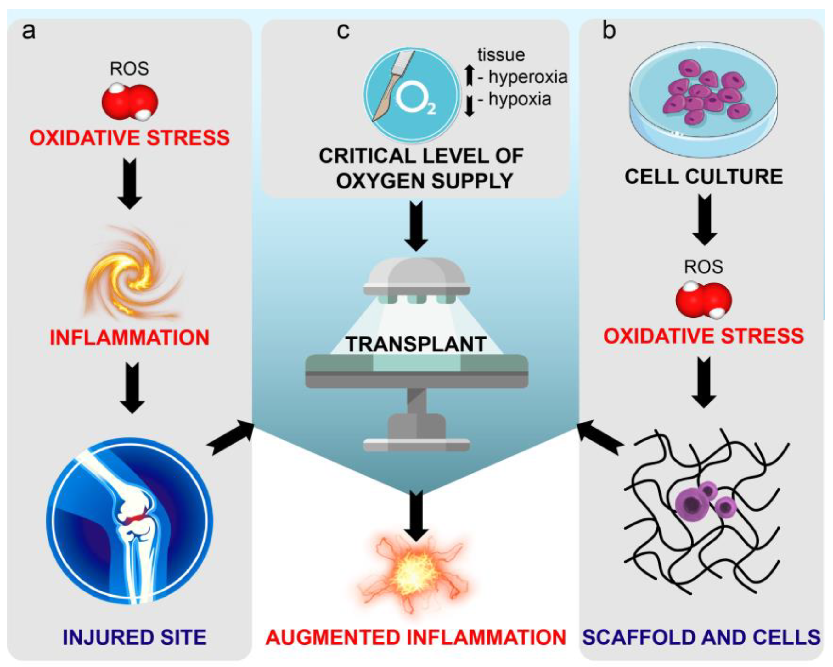
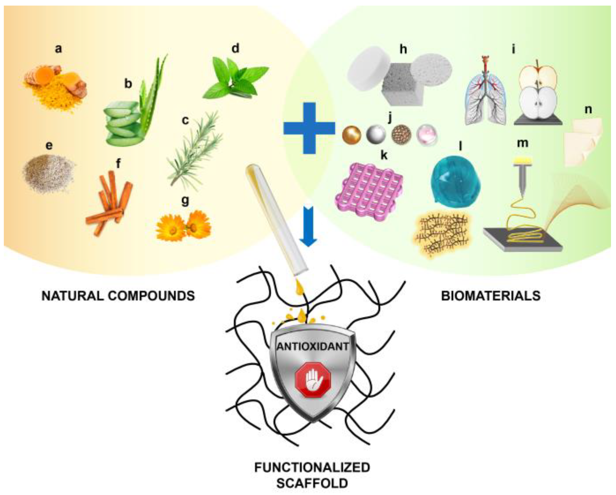
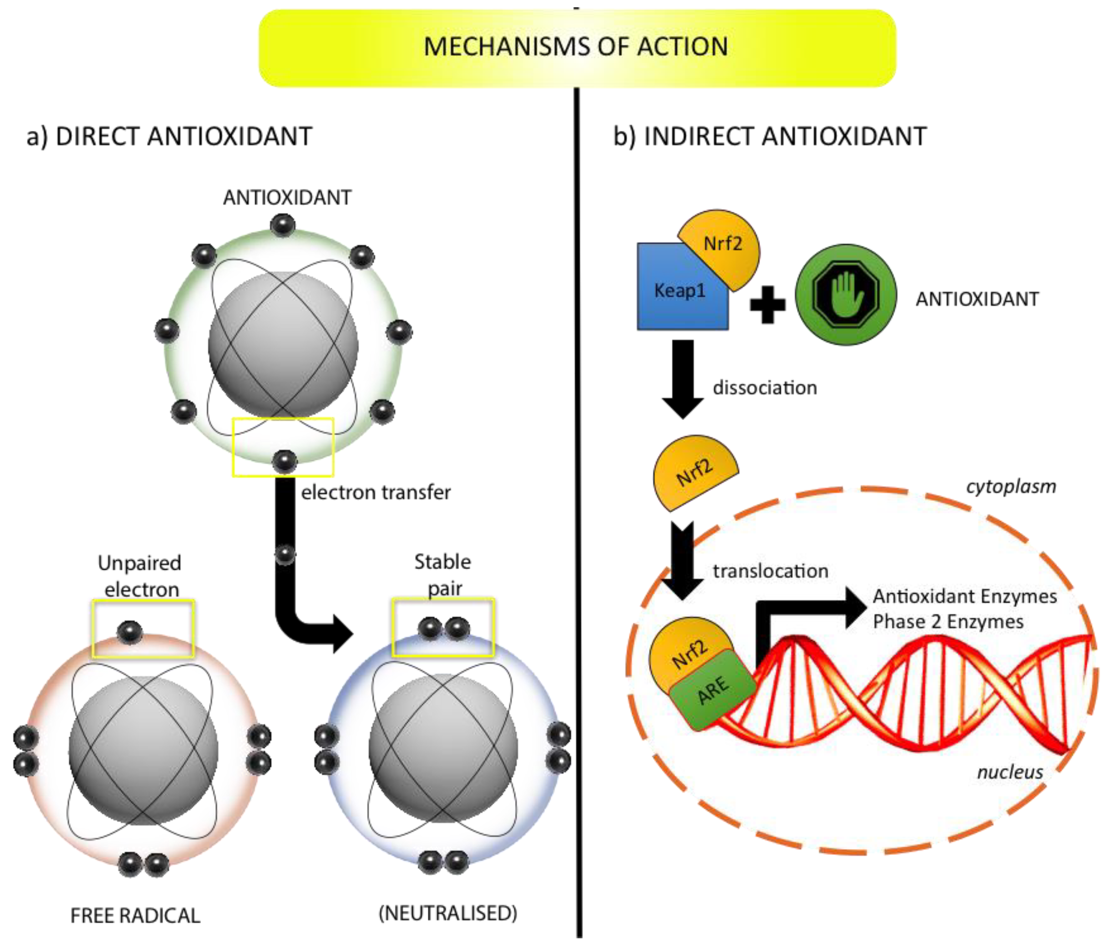
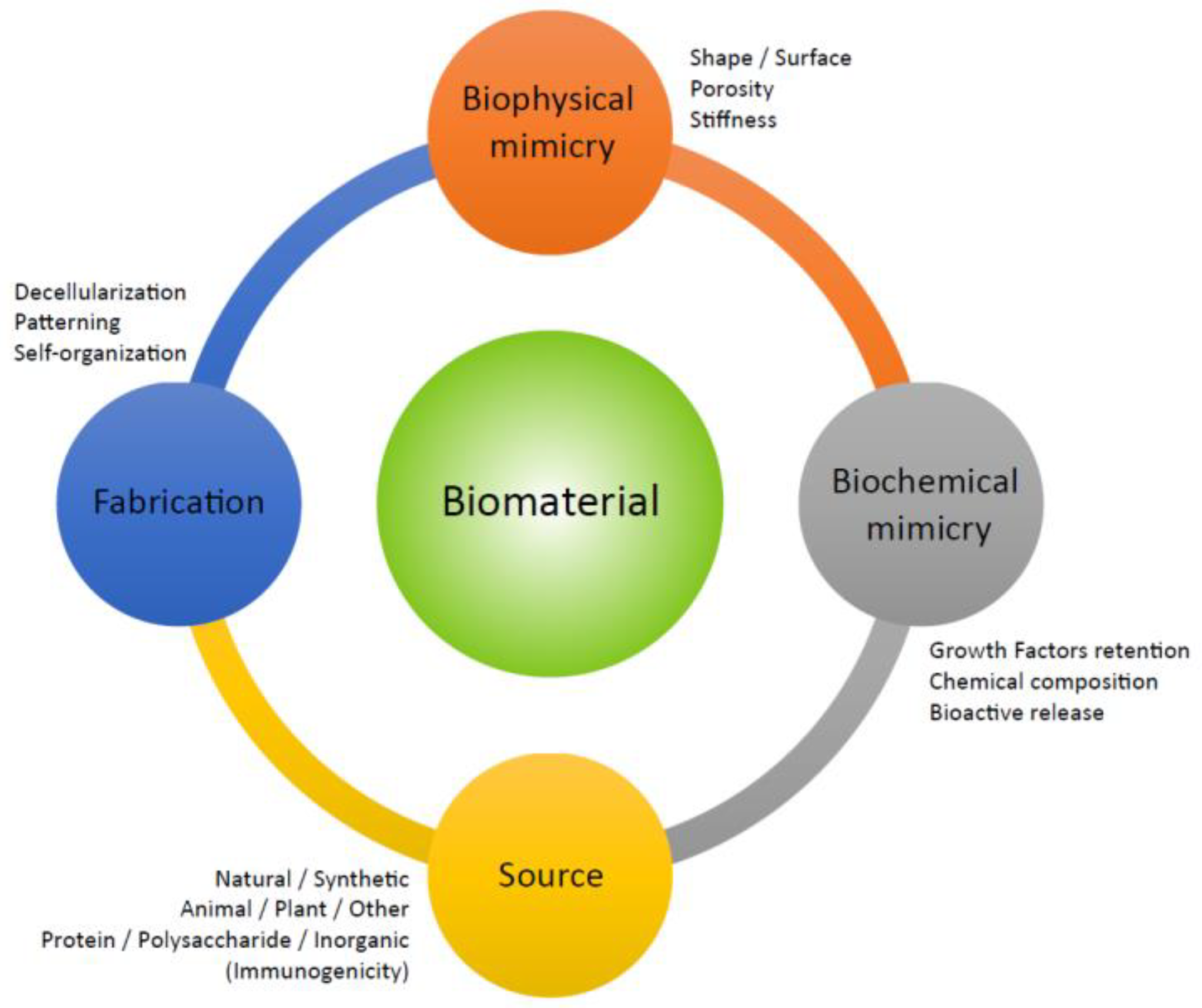
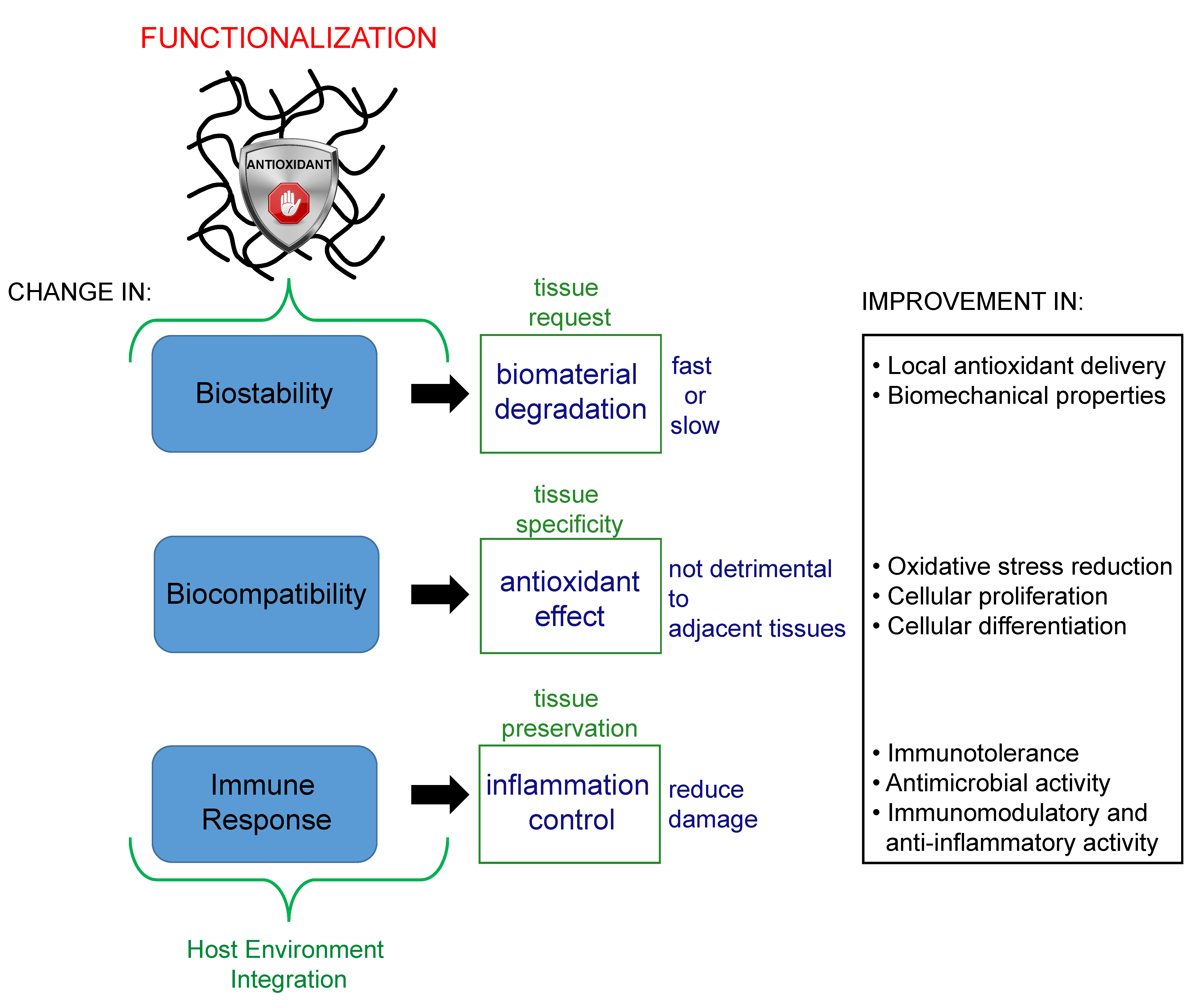
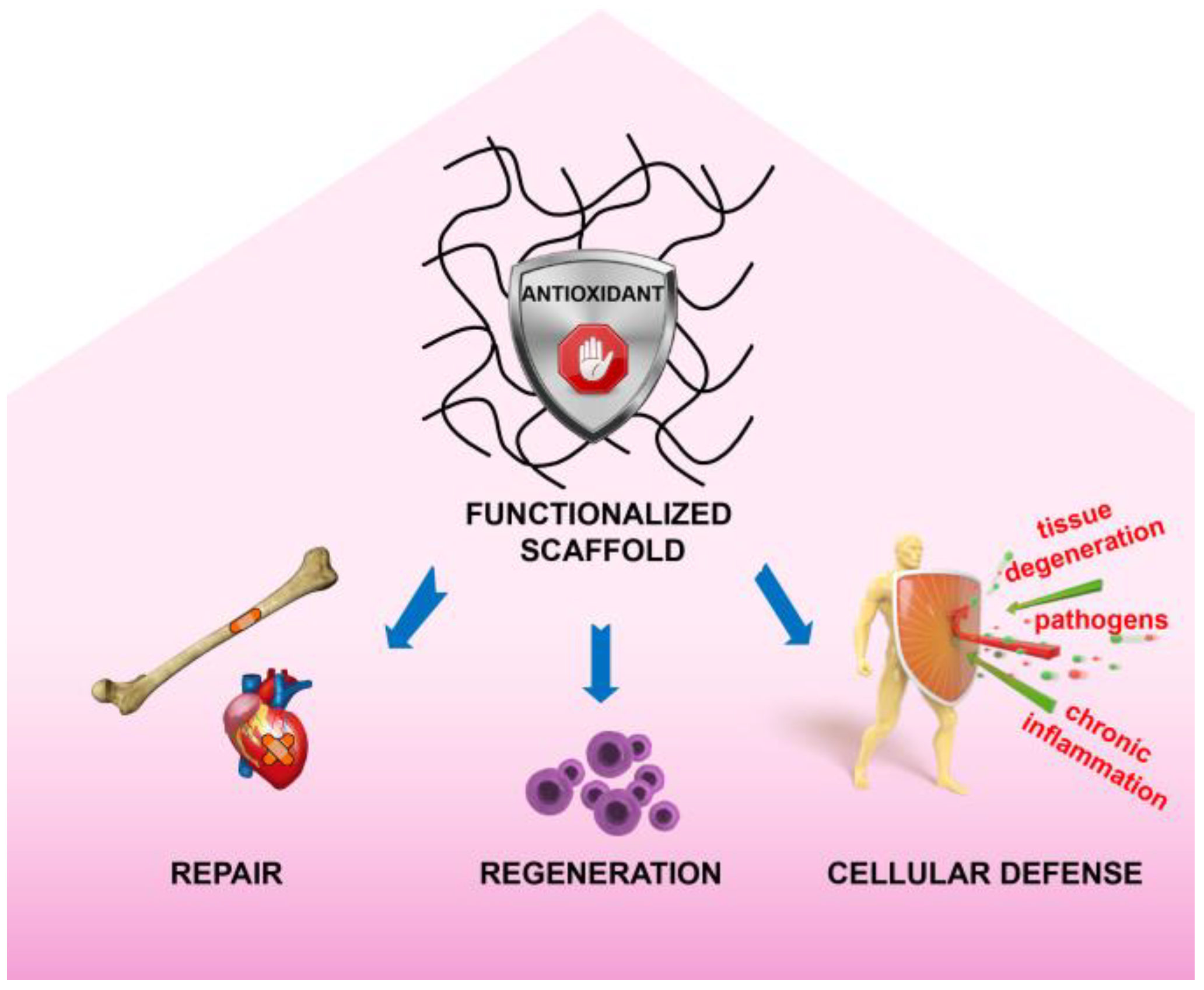
Table 1.
Studies demonstrating the advantageous properties provided by the incorporation of specific natural antioxidants into various biomaterials.
Table 1.
Studies demonstrating the advantageous properties provided by the incorporation of specific natural antioxidants into various biomaterials.
| Property | Antioxidant | Biomaterial (Scaffold) |
|---|---|---|
| Scavenging activity | Aloe vera (extract) [109] Astaxanthin [110] Caffeic acid [111] Carvacrol [112] Catechin [113] Chlorogenic acid [114] Cinnamon E.O. [115] Curcumin [116] Eugenol [112] Fenugreek (seed absolut) [117] Gallic acid [118] Hydrolyzed spent coffee grounds [119] Lemongrass E.O. [120] Mango (extract) [121,122] Melanin [123] Moringa oleifera (extract) [109] Quercetin [113,124,125] Rosmarinic acid [126] Rosemary (extract) [127] Tannic acid [128,129] Teucrium polium (extract) [130] Thyme (polyphenol extract) [131] Thymol [132] Vitamin C [133] | Cellulose-PEG [120] CH [112,113,122,132] CH-derivates [118,131] CH-gum [115] HA [129] Halloyste [124] Magnesium alloy [128] P(3HB-co-3HV) [133] PDMS [125] PE [119] PEG–ALG [109] PEG-PCL [134] PLA –PEG [130] PLA [127] PVP [121] SF -collagen [117] SF [123] Silica [111,126] Silica-PEG [114] |
| Indirect antioxidant effects (including cytoprotection) | Citric acid [135] Curcumin [136,137] Cystamine [73] Gallic acid [138] Hydrolyzed spent coffee grounds [119] Lignin (alkali) [139] Quercetin [136] Rapeseed flower (pollen extract) [140] Vitamin C [141] Vitamin E [142] White tea (leaves extract) [143] | CH [138] CH-derivates [116] Hyaluronic acid-β-cyclodextrin [142] Montmorillonite [143] PBAE [136] PCL [137,139] PE [119] PU [141] PVA [135] |
| Biodegradation modulation | Aloe vera [144] EGCG [145] Soybean (extract) [146] Tannic acid [129] Vitamin C [133] | Gelatin-Hap [146] HA [129,145] P(3HB-co-3HV) [133] SF [144] |
| Biomechanical stability | Aloe vera (extract) [147,148] Caffeic acid [149,150] Calendula Officinalis [151] Cinnamaldhyde [152] Citric acid [153] Curcumin [154] EGCG [155] Ellagic acid [155] Equisetum arvense (extract) [156] Grape (pomace) [157] Lignin (kraft) [100] Ostholamide [158] Plumbagin [159] Resveratrol [160] Soybean (extract) [146] Tannic acid [131,155] Vitamin C [133]. | CH-Starch [131] Collagen [159] Collagen type I–Gelatin [148,151] Gelatin [153] Gelatin derivates-collagen [158] Gelatin-Hap [146] HA [160] P(3HB-co-3HV) [133] PCL [100,147] PCL-CH [149,150] PEG derivatives [155] PLA–derivates [152] PLA-Hap [156] PLGA [154] Porcine pericardium (decellularized) [157] |
| Hydrophilicity and wettability/swelling | Aloe vera (extract) [48,109] Astaxanthin [161] Berberine [162] Cinnamaldhyde [152] Curcumin [163,164] Lignin (kraft) [100] Ostholamide [158] Peppermint E.O. [165] Plumbagin [159] Rosemary (extract) [127] Sesamol [166] Soybean (oil) [167] Thymus (polyphenol extract) [168] Zataria Multiflora E.O. [169] | Cellulose acetate-gelatin [162] CH [168] Collagen [159,161] Gelatin derivates-collagen [158] HPG [164] PA-6 [167] PCL [48,100,165] PCL-gum [163] PEG-ALG [109] PLA [127,166] PLA-derivatives [152] PVA derivates [169] |
| Controlled release | Astaxanthin [170,171,172] Caffeic acid [173] Curcumin [116,174,175,176] Cystamine [73] EGCG [177,178] Morin [126] Ostholamide [158] Quercetin [157] Rosmarinic acid [179] Sage E.O. [180] Thymol [181] Thymus (polyphenol extract) [168] | ALG [170,177] CH-derivates [116,171] CH [168,180] Gelatin [178] Gelatin derivates-collagen [158] PBAE [73,176] PCL [175] Porcine pericardium (decellularized) [157] PVA [174] PVA derivates [181] Silica [126] |
| Biocompatibility | Aloe Vera (extract) [182,183,184,185,186] Astaxanthin [187,188,189] Calendula Officinalis (extract) [190,191,192] Cinnamon, E.O. [193] Curcumin [116,134,154,163,164,186] Lemongrass E.O. [193] Peppermint E.O. [193] Plumbagin [159] Rosmarinic acid [194] Soybean (oil) [167] Tannic acid [195] Tyramine [145] Vitamin E [196] | Cellulose acetate [193] CH [184,194,196] CH-derivates [116] Collagen [159] Gelatin [186] HA [145] HPG [164,190] PA-6 [167] PCL [185] PCL-gum [163] PCL-gum [191,192] PEG-PCL [134] PLGA [154,182,183,187,188] Poly-tannic acid [195] PVA derivatives [189] |
| Cell proliferation | Aloe Vera (extract) [48,109,144,197,198,199] Caffeic acid [149,200] Curcumin [154] Lignin (alkali) [139] Moringa oleifera (extract) [109] p-coumaric acid [200] Quercetin [201,202] Salvianolic acid B [203] Thymol [132] Vitamin B6 [200] Vitamin C (Magnesium phosphate) [203] | Amnion (decellularized hydrogel) [198] CH [132] Hap [201,202] PCL [48,139] PCL-gelatin [203] PDX [199] PEG-ALG [109] PLA [200] PLGA [154] SF [144] SF-PVA [197] |
| Cell differentiation | Aloe vera (extract) [204,205] Astaxanthin [206] EGCG [178,207] Lignin (alkali) [139] (kraft) [100] Melanin [123] Quercetin [201] Salvianolic acid B [203] Soybean lecithin [208] Vitamin C (Magnesium phosphate) [203] Vitamin C [141] | Gelatin [178,207] Gelatin derivatives [206] Hap [201] PCL [100,139] PCL-gelatin [203] PHBV [204] PLA-PCL-SF [205] PLGA [208] PU [141] SF [123] |
| Antimicrobial activity | Aloe vera (extract) [209] Berberine [162] Caffeic acid [149] Calendula Officinalis (extract) [192,210] Carvacrol [112,211] Chlorogenic acid [114] Cinnamaldhyde [152] Cinnamon E.O. [193,212,213] Curcumin [174] Eugenol [112] Lemongrass E.O. [193] Mango (silver particles) [214] Moringa Oleifera (extract) [109] Ostholamide [158] Peppermint E.O. [165,193] Plumbagin [159] Punica granatum (peel extract) [215] Quercetin [125,201] Rosemary (extract) [127] Rosmarinic acid [216] Sage E.O. [180] Teucrium polium (extract) [130] Thyme and Oregano E.O. [217,218,219] Thymol [132,220,221] Vitamin E [222] Zataria Multiflora E.O. [169] | ALG [211] Cellulose acetate [193,221] Cellulose acetate-gelatin [162] Cellulose-PEG [120] CH [112,210,220] CH-derivatives [215,218] CH-gelatin [219] Collagen [159] Gelatin derivates-collagen [158] Gelatin-gum [216] Hap [201] Ionomer cement [214] PCL [165] PCL-CH [149] PCL-gum [191,192] PDMS [125] PEG-ALG [109] PLA [127] PLA-CH [212,213] PLA-derivatives [152,222] PLA-PEG [130] PVA [174] PVA derivates [169] PVP [209] Silica-PEG [114] β-cyclodextrin [217] |
| Immunomodulation and anti-inflammatory effect | Aloe vera (extract) [223,224] Citric acid [135] Curcumin [154] EGCG [145] Quercetin [202] Resveratrol [225,226] Soybean [227,228] | ALG [223] CH [227,228] Cow bone matrix (decellularized) [224] HA [145] Hap [202] PLGA [154] PVA [135] SF [225,226] |
ALG, alginate; CH, chitosan; E.O., essential oil; HA, hyaluronic acid; Hap, hydroxyapatite; P(3HB-co-3HV), poly(3-hydroxybutyrate-co-3-hydroxyvalerate); PBAE, poly(β-amino ester); PCL polycaprolactone; PDMS, polydimethylsiloxane: PDX, polydioxanone; PE, polyethylene, PEG, polyethylene glycol; PHBV, poly(3-hydroxybutyrate-co-3-hydroxyvalerate); PLA, polylactic acid; PLGA, poly(lactic-co-glycolic acid); PU, polyuretane; PVA, polyvinyl alcohol; PVP, polyvinylpyrrolidone; SF, silk fibroin.
Table 2.
Studies demonstrating the potential therapeutic application of biomaterials including natural antioxidants.
Table 2.
Studies demonstrating the potential therapeutic application of biomaterials including natural antioxidants.
| Therapeutic Application | Antioxidant | Biomaterial |
|---|---|---|
| Wound healing | Aloe vera [147,183,186,198,224,230] Astaxanthin [161] Berberine [162] Bixin [233] Calendula officinalis [190] Curcumin [134,147,164,186,230,233] Dopamine [125,142] EGCG [145] Moringa oleifera (extract) [109] Ostholamide [158] Quercetin [125] Sesamol [232] Soybean (extract) [167] Thymol [234] Tyramine [145] Vitamin E [142] β-Carotene [234] | ALG derivatives [109] Amnion (decellularized hydrogel) [198] Bone matrix (decellularized) [224] .Cellulose acetate–gelatin [162] Cellulose acetate derivatives [232] CH derivatives [116] CH-ALG [234] Collagen [161] Gelatin derivative [186] Gelatin derivatives-collagen [158] HA [145] HA-β-cyclodextrin [142] HPG [190] PA-6 [167] PCL [148,236] PDMS [125] PEG-PCL [134] PLA-HPG [164] PLGA derivatives [183] PMAA [230] |
| Bone defects | Aloe vera (acemannan/glucomannan) [199] (extract) [204] Broussonetia kazinoki (extract) [242] Cissus quadrangularis (extract) [245] Curcumin [175] EGCG [178,207] Equisetum arvense (extract) [156] Fenugreek (seed absolut) [117] Quercetin [204,205,243] Soybean (extract) [240] Soybean (oil) [241] | CH derivatives [245] Gelatin [178,207] Gelatin-Hap [240] Hap [201,202] PCL [175] PDX derivates [199] PHBV [204] PLA-Hap [156] PS [241] SF [242] SF-collagen [117] SF-Hap [239] |
| Cardiovascular diseases | Aloe Vera [205] Astaxanthin [189] Curcumin [154] Salvianolic acid B [203] Tannic acid [128] Vitamin C (Magnesium phosphate) [203] Vitamin E [196,246] | CH [196] Magnesium alloy [128] PCL-gelatin [203] PLA [246] PLA-PCL-SF [205] PLGA [154] PVA derivatives [189] |
| Neurological disorders | Lignin (alkali) [139] (kraft) ([100]) Melanin [123] | PCL [100,139] SF [123] |
| Other tissue disorders | Aloe vera [223] Resveratrol [160] | ALG [223] HA [160] |
ALG, alginate; CH, chitosan; HA, hyaluronic acid; Hap, hydroxyapatite; P(3HB-co-3HV), poly(3-hydroxybutyrate-co-3-hydroxyvalerate); PA-6, polyammide 6; PCL, polycaprolactone; PDMS, polydimethylsiloxane: PDX, polydioxanone; PE, polyethylene, PEG, polyethylene glycol; PHBV, poly(3-hydroxybutyrate-co-3-hydroxyvalerate); PLA, polylactic acid; PLGA, poly(lactic-co-glycolic acid); PMAA, polymethacrylic acid; PS, polystyrene; PVA, polyvinyl alcohol; PVP, polyvinylpyrrolidone; SF, silk fibroin.
© 2020 by the authors. Licensee MDPI, Basel, Switzerland. This article is an open access article distributed under the terms and conditions of the Creative Commons Attribution (CC BY) license (http://creativecommons.org/licenses/by/4.0/).
Share and Cite
MDPI and ACS Style
Marrazzo, P.; O’Leary, C. Repositioning Natural Antioxidants for Therapeutic Applications in Tissue Engineering. Bioengineering 2020, 7, 104. https://0-doi-org.brum.beds.ac.uk/10.3390/bioengineering7030104
AMA Style
Marrazzo P, O’Leary C. Repositioning Natural Antioxidants for Therapeutic Applications in Tissue Engineering. Bioengineering. 2020; 7(3):104. https://0-doi-org.brum.beds.ac.uk/10.3390/bioengineering7030104
Chicago/Turabian StyleMarrazzo, Pasquale, and Cian O’Leary. 2020. "Repositioning Natural Antioxidants for Therapeutic Applications in Tissue Engineering" Bioengineering 7, no. 3: 104. https://0-doi-org.brum.beds.ac.uk/10.3390/bioengineering7030104
Note that from the first issue of 2016, this journal uses article numbers instead of page numbers. See further details here.