
Swine Small Intestine Sealing Performed by Different Vessel Sealing Devices: Ex-Vivo Test

 , , ,
, , ,
Abstract
:1. Introduction
2. Materials and Methods
2.1. Samples
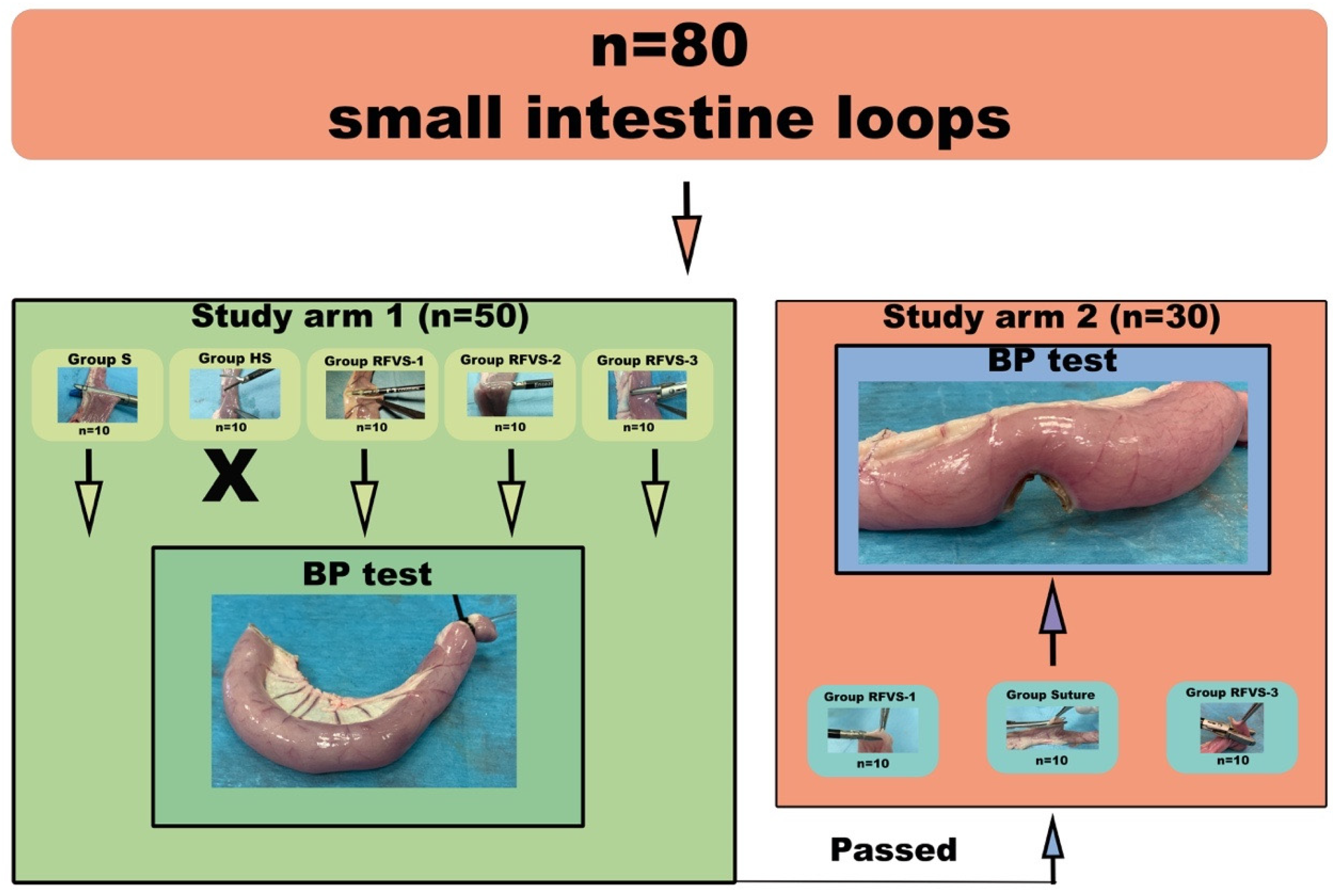
2.2. Study Design
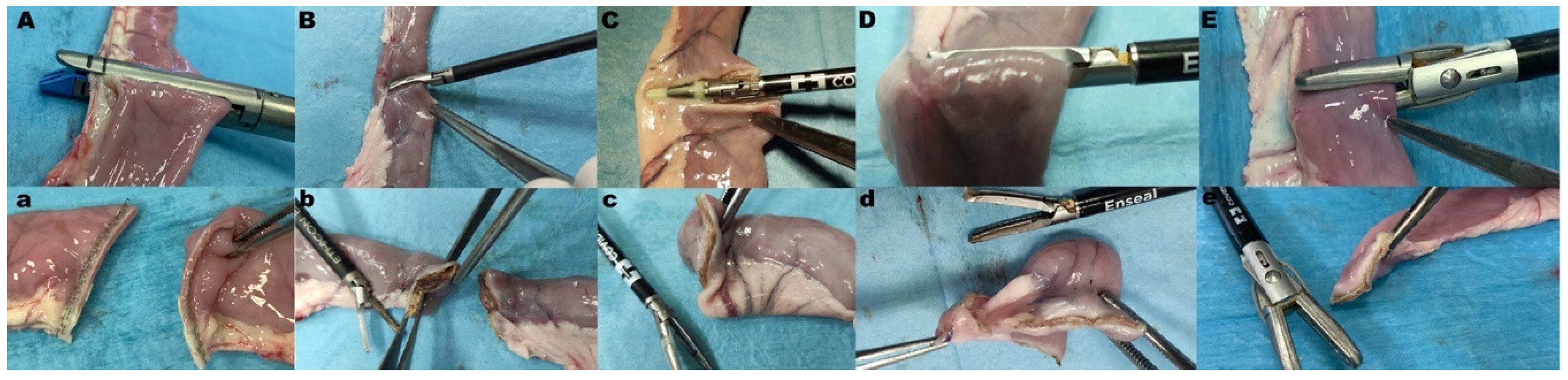
2.3. Experimental Groups of Study Arm 1
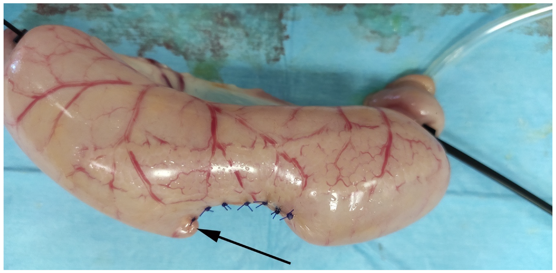
2.4. Sample Constructs
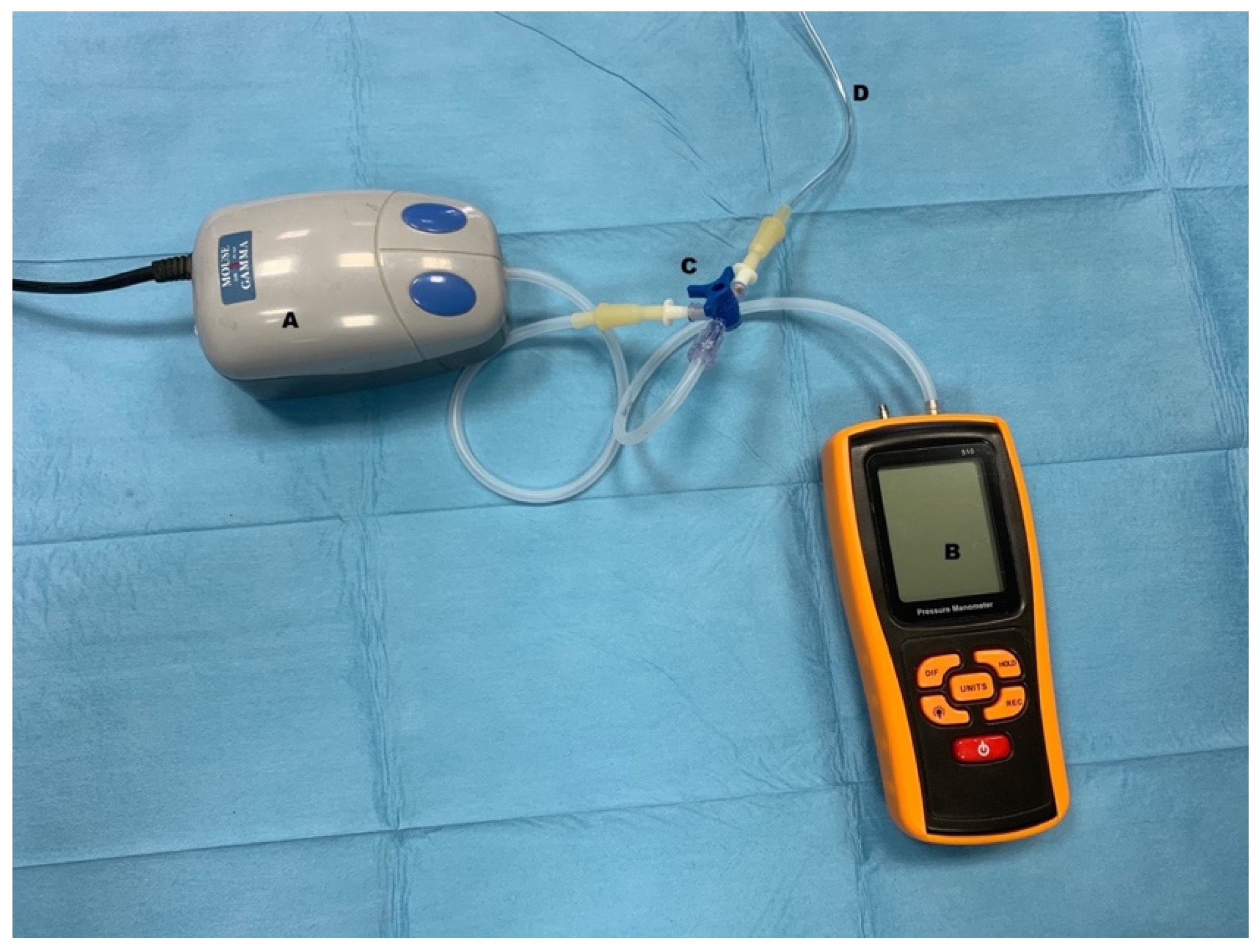
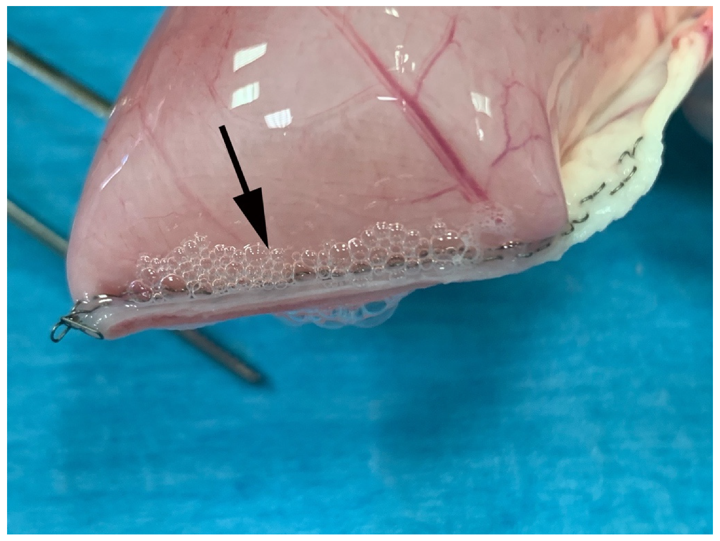
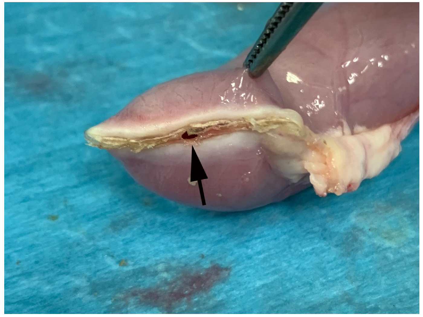
2.5. Burst and Leak Pressures
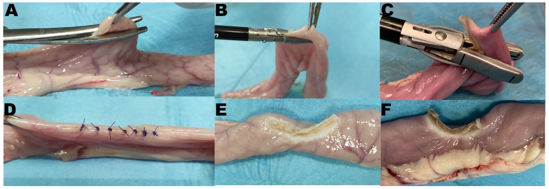
2.6. Experimental Groups of Study Arm 2
2.7. Histology
2.8. Statistical Analysis
3. Results
3.1. Study Arm 1
3.2. Study Arm 2
3.3. Histology
4. Discussion
5. Conclusions
Author Contributions
Funding
Institutional Review Board Statement
Informed Consent Statement
Data Availability Statement
Acknowledgments
Conflicts of Interest
References
- Latimer, C.A.; Nelson, M.; Moore, C.M.; Martin, K.E. Effect of collagen and elastin content on the burst pressure of human blood vessel seals formed with a bipolar tissue sealing system. J. Surg. Res. 2014, 186, 73–80. [Google Scholar] [CrossRef] [PubMed]
- Arya, S.; Hadjievangelou, N.; Lei, S.; Kudo, H.; Goldin, R.D.; Darzi, A.W.; Elson, D.S.; Hanna, G.B. Radiofrequency-induced small bowel thermofusion: An ex vivo study of intestinal seal adequacy using mechanical and imaging modalities. Surg. Endosc. 2013, 27, 3485–3496. [Google Scholar] [CrossRef] [PubMed]
- Arya, S.; Mackenzie, H.; Hanna, G.B. Non-vascular experimental and clinical applications of advanced bipolar radiofrequency thermofusion technology in the thorax and abdomen: A systematic review. Surg. Endosc. 2015, 29, 1659–1678. [Google Scholar] [CrossRef] [PubMed]
- Santini, M.; Fiorelli, A.; Messina, G.; Laperuta, P.; Mazzella, A.; Accardo, M. Use of the LigaSure device and the Stapler for closure of the small bowel: A comparative ex vivo study. Surg. Today 2013, 43, 787–793. [Google Scholar] [CrossRef]
- Smulders, J.F.; de Hingh, I.H.; Stavast, J.; Jackimowicz, J.J. Exploring new technologies to facilitate laparoscopic surgery: Creating intestinal anastomoses without sutures or staples, using a radio-frequency-energy-driven bipolar fusion device. Surg. Endosc. 2007, 21, 2105–2109. [Google Scholar] [CrossRef] [PubMed] [Green Version]
- Tagkalos, E.; Heimann, A.; Gaiser, T.; Hirsch, D.; Gockel, I.; Lang, H.; Jansen-Winkeln, B. Cecal Resection with Bipolar Sealing Devices in a Rat Model. J. Investig. Surg. 2020, 33, 59–66. [Google Scholar] [CrossRef] [PubMed]
- Wei, M.T.; Yang, T.H.; Deng, X.B.; Meng, W.J.; Han, J.H.; Zhou, Z.G.; Wang, Z.Q. Laparoscopic colorectal anastomosis technique without ‘‘dog ear’’ formation using LigaSure for bowel transection. Tech. Coloproctol. 2019. [Google Scholar] [CrossRef] [PubMed]
- Yavuz, A.; Bulus, H.; Tas, A.; Aydin, A. Evaluation of Stump Pressure in Three Types of Appendectomy: Harmonic Scalpel, LigaSure, and Conventional Technique. J. Laparoendosc. Adv. Surg. Tech. A 2016, 26, 950–953. [Google Scholar] [CrossRef] [PubMed]
- Gehrig, T.; Billeter, A.T.; Wekerle, A.L.; Shevchenko, M.; Brand, K.; Muller-Stich, B.P. Evaluation of the LigaSure() Vessel Sealing System for bowel transection and intestinal anastomosis-an experimental study in a porcine model. Langenbecks Arch. Surg. 2016, 401, 381–387. [Google Scholar] [CrossRef] [PubMed]
- Himpens, J.; Leman, G.; Sonneville, T. Laparoscopic Roux-en-Y gastric bypass performed without staples. Surg. Endosc. 2005, 19, 1003. [Google Scholar] [CrossRef] [PubMed]
- Kawahara, H.; Watanabe, K.; Tomoda, M.; Enomoto, H.; Akiba, T.; Yanaga, K. Single-incision clipless laparoscopic total colectomy. Hepatogastroenterology 2014, 61, 453–455. [Google Scholar] [PubMed]
- Moreno-Sanz, C.; Picazo-Yeste, J.; Seoane-Gonzales, J.; Manzanera-Diaz, M.; Tadeo-Ruiz, G. Division of the small bowel with the LigaSure Atlas device during the right laparoscopic colectomy. J. Laparoendosc. Adv. Surg Tech. A 2008, 18, 99–101. [Google Scholar] [CrossRef]
- Rumbaugh, M.L.; Burba, D.J.; Natalini, C.; Hosgood, G.; Moore, R.M. Evaluation of a vessel-sealing device for small intestinal resection and anastomosis in normal horses. Vet. Surg. 2003, 32, 574–579. [Google Scholar] [CrossRef] [PubMed]
- Sanchez Trejo, H.A.; Hakakian, D.; Rolandelli, R.H.; Nouri, A.M.; Antonioli, L.; Nemeth, Z.H. “Cecal Resection with Bipolar Sealing in a Rat Model”: A Promising Approach for Future Human Studies. J. Investig. Surg. 2020, 33, 67–68. [Google Scholar] [CrossRef]
- Sanchez-De Pedro, F.; Moreno-Sanz, C.; Morandeira-Rivas, A.; Tenias-Burillo, J.M.; Alhambra-Rodriguez De Guzman, C. Colorectal anastomosis facilitated by the use of the LigaSure((R)) sealing device: Comparative study in an animal model. Surg. Endosc. 2014, 28, 508–514. [Google Scholar] [CrossRef] [PubMed]
- Santini, M.; Fiorelli, A.; Messina, G.; Mazzella, A.; Accardo, M. The Feasibility of LigaSure to Create Intestinal Anastomosis: Results of Ex Vivo Study. Surg. Innov. 2015, 22, 266–273. [Google Scholar] [CrossRef]
- Winter, H.; Holmer, C.; Buhr, H.J.; Lindner, G.; Lauster, R.; Kraft, M.; Ritz, J.P. Pilot study of bipolar radiofrequency-induced anastomotic thermofusion-exploration of therapy parameters ex vivo. Int. J. Colorectal Dis. 2010, 25, 129–133. [Google Scholar] [CrossRef] [PubMed]
- Baron, J.; Giuffrida, M.; Mayhew, P.D.; Singh, A.; Case, J.B.; Culp, W.T.N.; Holt, D.E.; Mayhew, K.N.; Runge, J.J. Minimally invasive small intestinal exploration and targeted abdominal organ biopsy with a wound retraction device in 42 cats (2005-2015). Vet. Surg. 2017, 46, 925–932. [Google Scholar] [CrossRef] [PubMed] [Green Version]
- Case, J.B.; Ellison, G. Single incision laparoscopic-assisted intestinal surgery (SILAIS) in 7 dogs and 1 cat. Vet. Surg. 2013, 42, 629–634. [Google Scholar] [CrossRef]
- Ziegler, A.; Gonzalez, L.; Blikslager, A. Large Animal Models: The Key to Translational Discovery in Digestive Disease Research. Cell Mol. Gastroenterol. Hepatol. 2016, 2, 716–724. [Google Scholar] [CrossRef] [PubMed] [Green Version]
- Barnes, R.F.; Greenfield, C.L.; Schaeffer, D.J.; Landolfi, J.; Andrews, J. Comparison of biopsy samples obtained using standard endoscopic instruments and the harmonic scalpel during laparoscopic and laparoscopic-assisted surgery in normal dogs. Vet. Surg. 2006, 35, 243–251. [Google Scholar] [CrossRef]
- Holmer, C.; Winter, H.; Kroger, M.; Nagel, A.; Jaenicke, A.; Lauster, R.; Kraft, M.; Buhr, H.J.; Ritz, J.P. Bipolar radiofrequency-induced thermofusion of intestinal anastomoses--feasibility of a new anastomosis technique in porcine and rat colon. Langenbecks Arch. Surg. 2011, 396, 529–533. [Google Scholar] [CrossRef] [PubMed]
- Okhunov, Z.; Yoon, R.; Lusch, A.; Spradling, K.; Suarez, M.; Kaler, K.S.; Patel, R.; Hwang, C.; Osann, K.; Huang, J.; et al. Evaluation and Comparison of Contemporary Energy-Based Surgical Vessel Sealing Devices. J. Endourol. 2018, 32, 329–337. [Google Scholar] [CrossRef] [PubMed] [Green Version]
- Lyons, S.D.; Law, K.S. Laparoscopic vessel sealing technologies. J. Minim. Invasive Gynecol. 2013, 20, 301–307. [Google Scholar] [CrossRef] [PubMed]
- Chekan, E.G.; Davison, M.A.; Singleton, D.W.; Mennone, J.Z.; Hinoul, P. Consistency and sealing of advanced bipolar tissue sealers. Med. Devices (Auckl.) 2015, 8, 193–199. [Google Scholar] [CrossRef] [Green Version]
- Chen, R.K.; Chastagner, M.W.; Geiger, J.D.; Shih, A.J. Bipolar electrosurgical vessel-sealing device with compressive force monitoring. J. Biomech. Eng. 2014, 136, 061001. [Google Scholar] [CrossRef]
- Eick, S.; Loudermilk, B.; Walberg, E.; Wente, M.N. Rationale, bench testing and in vivo evaluation of a novel 5 mm laparoscopic vessel sealing device with homogeneous pressure distribution in long instrument jaws. Ann. Surg. Innov. Res. 2013, 7, 15. [Google Scholar] [CrossRef] [Green Version]
- Govekar, H.R.; Robinson, T.N.; Stiegmann, G.V.; McGreevy, F.T. Residual heat of laparoscopic energy devices: How long must the surgeon wait to touch additional tissue? Surg. Endosc. 2011, 25, 3499–3502. [Google Scholar] [CrossRef] [PubMed]
- Slam, K.D.; Calkins, S.; Cason, F.D. LaPlace’s law revisited: Cecal perforation as an unusual presentation of pancreatic carcinoma. World J. Surg. Oncol. 2007, 5, 14. [Google Scholar] [CrossRef] [Green Version]
- Matz, B.M.; Boothe, H.W.; Wright, J.C.; Boothe, D.M. Effect of enteric biopsy closure orientation on enteric circumference and volume of saline needed for leak testing. Can. Vet. J. 2014, 55, 1255–1257. [Google Scholar]
- Tasaka, K.; Farrar, J.T. Intraluminal pressure of the small intestine of the unanesthetized dog. Pflug. Arch. 1976, 364, 35–44. [Google Scholar] [CrossRef] [PubMed]
- Chaikomin, R.; Wu, K.L.; Doran, S.; Jones, K.L.; Smout, A.J.; Renooij, W.; Holloway, R.H.; Meyer, J.H.; Horowitz, M.; Rayner, C.K. Concurrent duodenal manometric and impedance recording to evaluate the effects of hyoscine on motility and flow events, glucose absorption, and incretin release. Am. J. Physiol. Gastrointest. Liver Physiol. 2007, 292, G1099–G1104. [Google Scholar] [CrossRef] [PubMed]
- Imam, H.; Sanmiguel, C.; Larive, B.; Bhat, Y.; Soffer, E. Study of intestinal flow by combined videofluoroscopy, manometry, and multiple intraluminal impedance. Am. J. Physiol. Gastrointest. Liver Physiol 2004, 286, G263–G270. [Google Scholar] [CrossRef] [Green Version]
- Paral, J.; Lochman, P.; Blazej, S.; Pavlik, M. Glued versus stapled anastomosis of the colon: An experimental study to determine comparative resistance to intraluminal pressure. Asian J. Surg. 2014, 37, 154–161. [Google Scholar] [CrossRef] [PubMed] [Green Version]
- Ellison, G.W. Intestinal obstruction. In Disease Mechanisms in Small Animal Surgery, 2nd ed.; Bojrab, M.J., Ed.; Lea & Febiger: Philadelphia, PA, USA, 1993; pp. 252–257. [Google Scholar]
- Shikata, J.; Shida, T.; Amino, K.; Ishioka, K. Experimental studies on the hemodynamics of the small intestine following increased intraluminal pressure. Surg. Gynecol. Obstet. 1983, 156, 155–160. [Google Scholar]
- Boscan, P.; Cochran, S.; Monnet, E.; Webb, C.; Twedt, D. Effect of prolonged general anesthesia with sevoflurane and laparoscopic surgery on gastric and small bowel propulsive motility and pH in dogs. Vet. Anaesth. Analg. 2014, 41, 73–81. [Google Scholar] [CrossRef]
- Hayami, M.; Watanabe, M.; Mine, S.; Imamura, Y.; Okamura, A.; Yuda, M.; Yamashita, K.; Toihata, T.; Shoji, Y.; Ishizuka, N. Lateral thermal spread induced by energy devices: A porcine model to evaluate the influence on the recurrent laryngeal nerve. Surg. Endosc. 2019, 33, 4153–4163. [Google Scholar] [CrossRef] [PubMed]
- Elemen, L.; Yazir, Y.; Akay, A.; Boyacioglu, Z.; Ceyran, B.; Ceylan, S. Comparison of bipolar electrosurgical devices with ligatures and endoclips in the rat appendicitis model. J. Pediatr. Surg. 2011, 46, 1923–1929. [Google Scholar] [CrossRef]
- Pan, H.; Leung, K.K.C.; Ng, E.K.W. Tissue fusion technology versus suture and staple in porcine bowel anastomosis: An in vivo study. Braz J. Med. Biol. Res. 2020, 53, e9305. [Google Scholar] [CrossRef] [PubMed]
- Kramer, E.A.; Rentschler, M.E. Energy-Based Tissue Fusion for Sutureless Closure: Applications, Mechanisms, and Potential for Functional Recovery. Annu. Rev. Biomed. Eng. 2018, 20, 1–20. [Google Scholar] [CrossRef] [PubMed]
- Sorgato, N.; Bernante, P.; Pelizzo, M.R. Application of the LigaSure tissue sealing system to intestinal resection. Experimental and clinical trial. Ann. Ital. Chir. 2008, 79, 383–388. [Google Scholar] [PubMed]
- Aeschlimann, K.A.; Mann, F.A.; Middleton, J.R.; Belter, R.C. Comparison of enterotomy leak pressure among fresh, cooled, and frozen-thawed porcine jejunal segments. Am. J. Vet. Res. 2018, 79, 576–580. [Google Scholar] [CrossRef] [PubMed] [Green Version]


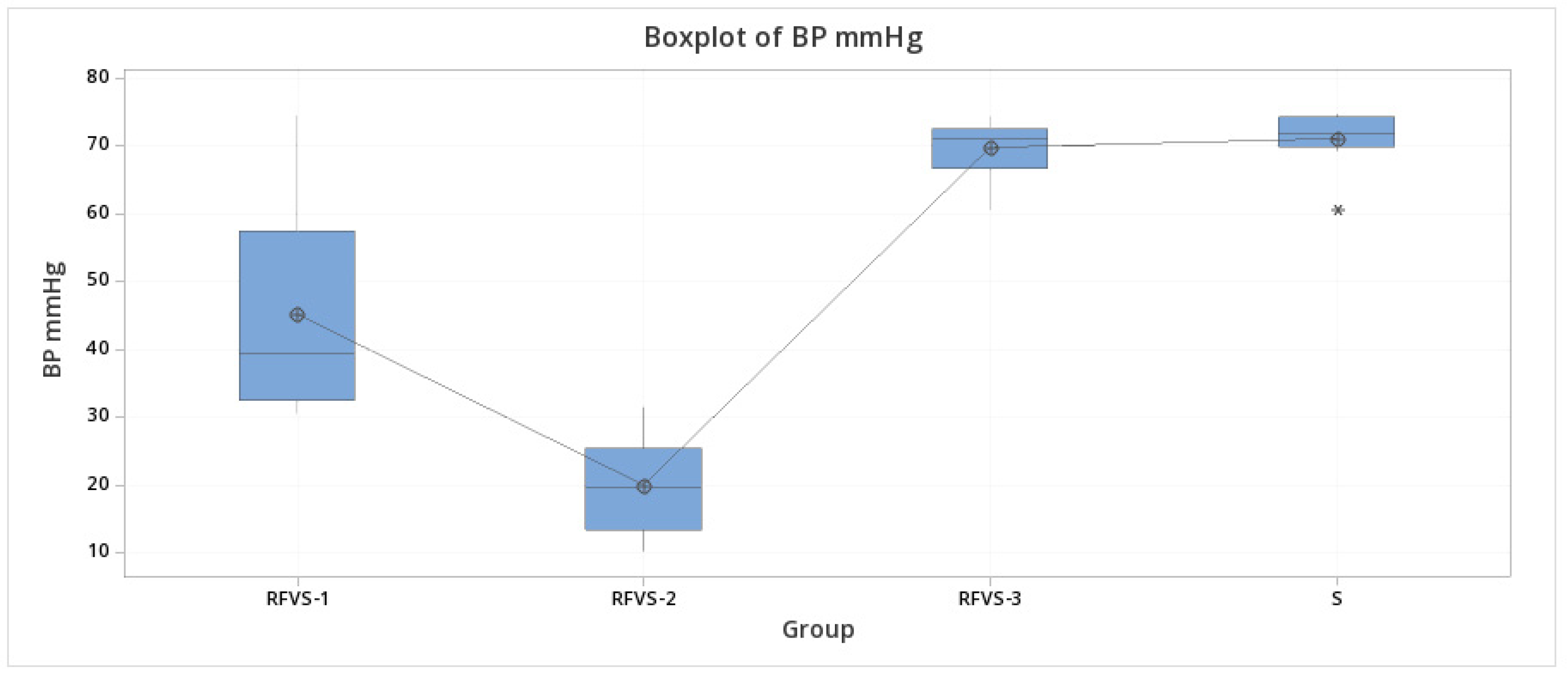
| BP (mmHg) | |||
|---|---|---|---|
| Group | Mean | St Dev | IQR |
| HS | * | * | * |
| RFVS-1 | 45.28 | 15.23 | 24.95 |
| RFVS-2 | 20.16 | 7.19 | 12.02 |
| RFVS-3 | 69.78 | 4.23 | 5.8 |
| S | 71.09 | 4.22 | 4.38 |
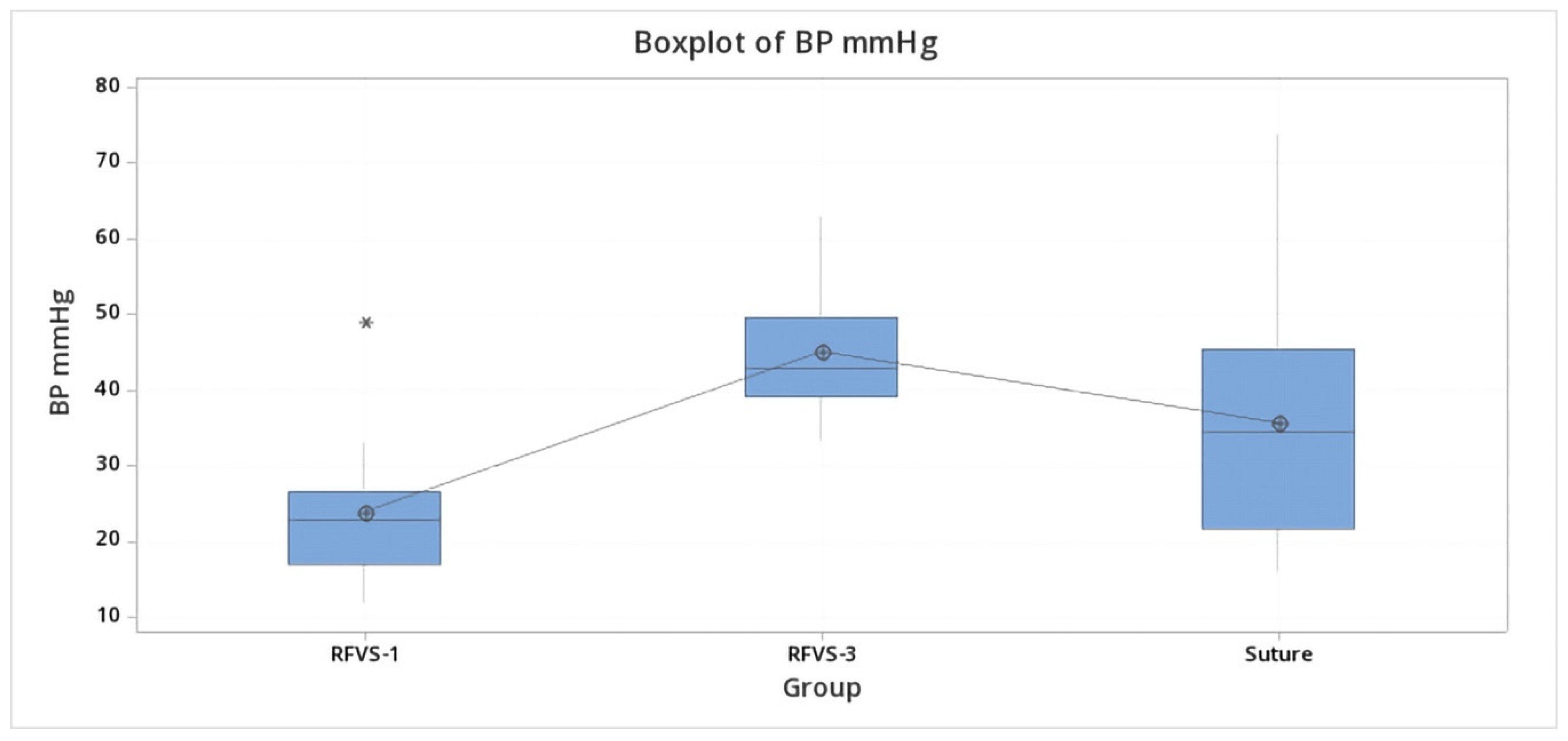
| BP mmHg | |||
|---|---|---|---|
| Group | Mean | St Dev | IQR |
| RFVS-1 | 23.96 | 10.63 | 9.62 |
| RFVS-3 | 45.09 | 8.75 | 10.48 |
| Suture | 35.71 | 17.51 | 23.77 |
| Biopsy Length (mm) | |||
|---|---|---|---|
| Group | Mean | St Dev | IQR |
| RFVS-1 | 14.7 | 2.111 | 4.25 |
| RFVS-3 | 21.5 | 1.65 | 3.25 |
| Suture | 23.1 | 4.04 | 5 |
| BP (mmHg) | |||
|---|---|---|---|
| Mean | St Dev | IQR | |
| RFVS-1 | |||
| Biopsy | 23.96 | 10.63 | 9.62 |
| Transection | 45.28 | 15.23 | 24.95 |
| RFVS-3 | |||
| Biopsy | 45.09 | 8.75 | 10.48 |
| Transection | 69.78 | 4.23 | 5.8 |
Publisher’s Note: MDPI stays neutral with regard to jurisdictional claims in published maps and institutional affiliations. |
© 2021 by the authors. Licensee MDPI, Basel, Switzerland. This article is an open access article distributed under the terms and conditions of the Creative Commons Attribution (CC BY) license (http://creativecommons.org/licenses/by/4.0/).
Share and Cite
Lacitignola, L.; Imperante, A.; Trisciuzzi, R.; Zizzo, N.; Crovace, A.M.; Staffieri, F. Swine Small Intestine Sealing Performed by Different Vessel Sealing Devices: Ex-Vivo Test. Vet. Sci. 2021, 8, 34. https://0-doi-org.brum.beds.ac.uk/10.3390/vetsci8020034
Lacitignola L, Imperante A, Trisciuzzi R, Zizzo N, Crovace AM, Staffieri F. Swine Small Intestine Sealing Performed by Different Vessel Sealing Devices: Ex-Vivo Test. Veterinary Sciences. 2021; 8(2):34. https://0-doi-org.brum.beds.ac.uk/10.3390/vetsci8020034
Chicago/Turabian StyleLacitignola, Luca, Annarita Imperante, Rodrigo Trisciuzzi, Nicola Zizzo, Alberto Maria Crovace, and Francesco Staffieri. 2021. "Swine Small Intestine Sealing Performed by Different Vessel Sealing Devices: Ex-Vivo Test" Veterinary Sciences 8, no. 2: 34. https://0-doi-org.brum.beds.ac.uk/10.3390/vetsci8020034