1. Introduction
The word teratoma was coined by Rudolph Virchow in 1863 from the Greek words “teras and onkoma” that respectively mean “monster and tumor [swelling]”. Teratoma is a rare embryonic tumor that can be both benign or malignant, most often diagnosed as benign. Teratoma is composed of mature somatic tissue that is not well organized and composed of neoplastic germ cells [
1] originating from all three layers of embryonic germ cells: ectoderm, mesoderm and endoderm (ex: epithelium, brain, bone, cartilage) [
1,
2,
3,
4,
5,
6,
7,
8]. In human medicine these tumors that originate from all three germ layers are also called “tridermomas”; this term is not currently used in veterinary medicine. Furthermore, it has been demonstrated that most of these neoplasms derive from anomalies in the ectodermal layer and in a minority from the endodermal layer. Teratomas can be classified as mature and immature based on cell differentiation, therefore they can consist of different mature and immature tissues derived from the three layers of germ cells [
9]. Testicular cancer accounts for only 1% of malignant tumors in both humans and animals [
10]. Testicular teratomas are rare in horses, and are generally diagnosed accidentally during autopsy or surgery for cryptorchidism, an incomplete descent of one or two testicles in the scrotum. As they do not have any clinical manifestations, it is usually an incidental finding. A greater probability of detecting teratomas in horses occurs when testicles are localized in the abdomen. Cryptorchid testicles commonly remain localized in the abdominal cavity, where they are more prone to the onset of neoplastic conditions such as Seminomas, Sertoli cell tumors and Leydig cell tumors; these neoplasms have an approximate equal frequency in mammalian species [
11,
12,
13,
14].
To date, there is no effective therapy for this tumor, other than surgical removal. In this report we describe the clinical and histopathological characteristics of a rare case of unilateral abdominal testicular dentigerous teratoma in a horse.
2. Case Report
A five-year-old, 450 kg, thoroughbred stallion was admitted for standing laparoscopy for cryptorchidectomy. The left testis was in the scrotum but the right testis was not palpable. The horse was for 48 h and before surgery, sodium penicillin (22,000 IU/kg IM) and flunixin meglumine (1.1 mg/kg IV) were administered as per our pre-operative protocols. The horse was restrained and the right flank was clipped and aseptically prepared for surgery. Sedation was achieved with a continuous rate of infusion of detomidine hydrochloride (8.4 μg/kg IV) and butorphanol tartrate (10 μg/kg IV). The first portal was positioned midway between the tuber coxae and the 18th rib, at the dorsal border of the internal abdominal oblique muscle. The second portal was placed 5 cm ventral and 3 cm caudal to the first one, and the third portal was located in the 17th intercostal space at the level of the ventral aspect of the tuber coxae. A 15-mm skin incision was made at each site for laparoscope and instrument portals, following the infiltration of 20 mL of 2% mepivacaine subcutaneously. A 12-mm trocar-cannula unit (VersaOneTM, Medtronic-Minneapolis, MN, USA) was introduced at portal one into the abdomen with a 10-mm laparoscope 0° inside to provide a safe access. The abdomen was then distended using an automatic high-flow CO2 insufflator up to an intra-abdominal pressure of 8–10 mmHg. The accessory trocars were inserted at portals two and three under laparoscopic supervision. The laparoscope was then placed in the third portal and directed caudally toward the vaginal ring. Only a portion of the mesorchium with the spermatic cord and the ducts deferens was visualized, since the identification of the retained testis was not possible due to the interposition of the large colon. The mesorchium was desensitized with 20 mL of 2% mepivicaine via a laparoscopic needle inserted at the first portal, before starting its manipulation. Ten-millimeter Teeth Claw Jaw Grasping Forceps were inserted through portal two, and placed at the more distal portion of the spermatic cord, which was held under slight tension. A 10-mm LigasureTM was inserted through portal 1 and was applied to seal and transect the spermatic cord and ducts deferens just dorsal to the grasping forceps. The mesorchium, freed of its dorsal attachment, was pulled up, revealing the presence of a large mass, compatible with the right retained testis. The LigasureTM was removed and another of the 10-mm Teeth Claw Jaw Grasping Forceps was placed at portal one; this grasped the spermatic cord as close as possible to the testis, which was then removed from the abdomen by enlarging the incision up to 10 cm. The abdominal cavity was checked for any abnormal bleeding, then it was deflated and the trocars were removed. Portal one was closed in two layers: the external abdominal oblique muscle was closed with size 0 polydioxanone suture and the skin with size 0 polypropylene. Only the skin of portals two and three were closed with size 0 polypropylene in a simple interrupted pattern. The retained testis was submitted for histopathology examination, while the other was left in the scrotal cavity by the will of the owner. Post-operatively, the horse received sodium penicillin (22,000 IU/kg IM every 12 h) and flunixin meglumine (1.1 mg/kg IV every 24 h) for 5 days as required by our post-operative prevention and prophylaxis protocols, to avoid complications of a mainly infectious nature. The horse was confined then to a stable for one week with daily hand walking. He was then discharged and normal activity resumed.
2.1. Macroscopic Features and Methods
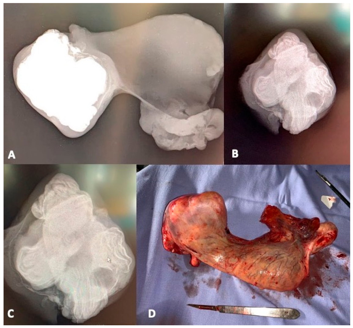
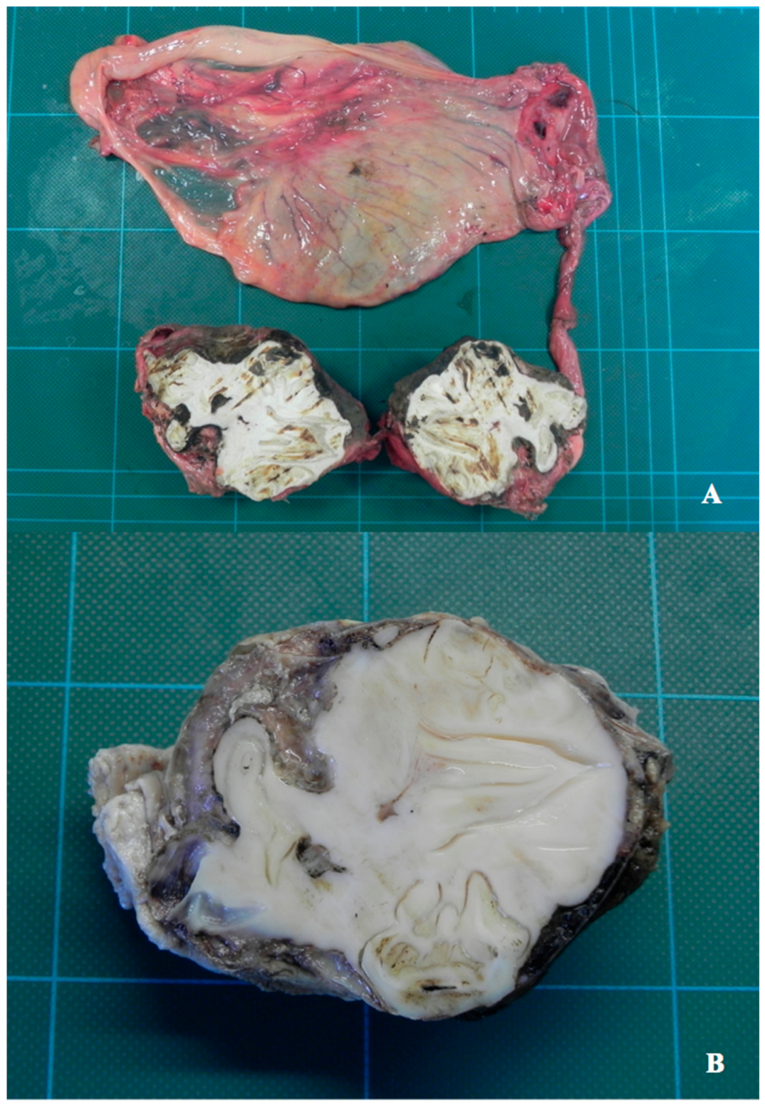
Once the testicle was extracted from the abdomen, considering its anomalous shape and its hard and non-deformable state of consistency, we decided to take radiographs. The teratoma appeared to predominantly involve the structure of the epididymis. Macroscopic examination showed the removed testicle with an enlarged oval shape of 9 × 6.5 × 6 cm, with a very hard “bony” compact consistency due to the internally formed component (teeth). After a necessary cross cut made with a band saw, the cut surface allowed us to detect the presence of four morphologically distinct elements, whitish in color, hard, and apparently normal shaped, referable to well differentiated dental structures, immersed in a hard dentinal-osseous amorphous matrix, which occupied most of the entire cut surface, replacing what must have been the normal structural tissue of the testis (
Figure 1 and
Figure 2).
Several tissue samples were collected and fixed in 10% buffered formalin, decalcified with aqueous solution of formic and hydrochloric acid for more than 20 days; subsequently, they were embedded in paraffin, cut into sections of 4–5 µm, and then stained with Hematoxylin-Eosin.
2.2. Histological Features
In general, the histopathological investigations revealed a neoplastic lesion composed mainly of different types of mature tissues by many germinal layers. In this case bone, enamel, dentin and mineralized areas represented the predominant part of the tumor which were also associated with separate and more marginal foci of mature fibrous tissue and tubular-like and glandular-like structures lined with mucosa, arranged in a markedly disorganized way, unlike the teeth which showed a well-ordered structural organization especially in the enamel and dentin components. In this case, no malignancy was detected at histopathological investigations [
Figure 3].
3. Discussion
Reproductive disorders are common in horses, and cryptorchidism is one of the most common conditions where surgical approaches to remove the testes are the most effective and low risk treatment.
Teratomas are tumors consisting of mature and immature tissues derived from pluripotent cells of all three germ layers, and which arise from multipotential germ cells that have undergone partial differentiation. In teratomas, ectodermal (skin derivatives and neural tissue), mesodermal (bone, fat, cartilage and muscle) and endodermal (gastrointestinal and bronchial epithelium, thyroid) tissues can coexist.
Teratoma occurs mostly in cryptorchid testicles in horses, where it is reported in an age range of under one year to a maximum of five years, considering it is most likely congenital [
15,
16,
17,
18]. It can be macroscopically differentially diagnosed with tumors such as extraskeletal osteosarcoma, adamantinoma, and undifferentiated teratocarcinoma, but histopathology analysis is able to clarify the exact nature of the tumor under examination [
19].
4. Conclusions
Based on our information, this case-report represents the first case described in literature of a dentigerous equine teratoma in this species. Our bibliographic research found only very few cases of teratomas with ectopic teeth described in the human species, mainly localized in the orbital region or in the lung, and never at testicular level and with related primitive onset. Laparoscopy is the best surgical technique to approach this type of condition because it is minimally invasive and offers good visibility with very few and infrequent complications as described by D. Hendrickson [
20].
To our knowledge the surgery was well tolerated by the horse [
20], which is in good health and has returned to regular racing performance. The other scrotal testicle was left at the behest of the owner and showed no clinical signs of disease.
Author Contributions
For this research the individual contributions by singular author are represented by: L.L.: conceptualization, methodology, writing original draft preparation, investigations, supervision, writing—review editing. A.B.: conceptualization, writing—review and editing. E.B.: conceptualization. I.P.: conceptualization, methodology. R.G.: conceptualization, methodology, clinical investigations, writing-review editing. All authors have read and agreed to the published version of the manuscript.
Funding
This research received no external funding.
Institutional Review Board Statement
Ethical review and approval were waived for this study We declare that the biological and surgical material used and described in this manuscript is represented by primary neoformed tissues, taken from the animal in question for exclusively diagnostic and therapeutic uses.
Informed Consent Statement
Informed consent was obtained from all subjects involved in the study. An experimental use of the material is categorically excluded and that the horse affected by the tumor was brought personally and spontaneously by the owner who also gave his approval to the subsequent clinical-diagnostic and therapeutic investigations.
Data Availability Statement
The data presented in this study are available in the manuscript.
Acknowledgments
The authors wish to thank Alberto Leonardi for graphic work, Sara Leto, Valeria Migni, Giampaolo Ceccarani, and Luca Stefanelli for their precious technical scientific support and Katarzyna Malgorzata Walczak (Poland) for her English editing support.
Conflicts of Interest
The authors declare that the research was conducted in the absence of any commercial or financial relationships that could be construed as a potential conflict of interest.
References
- Ramkumar, J.; Best, A.; Gurung, A.; Dufresne, A.M.; Melich, G.; Vikis, E.; MacKenzie, S. Resection of ruptured hepatic tera-toma in an adult. Int. J. Surg. Case Rep. 2018, 53, 414–419. [Google Scholar] [CrossRef] [PubMed]
- Ashley, D.J.B. Origin of teratomas. Cancer 1973, 32, 390–394. [Google Scholar] [CrossRef]
- AlQurashi, A.; Bakry, E.; Straube, M.; Rickert, C.H.; Mir-Salim, P. Mature Teratoma of the Temporal Bone in 3.5-Month-Old Baby Girl. Case Rep. Otolaryngol. 2015, 2015, 372089. [Google Scholar] [CrossRef] [PubMed] [Green Version]
- Lefebvre, R.; Theoret, C.; Doré, M.; Girard, C.; Laverty, S.; Vaillancourt, D. Ovarian teratoma and endometritis in a mare. Can. Veter. J. La Rev. Veter. Can. 2005, 46, 1029–1033. [Google Scholar]
- Maghrabi, Y.; Kurdi, M.E.; Baeesa, S.S. Infratentorial immature teratoma of congenital origin can be associated with a 20-year survival outcome: A case report and review of literature. World J. Surg. Oncol. 2019, 17, 22. [Google Scholar] [CrossRef] [Green Version]
- Ting, J.; Bell, D.; Ahmed, S.; Ying, A.; Waguespack, S.G.; Tu, S.M.; Weber, R.; Zafereo, M. Primary Malignant Thyroid Teratoma: An Institutional Experience. Thyroid 2019, 29, 229–236. [Google Scholar] [CrossRef]
- Cribb, N.C.; Bourã, L.P.; Bouré, L.P. Laparoscopic Removal of a Large Abdominal Testicular Teratoma in a Standing Horse. Veter. Surg. 2010, 39, 131–135. [Google Scholar] [CrossRef] [PubMed]
- Meliti, A.; Hafiz, B.; Al-Maghrabi, H.; Gari, A. Collision Glial Neoplasms Arising in an Ovarian Mature Cystic Teratoma: A Rare Event. Case Rep. Pathol. 2020, 2020, 7568671–7568674. [Google Scholar] [CrossRef] [PubMed]
- Fox, L.; Hanley, C.S.; Padilla, L.R.; Duncan, M. Bladder teratoma in a maned wolf (Chrysocyon brachyurus). Open Veter. J. 2019, 9, 259–262. [Google Scholar] [CrossRef] [PubMed] [Green Version]
- Meuten, D.J. Tumors of the Genital System (Testicle). In Tumors in Domestic Animals, 5th ed.; 1606 Golden Aspen Drive, Suites 103 and 104: Ames, IA, USA, 2017; pp. 706–721. [Google Scholar]
- Kumar, V.; Abbas, A.K.; Aster, J.C. Robbins—Fondamenti di Patologia e di Fisiopatologia, 9th ed.; Edra Masson LSWR Srl: Milano, Italy, 2013; p. 646. [Google Scholar]
- Ugolini, L.W.; Cunha dos Santos, F.C.; da Costa, G.V.; Oliveira, H.R.; Folchini, N.; Machad, T.P.; Zannella, R.; Porto Alves, L. Testicular Teratoma in a Unilateral RightSided Abdominal Cryptorchid Horse. Acta Sci. Vet. 2019, 47, 409. [Google Scholar]
- Ulbright, T.M. Germ cell tumors of the gonads: A selective review emphasizing problems in differential diagnosis, newly appreciated, and controversial issues. Mod. Pathol. 2005, 18, S61–S79. [Google Scholar] [CrossRef] [PubMed]
- Nielsen, S.W.; Misdorp, W.; Mcentee, K. Tumours of the ovary. Bull. World Health Organ. 1976, 53, 203–215. [Google Scholar] [PubMed]
- Pasolini, M.P.; Della Valle, G.; Pagano, T.B.; Miele, F.; Paciello, O.; Fatone, G.; Greco, M. Mature teratoma arising from an un-descended testis in a horse: Comparison between ultrasonographic and morphological features. Folia Morphol. 2016, 75, 211–215. [Google Scholar] [CrossRef] [PubMed] [Green Version]
- Smyth, G.B. Testicular Teratoma in an Equine Cryptorchid. Equine Veter. J. 1979, 11, 21–23. [Google Scholar] [CrossRef] [PubMed]
- Chowdhury, W.; Lodhi, M.U.; Syed, I.A.; Rahim, U.; Rahim, M. Mature Testicular Teratoma with a Focus of Embryonal Car-cinoma: A Case Report and Review of Literature. Cureus 2018, 10, e2329. [Google Scholar]
- Baidya, K.P.; Ghosh, S.; Datta, A.; Mukhopadhyay, S.; Bhaduri, G. Huge congenital teratoma containing tooth in a three-day-old neonate. Oman J. Ophthalmol. 2014, 7, 13–15. [Google Scholar] [CrossRef] [PubMed]
- Bouzoubaa, W.; Jayi, S.; Alaoui, F.Z.F.; Chaara, H.; Melhouf, M.A. Tératome immature de l’ovaire: À propos d’un cas [Imma-ture teratoma of the ovary: About a case. Pan. Afr. Med. J. 2017, 27, 263. [Google Scholar] [CrossRef] [PubMed]
- Hendrickson, D. Laparoscopic Cryptorchidectomy and Ovariectomy in Horses. Veter. Clin. N. Am. Equine Pr. 2006, 22, 777–798. [Google Scholar] [CrossRef] [PubMed]
| Publisher’s Note: MDPI stays neutral with regard to jurisdictional claims in published maps and institutional affiliations. |
© 2021 by the authors. Licensee MDPI, Basel, Switzerland. This article is an open access article distributed under the terms and conditions of the Creative Commons Attribution (CC BY) license (https://creativecommons.org/licenses/by/4.0/).


