Article

The SCCmec Types and Antimicrobial Resistance among Methicillin-Resistant Staphylococcus Species Isolated from Dogs with Superficial Pyoderma

by
Yuttana Chanayat
1,
Areerath Akatvipat
2,
Jeff B. Bender
3,
Veerasak Punyapornwithaya
4,
Tongkorn Meeyam
4,5,
Usanee Anukool
6,7 and
Duangporn Pichpol
4,5,*
1
Master’s Degree Program in Veterinary Science, Faculty of Veterinary Medicine, Chiang Mai University, Chiang Mai 50100, Thailand
2
Department of Companion Animals and Wildlife Clinic, Faculty of Veterinary Medicine, Chiang Mai University, Chiang Mai 50100, Thailand
3
College of Veterinary Medicine and the School of Public Health, University of Minnesota, Minneapolis, MN 55455, USA
4
Veterinary Public Health and Food Safety Centre for Asia Pacific, Faculty of Veterinary Medicine, Chiang Mai University, Chiang Mai 50100, Thailand
5
Department of Veterinary Biosciences and Veterinary Public Health, Faculty of Veterinary Medicine, Chiang Mai University, Chiang Mai 50100, Thailand
6
Division of Clinical Microbiology, Department of Medical Technology, Faculty of Associated Medical Sciences, Chiang Mai University, Chiang Mai 50200, Thailand
7
Infectious Diseases Research Unit (IDRU), Faculty of Associated Medical Sciences, Chiang Mai University, Chiang Mai 50200, Thailand
*
Author to whom correspondence should be addressed.
Submission received: 4 March 2021 / Revised: 7 May 2021 / Accepted: 10 May 2021 / Published: 13 May 2021
(This article belongs to the Special Issue Resistant Staphylococci in Animals)

Abstract

:
This study characterizes clinical methicillin-resistant staphylococcal (MRS) isolates obtained from superficial pyoderma infections in dogs. Our interest was to determine the staphylococcal cassette chromosome mec (SCCmec) type and the antimicrobial susceptibility among MRS isolates from clinical cases. Skin swabs were collected and cultured. Staphylococcus species were identified and characterized with biochemical tests and MALDI-TOF-MS and antimicrobial susceptibility testing by disk diffusion. mecA detection and staphylococcal cassette chromosome mec (SCCmec) typing were achieved by PCR. Of the 65 clinical samples, 56 (86.2%) staphylococcal infections were identified. Twelve (21%) of 56 isolates were MRS infections. All MRS isolates were multidrug resistant. The ccrC and class-C2 mec, which were SCCmec type V, were the most prevalent (66.7%) among the 12 MRS isolates. The predominant SCCmec type V was found in S. aureus, S. intermedius group, S. lentus, S. xylosus, and S. arlettae. Treatment failure is a concern with the emergence of highly resistant MRS in dogs associated with superficial pyoderma. The detection of type V SCCmec MRS has previously been reported among veterinarians and dog owners but not in Northern Thailand. These infections serve as a reminder to improve infection prevention and control measures including reducing environmental contamination and potential zoonotic exposures to MRS. In addition, educational awareness of these risks in small animal hospitals needs to be increased among veterinary hospital staff, clients, and patients.

1. Introduction

Infection with methicillin-resistant staphylococci (MRS) is an ongoing and emerging health concern among both humans and animals. Methicillin-resistant Staphylococcus aureus (MRSA) not only causes hospital-, community-, and livestock-associated infections in humans, but it can also lead to infections in pet animals [1]. Methicillin-resistant Staphylococcus pseudintermedius, which belongs to the S. intermedius group (MRSIG), can cause serious wound infections in domestic companion animals such as dogs and cats. It may also be capable of spreading between animals and humans, including veterinarians, companion animal owners, and veterinary nurses [2,3]. Methicillin-resistant coagulase-negative staphylococci can persist in the patient environment and serve as a source of human infection in hospital settings [4]. MRS is recognized as a public health threat and is a significant opportunistic pathogen in both human and veterinary medicine [5].
The evolution of MRS emerged from the development of the penicillin-binding protein 2a (PBP2a), which is encoded by the mecA gene [1]. This protein has a significantly lower affinity for beta-lactams and thus cell wall synthesis can continue even in the presence of normally inhibitory concentrations of beta-lactam antibiotics. Thus, the detection of mecA by polymerase chain reaction (PCR) is used to characterize MRS [2,5]. The mecA gene is the mobile genetic element located in the chromosome of Staphylococcus known as the staphylococcal cassette chromosome mec, or SCCmec [6]. There are four important components of the SCCmec element: (1) the group comprised of the mec genes; the mecA gene and the regulatory genes, (2) the group comprised of the ccr genes responsible for the mobility of the SCCmec element, (3) the direct repeated nucleotide at the end of both sides of SCCmec making the movable structure of the SCCmec element, and (4) the 3′ ending part of the orfX gene [7]. Currently, SCCmec is classified into 13 types, type I to type XIII, according to the differences of the mec gene and the ccr gene [8]. A multiplex PCR is used to determine the structure of the mec complex, while the presence of different ccr genes is a popular method for SCCmec typing [9]. Staphylococcal infections in companion animals, particularly in dogs and cats, include superficial pyoderma, otitis externa, and superficial bacterial folliculitis. Occasionally, MRS is also detected among companion animal owners. These are mostly multidrug-resistant MRS infections [10,11]. According to prior reports, the identification of various MRS isolates, such as MRSA, MRSIG, and MRCNS, are rarely reported in Thailand. The objectives of this study were to characterize the type of staphylococcal infections associated with superficial pyoderma among dogs at the Small Animal Teaching Hospital, Chiang Mai University, Thailand.

2. Materials and Methods

2.1. Data and Sample Collection

Cases of superficial pyoderma in dogs were identified by clinical staff at our teaching hospital from 2015 to 2017. Sixty-five dogs presenting with superficial pyoderma at the Small Animal Teaching Hospital, Faculty of Veterinary Medicine, Chiang Mai University, Thailand were swabbed and submitted to the veterinary diagnostic laboratory for microbiological analysis.

2.2. Bacterial Isolation and Identification

The skin swabs were kept in Stuart transport medium (BD BBLTM, US) and submitted to the microbiology laboratory within 4–6 h for bacterial identification and antimicrobial susceptibility testing. Standard bacterial isolation procedures were used as follows. Staphylococcal isolates were identified in cultures using 5% of sheep blood agar (Merck, Darmstadt, Germany) along with a biochemical testing process that employed Gram stain, catalase, tube coagulase, Voges–Proskauer (VP) test, and the clumping factor [12]. All staphylococcal species were confirmed by MALDI-TOF-MS analysis.

2.3. Antimicrobial Susceptibility Testing

Antimicrobial susceptibility testing was performed by the disk diffusion method according to the Clinical and Laboratory Standards Institute (CLSI) recommendations [13]. Sixteen antimicrobial agents, including penicillin, ampicillin, cefoxitin, amoxicillin/clavulanate, cefazolin, cefpodoxime, amikacin, gentamicin, doxycycline, ciprofloxacin, chloramphenicol, clindamycin, erythromycin, linezolid, rifampin, and trimethoprim/sulfamethoxazole, were used in this study. The minimum inhibitory concentration (MIC) of oxacillin was determined for all Staphylococcus isolates by Vitek 2 system. All MRS isolates were identified using the CLSI oxacillin MIC breakpoints: MIC ≥ 4 µg/mL for S. aureus and MIC ≥ 0.5 µg/mL for other Staphylococcus spp. [13]. Staphylococcus aureus ATCC 25923 was used as a quality control strain.

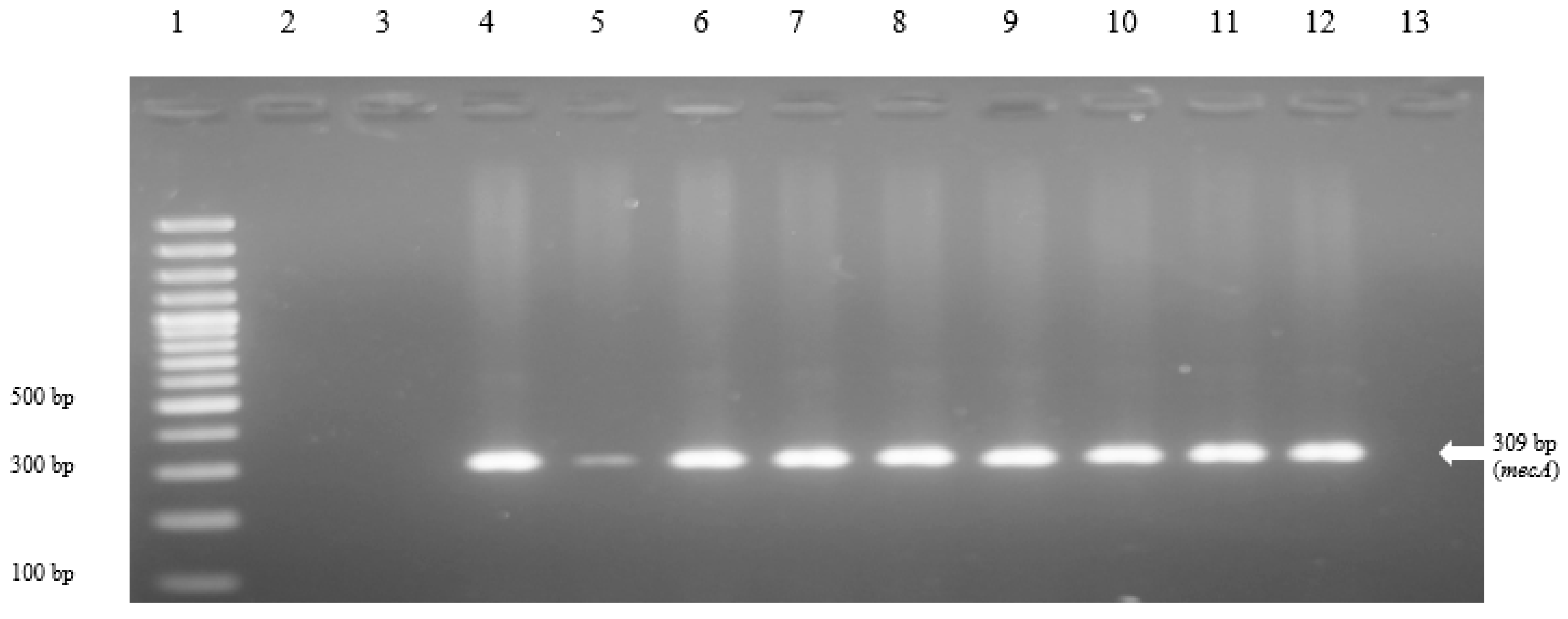
2.4. PCR Detection of mecA Gene
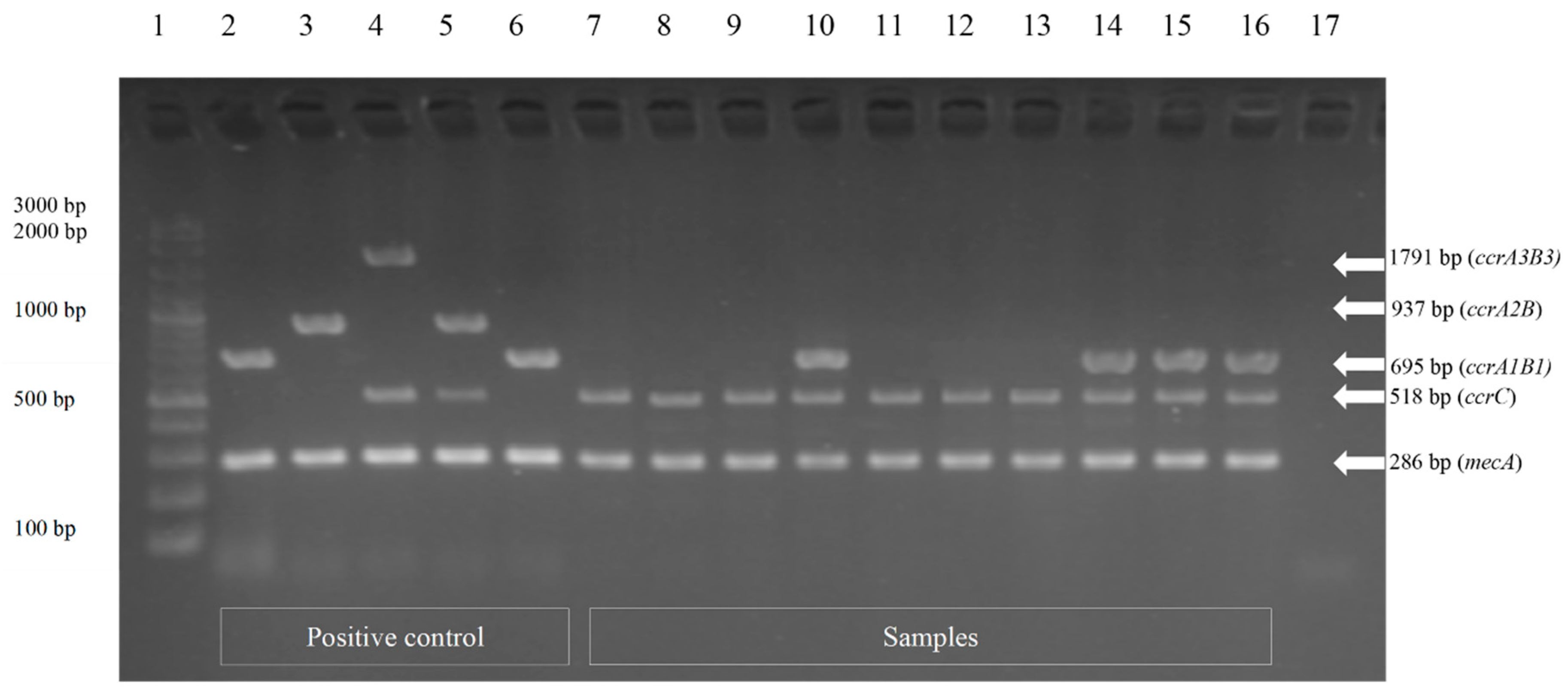
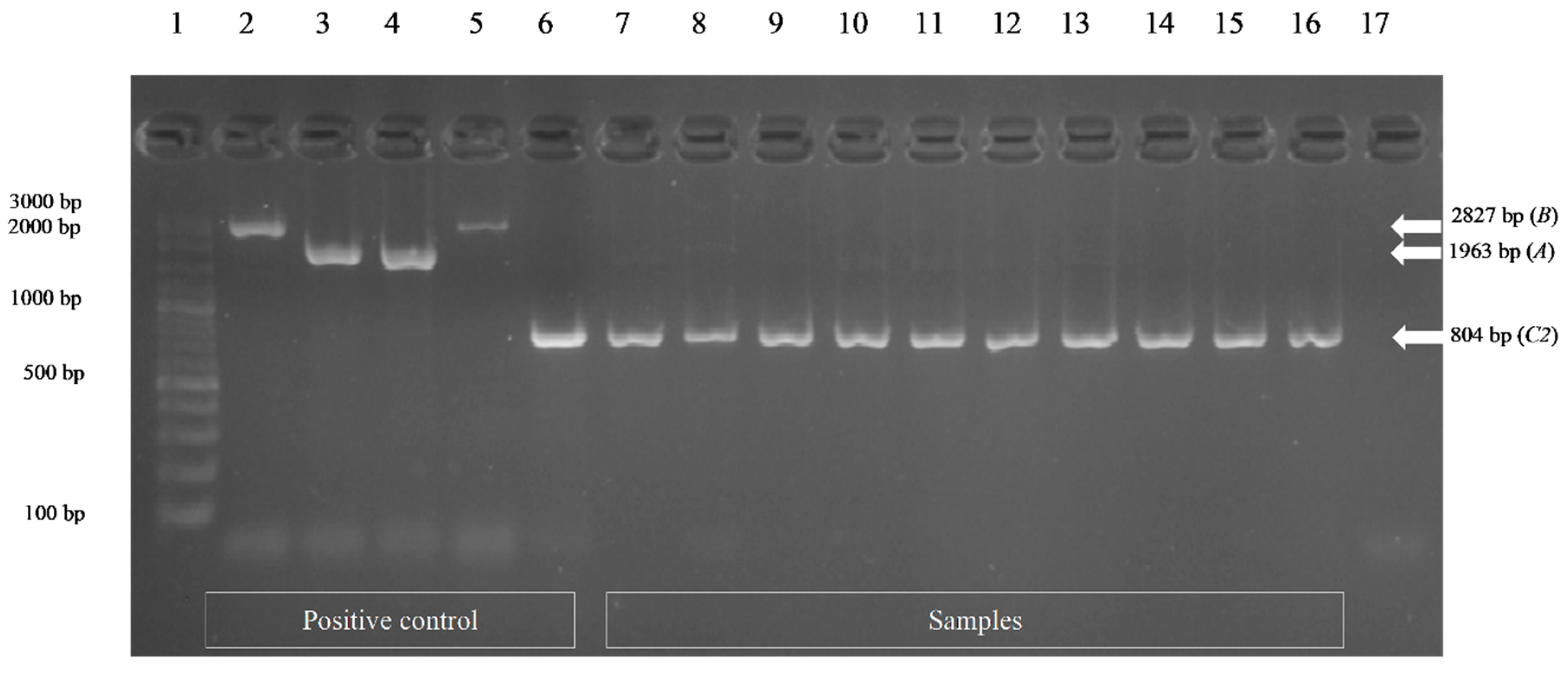
DNA was extracted using a commercial test kit for genomic DNA obtained from tissue samples (NucleoSpin R Tissue). The mecA genotype of staphylococci was characterized. The 50-µL PCR reaction mixtures contained 100 ng chromosomal DNA, oligonucleotide primers (10 pmols), 2.5 mM each deoxynucleotide triphosphates, Taq buffer, and 5 U Taq polymerase (I-TagTM plus iNtRON Biotechnology, Korea) at a final volume of 50 µL. A PCR thermal cycler was used for amplification with an initial denaturation step (94 °C, 5 min) comprised of 40 cycles of denaturation (95 °C, 45 s), an annealing step (59 °C, 45 s), and an extension step (72 °C, 45 s); and a final extension step at 72 °C for 5 min. Regarding the primer pairs used for PCR experiments, the forward primer was 5′-TGGCTATCGTGTCACAATCG-3′, the reverse primer was 5′-CTGGAACTTGT TGAGCAGAG-3′, and the product size was 309 base pairs [14].

2.5. SCCmec Typing

The SCCmec typing of all mecA gene-positive staphylococcal isolates was performed using the multiplex PCR, M-PCR1, and MPCR2, as previously described by [15]. The M50-µL reaction mixtures of M-PCR1 contained 100 ng chromosomal DNA, oligonucleotide primers (0.1 µM), 200 µM each deoxynucleotide triphosphates, Taq buffer, and 2.5 U DreamTaq DNA polymerase (Thermo Fisher Scientific UAB, Vilnius, Lithuania) at a final volume of 50 µL. The concentration of MgCl2 was 3.2 mM. A PCR thermal cycler was used for amplification with an initial denaturation step (94 °C, 2 min) comprised of 30 cycles of denaturation (94 °C, 2 min), an annealing step (57 °C, 2 min), and an extension step (72 °C, 2 min); and a final extension step at 72 °C for 2 min. The 50-µL reaction mixture of M-PCR2 contained the same components as the M-PCR1 except that the concentration of MgCl2 was 2 mM and the annealing temperature was raised to 60 °C for 2 min. The primer pairs used for PCR experiments are listed in Tables S1 and S2. The M-PCR 1 for ccr type assignment contained two primers used to detect mecA and eight primers used for the identification of five ccr genes: four primers including a common forward primer (common to ccrB1–3) and three reverse primers specific for ccrA1, ccrA2, and ccrA3 used to identify ccr1–3 based on the differences present in the ccrA genes; two primers used to identify ccr4; and two primers used to identify ccr5. Furthermore, the M-PCR2 for mec class assignment contained four primers that were used to identify the gene lineages of mecA–mecI (class A mec), mecA-IS1272 (class B mec) and mecAIS431 (class C mec). The positive control strains for SCCmec typing were 4 MRSA strains, including epidemic MRSA (EMRSA)-8 (SCCmec type I), N315 (SCCmec type II), EMRSA-4 (SCCmec type III), and EMRSA-10 (SCCmec type IV).

2.6. Statistical Analysis

Percentages of antimicrobial susceptibility were calculated for staphylococci and MRS isolates. The antimicrobial susceptibility was categorized as 3 groups, including susceptible, intermediate, or resistant, according to the breakpoint of the inhibition zone or MIC as recommended by CLSI M100-S26 guidelines [13]. Data were visualized and analyzed using R version 4.0.3 [16].

3. Results

3.1. Staphylococcal Infections in Dogs with Superficial Pyoderma

Staphylococci were isolated from 56 (86.2%) of 65 clinical samples. Two isolates (3.1%) were identified as Staphylococcus aureus and 50 isolates (76.9%) as the S. intermedius group. In addition, one isolate each of coagulase-negative staphylococci, S. lentus, S. xylosus, S. lugdunensis, and S. arlettae, were found. For nine culture-negative samples, non-Staphylococcus bacteria were detected including beta-hemolytic Streptococcus Group C, Escherichia coli, Aerococcus viridans, Proteus mirabilis, Pseudomonas aeruginosa, Rothia nasimurium, Klebsiella pneumoniae, Moraxella sp., Corynebacterium auriscanis, and Enterococcus faecalis. From available information, the mean age of the patients was 6.2 years of age (range from 0.5 to 14 years). Twenty-eight dogs were intact males, 20 were female, and one was a spayed female. Eight (16%) dogs presented with papules, 19 (34%) presented with pustules, 35 (63%) presented with crusts, and 20 (36%) presented with epidermal collarettes. Eleven patients had recurring pyoderma and 37 had other underlying medical conditions such as atopic dermatitis, food allergies, demodicosis, hyperadrenocorticism, or hypothyroidism. Fifteen (31%) of 49 dogs with reviewable records were previously treated with antibiotics (cephalexin (n = 8), amoxicillin–clavulanate (n = 4), doxycycline (n = 3).

3.2. Antimicrobial Susceptibility Testing Results

Most staphylococci were found to be resistant to penicillin and ampicillin. All staphylococci were susceptible to linezolid and rifampin (Table 1). Twelve staphylococcal isolates were resistant to oxacillin and cefoxitin and were confirmed as MRS isolates. One isolate was S. aureus, eight isolates were in the S. intermedius group, and three isolates were identified as coagulase-negative staphylococci: S. lentus, S. xylosus, and S. arlettae. All MRS isolates were resistant to penicillin, cephalosporin, and fluoroquinolone. Notably, the mecA gene was found to be present in all methicillin-resistant staphylococci (Figure 1). Epidemiologic data were available for 10 dogs. The mean age of these 10 dogs was 6.3 years of age (ranging from 1 to 13 years). Six of the dogs were intact males and four were female dogs. Two (20%) dogs presented with papules, four (40%) presented with pustules, eight (80%) presented with crusts, and four (40%) presented with epidermal collarettes. Five of ten dogs had recurring pyoderma and five had underlying medical conditions. Furthermore, eight of the ten subjects had previously been prescribed antibiotics.

3.3. SCCmec Types of Methicillin-Resistant Staphylococcal Isolates

There were 12 isolates of MRS that were SCCmec typed using the multiplex PCR. M-PCR1 successfully amplified DNA fragments corresponding in size to each ccr gene (Figure 2) and M-PCR2 was applied to assign the mec class (Figure 3). Characterization results from all 12 MRS isolates revealed that ccrC and class-C2 mec were found from one isolate (8.3%) of S. aureus; thus, it could be classified as SCCmec type V. These components were also found in four isolates (33.3%) of the S. intermedius group. The ccrA1B1, ccrC, and class-C2 mec were found in four isolates (33.3%) of the S. intermedius group and they were categorized as SCCmec non-typeable strains. The coagulase-negative staphylococci, including S. lentus, S. xylosus, and S. arlettae, were found to carry ccrC and class-C2 mec. These strains were classified as SCCmec type V (Table 2).

4. Discussion

This study represents, to our knowledge, the first molecular characterization of MRS isolates obtained from canines with superficial pyoderma in Northern Thailand. The presence of mecA gene encoding PBP2a in all MRS isolates was confirmed along with the antimicrobial susceptibility profiles highlighting potential treatment failures with commonly used antimicrobials [17]. One MRSA isolate was recovered from a dog with superficial pyoderma. This emphasizes the need for clinical awareness of antimicrobial-resistant organisms and potential treatment failures and the need for optimum infection control measures in clinical settings to prevent healthcare-associated infections. Direct contact with an infected animal can be a risk for human pet owners, especially those with underlying health conditions [18]. Dogs are at risk for carrying MRS and MRSA and can be colonized with resistant strains without showing clinical signs [10,19,20]. Transmission can occur through close contact [21,22]. These findings highlight the need for veterinarian awareness of potential multidrug-resistant infections, and the importance of providing client information about the management and care of patients and the appropriate hand hygiene practices to reduce the transmission risk of multidrug-resistant infections between animals and humans.
Staphylococcal infections in domestic companion animals, especially in dogs and cats, can commonly cause superficial pyoderma, otitis externa, superficial bacterial folliculitis, and bacterial rhinitis. The main cause of these infections are often S. pseudintermedius [10,23,24]. The S. intermedius group (SIG) accounted for the majority of the isolates recovered from clinical pyoderma cases in this study. These SIG isolates from dogs with pyoderma could not be conclusively identified in the absence of molecular testing and they were indicated as S. pseudintermedius/S. intermedius. Subsequent analysis of two strains by MALDI-TOF-MS systems (VITEK MS, bioMérieux, Marcy l’Etoile France) gave the same results. Although S. pseudintermedius has been recognized as a major pathogen among SIG isolates, the multiplex PCR method for species identification of coagulase-positive staphylococci by targeting the nuc gene locus [25] should be performed. Currently, the concept of SIG is becoming more and more broad. Recently, two species, S. cornubiensis and S. ursi, have been added to the SIG [26,27]; however, they were not present in the dogs tested here. There are some reports which document MRS infection in companion animals, their owners, veterinarians, and other animal caretakers [22,28,29,30,31]. MRS can readily spread and can contaminate the immediate environment, including surfaces in veterinary clinics [32].
Previous studies have documented the increasing frequency of the recovery of MRS in canine pyoderma [33,34]. These multidrug-resistant infections identified in companion animals are difficult to treat [2,35]. For our study, MRS isolates were resistant to fluoroquinolones. The International Society for Companion Animal Infectious Diseases (ISCAID) has devised some important diagnostic and treatment approaches [36]. It is, therefore, necessary and important for Thailand and other South East Asian countries with limited culture and epidemiologic data to recognize the emergence of resistant staphylococcal infections and to follow appropriate antimicrobial stewardship principles. Awareness of the emergence of MRS is important to veterinary practitioners and future veterinarians. Due to the limited effectiveness of antimicrobials to treat MRS, good hygienic and infection control practices are needed to prevent potential environmental contamination and spread to other animals and humans.
Our study also documented that staphylococcal isolates contained SCCmec types V. This SCCmec type has previously been found in dogs living in the central part of Thailand [10]. However, these MRS strains were isolated by swabbing the nares and perineum of healthy dogs and not from the dogs with pyoderma. In addition, SCCmec type V MRS samples were isolated from veterinarians and the owners of dogs in the study area [10]. Recently, studies of SCCmec typing in cats and dogs from other geographic areas indicated the presence of type V and type VII SCCmec MRS in Asia. The type V SCCmec was the most commonly detected type in MRS in Europe and America [11,37,38,39,40].

5. Conclusions

MRS, including S. aureus, S. intermedius group, and coagulase-negative Staphylococcus with multidrug resistance phenotypes, was isolated from dogs with superficial pyoderma. These MRS infection findings pose certain diagnostic and treatment challenges for South East Asian veterinary practitioners and highlight the need for improved antimicrobial stewardship and hygienic practices. This includes diagnostic recognition and new treatment approaches. It also reminds veterinarians about the potential for zoonotic transmission of multidrug-resistant organisms. Veterinarians should consider appropriate treatment options, including effective topical treatments for mild cases to reserve the use of important parenteral treatments if needed. Newly released recommendations can serve as established approaches for the treatment of superficial pyoderma. The emergence of type V SCCmec MRS has broad implications for companion animals, pet owners, veterinarians, and animal caretakers. The emergence of these MRS strains can contaminate the hospital environment and are a public health concern.

Supplementary Materials

The following are available online at https://0-www-mdpi-com.brum.beds.ac.uk/article/10.3390/vetsci8050085/s1, Table S1: M-PCR1 for amplification of ccr gene complex type with mecA, Table S2: M-PCR2 for amplification of mec gene complex class.

Author Contributions

Conceptualization, A.A., J.B.B., V.P. and D.P.; methodology, J.B.B., U.A. and D.P.; software, V.P.; validation, D.P. and U.A.; formal analysis, Y.C.; investigation, Y.C. and A.A.; resources, D.P.; acquisition of data, A.A.; data curation, Y.C.; writing—original draft preparation, Y.C.; writing—review and editing, D.P., T.M., U.A., J.B.B. and V.P.; visualization, D.P.; supervision, D.P.; project administration, V.P.; funding acquisition, D.P. and V.P. All authors have read and agreed to the published version of the manuscript.

Funding

This research was funded by the Project of Antibiotic Stewardship in Companion Animal Practice jointly with Chiang Mai University/the University of Minnesota, College of Veterinary Medicine (R000012898), Excellent Center of Veterinary Public Health (R000016652), and Research Group for Veterinary Public Health (R000026062), Faculty of Veterinary Medicine, Chiang Mai University, Thailand.

Institutional Review Board Statement

Animal ethical approval for this study was granted as follows: CMU-ACUC Ref. No. R3/2558 Animal Use Protocol Titles: Antibiotic Stewardship in Companion Animal Practice by Animal Care and Use Committee, the Faculty of Veterinary Medicine, Chiang Mai University, Thailand (FVM-ACUC).

Informed Consent Statement

Informed Consent Statement was obtained from the owners of Sixty-five dogs presenting with superficial pyoderma.

Data Availability Statement

The data presented in this study are available on request from the corresponding author. The data are not publicly available due to institutional privacy policy.

Acknowledgments

The authors gratefully acknowledge the Small Animal Hospital, Faculty of Veterinary Medicine, Chiang Mai University, Thailand for supporting the process of obtaining samples of skin swabs from superficial pyoderma dogs. We further thank the College of Veterinary Medicine and the School of Public Health, University of Minnesota Marshfield Medical Research Laboratory, which confirmed the results of staphylococci and methicillin-resistant staphylococci detection. We also express our appreciation to the Division of Clinical Microbiology, Department of Medical Technology, Faculty of Associated Medical Sciences, Chiang Mai University, Thailand for kindly providing the facilities essential for SCCmec typing.

Conflicts of Interest

The authors declare no conflict of interest. The funders had no role in the design of the study; in the collection, analyses, or interpretation of data; in the writing of the manuscript, or in the decision to publish the results.

References

  1. Pomba, C.; Rantala, M.; Greko, C.; Baptiste, K.E.; Catry, B.; van Duijkeren, E.; Mateus, A.; Moreno, M.A.; Pyörälä, S.; Ružauskas, M.; et al. Public health risk of antimicrobial resistance transfer from companion animals. J. Antimicrob. Chemother. 2017, 72, 957–968. [Google Scholar] [CrossRef] [PubMed]
  2. Couto, N.; Monchique, C.; Belas, A.; Marques, C.; Gama, L.T.; Pomba, C. Trends and molecular mechanisms of antimicrobial resistance in clinical staphylococci isolated from companion animals over a 16 year period. J. Antimicrob. Chemother. 2016, 71, 1479–1487. [Google Scholar] [CrossRef] [PubMed]
  3. Wu, M.T.; Burnham, C.A.; Westblade, L.F.; Dien Bard, J.; Lawhon, S.D.; Wallace, M.A.; Stanley, T.; Burd, E.; Hindler, J.; Humphries, R.M. Evaluation of oxacillin and cefoxitin disk and MIC Breakpoints for prediction of methicillin resistance in human and veterinary isolates of Staphylococcus intermedius group. J. Clin. Microbiol. 2016, 54, 535–542. [Google Scholar] [CrossRef] [PubMed] [Green Version]
  4. Wilson, A.P.; Smyth, D.; Moore, G.; Singleton, J.; Jackson, R.; Gant, V.; Jeanes, A.; Shaw, S.; James, E.; Cooper, B.; et al. The impact of enhanced cleaning within the intensive care unit on contamination of the near-patient environment with hospital pathogens: A randomized crossover study in critical care units in two hospitals. Crit. Care Med. 2011, 39, 651–658. [Google Scholar] [CrossRef] [PubMed]
  5. Igbinosa, E.O.; Beshiru, A.; Akporehe, L.U.; Ogofure, A.G. Detection of methicillin-resistant staphylococci isolated from food producing animals: A public health implication. Vet. Sci. 2016, 3, 14. [Google Scholar] [CrossRef] [PubMed] [Green Version]
  6. IWG-SCC. Classification of staphylococcal cassette chromosome mec (SCCmec): Guidelines for reporting novel SCCmec elements. Antimicrob. Agents Chemother. 2009, 53, 4961–4967. [Google Scholar] [CrossRef] [PubMed] [Green Version]
  7. Turlej, A.; Hryniewicz, W.; Empel, J. Staphylococcal cassette chromosome mec (Sccmec) classification and typing methods: An overview. Pol. J. Microbiol. 2011, 60, 95–103. [Google Scholar] [CrossRef]
  8. Baig, S.; Johannesen, T.B.; Overballe-Petersen, S.; Larsen, J.; Larsen, A.R.; Stegger, M. Novel SCCmec type XIII (9A) identified in an ST152 methicillin-resistant Staphylococcus aureus. Infect. Genet. Evol. 2018, 61, 74–76. [Google Scholar] [CrossRef]
  9. Deurenberg, R.H.; Stobberingh, E.E. The evolution of Staphylococcus aureus. Infect. Genet. Evol. 2008, 8, 747–763. [Google Scholar] [CrossRef]
  10. Chanchaithong, P.; Perreten, V.; Schwendener, S.; Tribuddharat, C.; Chongthaleong, A.; Niyomtham, W.; Prapasarakul, N. Strain typing and antimicrobial susceptibility of methicillin-resistant coagulase-positive staphylococcal species in dogs and people associated with dogs in Thailand. J. Appl. Microbiol. 2014, 117, 572–586. [Google Scholar] [CrossRef] [Green Version]
  11. Van Duijkeren, E.; Catry, B.; Greko, C.; Moreno, M.A.; Pomba, M.C.; Pyörälä, S.; Ruzauskas, M.; Sanders, P.; Threlfall, E.J.; Torren-Edo, J.; et al. Review on methicillin-resistant Staphylococcus pseudintermedius. J. Antimicrob. Chemother. 2011, 66, 2705–2714. [Google Scholar] [CrossRef] [PubMed] [Green Version]
  12. Baron, E.J.; Peterson, L.R.; Finegold, S.M. Baily & Scotts Diagnostic Microbiology, 9th ed.; Mosby-Yearbook, Inc.: Missouri, MO, USA, 1994; pp. 321–330. ISBN 0-8016-6987-1. [Google Scholar]
  13. CLSI. Performance Standards for Antimicrobial Susceptibility Testing. CLSI Supplement M100, 30th ed.; Clinical and Laboratory Standards Institute: Wayne, PA, USA, 2020. [Google Scholar]
  14. Alipour, F.; Ahmadi, M.; Javadi, S. Evaluation of different methods to detect methicillin resistance in Staphylococcus aureus (MRSA). J. Infect. Public Health 2014, 7, 186–191. [Google Scholar] [CrossRef] [PubMed] [Green Version]
  15. Kondo, Y.; Ito, T.; Ma, X.X.; Watanabe, S.; Kreiswirth, B.N.; Etienne, J.; Hiramatsu, K. Combination of multiplex PCRs for staphylococcal cassette chromosome mec type assignment: Rapid identification system for mec, ccr, and major differences in junkyard regions. Antimicrob. Agents Chemother. 2007, 51, 264–274. [Google Scholar] [CrossRef] [Green Version]
  16. RCoreTeam. R: A Language and Environment for Statistical Computing. Available online: https://www.R-project.org/ (accessed on 11 February 2021).
  17. Loeffler, A.; Lloyd, D.H. What has changed in canine pyoderma? A narrative review. Vet. J. 2018, 235, 73–82. [Google Scholar] [CrossRef] [PubMed] [Green Version]
  18. Köck, R.; Ballhausen, B.; Bischoff, M.; Cuny, C.; Eckmanns, T.; Fetsch, A.; Harmsen, D.; Goerge, T.; Oberheitmann, B.; Schwarz, S.; et al. The impact of zoonotic MRSA colonization and infection in Germany. Berl. Munch. Tierarztl. Wochenschr. 2014, 127, 384–398. [Google Scholar] [PubMed]
  19. Catry, B.; Van Duijkeren, E.; Pomba, M.C.; Greko, C.; Moreno, M.A.; Pyörälä, S.; Ruzauskas, M.; Sanders, P.; Threlfall, E.J.; Ungemach, F.; et al. Reflection paper on MRSA in food-producing and companion animals: Epidemiology and control options for human and animal health. Epidemiol. Infect. 2010, 138, 626–644. [Google Scholar] [CrossRef] [PubMed]
  20. Bramble, M.; Morris, D.; Tolomeo, P.; Lautenbach, E. Potential role of pet animals in household transmission of methicillin-resistant Staphylococcus aureus: A narrative review. Vector Borne Zoonotic Dis. 2011, 11, 617–620. [Google Scholar] [CrossRef] [Green Version]
  21. Kaspar, U.; von Lützau, A.; Schlattmann, A.; Roesler, U.; Köck, R.; Becker, K. Zoonotic multidrug-resistant microorganisms among small companion animals in Germany. PLoS ONE 2018, 13, e0208364. [Google Scholar] [CrossRef] [Green Version]
  22. Ishihara, K.; Saito, M.; Shimokubo, N.; Muramatsu, Y.; Maetani, S.; Tamura, Y. Methicillin-resistant Staphylococcus aureus carriage among veterinary staff and dogs in private veterinary clinics in Hokkaido, Japan. Microbiol. Immunol. 2014, 58, 149–154. [Google Scholar] [CrossRef]
  23. Menandro, M.L.; Dotto, G.; Mondin, A.; Martini, M.; Ceglie, L.; Pasotto, D. Prevalence and characterization of methicillin-resistant Staphylococcus pseudintermedius from symptomatic companion animals in Northern Italy: Clonal diversity and novel sequence types. Comp. Immunol. Microbiol. Infect. Dis. 2019, 66, 101331. [Google Scholar] [CrossRef]
  24. González-Domínguez, M.S.; Carvajal, H.D.; Calle-Echeverri, D.A.; Chinchilla-Cárdenas, D. Molecular Detection and Characterization of the mecA and nuc genes from staphylococcus species (S. aureus, S. pseudintermedius, and S. schleiferi) isolated from dogs suffering superficial pyoderma and their antimicrobial resistance profiles. Front. Vet. Sci. 2020, 7, 376. [Google Scholar] [CrossRef]
  25. Sasaki, T.; Tsubakishita, S.; Tanaka, Y.; Sakusabe, A.; Ohtsuka, M.; Hirotaki, S.; Kawakami, T.; Fukata, T.; Hiramatsu, K. Multiplex-PCR method for species identification of coagulase-positive staphylococci. J. Clin. Microbiol. 2010, 48, 765. [Google Scholar] [CrossRef] [Green Version]
  26. Perreten, V.; Kania, S.A.; Bemis, D. Staphylococcus ursi sp. nov., a new member of the ‘Staphylococcus intermedius group’ isolated from healthy black bears. Int. J. Syst. Evol. Microbiol. 2020, 70, 4637–4645. [Google Scholar] [CrossRef]
  27. Murray, A.K.; Lee, J.; Bendall, R.; Zhang, L.; Sunde, M.; Schau Slettemeås, J.; Gaze, W.; Page, A.J.; Vos, M. Staphylococcus cornubiensis sp. nov., a member of the Staphylococcus intermedius Group (SIG). Int. J. Syst. Evol. Microbiol. 2018, 68, 3404–3408. [Google Scholar] [CrossRef]
  28. Aires-de-Sousa, M. Methicillin-resistant Staphylococcus aureus among animals: Current overview. Clin. Microbiol. Infect. 2017, 23, 373–380. [Google Scholar] [CrossRef] [PubMed] [Green Version]
  29. Loncaric, I.; Lepuschitz, S.; Ruppitsch, W.; Trstan, A.; Andreadis, T.; Bouchlis, N.; Marbach, H.; Schauer, B.; Szostak, M.P.; Feßler, A.T.; et al. Increased genetic diversity of methicillin-resistant Staphylococcus aureus (MRSA) isolated from companion animals. Vet. Microbiol. 2019, 235, 118–126. [Google Scholar] [CrossRef]
  30. Worthing, K.A.; Brown, J.; Gerber, L.; Trott, D.J.; Abraham, S.; Norris, J.M. Methicillin-resistant staphylococci amongst veterinary personnel, personnel-owned pets, patients and the hospital environment of two small animal veterinary hospitals. Vet. Microbiol. 2018, 223, 79–85. [Google Scholar] [CrossRef]
  31. Van Balem, J.C.; Landers, T.; Nutt, E.; Dent, A.; Hoet, A.E. Molecular epidemiological analysis to assess the influence of pet-ownership in the biodiversity of Staphylococcus aureus and MRSA in dog- and non-dog-owning healthy households. Epidemiol. Infect. 2017, 145, 1135–1147. [Google Scholar] [CrossRef] [Green Version]
  32. Bender, J.B.; Schiffman, E.; Hiber, L.; Gerads, L.; Olsen, K. Recovery of staphylococci from computer keyboards in a veterinary medical centre and the effect of routine cleaning. Vet. Rec. 2012, 170, 414. [Google Scholar] [CrossRef]
  33. Joffe, D.; Goulding, F.; Langelier, K.; Magyar, G.; McCurdy, L.; Milstein, M.; Nielsen, K.; Villemaire, S. Prevalence of methicillin-resistant staphylococci in canine pyoderma cases in primary care veterinary practices in Canada: A preliminary study. Can. Vet. J. 2015, 56, 1084–1086. [Google Scholar]
  34. Nakaminami, H.; Okamura, Y.; Tanaka, S.; Wajima, T.; Murayama, N.; Noguchi, N. Prevalence of antimicrobial-resistant staphylococci in nares and affected sites of pet dogs with superficial pyoderma. J. Vet. Med. Sci. 2021, 83, 214–219. [Google Scholar] [CrossRef]
  35. Ference, E.H.; Danielian, A.; Kim, H.W.; Yoo, F.; Kuan, E.C.; Suh, J.D. Zoonotic Staphylococcus pseudintermedius sinonasal infections: Risk factors and resistance patterns. Int. Forum Allergy Rhinol. 2019, 9, 724–729. [Google Scholar] [CrossRef]
  36. Hillier, A.; Lloyd, D.H.; Weese, J.S.; Blondeau, J.M.; Boothe, D.; Breitschwerdt, E.; Guardabassi, L.; Papich, M.G.; Rankin, S.; Turnidge, J.D.; et al. Guidelines for the diagnosis and antimicrobial therapy of canine superficial bacterial folliculitis (antimicrobial guidelines working group of the international society for companion animal infectious diseases). Vet. Dermatol. 2014, 25, 163-e143. [Google Scholar] [CrossRef] [PubMed]
  37. Feng, Y.; Tian, W.; Lin, D.; Luo, Q.; Zhou, Y.; Yang, T.; Deng, Y.; Liu, Y.H.; Liu, J.H. Prevalence and characterization of methicillin-resistant Staphylococcus pseudintermedius in pets from South China. Vet. Microbiol. 2012, 160, 517–524. [Google Scholar] [CrossRef]
  38. Kern, A.; Perreten, V. Clinical and molecular features of methicillin-resistant, coagulase-negative staphylococci of pets and horses. J. Antimicrob. Chemother. 2013, 68, 1256–1266. [Google Scholar] [CrossRef] [PubMed]
  39. Park, Y.K.; Paik, Y.H.; Yoon, J.W.; Fox, L.K.; Hwang, S.Y.; Park, Y.H. Dissimilarity of ccrAB gene sequences between methicillin-resistant Staphylococcus epidermidis and methicillin-resistant Staphylococcus aureus among bovine isolates in Korea. J. Vet. Sci. 2013, 14, 299–305. [Google Scholar] [CrossRef] [Green Version]
  40. Youn, J.-H.; Koo, H.C.; Ahn, K.J.; Lim, S.-K.; Park, Y.H. Determination of staphylococcal exotoxins, SCCmec types, and genetic relatedness of Staphylococcus intermedius group isolates from veterinary staff, companion animals, and hospital environments in Korea. J. Vet. Sci. 2011, 12, 221–226. [Google Scholar] [CrossRef] [PubMed]
Figure 1. Results of mecA gene (309 bp) detection by the conventional PCR among 12 MRS strains. Lane 1, 100 bp plus marker; lane 2 (CMU1) and lane 3 (CMU2), samples with undetected mecA gene; lane 4 (CMU3), lane 5 (CMU68), lane 6 (CMU27), lane 7 (CMU29), lane 8 (CMU52), lane 9 (CMU62), lane 10 (CMU88), and lane 11 (CMU71), samples with detected mecA genes; lane 12 (CMU64) as a positive control for which mecA was detected using DNA sequence method; lane 13, negative control (distilled water).
Vetsci 08 00085 g001
Figure 2. M-PCR1 for identification of ccr genes for assignment of the type of ccr gene complex. Lane 1, 100 bp plus marker; lane 2, positive control types I ccrA1B1 (695 bp); lane 3, positive control type II ccrA2B2 (937 bp); lane 4, positive control type III ccrA3B3 (1791 bp) and ccrC (518 bp); lane 5, positive control type IV ccrA2B2 (937 bp) and ccrC (518 bp); lane 6, positive control type IX ccrA1B1 (695 bp); lane 7 (CMU64), lane 8 (CMU3), lane 9 (CMU27), lane 11 (CMU29), lane 12 (CMU33), and lane 13 (CMU42), samples with ccrC (518 bp); lane 10 (CMU52), lane 14 (CMU62), lane 15 (CMU68), and lane 16 (CMU88), samples with ccrA1B1 (695 bp) and ccrC (518 bp); lane 17, negative control (distilled water). The 286 bp amplification products that appear in each lane represent mecA.
Vetsci 08 00085 g002
Figure 3. M-PCR2 for identification of three gene alleles for assignment of the mec gene complex. Lane 1, 100 bp plus marker; lane 2, positive control type I mec class B (2827) bp; lane 3, positive control type II mec class A (1963 bp); lane 4, positive control type III mec class A (1963 bp); lane 5, positive control type IV mec class B (2827 bp); lane 6, positive control type IX mec class C2 (804 bp); lane 7 (CMU64), lane 8 (CMU3), lane 9 (CMU27), lane 10 (CMU52), lane 11 (CMU29), lane 12 (CMU33), lane 13 (CMU42), lane 14 (CMU62), lane 15 (CMU68), and lane 16 (CMU88), sample mec class C2 (804 bp); lane 17, negative control (distilled water).
Vetsci 08 00085 g003
Table 1. Antimicrobial susceptibility test result of 56 staphylococcal isolates from superficial pyoderma dogs.
Antimicrobial AgentAntimicrobial Susceptibility n (%)
SusceptibleIntermediateResistant
Penicillin19 (33.93%)0 (0%)37 (66.07%)
Ampicillin21 (37.50%)0 (0%)35 (62.50%)
Cefoxitin44 (78.57%)0 (0%)12 (21.43%)
Amoxicillin/clavulanate43 (76.79%)0 (0%)13 (23.21%)
Cefazolin43 (76.79%)0 (0%)13 (23.21%)
Cefpodoxime43 (76.79%)0 (0%)13 (23.21%)
Amikacin38 (67.86%)15 (26.79%)3 (5.36%)
Gentamicin39 (69.64%)14 (25%)3 (5.36%)
Doxycycline52 (92.86%)4 (7.14%)0 (0%)
Ciprofloxacin44 (78.57%)0 (0%)12 (21.43%)
Chloramphenicol46 (82.14%)2 (3.57%)8 (14.29%)
Clindamycin38 (67.86%)6 (10.71%)12 (21.43%)
Erythromycin37 (66.07%)5 (8.93%)14 (25%)
Linezolid56 (100%)0 (0%)0 (0%)
Rifampin56 (100%)0 (0%)0 (0%)
Trimethoprim/Sulfamethoxazole38 (67.86%)7 (12.50%)17 (30.36%)
Table 2. Results of methicillin-resistant staphylococci by oxacillin MIC, cefoxitin disk diffusion test, mecA gene detection, and SCCmec typing.
NO.Isolate IDBacteriaOxacillin MIC (µg/mL)Cefoxitin (30 µg) Disk Diffusion Test mecA Geneccr Gene Complexmec Gene ComplexSCCmec Typing
1CMU 3S. pseudintermedius/S. intermedius>4, ResistantNAPositiveCC2V
2CMU 27S. pseudintermedius/S. intermedius>4, ResistantNAPositiveCC2V
3CMU 29S. pseudintermedius/S. intermedius>4, ResistantNAPositiveCC2V
4CMU 33Staphylococcus lentus0.5, ResistantResistantPositiveCC2V
5CMU 42Staphylococcus arlettae1, ResistantResistantPositiveCC2V
6CMU 52S. pseudintermedius/S. intermedius>4, ResistantNAPositiveA1B1, CC2Non-typeable
7CMU 61Staphylococcus xylosus0.5, ResistantResistantPositiveCC2V
8CMU 62S. pseudintermedius/S. intermedius1, ResistantNAPositiveA1B1, CC2Non-typeable
9CMU 64Staphylococcus aureus>4, ResistantResistantPositiveCC2V
10CMU 68S. pseudintermedius/S. intermedius1, ResistantNAPositiveA1B1, CC2Non-typeable
11CMU 71S. pseudintermedius/S. intermedius>4, ResistantNAPositiveCC2V
12CMU 88S. pseudintermedius/S. intermedius>4, ResistantNAPositiveA1B1, CCNon-typeable
NA, not applicable.
Publisher’s Note: MDPI stays neutral with regard to jurisdictional claims in published maps and institutional affiliations.

