Article

Trichoderma asperellum T76-14 Released Volatile Organic Compounds against Postharvest Fruit Rot in Muskmelons (Cucumis melo) Caused by Fusarium incarnatum

by
Warin Intana
1,
Suchawadee Kheawleng
2,3 and
Anurag Sunpapao
3,*
1
School of Agricultural Technology and Food Industry, Walailak University, Tha Sala, Nakhon Si Thammarat 80161, Thailand
2
Graduate School, Prince of Songkla University, Hatyai 90112, Thailand
3
Agricultural Innovation and Management Division, Faculty of Natural Resources, Prince of Songkla University, Hatyai 90112, Thailand
*
Author to whom correspondence should be addressed.
Submission received: 21 December 2020 / Revised: 7 January 2021 / Accepted: 11 January 2021 / Published: 12 January 2021
(This article belongs to the Special Issue Exploiting Fungal Solutions for Today's Challenges)

Abstract

:
Postharvest fruit rot caused by Fusarium incarnatum is a destructive postharvest disease of muskmelon (Cucumis melo). Biocontrol by antagonistic microorganisms is considered an alternative to synthetic fungicide application. The aim of this study was to investigate the mechanisms of action involved in the biocontrol of postharvest fruit rot in muskmelons by Trichoderma species. Seven Trichoderma spp. isolates were selected for in vitro testing against F. incarnatum in potato dextrose agar (PDA) by dual culture assay. In other relevant works, Trichoderma asperellum T76-14 showed a significantly higher percentage of inhibition (81%) than other isolates. Through the sealed plate method, volatile organic compounds (VOCs) emitted from T. asperellum T76-14 proved effective at inhibiting the fungal growth of F. incarnatum by 62.5%. Solid-phase microextraction GC/MS analysis revealed several VOCs emitted from T. asperellum T76-14, whereas the dominant compound was tentatively identified as phenylethyl alcohol (PEA). We have tested commercial volatile (PEA) against in vitro growth of F. incarnatum; the result showed PEA at a concentration of 1.5 mg mL−1 suppressed fungal growth with 56% inhibition. Both VOCs and PEA caused abnormal changes in the fungal mycelia. In vivo testing showed that the lesion size of muskmelons exposed to VOCs from T. asperellum T76-14 was significantly smaller than that of the control. Muskmelons exposed to VOCs from T. asperellum T76-14 showed no fruit rot after incubation at seven days compared to fruit rot in the control. This study demonstrated the ability of T. asperellum T76-14 to produce volatile antifungal compounds, showing that it can be a major mechanism involved in and responsible for the successful inhibition of F. incarnatum and control of postharvest fruit rot in muskmelons.

1. Introduction

The muskmelon (Cucumis melo) is a species of melon that has been bred into several cultivated varieties worldwide. In Thailand, the cultivation of muskmelons has increased due to market demand, as observed by the Department of Agricultural Extension, Ministry of Agriculture and Cooperative, Thailand. However, Thailand is located in tropical and subtropical zones, in which the weather is favorable for pathogen germination and disease dispersal. The cultivation of muskmelons is complicated by several diseases that reduce both the quality and quantity of pre-harvest and postharvest production. Postharvest fruit rot is considered one of the most destructive diseases that negatively impact saleable stock. This disease has recently been reported to be caused by Fusarium incarnatum [1] in Thailand.
The application of high doses of chemical fungicide has effectively controlled plant fungal diseases. However, they cause a negative effect in terms of the pesticide resistance of pathogens [2], and can have harmful side effects for humans [3]. Biological control is considered an alternative way to control postharvest fruit rot disease. It is well known that Trichoderma species have many helpful qualities, including antibiosis [4,5,6,7], competition for nutrients and space [8], mycoparasitism [9], and induction of systemic resistance [10,11,12] to control disease pathogens, as well as to promote plant growth ability [13].
Endophytes are well known as a source of novel biologically active secondary metabolites that confer major ecological benefits on their host plants [14]. Antibiosis is one of the strongest mechanisms of the Trichoderma species, and its endophytic relationship in nature provides a better combination of defense responses against several plant pathogenic fungi [11,15]. The compounds released by endophytic fungi may benefit the host plant by contributing antifungal activity [16], and offer a defense against plant pathogens [17]. Endophytes are a successful biological control agent (BCA) also capable of secreting volatile organic compounds (VOCs) and cell-wall-degrading enzymes to inhibit the fungal growth involved in antibiosis. Some Trichoderma species produce nonvolatile and volatile compounds responsible for inhibiting pathogens [4,5,6,16,18,19,20,21], and may directly trigger the defense system in host plants [6,11,22].
Trichoderma species play an important role as a BCA controlling diseases in a wide variety of plants, but the mode of action for this antagonism remains unclear. It is known that several Trichoderma species produce volatile organic compounds (VOCs) responsible for inhibiting pathogens, activating a defense response as well as increased plant growth. Therefore, the present study focuses on the potential of VOCs emitted by endophytic Trichoderma as a biofumigant against postharvest fruit rot disease in muskmelons caused by F. incarnatum. Endophytic fungi Trichoderma spp. were screened for their antifungal activities, and attempts were made to determine the antagonistic mechanisms that may be responsible for suppressing F. incarnatum and controlling postharvest fruit rot.

2. Materials and Methods

2.1. Trichoderma Species and Pathogen Isolates

In this study, seven Trichoderma species were tested for their ability to inhibit F. incarnatum [1]. Six endophytic fungal isolates of the Trichoderma species, Trichoderma asperellum T1 [11]; T. asperellum T76-14 (LC158827); Trichoderma sp. PSU-P1; Trichoderma sp. T76-1 (LC158829), T76-12/2 (LC158830), and V76-12 (LC158826); one soil-borne T. harzianum TM2/1; and the fungal pathogen F. incarnatum, were obtained from the Culture Collection of Pest Management, Faculty of Natural Resources, Prince of Songkla University, Thailand. The Trichoderma species and F. incarnatum were cultured on potato dextrose agar (PDA) (Himedia, Mumbai, India) for three days prior to this study.

2.2. In Vitro Screening of Trichoderma spp. against F. incarnatum by Dual-Culture Assay

Seven isolates of Trichoderma spp. were screened for their inhibitory effects on the growth of F. incarnatum through a dual-culture assay on PDA plates [23]. The dual-culture plates were then incubated at an ambient temperature (28 ± 2 °C) for seven days. The experiment was subjected to complete randomized design (CRD) with five replicates, and the experiment was repeated twice. Colony radii of F. incarnatum on the control and test plates were measured, and the percentage of inhibition was calculated by the formula previously described by Rahman et al. [24]:
Percentage   inhibition   ( % )   =   R 1 R 2 R 1   × 100 ,
where R1 = radial growth of F. incarnatum in control and R2 = radial growth of F. incarnatum with treatment.

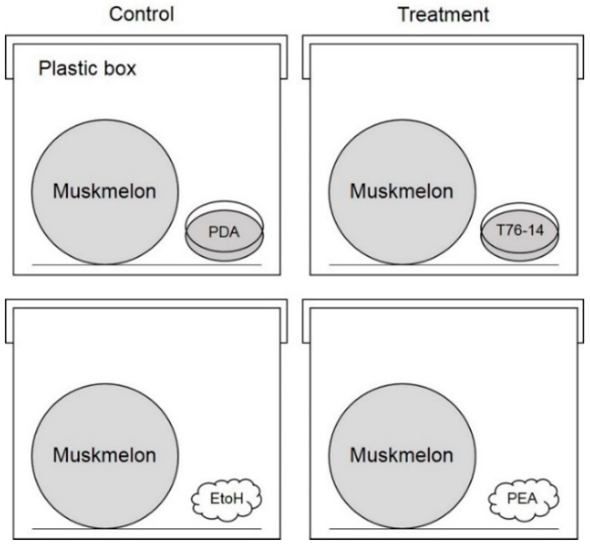
2.3. Volatile Antifungal Bioassay

In order to test the volatile effects of the Trichoderma species on the growth of F. incarnatum, the sealed plate method was conducted [25], with some modifications. The Trichoderma species were grown on potato dextrose agar (PDA) in 9-cm Petri dishes (Citotest, Jiangsu, China) for three days. An agar plug (0.5 cm in diameter) was cut from the culture plate, inserted centrally, and the lid of each petri dish was removed. We replaced the bottom with one containing PDA inoculated with tested fungi, and the two bottom plates were then sealed together with Parafilm and incubated at 28 ± 2 °C for five days. For the control, the lids of the control plates without Trichoderma inoculation were replaced in the same manner. The tested plates were also incubated at ambient temperature (28 ± 2 °C) for five days. Colony diameters of F. incarnatum were measured and converted to the percentage of inhibition through the following formula:
Percentage   inhibition   ( % )   =   D c D t D c   × 100 ,
where Dc = mycelial growth of F. incarnatum on control plate, and Dt = mycelial growth of F. incarnatum on the test plate. Each treatment was performed in five replicates, and the results are presented as the mean ± standard error (n = 5).

2.4. GC/MS Analysis

In order to examine the volatile and semi-volatile antifungal compounds within the Trichoderma species, gas chromatography spectrophotometry (GC/MS) was conducted. The Trichoderma species was cultured in a 20-mL chromatography vial, 20 mm in diameter (PerkinElmer, Waltham, MA, USA), and incubated at ambient temperature (28 ± 2 °C) for 14 days according to the method of Wonglom et al. [6] Solid-phase microextraction (SPME) was performed to extract the volatile organic compounds (VOCs), as previously described by Arthur et al. [6,26] SPME fiber (DVB/CAR/PDMS fiber) was exposed to the vapors above each Trichoderma culture for 30 min, and inserted into the injection port of the gas chromatograph SQ8 (PerkinElmer, Waltham, MA, USA), equipped with a DB-Wax capillary column (30 m × 0.25 mm i.d., 0.25 μm film thickness). The oven temperature was initially maintained at 40 °C, then increased to 230 °C, at a rate of 7 °C min−1. The injector temperature was 230 °C. The carrier gas was ultra-high-purity helium with an initial column head pressure of 60 kPa, at a flow rate of 1 mL min−1. Electron impact (EI) mass spectra were collected at 70 eV ionization voltage over the m/z range 29–550. The ion source and quadrupole temperatures were both set to 200 °C. Volatiles and semivolatiles were identified based on the computer searches dictated by The National Institute of Standards and Technology (NIST) Mass Spectral Library Search Chromatogram.

2.5. Sealed Plate Method of Commercial Volatile against Mycelial Growth of F. incarnatum

To test the effect of dominant volatile compound participated in antifungal activity against F. incarnatum, the sealed plate method was conducted as shown in Section 2.3. The compound phenylethyl alcohol (PEA) was purchased from Sigma-Aldrich (St. Louis, MO, USA). The effect of commercial PEA versus the other volatile antifungal compounds 2-ethylhexanol, 1-nonanol, 6-PP, and 2-methyl-1-butanol (Sigma-Aldrich, St. Louis, MO, USA) [6] was tested through the sealed plate method. PEA was dissolved in 95% ethanol and we adjusted the dilution to 10−1, 10−2, and 10−3. Each volatile compound was applied on a sterile cotton pad (20 µL) and subjected to the method of Wonglom et al. [6] Application of 95% ethanol served as a negative control. The tested plates were then incubated at 28 ± 2 °C for seven days. Each treatment was composed of five replicates and the experiment was repeated twice. Colony diameters of F. incarnatum were measured and the percentage inhibition was calculated as described in Section 2.2.

2.6. In Vivo Test of Volatile against Postharvest Fruit Rot

To test the effect of VOCs and commercial volatiles against postharvest fruit rot in muskmelon fruits, an in vivo test was conducted. F. incarnatum was cultured on PDA and incubated at ambient temperature (28 ± 2 °C) for seven days. Conidia of F. incarnatum were harvested and we adjusted the concentration to 1 × 10⁶ conidia mL−1. A conidia suspension (20 μL) was applied to muskmelon fruit, and each experiment, consisting of three fruits (three replicates), was repeated three times. Inoculated muskmelon fruits were incubated with VOCs from Trichoderma (eight petri dishes per treatment) or commercial volatile at 10−1 dilution in 50 μL applied on a cotton pad (four cotton pads) in a 24 × 35 × 18 cm plastic box (Figure 1) to share the atmosphere for three days. Inoculated muskmelon fruits incubated with PDA alone or 95% ethanol alone served as controls. Lesion development was measured and compared between the treatment and the control.

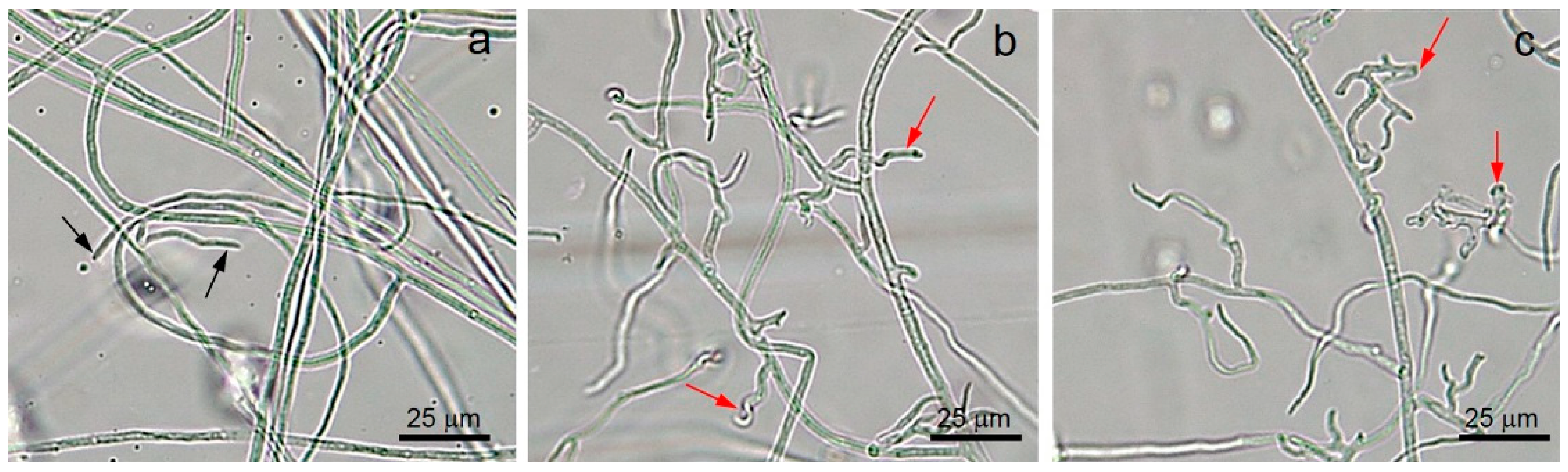
2.7. Morphological Study of F. incarnatum Exposed to Volatiles

To test the effect of VOCs or commercial volatiles on the morphology changes of F. incarnatum mycelia, the sealed plate method was conducted as described in Section 2.3. Changes in morphology between VOCs and commercial volatiles treated and untreated with mycelia from this study were observed by a Leica DM750 compound microscope (Leica Microsystems, Wetzlar, Germany).

2.8. Effect of VOCs Emitted from T. asperellum T76-14 on Fruit Rot

To test the effect of VOCs emitted from T. asperellum T76-14 on fruit rot in muskmelons, five muskmelon fruits were disinfected with 70% ethanol prior to VOC exposure. Muskmelon fruits were incubated with VOCs from Trichoderma or commercial volatiles, as described in Section 2.6, in a 24 × 35 × 18 cm plastic box to share the atmosphere for seven days. Fruit rot was observed from the day after treatment and recorded.

2.9. Statistical Analysis

Data including fungal growth, percentage inhibition, and lesion size were subjected to one-way analysis of variance (ANOVA). Statistically significant differences between treated samples and the untreated control were analyzed by Tukey’s test with a threshold of p < 0.05 [27].

3. Results

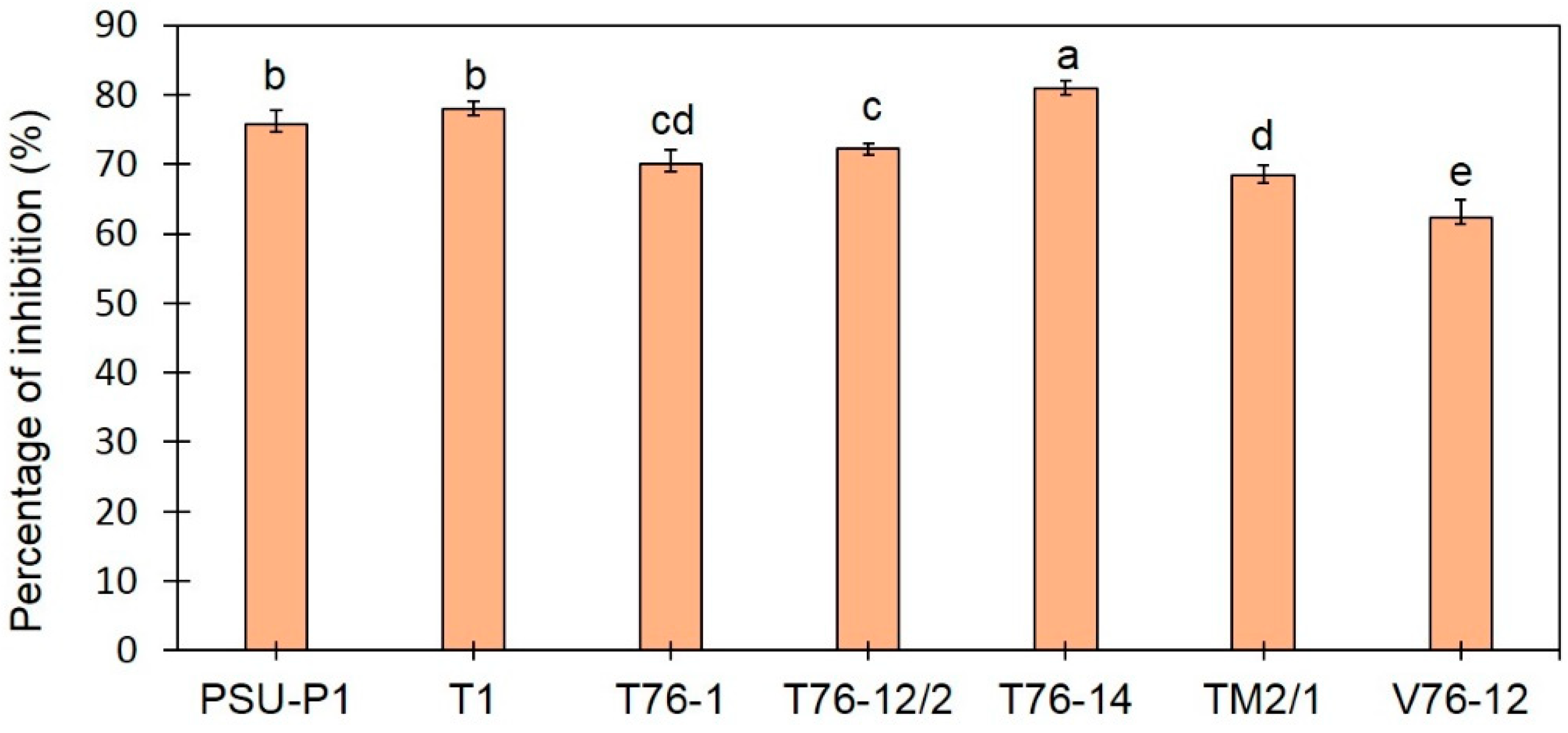
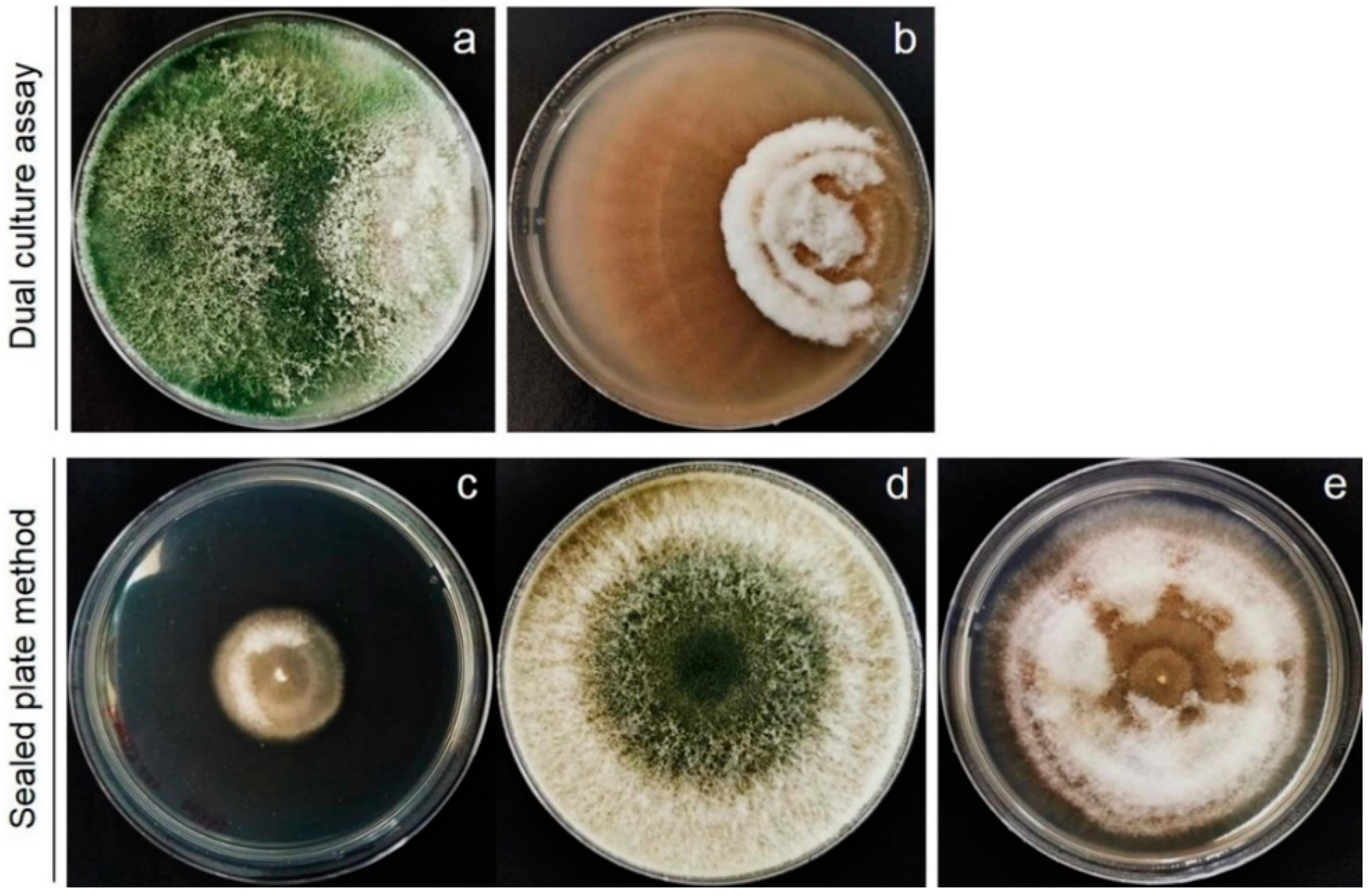
3.1. Trichoderma Species Inhibit Mycelial Growth of F. incarnatum

Based on the primary screening for BCA by dual-culture assay, Trichoderma spp. inhibited the fungal growth of F. incarnatum on the PDA plates. The percentage of inhibition ranged from 62% to 81%. Among the seven tested isolates, T. asperellum T76-14 effectively suppressed the growth of F. incarnatum by 81% (Figure 2 and Figure 3), statistically higher than those of the other isolates (p < 0.05), and was therefore selected for further analysis.

3.2. Volatiles Emitted by T. asperellum T76-14 Inhibit Mycelial Growth of F. incarnatum

To test the effects of the VOCs emitted by T. asperellum T76-14 on the fungal growth of F. incarnatum, the sealed plate method was conducted. After five days of incubation in ambient temperature, the colony diameters of F. incarnatum in the tested plates were smaller than those of the control bioassay plates (Figure 3). In the presence of VOCs emitted by T. asperellum T76-14, the growth of F. incarnatum was slower than without VOCs on PDA (Figure 3). The colony diameters of F. incarnatum in the control and test plates were measured and converted to represent the percentage of inhibition of fungal growth. The results demonstrated suppressed mycelial growth of F. incarnatum, with the greatest percentage of inhibition being 62.5%.

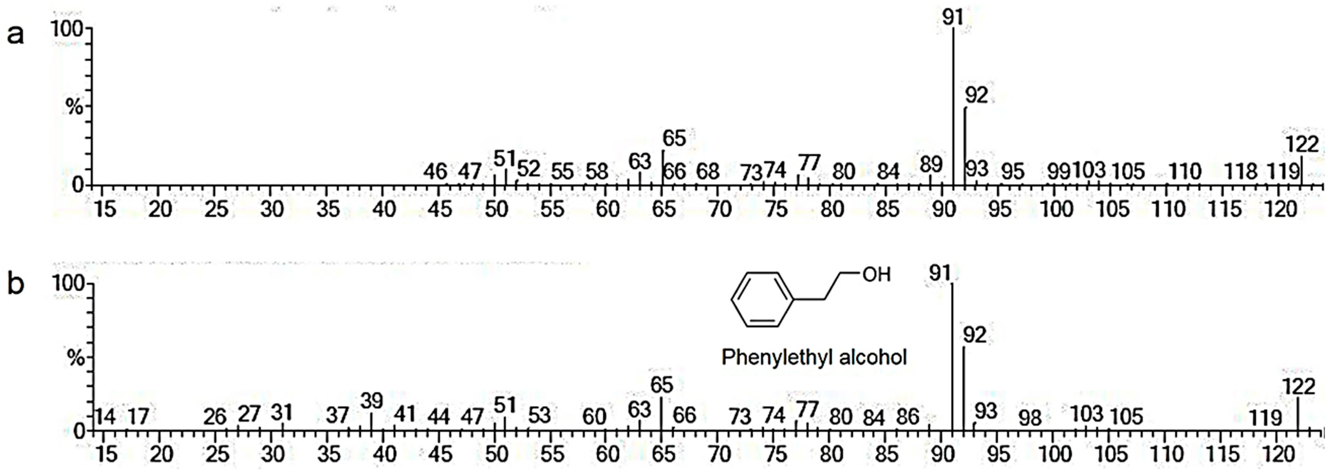
3.3. Identifying Volatile Organic Compounds

The results above demonstrate that the VOCs emitted by T. asperellum T76-14 inhibited the fungal growth of F. incarnatum, which suggests the antifungal activity of the volatile organic compounds. To characterize the VOCs of T. asperellum T76-14, SPME was performed to collect the VOCs, and they were analyzed by GC/MS. The results shown in Table 1 and Figure 4 and Figure 5 are from the preliminary screening of the VOCs produced by T. asperellum T76-14 that were responsible for inhibiting F. incarnatum. The chromatogram is presented in Figure 4. A total of 17 compounds with >70% were tentatively identified using the NIST library (Table 1). The compounds detected in T. asperellum T76-14 are members of the compound classes alcohols, alkane, and terpene, as illustrated in Table 1. The most dominant volatile compounds detected in this study were phenylethyl alcohol (PEA) at 6.52 min with 23.188% peak area, followed by fluorotrinitromethane, at 1.578 min with 8.778% peak area, and β-Curcumene, a sesquiterpene, at 11.641 min with 8.488% peak area. Figure 5 shows the mass spectrum of some of the major compounds, and the structure of the most dominant compound, PEA.

3.4. Effect of Commercial Volatile Compounds on Antifungal Activity against F. incarnatum

Commercial volatile compounds that have been reported as antifungal compounds (2-ethylhexanol, 1-nonanol, 6-PP, and 2-methyl-1-butanol) and PEA were tested against F. incarnatum growth in vitro on PDA plates. The results showed that all commercial volatile compounds inhibited the mycelial growth of F. incarnatum (Table 2). High percentage inhibition of volatile compounds depended on high concentrations. A dilution of 10−1 of all volatile compounds strongly inhibited F. incarnatum, with the percentage inhibition ranging from 21.68% to 74.29%. 2-ethylhexanol was found to be the best volatile compound for inhibiting F. incarnatum, with a percentage inhibition of 74.28%. The PEA showed a percentage inhibition of F. incarnatum of 56% at a dilution of 10−1, equivalent to a concentration of 1.5 mg mL−1. This confirmed that PEA is a volatile antifungal compound against F. incarnatum.
Fungal mycelia among the VOCs released from T. asperellum T76-14, PEA, and the control was observed by a compound microscope. The fungal mycelia of F. incarnatum exposed to VOCs and PEA showed an abnormal shape, whereas the control showed no change of mycelia and remained a normal shape (Figure 6).

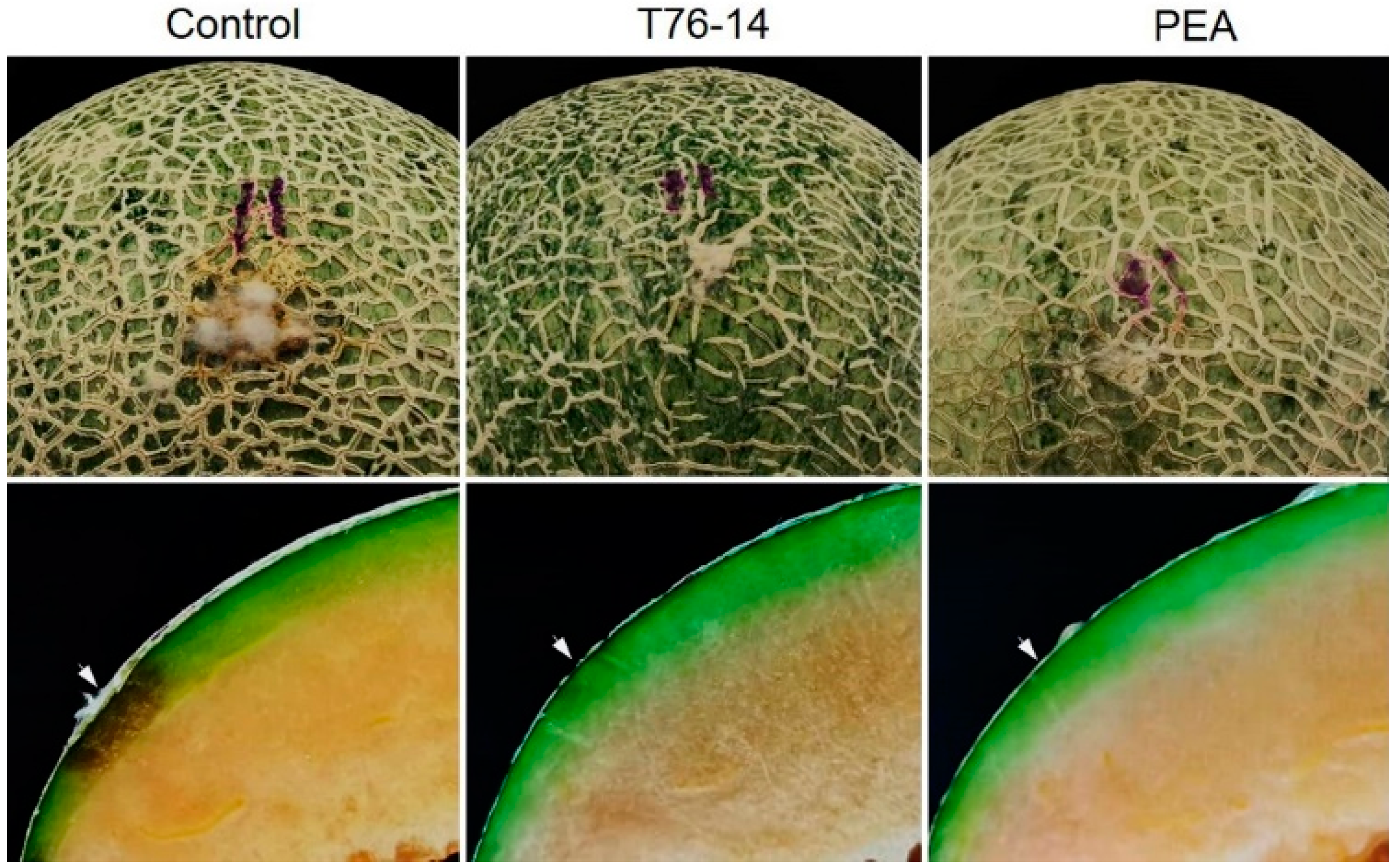
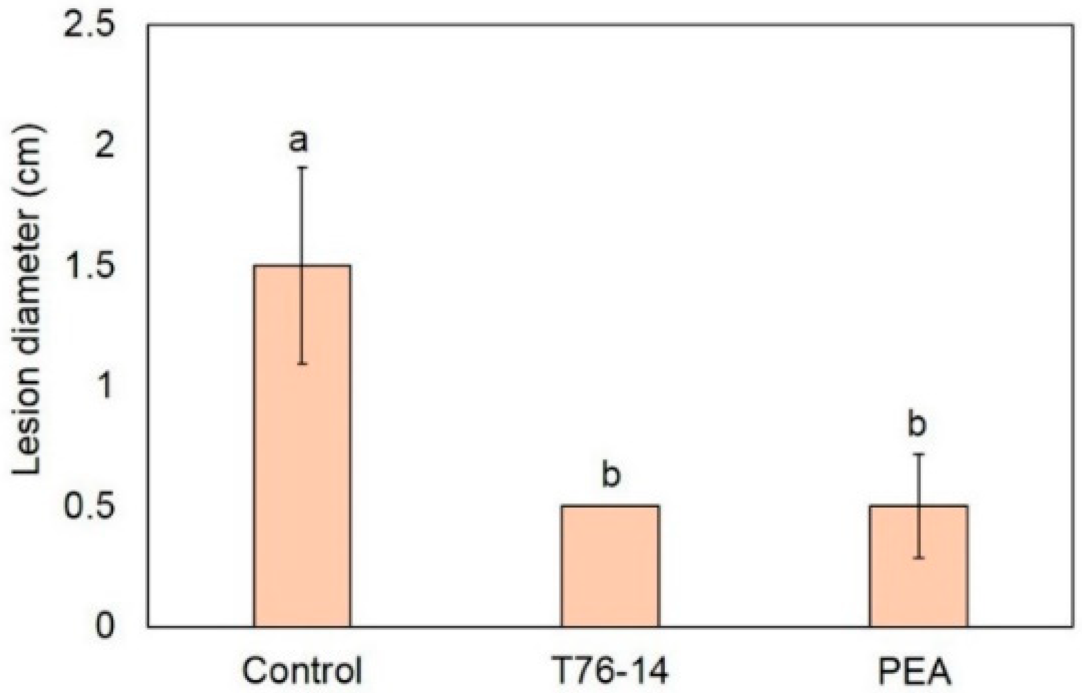
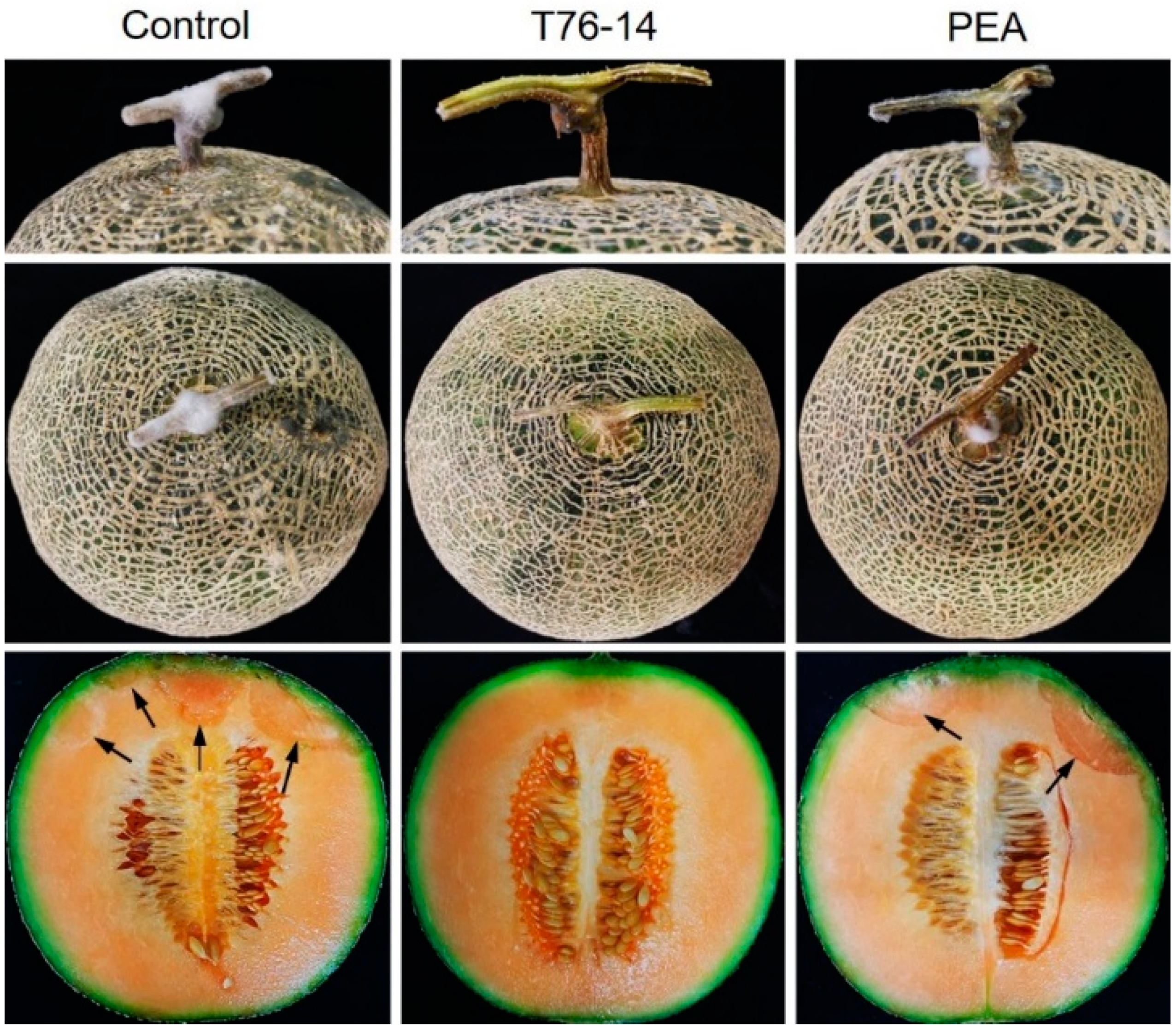
3.5. VOCs of T. asperellum T76-14 Reduced Postharvest Fruit Rot in Muskmelon Fruits

As seen above, the VOCs emitted from T. asperellum T76-14 and PEA showed antifungal activity against F. incarnatum, the pathogen of postharvest fruit rot in muskmelons. Therefore, they were selected to test in vivo against fruit rot in muskmelons by observing the lesion size after inoculation with a spore suspension of F. incarnatum. After incubation for three days, the lesions of VOCs- and PEA-treated muskmelons were statistically significantly smaller than in the control (Figure 7 and Figure 8). The lesion sizes of the control, VOCs-treated, and PEA-treated muskmelons were 1.5, 0.5, and 0.5 cm, respectively (Figure 8).

3.6. VOCs of T. asperellum T76-14 Suppressed Fruit Rot in Muskmelons

Based on the results above, we hypothesized that the VOCs emitted from T. asperellum may preserve muskmelons from fruit rot during postharvest storage. In order to test the effect of VOCs on muskmelons during postharvest, fruit rot was observed from the day after exposure with VOCs or PEA. The results showed that, after incubation at an ambient temperature for seven days, rotten tissue was observed in control muskmelons and muskmelons exposed to PEA, whereas VOC-treated muskmelons did not have rot (Figure 7). Fungal mycelia-covered tissues at peduncle were most prominent in the control, followed by PEA-treated muskmelons, causing tissue rot inside the muskmelon. Muskmelons exposed to VOCs from T. asperellum T76-14 showed less fungal mycelia on the fruit peduncle and no rotten tissue inside seven days after incubation.

4. Discussion

In the present study, T. asperellum T16-14 exhibited antifungal activity against the mycelial growth of F. incarnatum, the causal microorganism of postharvest fruit rot in muskmelons. T. asperellum T16-14 effectively inhibited mycelial growth of F. incarnatum in vitro by a dual-culture assay, which suggested a competition mechanism (Figure 2). The sealed plate method also revealed the antibiosis mechanism of T. asperellum T16-14 against F. incarnatum (Figure 3). The dominant volatile compound released by T. asperellum T16-14, tentatively identified as PEA based on SPME GC/MS, was involved in antifungal activity against F. incarnatum (Figure 4 and Figure 5). VOCs released by T. asperellum T16-14 caused abnormal changes in the F. incarnatum mycelia (Figure 6), reducing the postharvest fruit rot in muskmelons (Figure 7 and Figure 8).
In a primary screening by dual-culture assay, T. asperellum T76-14 grew faster than F. incarnatum on PDA (Figure 3), suggesting a competition mechanism. This mechanism is common in several species of Trichoderma against plant pathogens [4,5,28]. In the study above, T. asperellum T76-14 was found to produce VOCs (Figure 3 and Table 1) that slow the fungal growth of F. incarnatum. This production involved an antibiosis mechanism within the Trichoderma species. It has been established that several Trichoderma species emit volatile antifungal compounds that inhibit pathogen growth [4,5,6,25]. In this study, volatiles emitted from T. asperellum T76-14 inhibited the fungal growth of F. incarnatum by 62.5% (Figure 2), as determined through the sealed plate method. The VOCs emitted by T. asperellum T76-14 slow the fungal growth of F. incarnatum, similar to PEA. Both VOCs and PEA caused abnormal changes in the fungal mycelia (Figure 6), which may result in inhibiting the fungal growth of F. incarnatum. As shown in Figure 4 and Table 1, GC/MS revealed some minor peaks, which may have contributed to antifungal activity due to a low percent peak area. This revealed that the VOCs emitted by T. asperellum T76-14 were responsible for the antifungal activity against F. incarnatum. The GC/MS results presented 17 compounds; however, according to the literature, the only antifungal compound found in this study was PEA (Figure 4 and Figure 5).
PEA, also known as an aromatic alcohol, is an organic compound found as a colorless liquid with a rose-like odor. This compound occurs widely in nature as an essential oil from several plant species such as roses, jasmine, carnations, and hyacinths [29]. PEA has been used to modify certain flavor compositions of foods with low toxicity [30,31]. It has been shown that PEA has antimicrobial properties [29,30,32,33,34]. An in vitro study revealed that PEA inhibits fungal growth and the synthesis of RNA, DNA, and the protein of Neurospora crassa [32]. PEA has been reported to show strong antifungal activity against Ganoderma boninense [35], significantly reducing fungal growth to prolong the postharvest life of strawberries [34], and controlling the blue mold decay caused by Penicillium digitatum and P. italicum in citrus fruits [36]. Our result is in agreement with previous reports that PEA inhibited the fungal growth of F. incarnatum in vitro (Table 2) and reduced lesions on muskmelon fruits (Figure 7 and Figure 8). This also proved that PEA in VOCs released from T. asperellum T76-14 had antifungal activity against F. incarnatum.
PEA has been reported to be produced by several Trichoderma species. For instance, PEA emitted by Trichoderma sp. [29], T. atroviride [37], T. harzianum [38], T. spirale [5], and T. virens [36] displayed antifungal ability against several plant pathogens. In this study, PEA emitted by T. asperellum T76-14, is a dominant compound with 23.188% peak area (Table 2), and has been reported to have antifungal activities [34,35]. Therefore, the ability of T. asperellum T76-14 to produce volatile antifungal compounds would further promote antifungal activity against F. incarnatum. In this study, we investigated the effect of VOCs emitted by T. asperellum T76-14 on fungal growth, morphology changes of fungal mycelia, and the reduction in postharvest fruit rot, as well as the prolonged postharvest life of muskmelons. In comparison with commercial PEA, VOCs of T. asperellum T76-14 prevented fruit rot in muskmelons, which may be due to a complex mix of VOCs with other volatiles, working synergistically to inhibit fungal growth. Commercial volatile PEA can limit lesion development at three days after treatment (Figure 7); however, at seven days it showed fruit rot similar to the control (Figure 9). This may result from PEA vapor after incubation in a long period in a shared-atmosphere plastic box. The use of fresh Trichoderma may produce VOCs continuously; however PEA was applied once so the concentration may reduce after a long period of incubation.
Several reports have clarified the effect of microbial volatiles as a biofumigant controlling postharvest diseases and prolonging postharvest life. For instance, Li et al. [39] used VOCs produced by Ceratocystis fimbriata as biofumigation on postharvest disease of fruit. The authors showed that VOCs inhibited the growth of Monilinia fructicola and Penicilium digitatum (causing peach brown rot and citrus green mold, respectively) [39]. Volatile compounds produced by Aureobasidium pullulans increased fruit waxes’ complexity, reducing pathogen attacks on stone fruit [40]. Furthermore, VOCs emitted from T. atroviride inhibited the mycelial growth of Phytophthora infestans in postharvest potato tubers [41]. The authors investigated VOCs by GC/MS and found abundant compounds, namely 3-mrthyl-1-butanol, p-6-pentyl-2-pyrone, 2-mthyl-1-propanol, and acetone, which were responsible for serious morphological and ultrastructural damage of the pathogens [41]. Our findings are in agreement with [41]: T. asperellum T76-14 produced VOCs, which contained dominant volatiles of PEA. VOCs of T. asperellum T76-14 are responsible for inhibiting postharvest fruit rot pathogens and could prolong the postharvest life of muskmelons. During postharvest storage, some fungi or mold can infest the fruit surface and penetrate into tissues, causing fruit rot. This finding indicates that VOCs released from T. asperellum T76-14 contain PEA to clean the fruit surface and reduce fungal infection, which may prolong the postharvest life of muskmelons.

5. Conclusions

Herein, we described the ability of VOCs emitted by T. asperellum T76-14 involved in antifungal activity against the postharvest fruit rot pathogen (F. incarnatum) of muskmelons to reduce fruit rot during postharvest storage by inhibiting postharvest pathogens. However, the extraction of valuable compounds, and the quality and quantity of volatiles emitted by this strain, have yet to be investigated. In order to apply VOCs from T. asperellum T76-14 as a biofumigant to control postharvest disease and maintain postharvest quality on a large scale, further experiments need to be conducted.

Author Contributions

Conceptualization, W.I. and A.S.; methodology, W.I. and S.K.; software, W.I. and A.S.; validation, A.S; formal analysis, A.S.; investigation, W.I. and S.K.; resources, A.S.; data curation, A.S.; writing—original draft preparation, W.I. and A.S.; writing—review and editing, W.I., S.K. and A.S.; visualization, A.S.; supervision, A.S.; project administration, A.S.; funding acquisition, A.S. All authors have read and agreed to the published version of the manuscript.

Funding

This research was funded by the National Research Council of Thailand (NRCT) via grant NRCT5-RSA63022-01.

Institutional Review Board Statement

Not applicable.

Informed Consent Statement

Not applicable.

Data Availability Statement

Not applicable.

Acknowledgments

Special thanks go to Prince of Songkla University and the Center of Excellence in Agricultural and Natural Resources Biotechnology (CoE-ANRB) phase 3 for facilities and Kitsada Pitija, PerkinElmer Co. Ltd., Bangkok, Thailand for the GC/MS analysis and MDPI’s English editing service for English editing.

Conflicts of Interest

The authors declare no conflict of interest.

References

  1. Wonglom, P.; Sunpapao, A. Fusarium incarnatum is associated with postharvest fruit rot of muskmelon (Cucumis melo). J. Phytopath. 2020, 168, 204–210. [Google Scholar] [CrossRef]
  2. Yoon, M.Y.; Cha, B.; Kim, J.C. Recent trends in studies on botanical fungicides in agriculture. Plant Pathol. J. 2013, 29, 1–9. [Google Scholar] [CrossRef] [Green Version]
  3. Fattahi, E.; Mousavi Moghadam, M.; Khanbabaei, R. The effect of fricyclazole on testosterone changes and testicular structure in mice. J. Babol Univ. Med. Sci. 2015, 17, 43–49. [Google Scholar]
  4. Sunpapao, A.; Chairin, T.; Ito, S. The biocontrol by Streptomyces and Trichoderma of leaf spot disease caused by Curvularia oryzae in oil palm seedlings. Biol. Control. 2018, 123, 36–42. [Google Scholar] [CrossRef]
  5. Baiyee, B.; Pornsuriya, C.; Ito, S.; Sunapapo, A. Trichoderma spirale T76-1 displays biocontrol activity on lettuce (Lactuca sativa L.) caused by Corynespora cassiicola or Curvularia aeria. Biol. Control. 2019, 129, 195–200. [Google Scholar] [CrossRef]
  6. Wonglom, P.; Ito, S.; Sunpapao, A. Volatile organic compounds emitted from endophytic fungus Trichoderma asperellum T1 mediate antifungal activity, defense response and promote plant growth in lettuce (Lactuca sativa). Fungal Ecol. 2020, 43, 100867. [Google Scholar] [CrossRef]
  7. Sunpapao, A. Antagonistic Microorganisms: Current Research and Innovations; Lambert Academic Publishing: Saarbrücken, Germany, 2020; p. 120. [Google Scholar]
  8. Elad, Y.; David, D.R.; Levi, T.; Kapat, A.; Kirshner, B. Trichoderma harzianum T-39- mechanisms of biocontrol of foliar pathogens. In Modern Fungicides and Antifungal Compounds; Lyr, H., Russell, P.E., Dehne, H.W., Sisler, H.D., Eds.; Intercept: Andover, UK, 1999; pp. 459–467. [Google Scholar]
  9. Troian, R.F.; Steindorff, A.S.; Ramada, M.H.; Arruda, W.; Ulhoa, C.J. Mycoparasitism studies of Trichoderma harzianum against Sclerotinia sclerotiorum: Evaluation of antagonism and expression of cell wall-degrading enzymes genes. Biotechnol. Lett. 2014, 36, 2095–2101. [Google Scholar] [CrossRef]
  10. Howell, C.R. Mechanisms employed by Trichoderma species in the biological control of plant diseases: The history and evolution of current concepts. Plant Dis. 2003, 87, 4–10. [Google Scholar] [CrossRef] [PubMed] [Green Version]
  11. Baiyee, B.; Ito, S.; Sunapapo, A. Trichoderma asperellum T1 mediated antifungal activity and induced defense response against leaf spot fungi in lettuce (Lactuca sativa L.). Physiol. Molec. Plant Pathol. 2019, 106, 96–101. [Google Scholar] [CrossRef]
  12. Phoka, N.; Suwannarach, N.; Lumyong, S.; Ito, S.-I.; Matsui, K.; Arikit, S.; Sunpapao, A. Role of Volatiles from the Endophytic Fungus Trichoderma asperelloides PSU-P1 in Biocontrol Potential and in Promoting the Plant Growth of Arabidopsis thaliana. J. Fungi 2020, 6, 341. [Google Scholar] [CrossRef] [PubMed]
  13. Martínez-Medina, A.; Alguacil, M.D.M.; Pascual, J.A.; VanWees, S.C. Phytohormone profiles induced by Trichoderma isolates correspond with their biocontrol and plant growth-promoting activity on melon plants. J. Chem. Ecol. 2014, 40, 804–815. [Google Scholar] [CrossRef] [PubMed]
  14. Brader, G.; Compant, S.; Mitter, B.; Trognitz, F.; Sessitsch, A. Metabolic potential of endophytic bacteria. Curr. Opin. Biotechnol. 2014, 27, 30–37. [Google Scholar] [CrossRef] [PubMed] [Green Version]
  15. Shi, M.; Chen, L.; Wang, X.W.; Zhang, T.; Zhao, P.B.; Song, X.Y.; Sun, C.Y.; Chen, X.L.; Zhou, B.C.; Zhang, Y.Z. Antimicrobial petaibols from Trichoderma pseudokoningii induce programmed cell death in plant fungal pathogens. Microbiology 2012, 158, 166–175. [Google Scholar] [CrossRef] [PubMed] [Green Version]
  16. Reino, J.L.; Guerrero, R.F.; Hernández-Galán, R.; Collado, I.G. Secondary metabolites from species of the biocontrol agent Trichoderma. Phytochem. Rev. 2007, 7, 89–123. [Google Scholar] [CrossRef]
  17. Marcías-Rubalcava, M.L.; Hernández-Bautista, B.E.; Oropeza, F.; Duarte, G.; González, M.C.; Glenn, A.E.; Hanlin, R.T.; Anaya, A.L. Allelochemical effects of volatile compounds and organic extracts from Muscodor yucatanensis, a tropical endophytic fungus from Bursera simaruba. J. Chem. Ecol. 2010, 36, 1122–1131. [Google Scholar] [CrossRef]
  18. Wu, Q.; Sun, R.; Ni, M.; Yu, J.; Li, Y.; Yu, C.; Dou, K.; Ren, J.; Chen, J. Identification of a novel fungus, Trichoderma asperellum GDFS1009, and comprehensive evaluation of its biocontrol efficacy. PLoS ONE 2017, 12, e0179957. [Google Scholar] [CrossRef]
  19. Loulier, J.; Lefort, F.; Stocki, M.; Asztemborska, M.; Szmigielski, R.; Siwek, K.; Grzywacz, T.; Hsiang, T.; Ślusarski, S.; Oszako, T.; et al. Detection of Fungi and Oomycetes by Volatiles Using E-Nose and SPME-GC/MS Platforms. Molecules 2020, 25, 5749. [Google Scholar] [CrossRef]
  20. Stracquadanio, C.; Quiles, J.M.; Meca, G.; Cacciola, S.O. Antifungal Activity of Bioactive Metabolites Produced by Trichoderma asperellum and Trichoderma atroviride in Liquid Medium. J. Fungi 2020, 6, 263. [Google Scholar] [CrossRef]
  21. Tao, L.Y.; Zhang, Y.W.; Li, Y.Q.; Luo, L.P.; Zhang, Z.L.; Chen, J. Antagonistic activity of volatile metabolites from Trichoderma asperellum. Chin. J. Biotechnol. 2020, 36, 1181–1189. [Google Scholar]
  22. Vinale, F.; Sivasithamparam, K.; Ghisalberti, E.L.; Ruocco, M.; Woo, S.; Lorito, M. Trichoderma secondary metabolites that affect plant metabolism. Nat. Prod. Commun. 2012, 7, 1545–1550. [Google Scholar] [CrossRef] [Green Version]
  23. Castillo, F.D.H.; Padilla, A.M.B.; Morales, G.G.; Siller, M.C.; Herrera, R.R.; Gonzales, C.N.A.; Reyes, F.C. In vitro antagonistic action of Trichoderma strains against Sclerotinia sclerotiorum and Sclerotium cepivorum. Am. J. Agr. Biol. Sci. 2011, 6, 410–417. [Google Scholar] [CrossRef] [Green Version]
  24. Rahman, M.A.; Begum, M.F.; Alam, M.F. Screening of Trichoderma isolates as a biological control agent against Ceratocystis paradoxa causing pineapple disease of sugarcane. Microbiology 2009, 37, 277–285. [Google Scholar]
  25. Dennis, C.; Webster, J. Antagonistic properties of species-groups of Trichoderma, II. Production of volatile antibiotic. Trans. Br. Mycol. Soc. 1971, 57, 41–48. [Google Scholar] [CrossRef]
  26. Arthur, C.L.; Killam, L.M.; Buchholz, K.D.; Pawliszyn, J. Automation and optimization of solid-phase microextraction. Anal. Chem. 1992, 64, 1960–1966. [Google Scholar] [CrossRef]
  27. Gomez, K.A.; Gomez, A.A. Statistical Procedure for Agricultural Research; John Wiley and Sons: New York, NY, USA, 1984. [Google Scholar]
  28. Wonglom, P.; Daengsuwan, W.; Ito, S.; Sunpapao, A. Biological control of Sclerotium fruit rot of snake fruit and stem rot of lettuce by Trichoderma sp. T76-12/2 and the mechanisms involved. Physiol. Molec. Plant Pathol. 2019, 107, 1–7. [Google Scholar] [CrossRef]
  29. Zhu, Y.J.; Zhou, H.T.; Hu, Y.H.; Tang, J.Y.; Su, M.X.; Guo, Y.J.; Chen, Q.X.; Liu, B. Antityrosinase and antimicrobial activities of 2-phenylethanol, 2-phenylacetaldehyde and 2-phenylacetic acid. Food Chem. 2011, 124, 298–302. [Google Scholar] [CrossRef]
  30. Fraud, S.; Rees, E.L.; Mahenthiralingam, E.; Russell, A.D.; Maillard, J.Y. Aromatic alcohols and their effect on gram-negative bacteria, cocci and mycobacteria. J. Antimicrob. Chemother. 2003, 51, 1435–1436. [Google Scholar] [CrossRef]
  31. Gao, F.; Daugulis, A.J. Bioproduction of the aroma compound 2-phenylethanol in a solid–liquid two-phase partitioning bioreactor system by Kluyveromyces marxianus. Biotechnol. Bioeng. 2009, 104, 332–339. [Google Scholar] [CrossRef]
  32. Lester, G. Inhibition of growth, synthesis, and permeability in Neurospora crassa by phenethyl alcohol. J Bacteriol. 1965, 90, 29–37. [Google Scholar] [CrossRef] [Green Version]
  33. Lucchini, J.J.; Bonnaveiro, N.; Cremieux, A.; Le Goffic, F. Mechanism of bactericidal action of phenethyl alcohol in Escherichia coli. Curr. Microbiol. 1993, 27, 295–300. [Google Scholar] [CrossRef]
  34. Mo, E.K.; Sung, C.K. Phenylethyl alcohol (PEA) application slows fungal growth and maintains aroma in strawberry. Postharvest Biol. Technol. 2007, 45, 234–239. [Google Scholar] [CrossRef]
  35. Angel, L.P.L.; Yusof, M.T.; Ismail, I.S.; Ping, B.T.Y.; Azni, I.N.A.M.; Kamarudin, N.H.; Sundram, S. An in vitro study of antifungal activity of Trichoderma virens 7b and profile of its non-polar antifungal components released against Ganoderma boninense. J. Microbiol. 2016, 54, 732–744. [Google Scholar] [CrossRef] [PubMed]
  36. Liu, P.; Cheng, Y.; Yang, M.; Liu, Y.; Chen, K.; Long, C.; Deng, X. Mechanisms of action for 2-phenylethylethanol isolated from Kloeckera apiculata in control of Penicillium molds of citrus fruits. BMC Microbiol. 2014, 14, 242. [Google Scholar] [CrossRef] [PubMed] [Green Version]
  37. Stoppacher, N.; Kluger, B.; Zeilinger, S.; Krska, R.; Schuhmacher, R. Identification and profiling of volatile metabolites of the biocontrol fungus Trichoderma atroviride by HS-SPME-GC-MS. J. Microbiol. Methods 2010, 81, 187–193. [Google Scholar] [CrossRef]
  38. Siddiquee, S.; Cheong, B.E.; Taslima, K.; Kausar, H.; Hasan, M.M. Separation and identification of volatile compounds from liquid culture of Trichoderma hazianum by GC-MS using three different capillary columns. J. Chromatogr. Sci. 2012, 50, 358–367. [Google Scholar] [CrossRef] [Green Version]
  39. Li, Q.; Wu, L.; Hao, J.; Luo, L.; Cao, Y.; Li, J. Biofumigation on post-harvest diseases of fruits using a new volatile-producing fungus of Ceratocystis fimbriata. PLoS ONE 2015, 10, e0132009. [Google Scholar] [CrossRef]
  40. Di Francesco, A.; Di Foggia, M.; Baraldi, E. Aureobasidium pullulans volatile organic compounds as alternative postharvest method to control brown rot of stone fruits. Food Microbiol. 2020, 87, 103395. [Google Scholar] [CrossRef]
  41. Elsherbiny, E.A.; Amin, B.H.; Aleem, B.; Kingsley, K.L.; Bennett, J.W. Trichoderma volatile organic compounds as a biofumigation tool against late blight pathogen Phytophthora infestans in [ostharvest potato tubers. J. Agric. Food Chem. 2020, 68, 8163–8171. [Google Scholar] [CrossRef]
Figure 1. Overview of experimental setup and VOC effect against Fusarium incarnatum on muskmelons. Schematic overview of the plate-within-a-box system.
Jof 07 00046 g001
Figure 2. Percentage inhibition of Trichoderma spp. against Fusarium incarnatum in PDA by dual-assay plates. Different letters indicate statistically significant differences among treatments (p < 0.05) using Tukey’s test.
Jof 07 00046 g002
Figure 3. In vitro test antagonistic Trichoderma against Fusarium incarnatum, Trichoderma asperellum T76-14, and F. incarnatum in dual culture plate (a), F. incarnatum in PDA plate alone (b), F. incarnatum (c) and T. asperellum T76-14 (d) in sealed plate method and F. incarnatum in PDA plate alone (e).
Jof 07 00046 g003
Figure 4. Total ion chromatogram of volatile compounds identified from T. asperellum T76-14.
Jof 07 00046 g004
Figure 5. Mass spectrum for the compound at 6.52 min (a) and phenylethyl alcohol (PEA) with its structure (b).
Jof 07 00046 g005
Figure 6. Morphological observation of Fusarium incarnatum mycelia, in control (a), exposed to VOCs emitted by T. asperellum T76-14 (b), and PEA (c), black arrows indicate normal hyphal tips and red arrows indicate abnormal hyphal tips.
Jof 07 00046 g006
Figure 7. Lesion development after inoculation with F. incarnatum, control without VOCs and PEA, muskmelons exposed to VOCs of Trichoderma asperellum T76-14 and phenylethyl alcohol (PEA). Arrows indicate the inoculation points.
Jof 07 00046 g007
Figure 8. Lesion diameter on muskmelons in the control versus muskmelons exposed to Trichoderma asperellum T76-14 (T76-14) and phenylethyl alcohol (PEA). Different letters indicate statistically significant differences among treatments (p < 0.05) using Tukey’s test.
Jof 07 00046 g008
Figure 9. Effect of VOCs and PEA on postharvest fruit rot in muskmelons, T76-14 = Trichoderma asperellum T76-14 and PEA = phenylethyl alcohol. Arrows indicate the rotten tissue.
Jof 07 00046 g009
Table 1. Volatile compounds produced by Trichoderma asperellum T76-14 tentatively identified through SPME GC/MS analysis.
RT (min)Volatile CompoundsFormula% Math% Area
1.58Fluoro(trinitro)methane *CFN3O6958.78
2.29Pentan-1-olC5H12O86.42.98
2.313-MethylpentaneC6H1489.24.10
5.662,4,6-TrimethyloctaneC11H2483.71.34
6.285,7-DimethylundecaneC13H2884.41.60
6.52Phenylethyl alcoholC8H10O94.823.19
7.45Octadec-9-yneC18H3474.93.09
7.651-MethylideneindeneC10H1092.81.34
10.273-Isopropyl-6,8a-dimethyl-1,2,4,5,8,8a-hexahydroazuleneC15H2485.81.41
11.64β-CurcumeneC15H2484.48.49
11.83α-BisaboleneC15H2483.81.69
12.04Sesquisabinene BC15H2483.51.74
13.10ZingiberenolC15H26O81.83.11
13.30CubenolC15H26O83.16.04
13.61Undeca-3,4-diene-2,10-dione, 5,6,6-trimethylC14H22O275.11.44
14.03Allyldimethyl(prop-1-ynyl)silaneC8H14Si71.40.77
15.12cis-Z-α-Bisabolene epoxideC15H24O71.31.73
* The result of three replicates.
Table 2. Comparison of phenylethyl alcohol (PEA) and other commercial volatiles against Fusarium incarnatum.
Commercial VolatilesPercentage Inhibition (%) a
10−110−210−3
1-Nonanol55.42 ± 13.69 b29.71 ± 3.32 b19.67 ± 5.69 c
2-Ethylhexanol74.29 ± 11.12 a29.71 ± 5.01 b24.89 ± 9.04 a
2-Methyl-1-butanol39.12 ± 8.43 c20.88 ± 4.86 d16.86 ± 1.20 d
6-Pentyl-2H-pyran-2-one21.68 ± 5.25 d32.53 ± 14.65 a20.08 ± 6.18 b
Phenylethyl alcohol56.00 ± 5.25 b24.00 ± 6.92 c12.00 ± 4.00 e
a Percentage inhibition of mycelial growth was calculated using 95% ethyl alcohol as a negative control. Means within the same column followed by the same superscript letters are not significantly different at p < 0.05 using Tukey’s test.
Publisher’s Note: MDPI stays neutral with regard to jurisdictional claims in published maps and institutional affiliations.

