Article

Molecular Epidemiological Investigation of a Nosocomial Cluster of C. auris: Evidence of Recent Emergence in Italy and Ease of Transmission during the COVID-19 Pandemic

1
Department of Surgical Sciences and Integrated Diagnostics (DISC), University of Genoa, 16132 Genoa, Italy
2
Anesthesia and Intensive Care, San Martino Policlinico Hospital—IRCCS for Oncology and Neuroscience, 16132 Genoa, Italy
3
Infectious Diseases Unit, San Martino Policlinico Hospital—IRCCS for Oncology and Neuroscience, 16132 Genoa, Italy
4
Department of Health Sciences (DISSAL), University of Genoa, 16132 Genoa, Italy
5
Clinical Microbiology Unit, San Martino Policlinico Hospital—IRCCS for Oncology and Neuroscience, 16132 Genoa, Italy
*
Author to whom correspondence should be addressed.
Submission received: 13 January 2021 / Revised: 12 February 2021 / Accepted: 12 February 2021 / Published: 15 February 2021
(This article belongs to the Special Issue Candida auris 2.0)

Abstract

:
Candida auris is an emerging MDR pathogen raising major concerns worldwide. In Italy, it was first and only identified in July 2019 in our hospital (San Martino Hospital, Genoa), where infection or colonization cases have been increasingly recognized during the following months. To gain insights into the introduction, transmission dynamics, and resistance traits of this fungal pathogen, consecutive C. auris isolates collected from July 2019 to May 2020 (n = 10) were subjected to whole-genome sequencing (WGS) and antifungal susceptibility testing (AST); patients’ clinical and trace data were also collected. WGS resolved all isolates within the genetic clade I (South Asian) and showed that all but one were part of a cluster likely stemming from the index case. Phylogenetic molecular clock analyses predicted a recent introduction (May 2019) in the hospital setting and suggested that most transmissions were associated with a ward converted to a COVID-19-dedicated ICU during the pandemic. All isolates were resistant to amphotericin B, voriconazole, and fluconazole at high-level, owing to mutations in ERG11(K143R) and TACB1(A640V). Present data demonstrated that the introduction of MDR C. auris in Italy was a recent event and suggested that its spread could have been facilitated by the COVID-19 pandemic. Continued efforts to implement stringent infection prevention and control strategies are warranted to limit the spread of this emerging pathogen within the healthcare system.

1. Introduction

Candida species are one of the most common fungal pathogens causing invasive infections at a global scale [1]. Over past years, Candida auris has emerged as a fungal pathogen of major concern, frequently showing a multidrug-resistant (MDR) phenotype and being recognized as an important cause of nosocomial outbreaks of invasive infections worldwide [2]. The global dissemination of this pathogen has been initially attributed to the simultaneous emergence of four major phylogenetically distinct lineages, showing a specific geographical distribution and including: South Asian (I), East Asian (II), African (III), and South American (IV) clades [3]. A potential fifth clade of Iranian origin has been also described [4]. More recently, however, some evidence for an increased phylogeographic mixing has been provided [5], since isolates from countries of most global regions appeared interspersed in phylogenies (i.e., primarily in clade I and III), a phenomenon likely contributed by global travels of persons with prior healthcare exposures to C. auris.
Most of reported infections occurred in immunocompromised and/or critically ill patients with severe underlying diseases and were associated with high mortality rates, as the frequent resistance phenotype to multiple antifungal drugs accounts for high rates of clinical treatment failures [6]. Although factors predisposing to Candida infections appear similar, regardless of the species, it has been recently shown that longer stay in ICU, respiratory disease, vascular surgery, presence of CVC, urinary catheter, surgery within past 30 days, and prolonged exposure to antifungal drugs are the most common risk factors for acquisition of C. auris [6,7,8]. Moreover, the ability to persist in the hospital environment and to colonize multiple human anatomical sites poses major risks for the intrahospital transmission of this fungal pathogen and could represent an additional risk factor for development of invasive candidiasis [9,10]. For these reasons, the impact that C. auris could have in the clinical setting is an ever-growing concern.
Since 2015, either sporadic cases or nosocomial outbreaks have been documented in several Europe countries, primarily in Spain and the UK, where intra- and inter-facility transmissions have been described [11]. In Italy, the index case of C. auris was identified in 2019 in our hospital (San Martino Hospital, Genoa), which is a large teaching facility located in the northern area of the country [12]; subsequently, no more reports have been documented in our country.
In this study, we report on novel C. auris cases recently recognized in our hospital after the initial description of the index case and being likely associated with a nosocomial cluster. A molecular epidemiological investigation was carried out to provide insights into the introduction, the putative transmission dynamics, and the resistance traits of this fungal pathogen.

2. Materials and Methods

2.1. Candida auris Isolates

A total of 10 nonduplicate clinical isolates were collected from individual patients admitted to the San Martino Hospital (Genoa, Italy) during the period spanning July 2019–May 2020 and diagnosed with invasive infections (blood, n = 5) or colonization (broncho-alveolar lavage, urine, n = 5) by C. auris. All isolates were cultivated using Liofilchem Chromatic Candida (Liofilchem, Roseto degli Abruzzi, Italy) and grown for 48 h at 30 °C; frozen stocks were subsequently prepared with 15% sterile glycerol and stored at −80 °C. The species-level identification was carried out by matrix-assisted laser desorption ionization-time of flight mass spectrometry (MALDI-TOF MS) (Vitek MS; bioMérieux, Marcy-l’Etoile, France) using the VITEK MS software v4,0 and confirmed by PCR amplification of the species-specific GPI protein-encoding genes, as previously described [12,13].

2.2. Antifungal Susceptibility Testing (AFST)Share and Cite

Antifungal susceptibility testing of nine antifungal agents (i.e., amphotericin B, flucytosine, fluconazole, itraconazole, voriconazole, posaconazole, caspofungin, anidulafungin, and micafungin) was performed according to the CLSI M27-A3 guidelines [14], using commercial broth microdilution plates (Sensititre YeastOne, Thermo Scientific, Waltham, MA, USA) and the manufacturer’s instructions. As C. auris-specific susceptibility breakpoints have not yet been established, tentative breakpoints proposed by the CDC (https://www.cdc.gov/fungal/candida-auris/c-auris-antifungal.html, accessed 10 January 2021) or previously adopted by other studies were used [15]. In brief, resistance breakpoints were defined as follows: micafungin at ≥4 μg/mL, caspofungin at ≥2 μg/mL, anidulafungin at ≥4 μg/mL, amphotericin B at ≥2 µg/mL, 5-fluorocytosine at ≥ 128 μg/mL, fluconazole at ≥32 μg/mL, and voriconazole at ≥2 µg/mL.

2.3. Whole-Genome Sequencing (WGS)

Total genomic DNAs was extracted from broth cultures using the Qiagen DNeasy PowerLyzer PowerSoil Kit (Qiagen, Hilden, Germany). Shotgun libraries were prepared from purified DNAs and sequenced with a 2 × 150 bp paired-end approach using the Illumina HiSeq platform (Illumina, Inc., San Diego, CA, USA) at Novogene Co., Ltd. (Cambridge, United Kingdom). The C. auris index strain (FG_GE01) was further sequenced using the MinION sequencer (Oxford Nanopore Technologies, Oxford, UK) to generate a complete, cluster-specific, reference genome using Flye v2.8 [16], Medaka v1.0.3 (https://github.com/nanoporetech/medaka, accessed on 10 September 2020) and Pilon v1.22 [17], as previously described [18].

2.4. Phylogenomic Reconstruction and Comparative Analyses

Clonal relatedness was evaluated by core-genome single-nucleotide polymorphisms (SNPs) using BWA v0.7.17 to align the Illumina reads against the C. auris FG_GE01 reference genome [19], and NUCmer v3.1 and FreeBayes v1.1.0 to identify repetitive regions and call SNPs, respectively [20,21]. Maximum-likelihood phylogenetic trees were constructed on concatenated core-genome SNPs using IQ-TREE v1.6.12, using a general time reversible (GTR) nucleotide substitution model [22]. The tree topology was estimated using TempEst v1.5.159 and calibrated with sampling times [23]; a root-to-tip regression was then calculated, and the root of the tree was selected to maximize R2, as previously described [24]. Estimates of times of the most recent common ancestor (tMRCA) were performed in a Bayesian framework using a Markov chain Monte Carlo (MCMC) method implemented in BEAST v.1.10.4 [25], as previously described [5]. Raw reads of C. auris isolates used in comparative analyses were retrieved from the NCBI SRA database (https://0-www-ncbi-nlm-nih-gov.brum.beds.ac.uk/sra/?term=candida+auris, accessed on 9 December 2020), through the RunSelecter tool; a total of 871 samples were selected using the following inclusion criteria: assay type: WGS; organism: Candida auirs; host: homo sapiens; Instrument: Illumina MiSeq, iSeq, HiSeq, NextSeq, NovaSeq; sequenced megabases: > 200; available information about the geographic location (Table S1). The genome sequence of the C. auris strain B8441 (Acc. no: PEKT00000000.2) was used as reference to verify the presence of mutation possibly involved in fluconazole and amphotericin B resistance. Sequence comparisons were performed by BLAST (https://blast.ncbi.nlm.nih.gov/blast/, accessed on 4 February2021).

3. Results

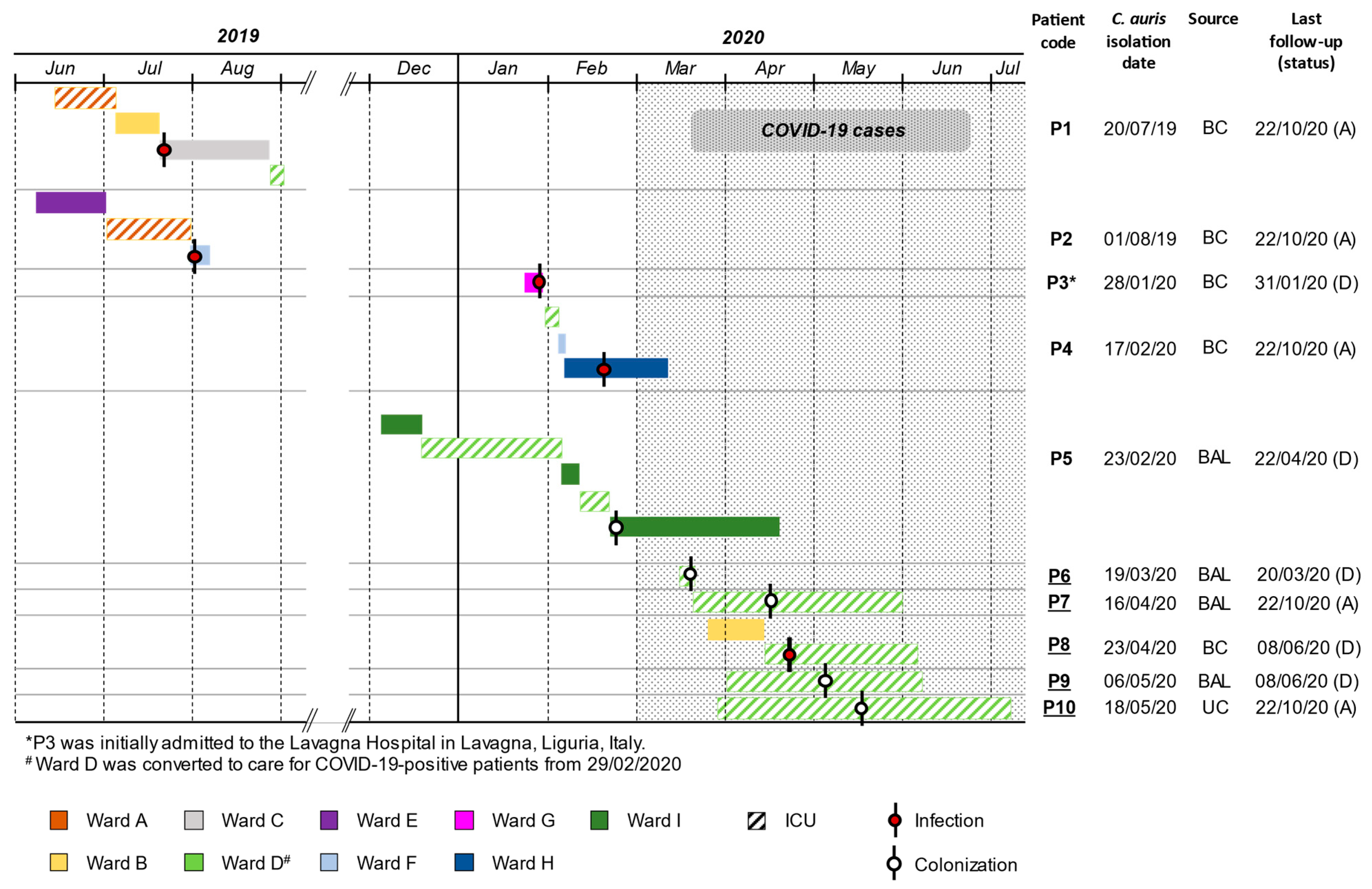
3.1. Case Series Description

The index case of C. auris has been reported in Italy in mid-July 2019 [11], from an intensive care unit (ICU) patient (P1) admitted to the San Martino Hospital (HSM) where, in early August 2019, a second patient (P2) was diagnosed with a bloodstream infection (BSI) by C. auris. After a few months, in late January 2020, an additional case was recorded in a patient (P3) initially diagnosed with a C. auris BSI (isolation date: 15/01/20) at the Lavagna Hospital (Lavagna, Italy), another healthcare facility located in the same regional area, and then transferred to the HSM for further treatment. Subsequently, following the emergence of the COVID-19 pandemic, episodes of BSI (P4, P8) and cases categorized as colonization (P5, P6, P7, P9, P10) by C. auris have been increasingly reported in patients admitted to the HSM from February to May 2020 (Figure 1). Overall, a total of 10 consecutive isolates of C. auris, accounting for 10 cases including the index patient, were collected and then subjected to further molecular and phenotypic investigations.
In addition, the clinical characteristics of COVID-19 patients diagnosed with infection or colonization by C. auris in our hospital were examined, and results were presented in a dedicated report [26].

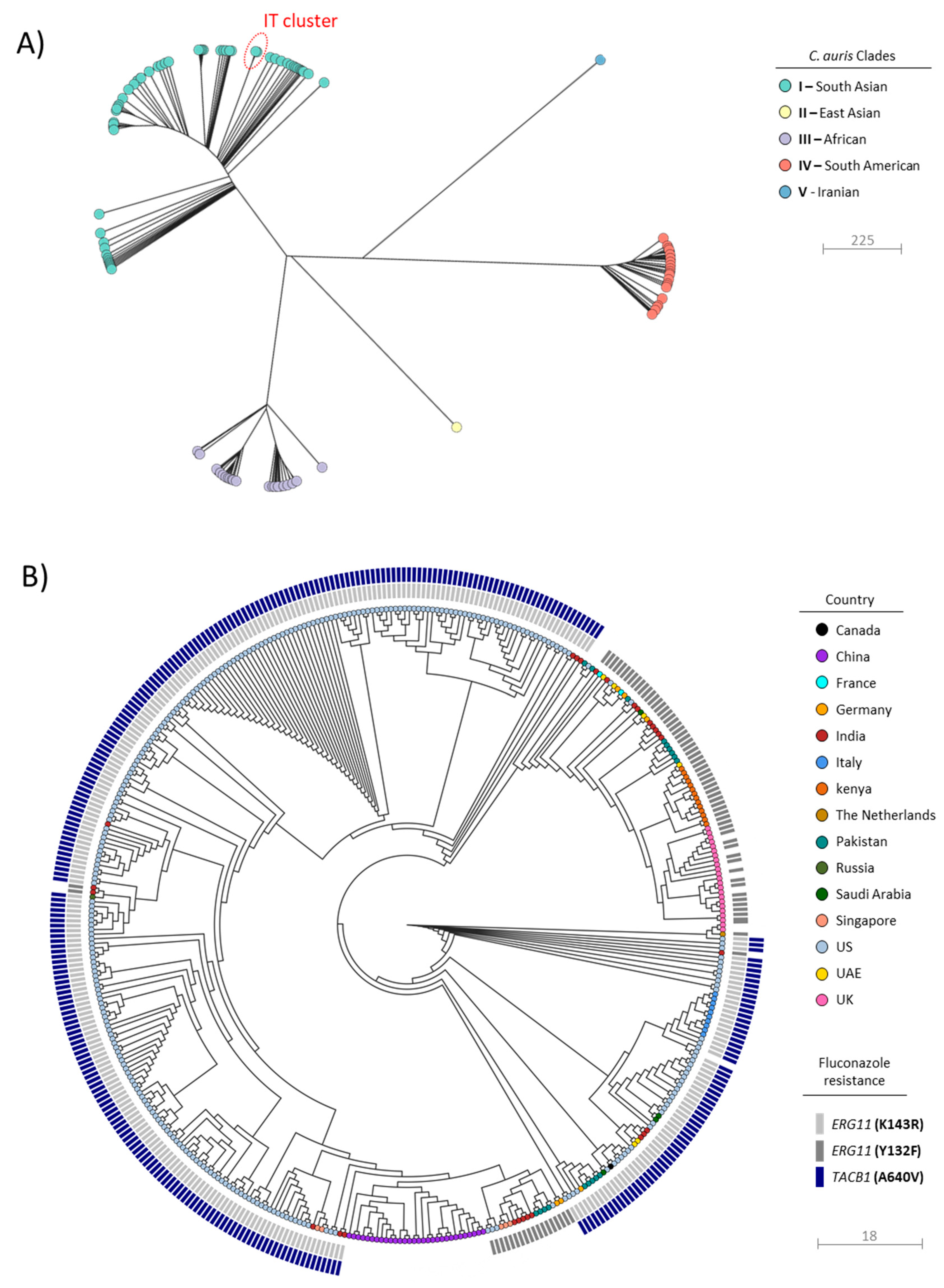
3.2. Inference of Putative Origin and Emergence of C. auris Isolates in Italy

In order to rule out the simultaneous emergence of multiple C. auris lineages and to further infer the origin of collected isolates, high-resolution genotyping through WGS was performed. According to sequencing results, the species-level identification as C. auris was confirmed for all isolates, and a complete reference genome (i.e., including seven linear chromosomes and one circular mitochondrial DNA molecule) to be used in downstream analyses was generated for C. auris FG_GE01 (Acc. Nos CP061160-CP061167), representing the Italian index case. A global phylogenetic analysis, including a collection of 871 genomes from 25 countries on six continents (Table S1), was then performed and results showed that all isolates from HSM were resolved within the South Asian clade (I) (Figure 2A). An additional phylodynamic analysis restricted to members of this clade, which included 416 isolates from Canada, China, France, Germany, India, Kenya, the Netherlands, Pakistan, Russia, Saudi Arabia, Singapore, the US, United Arab Emirates, and the UK, revealed that isolates from Italy segregated in a monophyletic clade (SNPs median\mean: 20) together with five isolates from the US (mainly from New Jersey). Previous epidemiological investigations including the latter isolates revealed their introduction route in the US was unknown, since no links to healthcare exposures abroad were documented [27].
Interestingly, following a root-to-tip regression analysis, a correlation between sampling dates and the genetic distances among sequence isolates in this study was observed (correlation coefficient: 0.89; R2 = 0.80), indicating that the sequence dataset had a sufficient phylogenetic temporal signal to perform further molecular clock analyses. A more accurate estimate of the time to the most recent common ancestor of sequenced isolates (i.e., the time since they last shared a common ancestor) corresponded to 27 May 2019 (95% highest posterior density interval: 27 December 2018–16 July 2019), less than two months prior to the identification of the C. auris index case in Italy, indicating a relatively recent introduction event.

3.3. Investigation of Clonal Relatedness and Putative Transmission Patterns of C. auris from HSM

A phylogenetic analysis restricted to the C. auris isolates from HSM suggested that 9 out of 10 of them were most likely associated with a nosocomial cluster (SNPs mean\median: 7) (Figure 3), consistent with previous evidence about the genetic diversity observed among pairs of epidemiologically linked cases of C. auris (median SNPs difference: 7) [26]. The remaining isolate, obtained from P3, most likely represented a member of the same C. auris lineage accounting for a separate introduction (SNPs against other isolates: mean, 12; median, 14), consistently with its original identification in a different hospital.
A combined analysis of clonal and patient trace data suggested that the intrahospital spread of C. auris likely stemmed from the index patient, occurred under different time frames, and involved two specific wards of HSM (Figure 1). The ward A was possibly related to the initial emergence and transmission of C. auris between the first two patients, P1 and P2, who were infected by genetically identical isolates (0 SNPs) and had a concomitant stay in this ICU. The ward D was associated with a putative horizontal transmission between the other seven patients, of which five (P6 to P10) were admitted as COVID-19 patients during the early phase of the pandemic (Figure 1). In this case, however, more complex dynamics were possibly involved, since transmissions supported by WGS were not fully explained by patients’ overlap. Independent transmissions from P1 to P4, P5, P6, and P9 (SNPs mean\median: 8) were deemed and likely involved intermediate patients/operators or contaminated environmental surfaces in ward D, where C. auris could have been primarily introduced by the index patient during its stay (Figure 1 and Figure 3). Former investigations, indeed, showed that C. auris was successfully cultivated from axillary and ear swabs obtained from P1 [12], who could have shed large amounts of this organism from her skin, thus leading to contamination of surrounding environments. Furthermore, P4, P5, P6, and P9 occupied adjacent beds in a defined area of ward D, being in the proximity to the bed initially occupied by P1. A transmission chain, on the other hand, was predicted among the remaining patients, likely going from P6 through P7, P8 and P10 as suggested by the high genetic relatedness of corresponding C. auris isolates (SNPs mean\median: 3) and by their overlap in ward D (Figure 1 and Figure 3).

3.4. Antifungal Susceptibility Profiles

Results from AFST revealed that all tested isolates were resistant to fluconazole, voriconazole, and amphotericin B. Conversely, lower MIC values were observed for the other tested antifungal drugs (Table 1). Inspection of the ERG2, ERG3, ERG5, and ERG6 coding sequence in C. auris genomes from Italy revealed no mutations previously associated with amphotericin B resistance [28,29,30]. Conversely, the presence of the same amino acid substitution ERG11(K143R), being previously shown to reduce azole susceptibility [3], has been detected in all isolates. A genetic alteration recently associated with a high-level resistance to fluconazole, due to a transcriptional upregulation of ABC-type efflux pumps, was additionally identified in all sequenced isolates in TAC1B(A640V) [31]. Interestingly, following a large-scale analysis of WGS data aimed at evaluating the distribution and frequency of the TAC1B(A640V) substitution over 400 C. auris isolates belonging to the South Asian clade, a strong association with the ERG11(K143R) variant was observed at a global level (Figure 2B), corroborating a significant contribution of both alterations to clinical fluconazole resistance. Nevertheless, multiple factors seem to contribute to azole resistance in C. auris, since mutation in ERG11 have been also identified in azole-susceptible isolates [32]; further investigations are therefore warranted to elucidate the genetic bases of this resistance mechanism.

4. Discussion

The global spread of C. auris poses a major threat to healthcare system. Hence, tracking the dissemination of this emerging pathogen and understanding its introduction and transmission dynamics, as well as its resistance features, have become crucial issues.
Here we report on a molecular epidemiological investigation of a C. auris cluster occurring in the same hospital where this yeast was first and only identified in Italy during 2019 [12]. The conjunction of WGS and patient trace data provided some notable insights into the genomic epidemiology of this fungal pathogen in our setting: (i) at present, its emergence in Italy is driven by one of the four major phylogeographically distinct lineages, namely clade I; (ii) the temporal analysis performed on sequenced genomes suggested that the introduction in our country was a relatively recent event (late May 2019); (iii) the genetic relatedness observed among most isolates was consistent with a nosocomial cluster sustained by a single clone, despite the interval of more than 6 months between less (August 2019) and more recent (February 2020) cases; (iv) clade-specific genetic signatures associated with high-level fluconazole resistance were identified; and (v) the detection of closely related clones, from patients admitted to different hospitals, with no epidemiological links, underscores that the dissemination of C. auris in our country could be broader than expected.
Over the past decade, C. auris has been detected across all major continents, and recent studies showed that in some European countries, like the UK and Germany [3,33], and in the US [27], where a near-nationwide spread has been observed, the emergence of this MDR yeast was contributed by isolates belonging to multiple genetic lineages (i.e., clade I, II, III). Conversely, our findings depicted a different epidemiological scenario in Italy, where a single lineage was recognized. Although WGS provided some insights into the phylogeny of characterized C. auris isolates, the introduction of this fungal pathogen in our country remains to be clarified, since the index patient had no history of recent travel or hospital admission abroad [12].
Both genomic and epidemiological data shed light on the putative transmission pathways in our setting, suggesting that unrecognized contaminated environments and/or colonized patients played a major role in creating long-term reservoirs and sustaining silent transmission chains, despite the bundle of infection-control interventions implemented at HSM after recognition of the index case (July 2019). Furthermore, genomic data suggested that one isolate (i.e., FG_GE03 from P3) was apparently not related with the cluster recognized at HSM, and likely represented an additional reservoir of ongoing transmission occurring in other regional facilities (e.g., Lavagna), thus posing major concerns for the control of this organism.
More importantly, the COVID-19 pandemic may have provided ideal conditions for the spread of C. auris. Indeed, in a context where the existing ICU capacity had been overwhelmed by the large number of COVID-19 patients requiring critical care and the conventional infection prevention and control measures were challenging (e.g., cohorting), the prolonged use of personal protective equipment (PPE) by the healthcare personnel may have inadvertently mediated a silent dissemination of this fungal pathogen. Epidemiological alerts of C. auris outbreaks occurring in healthcare facilities in the context of the COVID-19 pandemic have been recently documented also in Florida, Mexico, and India [34,35,36]. Of note, these reports consistently remarked the role that a possible low compliance to the guidelines for the correct use of PPE (e.g., experienced during anticipated or existing shortages) may have played in environmental contamination and transmission of C. auris, thus providing further evidence about major risk factors likely associated with an enhanced nosocomial spread of this organism during the pandemic. Furthermore, considering that critically ill COVID-19 patients tend to share risk factors (e.g., indwelling catheters) and underlying comorbidities with C. auris infections (e.g., diabetes mellitus, chronic kidney disease, intubation/mechanical ventilation, administration of broad-spectrum antibiotics, and systemic steroids), the spread of this fungal pathogen in the ICU setting raises major concerns [37]. Real-life data on the possible negative impact of the COVID-19 pandemic on antimicrobial stewardship programs has been recently reported [26]. In our hospital, following the increased number of cases observed in ward D, a systematic screening for C. auris colonization was started in mid-May 2020. Nevertheless, despite our attempts to monitor and control its spread, C. auris is proving difficult to eradicate and infection and/or colonization episodes continue to appear until now (November 2020).
All isolates characterized in this study showed a reduced susceptibility to voriconazole and were highly resistant to fluconazole, owing to the presence of missense mutations in the ERG11(K143R) and TAC1B(A640V) genetic loci, consistently with the recently reported association between high-level resistance to fluconazole and co-occurrence of these mutations [31]. Different ERG11 hot-spot mutations (Y132F, K143R, and F126L or VF125AL) have been previously associated with different C. auris genetic lineages, with the ERG11(Y132F or K143R) being primarily found within the South Asian clade [15]. Interestingly, our findings underscored that a strong association between ERG11(K143R) and TAC1B(A640V) clearly subsists within the latter clade, corroborating the role of such mutations as lineage-specific resistance signatures. Besides azoles, all studied isolates showed a reduced susceptibility to amphotericin B, which represents a less common resistance trait in C. auris (Figure 2B) [38]. The variable susceptibility to amphotericin B is of particular concern and has been previously observed in outbreaks sustained by members of the South Asian clade [7].

5. Conclusions

Herein, we present the results of a molecular epidemiological investigation aimed at exploring the dynamics underlying the emergence and the dissemination of C. auris in a large teaching hospital in Italy. Present findings expand current knowledge on the population structure, genomic epidemiology, and resistance traits of this yeast. Its spread during the COVID-19 pandemic represents a worrisome phenomenon, and extreme caution is warranted for the management of critically ill COVID-19 patients developing infection or colonization by C. auris.
Continued efforts in identifying and in understanding the introduction and transmission dynamics of this emerging pathogen are critical to deliver timely and effective public health responses aimed at containing its spread within the healthcare system.

Supplementary Materials

The following are available online at https://0-www-mdpi-com.brum.beds.ac.uk/2309-608X/7/2/140/s1, Table S1: Metadata of C. auris genome sequences included in this study.

Author Contributions

V.D.P.: Conceptualization, formal analysis, investigation, methodology, visualization, writing—original draft, writing—review and editing; G.C.: Data curation, formal analysis, investigation, visualization, writing—original draft; L.B.: Data curation, investigation, writing— review and editing; D.R.G.: Data curation, investigation, writing—review and editing; E.W.: Data curation, methodology, investigation; M.M.: Data curation, writing—review and editing; L.M.: Data curation, investigation; F.C.: Data curation, methodology, Investigation; A.V.: Data curation, investigation; P.P.: Writing—review and editing, supervision; M.B.: Writing—review and editing, supervision; A.M.: Conceptualization, project administration, supervision, Writing—review and editing. All authors have read and agreed to the published version of the manuscript.

Funding

This study was partially supported by a research grant funded by the University of Genoa to A.M. (FRA2018). The funding sources had no role in the study design, data collection, analysis, interpretation, or writing of the report.

Institutional Review Board Statement

The study was conducted according to the guidelines of the Declaration of Helsinki, and approved by the Ethics Committee of the Liguria region (N. CER Liguria 163/2020).

Informed Consent Statement

All conscious patients included in this study signed an informed consent for the use of their anonymized clinical data for scientific purposes on hospital admission. A waiver of informed consent for unconscious patients included in this study was obtained within the local ethics committee approval for collection of anonymized clinical data of hospitalized patients with COVID-19 (Liguria Region Ethics Committee, registry number 163/2020).

Data Availability Statement

Raw Illumina reads, complete and draft genomes of C. auris isolates sequenced in this study have been deposited in NCBI databases under BioProject no. PRJNA655187.

Conflicts of Interest

Outside the submitted work, M.B. has participated in advisory boards and/or received speaker honoraria from Achaogen, Angelini, Astellas, Bayer, Basilea, BioMérieux, Cidara, Gilead, Menarini, MSD, Nabriva, Paratek, Pfizer, Roche, Melinta, Shionogi, Tetraphase, VenatoRx, and Vifor, and has received study grants from Angelini, Basilea, Astellas, Shionogi, Cidara, Melinta, Gilead, Pfizer, and MSD. Outside the submitted work, DRG reports honoraria from Stepstone Pharma GmbH and unconditional grants from MSD Italia and Correvio Italia.

References

  1. Brown, G.D.; Denning, D.W.; Gow, N.A.R.; Levitz, S.M.; Netea, M.G.; White, T.C. Hidden killers: Human fungal infections. Sci. Transl. Med. 2012, 4, 165rv13. [Google Scholar] [CrossRef] [PubMed] [Green Version]
  2. Jeffery-Smith, A.; Taori, S.K.; Schelenz, S.; Jeffery, K.; Johnson, E.M.; Borman, A.; Manuel, R.; Brown, C.S.; Browna, C.S. Candida auris: A review of the literature. Clin. Microbiol. Rev. 2018, 31, 1–18. [Google Scholar] [CrossRef] [PubMed] [Green Version]
  3. Chybowska, A.D.; Childers, D.S.; Farrer, R.A. Nine Things Genomics Can Tell Us About Candida auris. Front. Genet. 2020, 11, 1–18. [Google Scholar] [CrossRef]
  4. Chow, N.A.; De Groot, T.; Badali, H.; Abastabar, M.; Chiller, T.M.; Meis, J.F. Potential fifth clade of Candida auris, Iran, 2018. Emerg. Infect. Dis. 2019, 25, 1780–1781. [Google Scholar] [CrossRef] [PubMed] [Green Version]
  5. Chow, N.A.; Muñoz, J.F.; Gade, L.; Berkow, E.L.; Li, X.; Welsh, R.M.; Forsberg, K.; Lockhart, S.R.; Adam, R.; Alanio, A.; et al. Tracing the Evolutionary History and Global Expansion of Candida auris Using Population Genomic Analyses. MBio 2020, 11. [Google Scholar] [CrossRef]
  6. Lone, S.A.; Ahmad, A. Candida auris-the growing menace to global health. Mycoses 2019, 62, 620–637. [Google Scholar] [CrossRef] [Green Version]
  7. Taori, S.K.; Khonyongwa, K.; Hayden, I.; Athukorala, G.D.A.; Letters, A.; Fife, A.; Desai, N.; Borman, A.M. Candida auris outbreak: Mortality, interventions and cost of sustaining control. J. Infect. 2019, 79, 601–611. [Google Scholar] [CrossRef]
  8. Rudramurthy, S.M.; Chakrabarti, A.; Paul, R.A.; Sood, P.; Kaur, H.; Capoor, M.R.; Kindo, A.J.; Marak, R.S.K.; Arora, A.; Sardana, R.; et al. Candida auris candidaemia in Indian ICUs: Analysis of risk factors. J. Antimicrob. Chemother. 2017, 72, 1794–1801. [Google Scholar] [CrossRef] [PubMed] [Green Version]
  9. Garcia-Bustos, V.; Salavert, M.; Ruiz-Gaitán, A.C.; Cabañero-Navalon, M.D.; Sigona-Giangreco, I.A.; Pemán, J. A clinical predictive model of candidaemia by Candida auris in previously colonized critically ill patients. Clin. Microbiol. Infect. 2020, 26. [Google Scholar] [CrossRef]
  10. Yapar, N. Epidemiology and risk factors for invasive candidiasis. Clin. Risk Manag. 2014, 10, 95–105. [Google Scholar] [CrossRef] [PubMed] [Green Version]
  11. Kohlenberg, A.; Struelens, M.J.; Monnet, D.L.; Plachouras, D. Candida auris: Epidemiological situation, laboratory capacity and preparedness in European Union and European Economic Area countries, 2013 to 2017. Eurosurveillance 2018, 23, 18-00136. [Google Scholar] [CrossRef] [PubMed] [Green Version]
  12. Crea, F.; Codda, G.; Orsi, A.; Battaglini, A.; Giacobbe, D.R.; Delfino, E.; Ungaro, R.; Marchese, A. Isolation of Candida auris from invasive and non-invasive samples of a patient suffering from vascular disease, Italy, July 2019. Euro Surveill. 2019, 24, 1900549. [Google Scholar] [CrossRef] [PubMed]
  13. Ruiz-Gaitán, A.C.; Fernández-Pereira, J.; Valentin, E.; Tormo-Mas, M.A.; Eraso, E.; Pemán, J.; de Groot, P.W.J. Molecular identification of Candida auris by PCR amplification of species-specific GPI protein-encoding genes. Int. J. Med. Microbiol. 2018, 308, 812–818. [Google Scholar] [CrossRef]
  14. CLSI. Reference Method for Broth Dilution Antifungal Susceptibility Testing of Yeast, 4th ed.; CLSI standard M27; Clinical and Laboratory Standards Institute, Ed.; CLSI: Wayne, PA, USA, 2017; ISBN 1-56238-827-4. [Google Scholar]
  15. Lockhart, S.R.; Etienne, K.A.; Vallabhaneni, S.; Farooqi, J.; Chowdhary, A.; Govender, N.P.; Colombo, A.L.; Calvo, B.; Cuomo, C.A.; Desjardins, C.A.; et al. Simultaneous emergence of multidrug-resistant candida auris on 3 continents confirmed by whole-genome sequencing and epidemiological analyses. Clin. Infect. Dis. 2017, 64, 134–140. [Google Scholar] [CrossRef] [Green Version]
  16. Kolmogorov, M.; Yuan, J.; Lin, Y.; Pevzner, P.A. Assembly of long, error-prone reads using repeat graphs. Nat. Biotechnol. 2019, 37, 540–546. [Google Scholar] [CrossRef]
  17. Walker, B.J.; Abeel, T.; Shea, T.; Priest, M.; Abouelliel, A.; Sakthikumar, S.; Cuomo, C.A.; Zeng, Q.; Wortman, J.; Young, S.K.; et al. Pilon: An integrated tool for comprehensive microbial variant detection and genome assembly improvement. PLoS ONE 2014, 9, e112963. [Google Scholar] [CrossRef]
  18. Moss, E.L.; Maghini, D.G.; Bhatt, A.S. Complete, closed bacterial genomes from microbiomes using nanopore sequencing. Nat. Biotechnol. 2020, 38, 701–707. [Google Scholar] [CrossRef] [PubMed] [Green Version]
  19. Li, H.; Durbin, R. Fast and accurate long-read alignment with Burrows-Wheeler transform. Bioinformatics 2010, 26, 589–595. [Google Scholar] [CrossRef] [PubMed] [Green Version]
  20. Kurtz, S.; Phillippy, A.; Delcher, A.L.; Smoot, M.; Shumway, M.; Antonescu, C.; Salzberg, S.L. Versatile and open software for comparing large genomes. Genome Biol. 2004, 5, R12. [Google Scholar] [CrossRef] [PubMed] [Green Version]
  21. Garrison, E.G.; Marth, G. Haplotype-based variant detection from short-read sequencing. arXiv 2012, arXiv:1207.3907. [Google Scholar]
  22. Nguyen, L.-T.; Schmidt, H.A.; von Haeseler, A.; Minh, B.Q. IQ-TREE: A fast and effective stochastic algorithm for estimating maximum-likelihood phylogenies. Mol. Biol. Evol. 2015, 32, 268–274. [Google Scholar] [CrossRef]
  23. Rambaut, A.; Lam, T.T.; Max Carvalho, L.; Pybus, O.G. Exploring the temporal structure of heterochronous sequences using TempEst (formerly Path-O-Gen). Virus Evol. 2016, 2, vew007. [Google Scholar] [CrossRef] [PubMed] [Green Version]
  24. Rhodes, J.; Abdolrasouli, A.; Farrer, R.A.; Cuomo, C.A.; Aanensen, D.M.; Armstrong-James, D.; Fisher, M.C.; Schelenz, S. Genomic epidemiology of the UK outbreak of the emerging human fungal pathogen Candida auris article. Emerg. Microbes Infect. 2018, 7, 43. [Google Scholar] [CrossRef] [Green Version]
  25. Drummond, A.J.; Suchard, M.A.; Xie, D.; Rambaut, A. Bayesian phylogenetics with BEAUti and the BEAST 1.7. Mol. Biol. Evol. 2012, 29, 1969–1973. [Google Scholar] [CrossRef] [PubMed] [Green Version]
  26. Magnasco, L.; Mikulska, M.; Giacobbe, D.R.; Taramasso, L.; Vena, A.; Dentone, C.; Dettori, S.; Tutino, S.; Labate, L.; Di Pilato, V.; et al. Spread of Carbapenem-Resistant Gram-Negatives and Candida auris during the COVID-19 Pandemic in Critically Ill Patients: One Step Back in Antimicrobial Stewardship? Microorganisms 2021, 9, 95. [Google Scholar] [CrossRef]
  27. Chow, N.A.; Gade, L.; Tsay, S.V.; Forsberg, K.; Greenko, J.A.; Southwick, K.L.; Barrett, P.M.; Kerins, J.L.; Lockhart, S.R.; Chiller, T.M.; et al. Multiple introductions and subsequent transmission of multidrug-resistant Candida auris in the USA: A molecular epidemiological survey. Lancet Infect. Dis. 2018, 18, 1377–1384. [Google Scholar] [CrossRef]
  28. Yadav, A.; Singh, A.; Wang, Y.; van Haren, M.H.; Singh, A.; de Groot, T.; Meis, J.F.; Xu, J.; Chowdhary, A. Colonisation and Transmission Dynamics of Candida auris among Chronic Respiratory Diseases Patients Hospitalised in a Chest Hospital, Delhi, India: A Comparative Analysis of Whole Genome Sequencing and Microsatellite Typing. J. Fungi (BaselSwitz.) 2021, 7, 81. [Google Scholar] [CrossRef]
  29. Carolus, H.; Pierson, S.; Muñoz, J.F.; Subotić, A.; Cruz, R.B.; Cuomo, C.A.; Van Dijck, P. Genome-wide analysis of experimentally evolved Candida auris reveals multiple novel mechanisms of multidrug-resistance. bioRxiv 2020. [Google Scholar] [CrossRef]
  30. Arendrup, M.C.; Patterson, T.F. Multidrug-Resistant Candida: Epidemiology, Molecular Mechanisms, and Treatment. J. Infect. Dis. 2017, 216, S445–S451. [Google Scholar] [CrossRef] [Green Version]
  31. Rybak, J.M.; Muñoz, J.F.; Barker, K.S.; Parker, J.E.; Esquivel, B.D.; Berkow, E.L.; Lockhart, S.R.; Gade, L.; Palmer, G.E.; White, T.C.; et al. Mutations in TAC1B: A Novel Genetic Determinant of Clinical Fluconazole Resistance in Candida auris. MBio 2020, 11. [Google Scholar] [CrossRef]
  32. Chowdhary, A.; Prakash, A.; Sharma, C.; Kordalewska, M.; Kumar, A.; Sarma, S.; Tarai, B.; Singh, A.; Upadhyaya, G.; Upadhyay, S.; et al. A multicentre study of antifungal susceptibility patterns among 350 Candida auris isolates (2009-17) in India: Role of the ERG11 and FKS1 genes in azole and echinocandin resistance. J. Antimicrob. Chemother. 2018, 73, 891–899. [Google Scholar] [CrossRef]
  33. Borman, A.M.; Johnson, E.M. Candida auris in the UK: Introduction, dissemination, and control. PLoS Pathog. 2020, 16, e1008563. [Google Scholar] [CrossRef] [PubMed]
  34. Prestel, C.; Anderson, E.; Forsberg, K.; Lyman, M.; de Perio, M.A.; Kuhar, D.; Edwards, K.; Rivera, M.; Shugart, A.; Walters, M.; et al. Candida auris Outbreak in a COVID-19 Specialty Care Unit—Florida, July–August 2020. Mmwr. Morb. Mortal Wkly Rep. 2021, 56–57. [Google Scholar] [CrossRef]
  35. Villanueva-Lozano, H.; Treviño-Rangel, R.d.J.; González, G.M.; Ramírez-Elizondo, M.T.; Lara-Medrano, R.; Aleman-Bocanegra, M.C.; Guajardo-Lara, C.E.; Gaona-Chávez, N.; Castilleja-Leal, F.; Torre-Amione, G.; et al. Outbreak of Candida auris infection in a COVID-19 hospital in Mexico. Clin. Microbiol. Infect. 2021, in press. [Google Scholar] [CrossRef]
  36. Chowdhary, A.; Tarai, B.; Singh, A.; Sharma, A. Multidrug-Resistant Candida auris Infections in Critically Ill Coronavirus Disease Patients, India, April-July 2020. Emerg. Infect. Dis. 2020, 26, 2694–2696. [Google Scholar] [CrossRef]
  37. Chowdhary, A.; Sharma, A. The lurking scourge of multidrug resistant Candida auris in times of COVID-19 pandemic. J. Glob. Antimicrob. Resist. 2020, 22, 175–176. [Google Scholar] [CrossRef] [PubMed]
  38. Lockhart, S.R. Candida auris and multidrug resistance: Defining the new normal. Fungal Genet. Biol. 2019, 131, 103243. [Google Scholar] [CrossRef]
Figure 1. Overview of patient traces within different HSM hospital wards in the period from June 2019 to July 2020. Isolation of C. auris is indicated by black bars, marked with a red- or white-filled circle in case of infection or colonization events, respectively. Patients diagnosed with COVID-19 are underlined. Abbreviations: BC, blood culture; BAL, broncho-alveolar lavage; UC, urine culture; A, alive; D, dead.
Jof 07 00140 g001
Figure 2. Global phylogenetic analysis of C. auris, including genomes retrieved in this study (n = 10) and representative genomes from different countries and clades (n = 871) (Table S1). (A) Maximum-likelihood phylogenetic tree of C. auris whole-genome sequences (n = 881) clustering into four major clades. The colors of the isolate tips represent the different clades. The branch comprising the C. auris isolates from Italy is circled in red. (B) Maximum-likelihood phylogenetic tree of C. auris belonging to the clade I (n = 426). The colors of the isolate tips represent the country of isolation. Details about the genetic mutations (ERG11, TAC1B) associated with high-level resistance to fluconazole were also provided.
Jof 07 00140 g002
Figure 3. Phylogenetic analysis of C. auris isolates sequenced in this study. The phylogenetic tree topology was calibrated with sampling times and estimated using TempEst [23]. For each tip, the C. auris strain name and the patient’s code are provided. Patients diagnosed with COVID-19 are underlined, and the corresponding isolates’ tips are marked with dark blue tick borders.
Jof 07 00140 g003
Table 1. In vitro antifungal susceptibility profiles of characterized C. auris isolates (n = 10). The MIC range, MIC50, and MIC90 values (μg/mL) were reported.
Antifungal DrugsMIC RangeMIC50MIC90
Fluconazole>256>256>256
Itraconazole0.50.50.5
Voriconazole444
Posaconazole0.250.250.25
Caspofungin0.06–0.250.250.25
Anidulafungin0.12–0.250.250.25
Micafungin0.06–0.120.120.12
Amphotericin B2–424
Flucytosine0.12–0.50.250.5
Publisher’s Note: MDPI stays neutral with regard to jurisdictional claims in published maps and institutional affiliations.

