Article

Histidine Kinase Sln1 and cAMP/PKA Signaling Pathways Antagonistically Regulate Sporisorium scitamineum Mating and Virulence via Transcription Factor Prf1

1
College of Plant Protection, South China Agricultural University, Guangzhou 510642, China
2
Integrate Microbiology Research Center, Guangdong Province Key Laboratory of Microbial Signals and Disease Control, South China Agricultural University, Guangzhou 510642, China
3
Environmental Monitoring and Remediation Engineering Technology Research Center, School of Environmental Engineering, Yellow River Conservancy Technical Institute, Kaifeng 475004, China
*
Authors to whom correspondence should be addressed.
Co-first authors.
Submission received: 24 June 2021 / Revised: 16 July 2021 / Accepted: 26 July 2021 / Published: 28 July 2021
(This article belongs to the Special Issue Smut Fungi 2.0)

Abstract

:
Many prokaryotes and eukaryotes utilize two-component signaling pathways to counter environmental stress and regulate virulence genes associated with infection. In this study, we identified and characterized a conserved histidine kinase (SsSln1), which is the sensor of the two-component system of Sln1–Ypd1–Ssk1 in Sporisorium scitamineum. SsSln1 null mutant exhibited enhanced mating and virulence capabilities in S. scitamineum, which is opposite to what has been reported in Candida albicans. Further investigations revealed that the deletion of SsSLN1 enhanced SsHog1 phosphorylation and nuclear localization and thus promoted S. scitamineum mating. Interestingly, SsSln1 and cAMP/PKA signaling pathways antagonistically regulated the transcription of pheromone-responsive transcription factor SsPrf1, for regulating S. scitamineum mating and virulence. In short, the study depicts a novel mechanism in which the cross-talk between SsSln1 and cAMP/PKA pathways antagonistically regulates mating and virulence by balancing the transcription of the SsPRF1 gene in S. scitamineum.

1. Introduction

The basidiomycetous fungus Sporisorium scitamineum is a global pathogen of sugarcane smut disease that causes substantial losses in cane yield and the sugar industry. S. scitamineum, as Ustilago maydis, is bipolar and undergoes three life stages, of haploid sporidium, yeast-like, non-pathogenic, dikaryotic hypha and diploid teliospore [1]. Haploid sporidia of two opposite mating types, MAT-1 and MAT-2, can recognize each other to undergo fusion, a process known as sexual mating [2]. After sexual mating, the b locus encodes an active heterodimeric transcription factor complex composed of bE and bW proteins derived from different alleles to control filamentation in S. scitamineum [2]. The fusion of two haploid cells of opposite mating types is necessary to form invasively dikaryotic hyphae in S. scitamineum to infect the host [1]. Thus, mating/filamentation plays a key role in S. scitamineum pathogenicity.
The two-component phosphorelay system is widely found, which regulates a variety of cellular processes such as response to environmental stimuli, cell differentiation, secondary metabolite production, antibiotic resistance and virulence, in plant and animal pathogens [3,4]. The two-component phosphorelay system in most eukaryotes mainly consists of a sensor histidine kinase (HK) component, a His-containing phosphotransfer protein component and a response regulator (RR) component that transmits signals [5,6]. The two-component phosphorelay system in yeast is composed of Sln1–Ypd1–Ssk1 and a mitogen-activated protein kinase cascade consisting of Ssk2/Ssk22–Pbs2–Hog1 for the hypertonicity response through osmolarity signal transmission [7,8]. The histidine kinase (ortholog to Sln1) in filamentous fungi (Aspergillus nidulans and Neurospora crassa) also adapts to highly osmotic conditions by participating in the osmotic sensitive signal transduction pathway [9,10,11]. The lethality associated with the deletion of the SLN1 or YPD1 gene in Saccharomyces cerevisiae has highlighted this family of phosphate relay proteins as a potential prime antifungal target [12,13]. Three virulence-related conventional histidine kinase genes have been identified in C. albicans [14], whereas histidine kinase Sln1 in Magnaporthe oryzae senses and controls turgor-driven infection [6,15]. However, the two-component phosphorelay system among the smut fungi remains largely unknown.
The Hog1 activator has two key upstream osmotic response branches (SLN1 and SHO1 branches) for the high-osmolarity glycerol (HOG) pathway, which are redundant for cell survival under hyperosmosis [7,16,17,18]. However, the SLN1 branch seems to be more important in pathway response to low or extremely high osmotic stress [19,20,21]. In the SLN1 branch, the phosphorylated Sln1 triggers Ssk1 phosphorylation through intermediary phosphorelay protein Ypd1 [7], and Ssk1 phosphorylation further phosphorylates Ssk2/Ssk22 to transmit the signals [17,22]. In another branch, Sho1 senses hyperosmosis and activates the cytoplasmic MAPKKK Ste11 [23]. Finally, the activation of Ssk2 and Ste11 phosphorylates the MAPKK Pbs2, which in turn phosphorylates and activates the MAPK Hog1 [8,24]. Phosphorylation of Hog1 localizes in the nucleus, where the promoter region of the target gene(s) recruits it to activate several transcription factors [25,26,27,28]. Osm1 (ortholog to Hog1) activates Atf1 phosphorylation in M. oryzae, initiating the transcription of Ptp1/2 for the dephosphorylation of Osm1 [29]. The negative feedback loop of the Hog1 kinase was also noted in Cryptococcus neoformans [30].
The cAMP-dependent protein kinase A (PKA) signaling pathway is important and ubiquitously exists in eukaryotic organisms. This pathway participates in regulating physiological processes, such as morphological switch, response to stress, polarized cell growth, mating and virulence [31,32,33,34,35,36,37]. The core components of the cAMP/PKA pathway, such as G-protein, adenylyl cyclase and catalytic subunit of cAMP-dependent protein kinase A, contribute to mating and virulence in S. scitamineum [38]. The pheromone response factor SsPrf1, the downstream element of cAMP/PKA signaling, is also involved in the mating and virulence in S. scitamineum [39]. The interaction of the cAMP/PKA signaling pathway with other signal transduction pathways to exhibit a cross-talk relationship has been reported. For example, the Ime2-related protein encoded by CRK1 acted as a signal integrator of external signals that were transmitted via cAMP/PKA and Kpp2–MAPK pathways in U. maydis [40]. Sln1 interacts with Sum1 in M. oryzae, which is a regulatory subunit of cAMP-dependent protein kinase A (PKA) [15]. However, the relationship between cAMP/PKA and SsSln1 in the smut fungi is unclear.
Therefore, during this study, Sln1 ortholog (SsSln1) was identified and characterized in S. scitamineum to understand its contribution in the regulation of virulence through triggering downstream signaling. The study revealed that the deletion of the SsSLN1 gene enhanced the phosphorylation and nuclear localization of SsHog1. Further investigations depicted that the phosphorylation level of SsHog1 was essential for the mating and virulence of S. scitamineum. Intriguingly, we found that histidine kinase SsSln1 and cAMP/PKA pathways affected the transcription of SsPRF1 gene to antagonistically regulate mating and virulence. In short, our results indicate that deletion of the SsSLN1 gene enhances S. scitamineum mating and virulence capabilities, and reveal a cross-talk relationship between SsSln1 and the cAMP/PKA signaling pathway.

2. Materials and Methods

2.1. Strains and Growth Conditions

Two S. scitamineum wild-type haploid MAT-1 (a1 b1) and MAT-2 (a2 b2) were isolated and identified by Yan [2], and stored locally. The haploid sporidia were cultured on YePS liquid medium (yeast extract 1%, peptone 2%, sucrose 2%, pH 6.5) at 28 °C for 1–2 days as described [2,38]. For sexual mating assay, the fresh haploid sporidia were cultured in YePS medium to about O.D.600 = 1.0. The sporidia were collected and washed twice in sterilized double-distilled water (ddH2O), and the concentration was adjusted to O.D.600 = 1.0 with sterilized ddH2O. The haploid sporidia of opposite mating types were mixed at equal volume (O.D.600 = 1.0) and spotted on YePSA (YePS, containing 2% agar) or minimal medium [38], and allowed to incubate at 28 °C for 1–2 days.

2.2. Construction of Strains

The deletion, reintegration and overexpression of genes were performed by polyethylene glycol (PEG) mediate protoplast transformation using a split marker approach as described previously [38,41,42]. The Hygromycin resistance (HYGR) cassette was used as a resistance screening gene. The deletion mutants were generated by individually disrupting SsSLN1 and SsATF1 genes in the MAT-1 and MAT-2 strains. The left and right borders of SsSLN1 and SsATF1 genes were PCR amplified from S. scitamineum wild-type genomic DNA with the primers SsSLN1-LB-F/SsSLN1-LB-R, SsSLN1-RB-F/SsSLN1-RB-R, SsATF1-LB-F/SsATF1-LB-R and SsATF1-RB-F/SsATF1-RB-R, respectively. Two truncated and partially overlapped fragments of HYGR gene were separately PCR amplified from pDAN plasmid with the primers pDAN-F/LB-226-R and pDAN-R/RB-225-F. These PCR products served as templates in fusion PCR to generate two PCR fragments with the primers SsSLN1-LB-F/LB-226-R, RB-225-F/SsSLN1-RB-R, SsATF1-LB-F/LB-226-R and RB-225-F/SsATF1-RB-R, individually. The mixture of two fusion homologous fragments for each targeted gene was transformed into MAT-1 and MAT-2 protoplasts via PEG-mediated protoplast transformation. The transformants were recovered in a regeneration medium impregnated with 200 mg/mL hygromycin B (Merck, Saint Louis, MO, USA). Putative deletion mutants were screened and identified by PCR amplification using the following primers: SsSLN1-inside-F/SsSLN1-inside-R, SsSLN1-outside-F/SsSLN1-outside-R, SsATF1-inside-F/SsATF1-inside-R and SsATF1-outside-F/SsATF1-outside-R. This operation was used to generate the double-deletion mutants ss1sln1Δgpa3Δ, ss1sln1Δuac1Δ and ss1sln1Δadr1Δ, with the Zeocin resistance (ZEOR) cassette as selection marker in the ss1sln1Δ background.
SsSLN1 and SsATF1 reintegration was carried out, as previously described [38]. The HYGR gene in the mutant was replaced with the reintegrated gene, together with the ZEOR gene as a selection marker, by a split marker approach. A fragment containing the native promoter and SsSLN1 or SsATF1 gene was PCR amplified with wild-type genomic DNA as a template, using the primers SsSLN1-COM-F/SsSLN1-COM-R and SsATF1-COM-F/SsATF1-COM-R, respectively. These PCR products were ligated with vector pEASY-COM containing HYGR-LB (HYGR-left homologous arm), ZEOR gene and HYGR-RB (HYGR-right homologous arm). Two reintegrated homologous fragments, one fragment including HYGR-LB, a complete target gene and partially overlapped fragments of the ZEOR gene, and the other containing partially overlapped fragments of the ZEOR gene and HYGR-RB, were PCR amplified with pEASY-SsSLN1-COM or pEASY-SsATF1-COM as a template using the primers COM-LB-F/COM-LB-R and COM-RB-F/COM-RB-R, respectively. Two reintegrated homologous fragments were individually transformed into ss1sln1Δ, ss2sln1Δ, ss1atf1Δ and ss2atf1Δ protoplasts through PEG-mediated protoplast transformation. Putative complementation transformants were selected with 100 mg/mL zeocin and identified with the primers SsSLN1-inside-F/SsSLN1-inside-R and SsATF1-inside-F/SsATF1-inside-R.
The IP locus, a DNA sequence without a function in S. scitamineum genomic DNA, was used as the target sequence for the overexpression of SsPTP1, SsPTP2 and SsPRF1. It was replaced with the overexpressed gene together with the ZEOR gene as a selection marker by a split marker approach. The CD fragments of SsPTP1, SsPTP2 and SsPRF1 genes were PCR amplified with S. scitamineum cDNA as a template, using the primers SsPTP1-OE-F/SsPTP1-OE-R, SsPTP2-OE-F/SsPTP2-OE-R and SsPRF1-OE-F/SsPRF1-OE-R, individually. These PCR products were individually ligated with vector pEASY-OE containing IP-LB (IP-left homologous arm), a constitutive GPA promoter, ZEOR gene and IP-RB (IP-right homologous arm). Two overexpressed homologous fragments, one fragment including IP-LB, the GPA promoter fused with the target gene and partially overlapped fragments of the ZEOR gene, and the other containing partially overlapped fragments of the ZEOR gene and IP-RB, were PCR amplified with templates pEASY-SsPTP1-OE, pEASY-SsPTP2-OE or pEASY-SsPRF1-OE using the primers OE-LB-F/OE-LB-R and OE-RB-F/OE-RB-R, respectively. Two overexpressed homologous fragments were mixed and transformed into MAT-1, MAT-2 or ss1hog1Δ protoplasts by PEG-mediated protoplast transformation, respectively. Putative overexpression transformants were selected with 100 mg/mL zeocin and identified with RT-qPCR using the primers qRT-SsPTP1-F/qRT-SsPTP1-R, qRT-SsPTP2-F/qRT-SsPTP2-R and qRT-SsPRF1-F/qRT-SsPRF1-R.
Ss1Hog1:eGFP(sln1Δ), Ss1Hog1:RFP, Ss1Atf1:eGFP and Ss1Atf1:eGFP(hog1Δ) strains were generated by using the termination codon of SsHOG1 or SsATF1 as the target sequence, which was replaced with a DNA sequence containing eGFP/RFP and ZEOR genes by a split marker approach. The left and/or right borders of SsHOG1:eGFP, SsHOG1:RFP and SsATF1:eGFP were separately PCR amplified from wild-type MAT-1 genomic DNA, using the primers SsHog1:eGFP-LB-F/SsHog1:eGFP-LB-R, SsHog1:eGFP-RB-F/SsHog1: eGFP-RB-R, SsHog1:RFP-LB-F/SsHog1:RFP-LB-R, SsHog1:RFP-RB-F/SsHog1: RFP-RB-R, SsAtf1:eGFP-LB-F/SsAtf1:eGFP-LB-R and SsAtf1:eGFP-RB-F/SsAtf1: eGFP-RB-R. Two truncated and partially overlapped fragments of the ZEOR or HYGR gene, including eGFP or RFP, were PCR amplified from pEASY-eGFP or pEASY-RFP plasmid with the primers eGFP-LB-F/eGFP-LB-R, eGFP-RB-F/eGFP-RB-R, RFP-LB-F/RFP-LB-R and RFP-RB-F/RFP-RB-R, respectively. These PCR products served as templates in fusion PCR to generate two PCR fragments with the primers SsHog1:eGFP-LB-F/eGFP-LB-R, eGFP-RB-F/SsHog1:eGFP-RB-R, SsAtf1:eGFP-LB-F/eGFP-LB-R, eGFP-RB-F/SsAtf1:eGFP-RB-R and SsHog1:RFP-LB-F/RFP-LB-R, RFP-RB-F/SsHog1: RFP-RB-R, individually.
The mixture of two fusion homologous fragments was transformed into MAT-1 or ss1sln1Δ protoplasts via PEG-mediated protoplast transformation for each targeted gene. The transformants were recovered in a regeneration medium impregnated with 200 mg/mL hygromycin B or 100 mg/mL zeocin. Putative fluorescent strains were screened and identified by fluorescence microscope and PCR amplification, respectively. The primers and sequences used in this study are listed in Table S1. Details of the strains generated and used in this study are listed in Table 1.

2.3. Nucleic Acid Related Manipulation

The S. scitamineum strains were grown on YePSA medium for 1–2 days at 28 °C. Then, the strains were collected and rapidly ground in liquid nitrogen to extract the genomic DNA of S. scitamineum using a modified SDS-based method [2]. The fresh haploid sporidia grown on YePSA medium at 28 °C for 30 h were used for the total RNA extraction of S. scitamineum with TRIzol reagent (ThermoFisher Scientific, Carlsbad, CA, USA) following established protocol [2]. HiScript ® II 1st Strand cDNA Synthesis Kit (Vazyme, Nanjing, China) was used to synthesize the cDNA. Real-Time Quantitative PCR was performed using Fast SYBR™ Green Master Mix (ThermoFisher Scientific, Carlsbad, CA, USA) on QuantStudio 6 Flex (ThermoFisher Scientific, Carlsbad, CA, USA). The relative gene expression level was calculated by adopting the −ΔΔCt method [43] and the cytoskeletal protein gene ACTIN was used as an internal control [38]. The experiment was carried out in triplicate for three independent biological replicates. To carry out Southern blot analysis, genomic DNA of negative control MAT-1 and MAT-2, positive control pDAN plasmid and genomic DNA of deletion mutant were digested with the restriction enzyme Hind III. The HPT sequence was amplified as the probe with PCR DIG Labeling Mix (Roche, Mannheim, BW, Germany). Probe hybridization was performed with a DIG Easy Hyb (Roche, Mannheim, BW, Germany) and detected by CSPD (Roche, Mannheim, BW, Germany). Probed bands of >3.0 kb size in deletion mutants confirmed the correct gene replacement.

2.4. Sporidia Staining and Epifluorescence Microscopy

The haploid sporidia of Ss1Hog1:RFP, Ss1Hog1:eGFP(sln1Δ), Ss1Atf1:eGFP and Ss1Atf1:eGFP(hog1Δ) strains were cultured overnight in YePS liquid medium at 28 °C, diluted to O.D.600 = 0.2 with the fresh YePS liquid medium and grown up to O.D.600 = 1.0. The fresh haploid sporidia were collected for nucleic acid staining by centrifugation and washed twice with 1 × PBS. Then, the cells were re-suspended in 20 μL of the Antifade Mounting Medium with DAPI (Beyotime, Shanghai, China). Finally, the samples (10 μL) were mounted on the slide and photographed under a Leica DMI8 Inverted Fluorescence Microscope using DAPI, GFP and RFP filters. The pictures were taken through Leica Application Suite (LAS) v.X software. Scale bar = 10 μm.

2.5. SsHog1 Phosphorylation Assays

The total protein was extracted from the fresh haploid sporidia grown on YePSA medium at 28 °C for 30 h [38]. Protein samples were separated by 10% SDS-PAGE. Phosphorylation of SsHog1 was determined by Western blot analysis with an antibody Phospho-p38 MAPK (Thr180/Tyr182) (D3F9) XP ® Rabbit (Cell Signaling Technology, Boston, MA, USA). Total levels of SsHog1 were detected by probing with an anti-Hog1 antibody (Genecreate Biological Engineering Company, Wuhan, China). Blot signals were displayed using enhanced chemiluminescence (BIO-RAD, 170-5061) after the binding of an Anti-Rabbit IgG–Peroxidase secondary antibody (Sigma, Louis, MO, USA), as described previously [38].

2.6. Yeast Two-Hybrid (Y2H) Assays

A modified yeast two-hybrid (Y2H) assay was used in this experiment [29]. The cDNA of SsSLN1, SsADR1, SsUBC1 and SsPRF1 was PCR amplified with Phanta ® Max Super-Fidelity DNA Polymerase (Vazyme, Nanjing, China). The PCR products were individually cloned into pGBKT7 or pGADT7 vectors. After the sequence verification, the pGBKT7-SsSln1, pGADT7-SsAdr1, pGADT7-SsUbc1 and pGADT7-SsPrf1 plasmids were, respectively, introduced into the yeast Y2HGold strain. Finally, the transformants grown on a synthetic medium lacking tryptophan and leucine (SD-Trp-Leu) were transferred to a synthetic medium lacking tryptophan, leucine, histidine and adenine (SD-Trp-Leu-His-Ade) at 30 °C.

2.7. Assessment of Pathogenicity and Relative Fungal Biomass

A highly susceptible sugarcane cultivar ROC22 was used for the pathogenicity assay as described [2,39]. The haploid sporidia of S. scitamineum strains were grown on YePS medium in a shaking incubator at 28 °C for 1–2 days. The fresh haploid sporidia were collected, re-suspended in the sterilized ddH2O, adjusted to 1 × 10⁶ cells/mL and mixed with haploid sporidia of opposite mating types in equal volume. Sugarcane seedlings of ROC22 grown to 5–6 leaf stage were inoculated by injection with approximately 0.2 mL of the mixture per plant. MAT-1 and MAT-2 mixture served as a positive control. Inoculated plants were kept in the greenhouse and a natural cycle of day and night was followed for 3–6 months. Three biological repeats were performed in the inoculation and each replicate involved the infection of at least 15 plants. The symptoms of ‘black whip’ were documented and photographed at about six months post inoculation. Percentage (%) of ‘black whip’/total seedlings was estimated.
Fungal biomass assay of the inoculated sugarcane seedlings was carried out according to Sun [44]. The same amount of S. scitamineum sporidia (mixture of compatible mating types) was injected into sugarcane seedlings at 3 days post-inoculation (dpi), and total DNA of the inoculated sugarcane tissue was extracted. The relative fungal biomass was measured using the fungal ACTIN gene as a reference, whereas the sugarcane glyceraldehyde dehydrogenase (GAPDH) gene served as an internal control.

2.8. Statistical Analysis

Data were expressed as mean ± standard error (SE). Differences among different treatments were analyzed using GraphPad Prism v.5 software.

3. Results

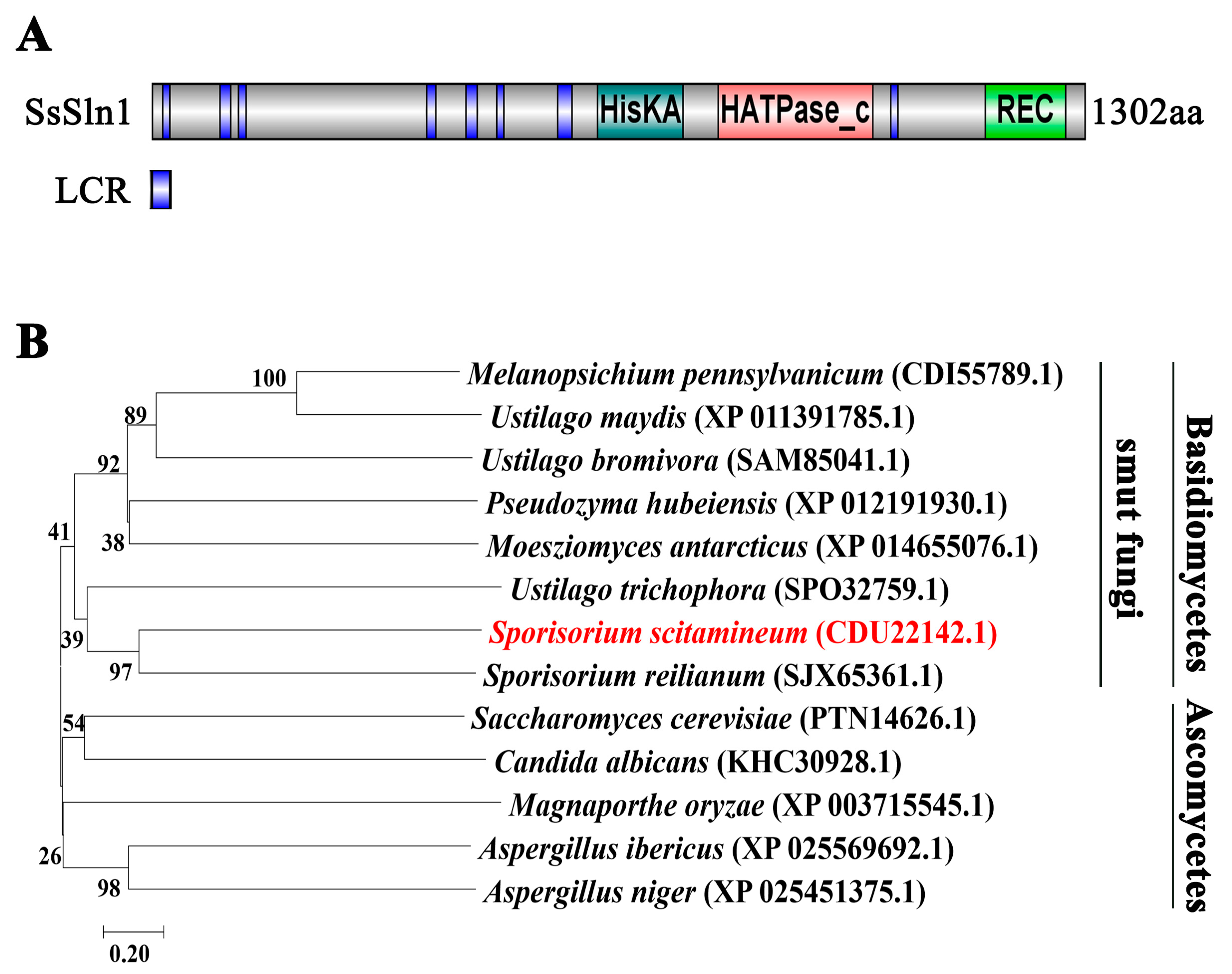
3.1. Identification of SsSln1 Protein in S. scitamineum

A BLASTp search of Sporisorium reilianum Sln1 (SJX65361.1) protein sequence revealed that the S. scitamineum proteome harbors a putative histidine kinase osmosensor protein (CDU22142.1), which we named SsSln1. SsSln1 was predicted as a peptide of 1302 amino acids. Putative domains of SsSln1 protein were further predicted through the SmartBLAST tool (https://blast.ncbi.nlm.nih.gov/smartblast/ (accessed on 8 June 2021)), which revealed that SsSln1 had a conserved histidine kinases (HisKA) domain, a histidine kinase-like ATPases (HATPase_c) domain and a cheY-homologous receiver (REC) domain (Figure 1A). Moreover, the SsSln1 protein and its orthologs from other fungal species, including basidiomycetous and ascomycetous, were selected for phylogenetic analysis. The results indicate that SsSln1 was closely related to its orthologs in S. reilianum and Ustilago trichophora, the smut fungi phylogenetic clade, whereas it was distant to Saccharomyces cerevisiae (TPN14626.1) and C. albicans (KHC30928.1), which were present in another clade of the phylogenetic tree (Figure 1B).
Overall, we identified the putative histidine kinase SsSln1 in S. scitamineum, and found that SsSln1 was highly conserved with its orthologs in basidiomycetous fungi.

3.2. Deletion of SsSLN1 Gene Promotes Mating and Pathogenicity of S. scitamineum

To characterize the role of SsSln1 in S. scitamineum, the deletion mutants of ss1sln1Δ in MAT-1 and ss2sln1Δ in MAT-2 background were separately generated, whereas reintegrated mutants of Ss1SLN1-COM in ss1sln1Δ and Ss2SLN1-COM in ss2sln1Δ background were generated by following the homologous recombination approach, as described before [38,41]. Mutants were confirmed by PCR amplification (Figure S1A,B) and Southern blotting (Figure S2A). Real-Time Quantitative PCR (RT-qPCR) analysis confirmed the complete deletion of SsSLN1 gene in ss1sln1Δ and ss2sln1Δ, and verified that SsSLN1 gene fully recovered in Ss1SLN1-COM and Ss2SLN1-COM strains, respectively (Figure S2B). S. scitamineum wild-type (WT) strains and generated mutants generated and used in this study are listed in Table 1.
To evaluate the impact of SsSln1 on mating, the haploid cells of MAT-1, MAT-2, ss1sln1Δ, ss2sln1Δ, Ss1SLN1-COM and Ss2SLN1-COM were mixed with the sporidia of opposite mating type and inoculated on YePSA medium. Successful formation of dikaryotic hyphae was observed as the appearance of white, fuzzy colonies at 30 h post inoculation. However, the ss1sln1Δ × ss2sln1Δ combination exhibited a stronger white and fuzzy colony as compared to MAT-1 × MAT-2 (Figure 2A). Meanwhile, the reintegrated strain of the Ss1SLN1-COM × Ss2SLN1-COM combination displayed a semblable mating of MAT-1 × MAT-2 on the same plate (Figure 2A). To examine the role of SsSln1 in virulence, the sporidial suspensions of MAT-1 × MAT-2, ss1sln1Δ × ss2sln1Δ and Ss1SLN1-COM × Ss2SLN1-COM combination were inoculated on the susceptible sugarcane cultivar ROC22. The results show that the typical symptoms of ‘black whip’ disease were observed in all strain-infected seedlings. However, the disease symptoms of ss1sln1Δ × ss2sln1Δ were more conspicuous than the wild-type or reintegrated strains (Figure 2B). Statistical results reveal that 68.15% of the total seedlings infected with ss1sln1Δ × ss2sln1Δ displayed ‘black whip’ symptoms, which was significantly higher (p < 0.05) than the incidence in other in treatments (Figure 2C).
In short, the SsSln1 negatively regulated the mating and virulence of S. scitamineum.

3.3. SsSln1 Negatively Regulates Phosphorylation and Nuclear Localization of SsHog1

The Sln1–Ypd1–Ssk1 “two-component” system regulates the Hog1–MAP kinase cascade in the budding yeast [7]. To explore the effects of SsSln1 on SsHog1 activity, we constructed a Ss1Hog1:RFP fusion strain in MAT-1 and Ss1Hog1:eGFP(sln1Δ) fusion strain in ss1sln1Δ. The localization analysis was carried out by staining the cell of Ss1Hog1:RFP and Ss1Hog1:eGFP(sln1Δ) fusion strains with 4′,6-diamidino-2-phenylindole (DAPI), photographed under a fluorescence microscope. In the absence of osmotic stress conditions, the SsHog1:RFP fusion protein was mainly distributed in the cytoplasm (Figure 3A), whereas the SsHog1 protein signal accumulated in the nucleus of about 23.3% of cells (Figure 3B). However, 0.8 M sorbitol treatment enhanced the entry of SsHog1:RFP fusion protein signals, and the number of cells increased up to 48.4% of cells (Figure 3A,B). Surprisingly, in ss1sln1Δ mutants, a large amount of SsHog1:eGFP fusion protein concentrated in the nucleus (Figure 3A), and the proportion of SsHog1 protein nuclear localization was noted in about 73.3% of cells (Figure 3B). These results suggest that SsHog1 was activated in the absence of SsSLN1 even without hyperosmosis treatment. To investigate the effect of SsSln1 on the phosphorylation level of SsHog1 in S. scitamineum, the level of SsHog1 phosphorylation was examined in MAT-1, ss1hog1Δ, ss1sln1Δ and Ss1SLN1-COM sporidia by Western blotting. The SsHog1 was found to be highly activated and phosphorylated in ss1sln1Δ mutants as compared to wild-type and Ss1SLN1-COM, and the phosphorylation of SsHog1 was abolished in ss1hog1Δ (Figure 3C).
Taken together, the SsSln1 negatively regulated the phosphorylation and nuclear localization of SsHog1 in S. scitamineum.

3.4. Phosphorylation of SsHog1 Is Necessary for Mating and Virulence of S. scitamineum

The role of protein tyrosine phosphatase Ptps in the dephosphorylation of Hog1 orthologs in C. neoformans was reported [30]. Therefore, we first identified the putative Ptps orthologs SsPtp1 (CDU22358.1) or SsPtp2 (CDU23562.1), and generated the overexpression mutants of SsPTP1-OE or SsPTP2-OE in MAT-1 and MAT-2 background, respectively (Figures S1E and S2D,E). As expected, the overexpression of Ss1PTP2-OE exhibited a lower level of SsHog1 phosphorylation (Figure 4A) in comparison to the wild type. To investigate the effects of the SsHog1 phosphorylation level on the mating and virulence of S. scitamineum, mating assays were performed by cospotting the compatible strains onto YePSA medium. The results reveal that during mating, the white hyphae was slightly reduced in the Ss1PTP1-OE × Ss2PTP1-OE combination but significantly reduced in the Ss1PTP2-OE × Ss2PTP2-OE combination, compared to the wild type (Figure 4B). In addition, the ss1hog1Δ mutant was also noted to be significantly deficient in mating in comparison to wild type and Ss1HOG1-COM (Figure 4B). Further pathogenicity analysis revealed that the typical symptoms of the disease ‘black whip’ were significantly (p < 0.05) reduced in ss1hog1Δ × MAT-2 combination, as compared to the wild-type and reintegrated strains (Figure 4C). Moreover, the percentage (%) of ‘black whip’/total seedlings was also markedly reduced in ss1hog1Δ × MAT-2 combination than wild-type or Ss1HOG1-COM × MAT-2 combination (Figure 4D). The inoculation of Ss1PTP1-OE × Ss2PTP1-OE and Ss1PTP2-OE × Ss2PTP2-OE combination also produced the ‘black whip’ symptoms but their virulence was significantly more reduced than the wild-type (Figure 4D).
Overall, the results demonstrate that SsPtp2 was involved in the dephosphorylation of SsHog1 and resulted in defective mating and virulence in S. scitamineum.

3.5. Phosphorylation Level of SsHog1 Positively Mediates Mating and Virulence of S. scitamineum

An Atf1 ortholog named SsAtf1 was identified and characterized in S. scitamineum. Deletion mutants of ss1atf1Δ in MAT-1 and ss2atf1Δ in MAT-2, and reintegrated mutants of Ss1ATF1-COM in ss1atf1Δ and Ss2ATF1-COM in ss2atf1Δ background, were individually obtained by homologous recombination approach as described before [38]. Mutants were confirmed by PCR amplification (Figure S1A,B) and Southern blot (Figure S2A). The results of RT-qPCR analysis revealed the complete deletion of the SsATF1 gene in ss1atf1Δ and ss2atf1Δ, and it was fully recovered in the reintegrated strains Ss1ATF1-COM and Ss2ATF11-COM, respectively (Figure S2C).
To characterize the potential influence of SsAtf1 on the phosphorylation level of SsHog1, the levels of SsHog1 phosphorylation in MAT-1, ss1atf1Δ and Ss1ATF1-COM sporidia were assessed. As expected, the SsHog1 was activated and phosphorylated in ss1atf1Δ mutants as compared to wild-type and Ss1ATF1-COM (Figure 5A). The phosphorylation of Atf1 by M. oryzae Hog1 ortholog has been reported [29]. Therefore, a Ss1Atf1:eGFP fusion strain was generated in MAT-1 or ss1hog1Δ to test its role in SsAtf1 nuclear localization. To study the localization, the Ss1Atf1:eGFP and Ss1Atf1:eGFP(hog1Δ) sporidia were stained with DAPI and observed under a fluorescence microscope. The results indicate that SsAtf1:eGFP fusion protein was mainly accumulated in the nucleus of both MAT-1 and ss1hog1Δ strains (Figure 5B), suggesting that SsHog1 did not affect the nuclear localization of SsAtf1. Furthermore, the mating with wild-type, deletion of SsATF1 and reintegrated strains was also assessed. The results depict an increase in the formation of white and fuzzy colonies in ss1atf1Δ × ss2atf1Δ combination in comparison to wild-type and reintegrated strains (Figure 5C). The relative fungal biomass continuously and significantly increased for up to 3 days in seedling stems inoculated with ss1atf1Δ × ss2atf1Δ, as compared to that MAT-1 × MAT-2 or Ss1ATF1-COM × Ss2ATF1-COM (Figure 5D).
These data demonstrate that SsAtf1 contributed to the dephosphorylation of SsHog1, enhanced SsHog1 phosphorylation level and facilitated the mating or virulence of S. scitamineum.

3.6. SsSln1 Negatively Regulates the Transcription of SsPRF1 by SsHog1 Phosphorylation, but Not through the cAMP/PKA Signaling Pathway

Previously, our study showed that the pheromone response factor SsPRF1 gene plays a key role in mating and pathogenicity [39], and was significantly down-regulated in cAMP/PKA defective mutants [38]. Therefore, RT-qPCR analysis was performed for assessing the expression of the SsPRF1 gene in MAT-1, ss1hog1Δ, Ss1PTP1-OE, Ss1PTP2-OE, ss1sln1Δ and ss1atf1Δ strains grown on YePSA medium. The results show that transcriptional expression of the SsPRF1 gene was significantly (p < 0.05) reduced in ss1hog1Δ and Ss1PTP2-OE, whereas it slightly decreased in Ss1PTP1-OE mutant, compared to wild type (Figure 6A). However, the transcription level of the SsPRF1 gene was significantly (p < 0.05) up-regulated in ss1sln1Δ mutant, whereas it slightly increased in ss1atf1Δ mutant (Figure 6A). To evaluate whether the reduced mating in ss1hog1Δ mutants was caused by the down-regulation of the SsPRF1 gene, a Ss1PRF1-OE(hog1Δ) mutant was generated, which overexpressed the SsPRF1 gene in ss1hog1Δ mutants. The mating was noted to be fully restored in Ss1PRF1-OE(hog1Δ) mutant compared to the ss1hog1Δ mutant (Figure 6B), and the transcriptional expression of SsPRF1 gene in Ss1PRF1-OE(hog1Δ) mutant was close to the level of wild-type MAT-1 (Figure S2F). Furthermore, the transcriptional profiling showed that SsGPA3, SsUAC1 and SsADR1 genes were not significantly different in wild-type and ss1sln1Δ strains grown in YePSA medium (Figure 6C). Moreover, SsSln1 did not interact with the regulatory subunit SsUbc1 or the catalytic subunit SsAdr1 of cAMP-dependent protein kinase A (PKA) through yeast two-hybrid analysis, indicating that SsSln1 also did not interact with the pheromone response factor SsPrf1 in yeast two-hybrid analysis (Figure 6D).
These results collectively suggest that SsSln1 negatively regulates the transcription of SsPRF1 by SsHog1 phosphorylation, but not through the cAMP/PKA signaling pathway.

3.7. Cross-Talk between the SsSln1 and cAMP/PKA Pathways Antagonistically Regulates Mating and Virulence by SsPRF1 Transcription

To investigate the potential relationship of SsSln1 with the cAMP/PKA pathway during mating and virulence of S. scitamineum, SsGPA3, SsUAC1 and SsADR1 deletion strains were individually generated in ss1sln1Δ mutants by homologous recombination and named as ss1sln1Δgpa3Δ, ss1sln1Δuac1Δ and ss1sln1Δadr1Δ (Figures S1C,D and S2G,H). Transcriptional profiling revealed that the expression of pheromone response factor gene SsPRF1 was significantly reduced in the cAMP/PKA defective mutants (Figure 7A), which is consistent with previous findings [38]. However, SsPRF1 gene expression was not significantly reduced in ss1sln1Δgpa3Δ, ss1sln1Δuac1Δ and ss1sln1Δadr1Δ mutants, as compared to the wild type (Figure 7A). To test the changes in the mating of double-deletion strains, we assessed the mating of MAT-1, ss1sln1Δ and cAMP/PKA single-deletion mutants (ss1gpa3Δ, ss1uac1Δ and ss1adr1Δ) and double-deletion strains (ss1sln1Δgpa3Δ, ss1sln1Δuac1Δ and ss1sln1Δadr1Δ) after mixing with MAT-2 sporidia on minimal medium. The results display that the mating capability of ss1sln1Δgpa3Δ, ss1sln1Δuac1Δ and ss1sln1Δadr1Δ mutants could be partially restored in the ss1gpa3Δ, ss1uac1Δ and ss1adr1Δ mutants by mixing with compatible wild-type MAT-2 (Figure 7B).
Pathogenicity analysis was also performed for assessing the virulence in wild-type, cAMP/PKA-defective mutants, and double-deletion strains. The results show that the cAMP/PKA-defective mutants failed to induce the disease symptoms in sugarcane seedlings, as previously described [38]. Contrarily, typical disease ‘black whip’ symptoms were observed in the double-deletion strains and WT-infected seedlings (Figure 7C). Statistical analysis depicted that the double-deletion strains presented the disease symptoms in more than 40% of the seedlings, whereas 55% of wild-type seedlings exhibited disease symptoms (Figure 7D).
Taken together, these data suggest that cross-talk between histidine kinase SsSln1 and cAMP/PKA pathways regulates the mating and virulence by affecting the transcription of the SsPRF1 gene.

4. Discussion

The two-component histidine kinases of fungi function as sensor proteins to mediate signal transduction events related to morphogenesis, cell growth, mycelium development, cell wall regulation, osmotic adaptation and virulence [7,15,46,47]. Ten histidine kinases have been reported in M. oryzae [6], one (Sln1) in S. cerevisiae [3,48] and three (Sln1, Hk1 and Nik1) in C. albicans [3,14]. During this study, a histidine kinase Sln1 was identified and functionally characterized in S. scitamineum. However, SLN1 is not essential for the growth of S. scitamineum, which contradicts the previous reports of S. cerevisiae [7,8,49].
Interestingly, our results show that the SsSln1 null mutant exhibited a stronger mating activity and increased virulence in S. scitamineum, which is opposite to what has been reported in C. albicans [14]. One question raised by this study is why enhanced mating and virulence occur in the SsSln1 null mutant. The model for regulating the mating and virulence after the loss of SsSln1 is summarized in Figure 8. In this study, an increased level of SsHog1 phosphorylation in the ss1sln1Δ mutant was found, indicating that SsSln1 negatively regulates SsHog1 phosphorylation in S. scitamineum. This is in line with previous reports about S. cerevisiae [7,48], which revealed the activation of Hog1 kinase through disruption of the SLN1 gene. However, ss1sln1Δ mutant displayed enhanced SsHog1 phosphorylation, mating and virulence that was similar to the Ypd1 null mutant of C. albicans, which blocked the YPD1 gene to enhance Hog1 phosphorylation and virulence [13]. This suggests that SsSln1-mediated SsHog1 might regulate the mating and virulence of S. scitamineum. The Hog1 kinase was reported to regulate virulence in various pathogenic fungi, such as C. albicans [49], C. neoformans [50,51] and Aspergillus fumigatus [9,52,53], but not in M. oryzae [54]. This study also elaborated that the elimination or reduction of SsHog1 phosphorylation affected the mating and virulence of S. scitamineum. Atf1 ortholog was also identified and characterized during the study, which was reported in M. oryzae [29,55] and C. neoformans [30] to suppress the hyperphosphorylation of Hog1 ortholog. As expected, the deletion of SsATF1 enhanced the phosphorylation of SsHog1, as compared to the wild-type and Ss1ATF1-COM strains. In M. oryzae, the Atf1 is required for virulence [29,55]. We found that SsAtf1 null mutant enhanced the mating and increased relative fungal biomass in inoculated seedlings in comparison to wild type. Nevertheless, the potential role of SsAtf1 in enhancing S. scitamineum virulence should be further investigated before assessing the pathogenicity with inoculation. In short, the results of this study demonstrate that the phosphorylation of SsHog1 was required for the mating and virulence of S. scitamineum.
On the other hand, several signal transduction pathways may interact with each other in fungi [37]. A significant down-regulation of pheromone response factor SsPRF1 gene in cAMP/PKA-defective mutants and sskpp2Δ mutant was reported in our previous study [38,56]. Moreover, the exogenous addition of cAMP could partially restore the mating defect in sskpp2Δ mutant [56], suggesting that there are essential connections between the cAMP/PKA and Kpp2 MAPK pathways in S. scitamineum. In this study, our findings reveal an intriguing underlying relationship between the histidine kinase SsSln1 and cAMP/PKA pathways during mating and virulence. We found that the histidine kinase SsSln1 did not interact with the regulatory subunit SsUbc1, catalytic subunit SsAdr1 of cAMP-dependent protein kinase A (PKA) and pheromone response factor SsPrf1 by yeast two-hybrid analysis. This is inconsistent with the results reported in M. oryzae, where interaction between Sln1 and the regulatory subunit of cAMP-dependent protein kinase A (Sum1) was demonstrated by yeast two-hybrid analyses and co-immunoprecipitation [15]. In addition, the abolition of SsHog1 phosphorylation led to the down-regulated transcription of the SsPRF1 gene. Contrarily, the enhanced SsHog1 phosphorylation up-regulated SsPRF1 transcription, suggesting that histidine kinase SsSln1 negatively regulated SsHog1 phosphorylation to activate the transcription of the SsPRF1 gene. However, the mechanism of SsHog1 phosphorylation that regulates the transcription of the SsPRF1 gene requires further elaboration. The pheromone response factor SsPrf1 was reported to play a key role in the virulence factor of S. scitamineum [39]. During this study, we found that the mating could be fully restored in Ss1PRF1-OE(hog1Δ) mutant, implying that the down-regulation of the SsPRF1 gene decreased the mating ability of ss1hog1Δ mutant. The transcription of the SsPRF1 gene was also partly restored in the double-deletion strains (ss1sln1Δgpa3Δ, ss1sln1Δuac1Δ and ss1sln1Δadr1Δ), as compared to down-regulation in cAMP/PKA-defective mutants, as previously described [38]. Meanwhile, the double-deletion strains exhibited remarkably enhanced mating and virulence capabilities in comparison to the cAMP/PKA-defective mutants. Taken together, we infer that cross-talk between the SsSln1 and cAMP/PKA pathways antagonistically regulates mating and virulence by balancing the transcription of the SsPRF1 gene in S. scitamineum.
In short, this study provides evidence that histidine kinase SsSln1 negatively regulates SsHog1 phosphorylation, which is essential for mating and virulence in S. scitamineum. Furthermore, we also reveal a novel mechanism by which histidine kinase SsSln1 and cAMP/PKA pathways antagonistically regulate mating and virulence via affecting the transcription of the SsPRF1 gene.

Supplementary Materials

The following are available online at https://0-www-mdpi-com.brum.beds.ac.uk/article/10.3390/jof7080610/s1, Figure S1: Identification of mutants by PCR amplification, Figure S2: Southern blot and RT-qPCR analysis of the mutant, Table S1: The primers and sequences used in this study.

Author Contributions

Conceptualization, Z.J., C.C. and E.C.; methodology, E.C. and S.S.; formal analysis, E.C. and S.S.; investigation, E.C., S.S., P.H., X.S. and Y.W.; resources, C.C. and Z.J.; writing—original draft preparation, E.C. and S.S.; writing—review and editing, Z.J., C.C., Y.D. and E.C.; supervision, Z.J., C.C. and Y.D.; project administration, Z.J. and C.C.; funding acquisition, Z.J. and C.C. All authors have read and agreed to the published version of the manuscript.

Funding

This research was supported by the Key Realm R&D Program of Guang Dong Province (2020B0202090001), the Natural Science Foundation of Guangdong Province (2021A1515011101) and Key Projects of Guangzhou Science and Technology Plan (201904020010).

Institutional Review Board Statement

Not applicable.

Informed Consent Statement

Not applicable.

Data Availability Statement

All data required to understand this article are presented in the study or the Supplementary Materials. Any raw data further requested will be provided by the corresponding authors.

Acknowledgments

We thank Xiaofan Zhou (South China Agricultural University) for the critical reading and helpful suggestions on our manuscript.

Conflicts of Interest

The authors declare no conflict of interest.

References

  1. Taniguti, L.M.; Schaker, P.D.; Benevenuto, J.; Peters, L.P.; Carvalho, G.; Palhares, A.; Quecine, M.C.; Nunes, F.R.; Kmit, M.C.; Wai, A.; et al. Complete Genome Sequence of Sporisorium scitamineum and Biotrophic Interaction Transcriptome with Sugarcane. PLoS ONE 2015, 10, e0129318. [Google Scholar] [CrossRef]
  2. Yan, M.; Zhu, G.; Lin, S.; Xian, X.; Chang, C.; Xi, P.; Shen, W.; Huang, W.; Cai, E.; Jiang, Z.; et al. The mating-type locus b of the sugarcane smut Sporisorium scitamineum is essential for mating, filamentous growth and pathogenicity. Fungal Genet. Biol. 2016, 86, 1–8. [Google Scholar] [CrossRef] [Green Version]
  3. Saito, H. Histidine phosphorylation and two-component signaling in eukaryotic cells. Chem. Rev. 2001, 101, 2497–2509. [Google Scholar] [CrossRef]
  4. Catlett, N.L.; Yoder, O.C.; Turgeon, B.G. Whole-genome analysis of two-component signal transduction genes in fungal pathogens. Eukaryot. Cell 2003, 2, 1151–1161. [Google Scholar] [CrossRef] [Green Version]
  5. Motoyama, T.; Ochiai, N.; Morita, M.; Iida, Y.; Usami, R.; Kudo, T. Involvement of putative response regulator genes of the rice blast fungus Magnaporthe oryzae in osmotic stress response, fungicide action, and pathogenicity. Curr. Genet. 2008, 54, 185–195. [Google Scholar] [CrossRef]
  6. Zhang, H.; Liu, K.; Zhang, X.; Song, W.; Zhao, Q.; Dong, Y.; Guo, M.; Zheng, X.; Zhang, Z. A two-component histidine kinase, MoSLN1, is required for cell wall integrity and pathogenicity of the rice blast fungus, Magnaporthe oryzae. Curr. Genet. 2010, 56, 517–528. [Google Scholar] [CrossRef]
  7. Posas, F.; Wurgler-Murphy, S.M.; Maeda, T.; Witten, E.A.; Thai, T.C.; Saito, H. Yeast HOG1 MAP kinase cascade is regulated by a multistep phosphorelay mechanism in the SLN1-YPD1-SSK1 “two-component” osmosensor. Cell 1996, 86, 865–875. [Google Scholar] [CrossRef] [Green Version]
  8. Hohmann, S. Osmotic stress signaling and osmoadaptation in yeasts. Microbiol. Mol. Biol. Rev. 2002, 66, 300–372. [Google Scholar] [CrossRef] [Green Version]
  9. Alex, L.A.; Borkovich, K.A.; Simon, M.I. Hyphal development in Neurospora crassa: Involvement of a two-component histidine kinase. Proc. Natl. Acad. Sci. USA 1996, 93, 3416–3421. [Google Scholar] [CrossRef] [Green Version]
  10. Schumacher, M.M.; Enderlin, C.S.; Selitrennikoff, C.P. The osmotic-1 locus of Neurospora crassa encodes a putative histidine kinase similar to osmosensors of bacteria and yeast. Curr. Microbiol. 1997, 34, 340–347. [Google Scholar] [CrossRef]
  11. Furukawa, K.; Katsuno, Y.; Urao, T.; Yabe, T.; Yamada-Okabe, T.; Yamada-Okabe, H.; Yamagata, Y.; Abe, K.; Nakajima, T. Isolation and functional analysis of a gene, tcsB, encoding a transmembrane hybrid-type histidine kinase from Aspergillus nidulans. Appl. Environ. Microbiol. 2002, 68, 5304–5310. [Google Scholar] [CrossRef] [Green Version]
  12. Fassler, J.S.; West, A.H. Histidine phosphotransfer proteins in fungal two-component signal transduction pathways. Eukaryot. Cell 2013, 12, 1052–1060. [Google Scholar] [CrossRef] [Green Version]
  13. Day, A.M.; Smith, D.A.; Ikeh, M.A.; Haider, M.; Herrero-de-Dios, C.M.; Brown, A.J.; Morgan, B.A.; Erwig, L.P.; MacCallum, D.M.; Quinn, J. Blocking two-component signalling enhances Candida albicans virulence and reveals adaptive mechanisms that counteract sustained SAPK activation. PLoS Pathog. 2017, 13, e1006131. [Google Scholar] [CrossRef] [PubMed]
  14. Yamada-Okabe, T.; Mio, T.; Ono, N.; Kashima, Y.; Matsui, M.; Arisawa, M.; Yamada-Okabe, H. Roles of three histidine kinase genes in hyphal development and virulence of the pathogenic fungus Candida albicans. J. Bacteriol. 1999, 181, 7243–7247. [Google Scholar] [CrossRef] [Green Version]
  15. Ryder, L.S.; Dagdas, Y.F.; Kershaw, M.J.; Venkataraman, C.; Madzvamuse, A.; Yan, X.; Cruz-Mireles, N.; Soanes, D.M.; Oses-Ruiz, M.; Styles, V.; et al. A sensor kinase controls turgor-driven plant infection by the rice blast fungus. Nature 2019, 574, 423–427. [Google Scholar] [CrossRef] [PubMed]
  16. Posas, F.; Saito, H. Osmotic activation of the HOG MAPK pathway via Ste11p MAPKKK: Scaffold role of Pbs2p MAPKK. Science 1997, 276, 1702–1705. [Google Scholar] [CrossRef] [PubMed]
  17. Posas, F.; Saito, H. Activation of the yeast SSK2 MAP kinase kinase kinase by the SSK1 two-component response regulator. EMBO J. 1998, 17, 1385–1394. [Google Scholar] [CrossRef] [Green Version]
  18. Tatebayashi, K.; Yamamoto, K.; Tanaka, K.; Tomida, T.; Maruoka, T.; Kasukawa, E.; Saito, H. Adaptor functions of Cdc42, Ste50, and Sho1 in the yeast osmoregulatory HOG MAPK pathway. EMBO J. 2006, 25, 3033–3044. [Google Scholar] [CrossRef]
  19. Maeda, T.; Takekawa, M.; Saito, H. Activation of yeast PBS2 MAPKK by MAPKKKs or by binding of an SH3-containing osmosensor. Science 1995, 269, 554–558. [Google Scholar] [CrossRef]
  20. Van Wuytswinkel, O.; Reiser, V.; Siderius, M.; Kelders, M.C.; Ammerer, G.; Ruis, H.; Mager, W.H. Response of Saccharomyces cerevisiae to severe osmotic stress: Evidence for a novel activation mechanism of the HOG MAP kinase pathway. Mol. Microbiol. 2000, 37, 382–397. [Google Scholar] [CrossRef]
  21. O’Rourke, S.M.; Herskowitz, I. Unique and redundant roles for HOG MAPK pathway components as revealed by whole-genome expression analysis. Mol. Biol. Cell 2004, 15, 532–542. [Google Scholar] [CrossRef]
  22. Horie, T.; Tatebayashi, K.; Yamada, R.; Saito, H. Phosphorylated Ssk1 prevents unphosphorylated Ssk1 from activating the Ssk2 mitogen-activated protein kinase kinase kinase in the yeast high-osmolarity glycerol osmoregulatory pathway. Mol. Cell. Biol. 2008, 28, 5172–5183. [Google Scholar] [CrossRef] [Green Version]
  23. Zarrinpar, A.; Bhattacharyya, R.P.; Nittler, M.P.; Lim, W.A. Sho1 and Pbs2 act as coscaffolds linking components in the yeast high osmolarity MAP kinase pathway. Mol. Cell 2004, 14, 825–832. [Google Scholar] [CrossRef]
  24. Hohmann, S. Control of high osmolarity signalling in the yeast Saccharomyces cerevisiae. FEBS Lett. 2009, 583, 4025–4029. [Google Scholar] [CrossRef] [Green Version]
  25. Ferrigno, P.; Posas, F.; Koepp, D.; Saito, H.; Silver, P.A. Regulated nucleo/cytoplasmic exchange of HOG1 MAPK requires the importin beta homologs NMD5 and XPO1. EMBO J. 1998, 17, 5606–5614. [Google Scholar] [CrossRef]
  26. Reiser, V.; Ruis, H.; Ammerer, G. Kinase activity-dependent nuclear export opposes stress-induced nuclear accumulation and retention of Hog1 mitogen-activated protein kinase in the budding yeast Saccharomyces cerevisiae. Mol. Biol. Cell 1999, 10, 1147–1161. [Google Scholar] [CrossRef] [Green Version]
  27. Sharma, P.; Mondal, A.K. Evidence that C-terminal non-kinase domain of Pbs2p has a role in high osmolarity-induced nuclear localization of Hog1p. Biochem. Biophys. Res. Commun. 2005, 328, 906–913. [Google Scholar] [CrossRef]
  28. Hersen, P.; McClean, M.N.; Mahadevan, L.; Ramanathan, S. Signal processing by the HOG MAP kinase pathway. Proc. Natl. Acad. Sci. USA 2008, 105, 7165–7170. [Google Scholar] [CrossRef] [Green Version]
  29. Liu, X.; Zhou, Q.; Guo, Z.; Liu, P.; Shen, L.; Chai, N.; Qian, B.; Cai, Y.; Wang, W.; Yin, Z.; et al. A self-balancing circuit centered on MoOsm1 kinase governs adaptive responses to host-derived ROS in Magnaporthe oryzae. eLife 2020, 9, e61605. [Google Scholar] [CrossRef] [PubMed]
  30. Lee, K.T.; Byun, H.J.; Jung, K.W.; Hong, J.; Cheong, E.; Bahn, Y.S. Distinct and redundant roles of protein tyrosine phosphatases Ptp1 and Ptp2 in governing the differentiation and pathogenicity of Cryptococcus neoformans. Eukaryot. Cell 2014, 13, 796–812. [Google Scholar] [CrossRef] [Green Version]
  31. Pan, X.; Harashima, T.; Heitman, J. Signal transduction cascades regulating pseudohyphal differentiation of Saccharomyces cerevisiae. Curr. Opin. Microbiol. 2000, 3, 567–572. [Google Scholar] [CrossRef]
  32. Thevelein, J.M.; Cauwenberg, L.; Colombo, S.; De Winde, J.H.; Donation, M.; Dumortier, F.; Kraakman, L.; Lemaire, K.; Ma, P.; Nauwelaers, D.; et al. Nutrient-induced signal transduction through the protein kinase A pathway and its role in the control of metabolism, stress resistance, and growth in yeast. Enzym. Microb. Technol. 2000, 26, 819–825. [Google Scholar] [CrossRef]
  33. Hogan, D.A.; Sundstrom, P. The Ras/cAMP/PKA signaling pathway and virulence in Candida albicans. Future Microbiol. 2009, 4, 1263–1270. [Google Scholar] [CrossRef]
  34. Choi, J.; Jung, W.H.; Kronstad, J.W. The cAMP/protein kinase A signaling pathway in pathogenic basidiomycete fungi: Connections with iron homeostasis. J. Microbiol. 2015, 53, 579–587. [Google Scholar] [CrossRef] [Green Version]
  35. Marroquin-Guzman, M.; Wilson, R.A. GATA-Dependent Glutaminolysis Drives Appressorium Formation in Magnaporthe oryzae by Suppressing TOR Inhibition of cAMP/PKA Signaling. PLoS Pathog. 2015, 11, e1004851. [Google Scholar] [CrossRef] [PubMed]
  36. Caza, M.; Kronstad, J.W. The cAMP/Protein Kinase a Pathway Regulates Virulence and Adaptation to Host Conditions in Cryptococcus neoformans. Front. Cell. Infect. Microbiol. 2019, 9, 212. [Google Scholar] [CrossRef]
  37. Martínez-Soto, D.; Ortiz-Castellanos, L.; Robledo-Briones, M.; León-Ramírez, C.G. Molecular Mechanisms Involved in the Multicellular Growth of Ustilaginomycetes. Microorganisms 2020, 8, 1072. [Google Scholar] [CrossRef]
  38. Chang, C.; Cai, E.; Deng, Y.Z.; Mei, D.; Qiu, S.; Chen, B.; Zhang, L.H.; Jiang, Z. cAMP/PKA signalling pathway regulates redox homeostasis essential for Sporisorium scitamineum mating/filamentation and virulence. Environ. Microbiol. 2019, 21, 959–971. [Google Scholar] [CrossRef]
  39. Zhu, G.; Deng, Y.; Cai, E.; Yan, M.; Cui, G.; Wang, Z.; Zou, C.; Zhang, B.; Xi, P.; Chang, C.; et al. Identification and Functional Analysis of the Pheromone Response Factor Gene of Sporisorium scitamineum. Front. Microbiol. 2019, 10, 2115. [Google Scholar] [CrossRef]
  40. Garrido, E.; Pérez-Martín, J. The crk1 gene encodes an Ime2-related protein that is required for morphogenesis in the plant pathogen Ustilago maydis. Mol. Microbiol. 2003, 47, 729–743. [Google Scholar] [CrossRef]
  41. Cai, E.; Li, L.; Deng, Y.; Sun, S.; Jia, H.; Wu, R.; Zhang, L.; Jiang, Z.; Chang, C. MAP kinase Hog1 mediates a cytochrome P450 oxidoreductase to promote the Sporisorium scitamineum cell survival under oxidative stress. Environ. Microbiol. 2021, 23, 3306–3317. [Google Scholar] [CrossRef]
  42. Cai, E.; Mei, D.; Zhang, X.; Sun, X.; Li, L.; Wu, R.; Deng, Y.; Jiang, Z.; Chang, C. A gene knockout method based on protoplast transformation with two PCR fragments in Sporisorium scitamineum. Mycosystema 2020, 39, 2314–2317. [Google Scholar] [CrossRef]
  43. Livak, K.J.; Schmittgen, T.D. Analysis of relative gene expression data using real-time quantitative PCR and the 2(-Delta Delta C(T)) Method. Methods 2001, 25, 402–408. [Google Scholar] [CrossRef]
  44. Sun, S.; Deng, Y.; Cai, E.; Yan, M.; Li, L.; Chen, B.; Chang, C.; Jiang, Z. The Farnesyltransferase β-Subunit Ram1 Regulates Sporisorium scitamineum Mating, Pathogenicity and Cell Wall Integrity. Front. Microbiol. 2019, 10, 976. [Google Scholar] [CrossRef]
  45. Kumar, S.; Stecher, G.; Tamura, K. MEGA7: Molecular Evolutionary Genetics Analysis Version 7.0 for Bigger Datasets. Mol. Biol. Evol. 2016, 33, 1870–1874. [Google Scholar] [CrossRef] [PubMed] [Green Version]
  46. Calderone, R.A.; Fonzi, W.A. Virulence factors of Candida albicans. Trends Microbiol. 2001, 9, 327–335. [Google Scholar] [CrossRef]
  47. Lipa, P.; Janczarek, M. Phosphorylation systems in symbiotic nitrogen-fixing bacteria and their role in bacterial adaptation to various environmental stresses. PeerJ 2020, 8, e8466. [Google Scholar] [CrossRef] [PubMed] [Green Version]
  48. Maeda, T.; Wurgler-Murphy, S.M.; Saito, H. A two-component system that regulates an osmosensing MAP kinase cascade in yeast. Nature 1994, 369, 242–245. [Google Scholar] [CrossRef] [PubMed]
  49. Alonso-Monge, R.; Navarro-García, F.; Molero, G.; Diez-Orejas, R.; Gustin, M.; Pla, J.; Sánchez, M.; Nombela, C. Role of the mitogen-activated protein kinase Hog1p in morphogenesis and virulence of Candida albicans. J. Bacteriol. 1999, 181, 3058–3068. [Google Scholar] [CrossRef] [PubMed] [Green Version]
  50. Bahn, Y.S.; Kojima, K.; Cox, G.M.; Heitman, J. Specialization of the HOG pathway and its impact on differentiation and virulence of Cryptococcus neoformans. Mol. Biol. Cell 2005, 16, 2285–2300. [Google Scholar] [CrossRef] [Green Version]
  51. Ko, Y.J.; Yu, Y.M.; Kim, G.B.; Lee, G.W.; Maeng, P.J.; Kim, S.; Floyd, A.; Heitman, J.; Bahn, Y.S. Remodeling of global transcription patterns of Cryptococcus neoformans genes mediated by the stress-activated HOG signaling pathways. Eukaryot. Cell 2009, 8, 1197–1217. [Google Scholar] [CrossRef] [Green Version]
  52. Winkelströter, L.K.; Bom, V.L.; de Castro, P.A.; Ramalho, L.N.; Goldman, M.H.; Brown, N.A.; Rajendran, R.; Ramage, G.; Bovier, E.; Dos Reis, T.F.; et al. High osmolarity glycerol response PtcB phosphatase is important for Aspergillus fumigatus virulence. Mol. Microbiol. 2015, 96, 42–54. [Google Scholar] [CrossRef] [PubMed]
  53. Bruder Nascimento, A.C.; Dos Reis, T.F.; de Castro, P.A.; Hori, J.I.; Bom, V.L.; de Assis, L.J.; Ramalho, L.N.; Rocha, M.C.; Malavazi, I.; Brown, N.A.; et al. Mitogen activated protein kinases SakA(HOG1) and MpkC collaborate for Aspergillus fumigatus virulence. Mol. Microbiol. 2016, 100, 841–859. [Google Scholar] [CrossRef] [Green Version]
  54. Dixon, K.P.; Xu, J.R.; Smirnoff, N.; Talbot, N.J. Independent signaling pathways regulate cellular turgor during hyperosmotic stress and appressorium-mediated plant infection by Magnaporthe grisea. Plant Cell 1999, 11, 2045–2058. [Google Scholar] [CrossRef] [Green Version]
  55. Guo, M.; Guo, W.; Chen, Y.; Dong, S.; Zhang, X.; Zhang, H.; Song, W.; Wang, W.; Wang, Q.; Lv, R.; et al. The basic leucine zipper transcription factor Moatf1 mediates oxidative stress responses and is necessary for full virulence of the rice blast fungus Magnaporthe oryzae. Mol. Plant Microbe Interact. 2010, 23, 1053–1068. [Google Scholar] [CrossRef] [Green Version]
  56. Deng, Y.Z.; Zhang, B.; Chang, C.; Wang, Y.; Lu, S.; Sun, S.; Zhang, X.; Chen, B.; Jiang, Z. The MAP Kinase SsKpp2 Is Required for Mating/Filamentation in Sporisorium scitamineum. Front. Microbiol. 2018, 9, 2555. [Google Scholar] [CrossRef] [PubMed] [Green Version]
Figure 1. Phylogenetic analysis of SsSln1 protein. (A) Domain arrangement of S. scitamineum SsSln1 protein was performed using the SmartBLAST tool (https://blast.ncbi.nlm.nih.gov/smartblast/ (accessed on 8 June 2021)). LCR represents low-complexity regions. (B) The amino acid sequence of SsSln1 protein was used to search orthologous proteins through the BLASTp tool. The phylogenetic tree was constructed from the Sln1 proteins of Melanopsichium pennsylvanicum (CDI55789.1), U. maydis (XP 011391785.1), Ustilago bromivora (SAM85041.1), Pseudozyma hubeiensis (XP 012191930.1), Moesziomyces antarcticus (XP 014655076.1), U. trichophora (SPO32759.1), S. scitamineum (CDU22142.1), S. reilianum (SJX65361.1), S. cerevisiae (PTN14626.1), C. albicans (KHC30928.1), M. oryzae (XP 003715545.1), Aspergillus ibericus (XP 025569692.1) and Aspergillus niger (XP 025451375.1) using neighbor-joining method in MEGA 7.0 software [45]. Gap extension = 0.2.
Jof 07 00610 g001
Figure 2. Effect of SsSln1 on mating and virulence. (A) Mating assay of mutants. Fresh haploid sporidia were grown up to O.D.600 = 1.0, and then mixed with an equal volume of the compatible WT strain or mutants and spotted onto YePSA medium to incubate at 28 °C. Images were taken after 30 h of cultivation. (B) Pathogenicity assay of SsSln1 null mutants. A susceptible sugarcane variety ROC22 was inoculated with mixed sporidia (1:1) of S. scitamineum and wild-type or mutant combination via injection at the 5–6-leaf seedling stage. The infection assays were performed with at least 15 seedlings. The symptoms of ‘black whip’ were documented and photographed at about six months post inoculation. The red dotted box regions were enlarged for a better view of whip symptoms. The symptoms of ‘black whip’ are denoted by red arrows. (C) Bar chart depicts the quantification of infection as shown in (B). Percentage (%) of ‘black whip’/total seedlings is indicated. Bar chart depicts the statistical differences among the mean values ( p < 0.05). Mean ± S.E. were derived from two independent biological repeats with three replications.
Jof 07 00610 g002
Figure 3. Effect of SsSln1 on phosphorylation and subcellular localization of SsHog1. (A) Subcellular localization of SsHog1 protein in S. scitamineum. The Ss1Hog1:RFP and Ss1Hog1:eGFP(sln1Δ) sporidia were grown up to O.D.600 = 1.0, and then treated with or without 0.8 M sorbitol in YePSA medium at 28 °C for 60 min. The nucleus was stained with DAPI and the images were taken under fluorescence microscope. Scale bar = 10 μm. (B) Bar chart depicts the quantification of nuclear localization as shown in (A). Percentage (%) of nuclear localization cells/total cells is indicated. Bar chart depicts the statistical difference among the mean values (★★ p < 0.01, ★★★ p < 0.001). Mean ± S.E. were derived from three independent biological repeats with three replications. (C) Phosphorylation of SsHog1 was enhanced in ss1sln1Δ mutant cells. The fresh haploid sporidia were grown in YePSA medium at 28 °C for 30 h. The total protein of sporidia was extracted with lysis buffer. Western blots depicting the levels of SsHog1 phosphorylation in the indicated strains. Above: blots were probed for phosphorylated SsHog1 (Hog1-P). Below: blots were probed for total SsHog1 (Hog1). Coomassie blue staining of total proteins served as loading control.
Jof 07 00610 g003
Figure 4. The effect of reduced SsHog1 phosphorylation level on mating and virulence. (A) Detection of SsHog1 phosphorylation in S. scitamineum. The fresh haploid sporidia were grown in YePSA medium at 28 °C for 30 h. The total protein of S. scitamineum was extracted with lysis buffer. Western blots depicting the levels of SsHog1 phosphorylation in the indicated strains. Above: blots were probed for phosphorylated SsHog1 (Hog1-P). Below: blots were probed for total SsHog1 (Hog1). Coomassie blue staining of total proteins served as loading control. (B) Mating assay of mutants. The fresh haploid sporidia were grown up to O.D.600 = 1.0, and then mixed with an equal volume of WT or mutant sporidia of opposite mating types and spotted onto YePSA medium to incubate at 28 °C. Images were taken after 30 h of cultivation. (C) Pathogenicity assay of mutants. MAT-1 × MAT-2, Ss1PTP1-OE × Ss2PTP1-OE, Ss1PTP2-OE × Ss2PTP2-OE, Ss1HOG1-COM × MAT-2 and ss1hog1Δ × MAT-2 combination were inoculated into the sugarcane seedlings of variety ROC22. The infection assays were performed with at least 15 seedlings. The symptoms of ‘black whip’ were documented and photographed at about six months post inoculation. The symptoms of ‘black whip’ are denoted by red arrows. The red dotted box regions were enlarged for a better view of whip symptoms. (D) Bar chart depicts the quantification of infection as shown in (C). Percentage (%) of ‘black whip’/total seedlings was indicated. Bar chart depicts the statistical difference among the mean values ( p < 0.05, ★★ p < 0.01, ★★★ p < 0.001). Mean ± S.E. were derived from two independent biological repeats with three replications.
Jof 07 00610 g004
Figure 5. Effect of SsAtf1 on SsHog1 phosphorylation and S. scitamineum mating. (A) Phosphorylation of SsHog1 was increased in ss1atf1Δ mutant cells. The fresh haploid sporidia were grown in YePSA medium at 28 °C for 30 h. The total protein of S. scitamineum was extracted with lysis buffer. Western blots depicting the levels of SsHog1 phosphorylation in the indicated strains. Above: blots were probed for phosphorylated SsHog1 (Hog1-P). Below: blots were probed for total SsHog1 (Hog1). Coomassie blue staining of total proteins served as loading control. (B) Subcellular localization of SsAtf1 protein in S. scitamineum. Ss1Atf1:eGFP and Ss1Atf1:eGFP(hog1Δ) sporidia were grown up to O.D.600 = 1.0, and stained with DAPI. Images were taken under the fluorescence microscope. Scale bar = 10 μm. (C) Mating assay of mutants. The fresh haploid sporidia were grown up to O.D.600 = 1.0, and then mixed with an equal volume of the compatible WT strain or mutants and spotted onto YePSA medium to incubate at 28 °C. Images were taken after 30 h of cultivation. (D) Measurement of relative fungal biomass. The fresh haploid sporidia were grown up to O.D.600 = 1.0, and then mixed with an equal volume of the compatible WT strain or mutants. The MAT-1 × MAT-2, ss1atf1Δ × ss2atf1Δ, and Ss1ATF1-COM × Ss2ATF1-COM combination was equally inoculated into the sugarcane seedlings of variety ROC22 to incubate at 28 °C for 3 days. Relative fungal biomass was measured by RT-qPCR with the total DNA isolated from infected sugarcane stems. The fungal ACTIN gene was used for the estimation of relative fungal biomass through the −ΔΔCt method with the plant GADPH gene as an internal control. Bar chart depicts the statistical difference among the mean values (★★ p < 0.01). Mean ± S.E. were derived from two independent biological repeats with three replications.
Jof 07 00610 g005
Figure 6. The relationship between SsSln1 and cAMP/PKA pathway in regulating the transcription of SsPRF1. (A) RT-qPCR analysis of SsPRF1 gene expression in the MAT-1, ss1hog1Δ, Ss1PTP1-OE, Ss1PTP2-OE, ss1sln1Δ and ss1atf1Δ strains under sporidial growth on YePSA plate for 30 h, respectively. The relative gene expression level was calculated by following the −ΔΔCt method with ACTIN as an internal control. Bar chart depicts the statistical difference among the mean values ( p < 0.05, ★★ p < 0.01). Mean ± S.E. were derived from three independent biological repeats with three replications. (B) Mating assay of mutants. Sporidia from MAT-1, ss1hog1Δ and SsPRF1-OE(hog1Δ) strains were separately mixed with an equal volume of MAT-2 sporidia and spotted to YePSA medium to incubate at 28 °C. Images were taken after 40 h of cultivation. (C) Transcriptional profile of SsGPA3, SsUAC1 and SsADR1 gene in the MAT-1 and ss1sln1Δ mutant. The relative gene expression level was calculated according to the −ΔΔCt method with ACTIN as an internal control. The NS represents no significance. Mean ± S.E. were derived from three independent biological repeats with three replications. (D) The yeast two-hybrid assay. Simultaneous co-transformation of positive-control vectors (pGBKT7-p53 and pGADT7-LargeT), negative-control vectors (pGADT7-LargeT and pGBKT7-LaminC), empty vectors (pGADT7 and pGBKT7) and pGBKT7-SsSln1 (bait vectors, BD) with pGADT7-SsAdr1, pGADT7-SsUbc1 and pGADT7-SsPrf1 into the Y2H Gold strain. The transformant was grown on SD-Trp-Leu (lacking tryptophan and leucine) and SD-Trp-Leu-His-Ade (lacking tryptophan, leucine, histidine and adenine) synthetic medium to incubate at 30 °C. Images were taken after 3 days of cultivation. Images are representative of n = 2 biological replications of the experiment.
Jof 07 00610 g006
Figure 7. Effect of cross-talk between SsSln1 and cAMP/PKA pathway on mating and virulence. (A) Transcriptional profile of SsPRF1 gene in the MAT-1 and mutants. The RT-qPCR assay was performed to assess the expression of SsPRF1 gene in MAT-1, ss1gpa3Δ, ss1sln1Δgpa3Δ, ss1uac1Δ, ss1sln1Δuac1Δ, ss1adr1Δ, and ss1sln1Δadr1Δ strains under sporidial growth on YePSA plate for 30 h, respectively. The relative gene expression was calculated by following the −ΔΔCt method with ACTIN as an internal control. Bar chart depicts the statistical difference among the mean values (★★ p < 0.01). The NS represents no significance. Mean ± S.E. were derived from three independent biological repeats with three replications. (B) Mating assay of mutants. Sporidia from MAT, ss1sln1Δ, ss1gpa3Δ, ss1sln1Δgpa3Δ, ss1uac1Δ, ss1sln1Δuac1Δ, ss1adr1Δ and ss1sln1Δadr1Δ strains were separately mixed with MAT-2 sporidia of equal volume and spotted onto minimal medium to incubate at 28 °C. Images were taken after 30 h of cultivation. Scale bar = 0.5 mm. (C) Pathogenicity assay of mutants. Sporidia from MAT-1, ss1gpa3Δ, ss1sln1Δgpa3Δ, ss1uac1Δ, ss1sln1Δuac1Δ, ss1adr1Δ and ss1sln1Δadr1Δ strains were separately mixed with an equal volume of MAT-2 sporidia and inoculated into the sugarcane seedlings of variety ROC22. The infection assays were performed with at least 15 seedlings. The symptoms of ‘black whip’ were documented and photographed at about six months post inoculation. The red dotted box regions were enlarged for a better view of whip symptoms. The symptoms of ‘black whip’ are denoted by red arrows. (D) Bar chart depicts the quantification of infection as shown in (C). Percentage (%) of ‘black whip’/total seedlings is indicated. Bar chart depicts the statistical difference among the mean values ( p < 0.05, ★★★ p < 0.001). Mean ± S.E. were derived from two independent biological repeats with three replications.
Jof 07 00610 g007
Figure 8. A proposed model depicting the outcomes after Sln1 loss in S. scitamineum. Loss of Sln1 results in increased phosphorylation of Hog1. The levels of Hog1 phosphorylation are suppressed via induction of the negative regulator Ptp2, which is initiated by the transcription factor Atf1. Loss of Sln1 function causes increased mating and virulence of S. scitamineum, by possibly enhancing Hog1 activity to promote the transcription of Prf1. Furthermore, the cAMP/PKA signaling pathway positively regulates mating and virulence by the transcription factor Prf1. TF represents a transcription factor.
Jof 07 00610 g008
Table 1. Details of strains generated in this study.
StrainsAccession Number for ProteinResistance Marker and Strain BackgroundReference or Source
MAT-1 a1, b1[2]
MAT-2 a2, b2[2]
ss1sln1Δ CDU22142.1Hygromycin, MAT-1This study
ss2sln1Δ CDU22142.1Hygromycin, MAT-2This study
Ss1SLN1-COMCDU22142.1Zeocin, ss1sln1ΔThis study
Ss2SLN1-COMCDU22142.1Zeocin, ss2sln1ΔThis study
ss1atf1Δ CDU21933.1Hygromycin, MAT-1This study
ss2atf1Δ CDU21933.1Hygromycin, MAT-2This study
Ss1ATF1-COMCDU21933.1Zeocin, ss1atf1ΔThis study
Ss2ATF1-COMCDU21933.1Zeocin, ss2atf1ΔThis study
ss1hog1Δ CDU23149.1Hygromycin, MAT-1[41]
Ss1PTP1-OECDU22358.1Zeocin, MAT-1This study
Ss2PTP1-OECDU22358.1Zeocin, MAT-2This study
Ss1PTP2-OECDU23562.1Zeocin, MAT-1This study
Ss2PTP2-OECDU23562.1Zeocin, MAT-2This study
Ss1PRF1-OE(hog1Δ)CDU22680.1Zeocin, ss1hog1ΔThis study
Ss1Hog1:RFPCDU23149.1Hygromycin, MAT-1This study
Ss1Hog1:eGFP(sln1Δ)CDU23149.1Zeocin, ss1sln1ΔThis study
Ss1Atf1:eGFPCDU21933.1Zeocin, MAT-1This study
Ss1Atf1:eGFP(hog1Δ)CDU21933.1Zeocin, ss1hog1ΔThis study
ss1sln1Δgpa3Δ CDU22378.1Zeocin, ss1sln1ΔThis study
ss1sln1Δuac1Δ CDU25762.1Zeocin, ss1sln1ΔThis study
ss1sln1Δadr1Δ CDU22361.1Zeocin, ss1sln1ΔThis study
Publisher’s Note: MDPI stays neutral with regard to jurisdictional claims in published maps and institutional affiliations.

