Article

Sorbicillinoid Derivatives with the Radical Scavenging Activities from the Marine-Derived Fungus Acremonium chrysogenum C10

1
State Key Laboratory of Mycology, Institute of Microbiology, Chinese Academy of Sciences, Beijing 100101, China
2
College of Life Sciences, University of Chinese Academy of Sciences, Beijing 100049, China
3
China Institutional Center for Shared Technologies and Facilities, Institute of Microbiology, Chinese Academy of Sciences, Beijing 100101, China
4
State Key Laboratory of Microbial Resources, Institute of Microbiology, Chinese Academy of Sciences, Beijing 100101, China
*
Authors to whom correspondence should be addressed.
These authors contributed equally to this work.
Submission received: 26 April 2022 / Revised: 14 May 2022 / Accepted: 16 May 2022 / Published: 20 May 2022

Abstract

:
Sorbicillinoids are a class of structurally diverse hexaketide metabolites with good biological activities. To explore new structural sorbicillinoids and their bioactivities, the marine-derived fungus Acremonium chrysogenum C10 was studied. Three new sorbicillinoid derivatives, acresorbicillinols A–C (13), along with five known ones, trichotetronine (4), trichodimerol (5), demethyltrichodimerol (6), trichopyrone (7) and oxosorbicillinol (8), were isolated. The structures of new sorbicillinoids were elucidated by analysis of nuclear magnetic resonance (NMR) and high-resolution electrospray ionization mass spectroscopy (HRESIMS). The absolute configurations of compounds 13 were determined by comparison of the experimental and calculated electronic circular dichroism (ECD) spectra. Compound 3 exhibited a strong 2,2-diphenyl-1-picrylhydrazyl (DPPH) radical scavenging activity, with the IC50 value ranging from 11.53 ± 1.53 to 60.29 ± 6.28 μM in 24 h. Additionally, compounds 2 and 3 showed moderate activities against Staphylococcus aureus and Cryptococcus neoformans, with IC50 values of 86.93 ± 1.72 and 69.06 ± 10.50 μM, respectively. The boundary of sorbicillinoid biosynthetic gene cluster in A. chrysogenum was confirmed by transcriptional analysis, and the biosynthetic pathway of compounds 18 was also proposed. In summary, our results indicated that A. chrysogenum is an important reservoir of sorbicillinoid derivatives, and compound 3 has the potential for new natural agents in DPPH radical scavenging.

1. Introduction

Marine-derived fungi can thrive in the extreme environments including salinity, high pressure, low temperature and oligotrophic conditions compared to their terrestrial coun-terparts, which makes them able to produce structurally diverse bioactive compounds more easily [1,2,3]. Meanwhile, these compounds usually have unique structures that also provide the possibility for structural design and modification of the leading compounds [4]. As one special marine-derived fungus, Acremonium chrysogenum has made irreplaceable contributions to controlling the bacterial infections and saving countless patients for production of the β-lactam antibiotic cephalosporin C (CPC) and its derivatives [5]. The genomic sequences and annotation of A. chrysogenum was first completed in 2014, and a total of 42 secondary metabolite biosynthetic gene clusters, including 14 polyketide synthetase (PKS) clusters, 10 terpene synthase clusters, 8 hybrid clusters, 7 nonribosomal peptide synthetase clusters and 3 non-identified secondary metabolite clusters, were predicted [6].
Sorbicillinoids are a class of structurally diverse hexaketide metabolites with a characteristic sorbyl side chain residue [7,8,9]. They were first isolated from Penicillium notatum in 1948 and structurally elucidated by Cram and Tishler [10,11]. Up until now, more than 159 naturally occurring sorbicillinoids have been isolated and have displayed good biological activities in cytotoxic, antimicrobial and phytotoxic activities [7,8,9]. Because free radicals play an important role in the development of aging and many diseases, including cancer, arthritis and atherosclerosis, exploring the novel radical scavengers is crucial for developing new drugs to slow down the aging process and treat these diseases. Some sorbicillinoid derivatives have shown great antioxidative application prospects, such as bisorbicillinol (ED50 = 31.4 μM) and bisorbibetanone (ED50 = 62.5 μM), etc. [8]. Additionally, there is an urgent need to find more novel compounds for the emergence of microbial resistance. Some sorbicillinoids showed significant antimicrobial activity, indicating their potential as candidates [7]. Meanwhile, the sorbicillinoid biosynthetic gene clusters from Penicillium chrysogenum and Trichoderma reesei have been identified and their biosynthetic pathway has been partially revealed [12,13,14]. Generally, two PKSs SorA and SorB are responsible for the formation of sorbicillin and dihydrosorbicillin, which are then oxidative dearomatized to give sorbicillinol and dihydrosorbicillinol by the FAD-dependent monooxygenase SorC, respectively [15]. Sorbicillinol is regarded as the precursor of most sorbicillinoids since it is condensed with its derivatives or other compounds to form the dimeric and hybrid sorbicillinoids by Diels-Alder or Michael-addition-like reactions [16,17]. The sorbicillinoid biosynthetic gene cluster in A. chrysogenum has been regarded as the most ancient, based on evolutionary origin, and carries more modifier than other species [13], and disruption of these two PKS encoding genes results in the abolishment of sorbicillinoids [18]. However, there is lack of a systematic investigation about sorbicillinoids produced by A. chrysogenum.
Based on the chemical investigations in this study, the resulting crude extracts of A. chrysogenum C10 from the rice solid fermentation, which has a higher accumulation of compounds and reproducibility than submerged fermentation [19], had afforded three structurally unique compounds: acresorbicillinols A–C (13) and five known sorbicillinoids including trichotetronine (4) [20], trichodimerol (5) [21], demethyltrichodimerol (6) [21], trichopyrone (7) [22] and oxosorbicillinol (8) [20] (Figure 1). Compounds 18 were evaluated for their DPPH radical scavenging abilities and antimicrobial activities. In addition, the boundary of sorbicillinoid biosynthetic gene cluster (Acsor) was confirmed and its biosynthetic pathway was proposed. This study reported the isolation, structural elucidation and bioactivities of the isolated compounds from A. chrysogenum C10.

2. Materials and Methods

2.1. General Experimental Procedure

Optical rotations, ECD spectra, UV and IR data were measured on the Austria Anton Paar MCP 200 Automatic Polarimeter, the Applied Photophysics Chirascan circular dichroism spectrometer, the Thermo Scientific GENESYS 10S UV-Vis and the Thermo Scientific Nicolet IS5 spectrophotometers, respectively. HRESIMS data and MS were obtained using an Agilent 6520B Q-TOF Mass instrument equipped with an ESI source. All MS experiments were performed in positive ion mode. NMR data were acquired with the AVANCE-500 spectrometer (Bruker, Bremen, Germany) using solvent signals (CD3OD, δH 3.30/δC 49.9, DMSO, δH 2.50, 3.30/δC 39.5, and CDCl3, δH 7.26/δC 77.16) as references. Octadecylsilyl (ODS, 50 μm, YMC Co., Ltd. Japan) and SephadexTM LH-20 (Cytiva, Uppsala, Sweden) were used for column chromatography. High performance liquid chromatography (HPLC) was performed on the SHIMADZU LC20AT system equipped with UV diode array detector using the Thermo Hypersil Gold-C18 columns (5 μm, 250 mm × 4.6 mm) at a flow rate of 1 mL/min. For semi-preparative HPLC, Waters 1525 system equipped with the UV/Visible detector and the Thermo Hypersil Gold-C18 columns (5 μm, 250 mm × 10 mm) was used and performed at a flow rate of 2 mL/min. Solvents including methanol and ethyl acetate (EtOAc) for extraction and chromatographic separation were analytical grade. HPLC-grade solvents (acetonitrile and formic acid) were used for the HPLC and semi-preparative HPLC analysis.

2.2. Fungal Materials and Fermentation

One high CPC-producing strain of A. chrysogenum C10 (ATCC 48272) was released by PanLab. This fungus was inoculated on the rice solid medium in 500 mL Erlenmeyer flasks containing 80 g of rice and 120 mL of H2O, and cultivated at 28 °C for 7 days for the production of sorbicillinoids. A total of 10 kg fermentation sample was harvested.

2.3. Extraction and Isolation

The rice solid fermentation of A. chrysogenum was extracted with EtOAc (3 × 5 L) under the ultrasonication processing. The organic solvents were filtered and evaporated by the vaccum to get the crude extracts (25 g). Extracts were fractionated by ODS reverse silica gel using the gradient MeOH/H2O (v/v, 10%, 20%, 30%, 40%, 50%, 55%, 60%, 65%, 70%, 75%, 80%, 85%, 90%, 95%, 100%) to afford 15 fractions (Fr.1–Fr.15). Fr.8 (MeOH/H2O (v/v, 65%)) (150 mg) was further subjected to the SephadexTM LH-20 and eluted with MeOH to give 30 subfractions. Fr.8–24 (30 mg) was purified by semi-preparative RP-HPLC using 50% acetonitrile in acidic water (0.1% formic acid) to give compounds 2 (5.0 mg, tR = 23 min), 4 (4.0 mg, tR = 24.1 min) and 8 (2.0 mg, tR = 22.2 min). Fr.10 (MeOH/H2O (v/v, 75%)) (133 mg) was subjected to the SephadexTM LH-20 and eluted with MeOH to give 30 subfractions. Fr.10–12 (56 mg) was purified by semi-preparative RP-HPLC using 58% acetonitrile in acidic water (0.1% formic acid) to yield compounds 1 (3.5 mg, tR = 27.2 min) and 6 (2.5 mg, tR = 26.4 min). Fr.11 (MeOH/H2O (v/v, 80%)) (220 mg) was subjected to the SephadexTM LH-20 and eluted with MeOH to give 30 subfractions. Fr.11–24 (60 mg) was purified by semi-preparative RP-HPLC using 60% acetonitrile in acidic water (0.1% formic acid) to yield compounds 3 (10.0 mg, tR = 28.5 min) and 5 (2.9 mg, tR = 28.3 min). Fr.5 (MeOH/H2O (v/v, 50%)) (96 mg) was subjected to the SephadexTM LH-20 and eluted with MeOH to give 25 subfractions. Fr.5-16 (10 mg) was purified by semi-preparative RP-HPLC using 37% acetonitrile in acidic water (0.1% formic acid) to yield compound 7 (2.0 mg, tR = 17.5 min).
Acresorbicillinol A (1): pale yellow solid; [α ] D 25 +81 (c 0.1, MeOH); UV (MeOH) λmax (log ε) 223 (3.65), 367 (1.72) nm; ECD (c 3.0 × 10⁻³ M, MeOH) λmaxε) 200 (+4.51), 228 (–8.29), 270 (–12.89), 315 (–34.60), 352 (+24.06) nm; IR (neat) νmax 3399, 2956, 2872, 1722, 1601, 1446, 1381, 1258 cm−1; 1H and 13C NMR, Table 1; HRESIMS at m/z 501.2850 [M + H]+ (calcd for C29H41O7, 501.2847).
Acresorbicillinol B (2): pale yellow solid; [α ] D 25 +5 (c 0.1, MeOH); UV (MeOH) λmax (log ε) 221 (2.72), 322 (0.68), 351 (0.54) nm; ECD (c 3.0 × 10⁻³ M, MeOH) λmaxε) 215 (–14.07), 245 (+36.00), 315 (–75.16) nm, 360 (+13.47) nm; IR (neat) νmax 3413, 1724, 1624, 1440, 1378, 1243 cm−1; 1H and 13C NMR, Table 1; HRESIMS at m/z 369.1696 [M + H]+ (calcd for C22H25O5, 369.1697).
Acresorbicillinol C (3): bright yellow solid; [α ] D 25 −1048 (c 0.1, MeOH); UV (MeOH) λmax (log ε) 207 (1.82), 37 (2.12), 278 (2.55), 375 (3.23) nm; ECD (c 3.0 × 10⁻³ M, MeOH) λmaxε) 221 (–22.24), 275 (+38.06), 345 (+51.25) nm, 405 (–88.67) nm; IR (neat) νmax 3420, 1664, 1606, 1556, 1412, 1347, 1209 cm−1; 1H and 13C NMR, Table 2; HRESIMS at m/z 513.2116 [M + H]+ (calcd for C28H33O9, 513.2119).

2.4. ECD Calculations

Conformational analyses were performed using Maestro 10.2 in the OPLS3 molecular mechanics force-field within an energy window of 5.0 or 3.0 kcal/mol. The conformers were then further optimized with the software package Gaussian 09 at the B3LYP/6-31G(d) level for compounds 13, respectively, and the harmonic vibrational frequencies were also calculated to confirm their stability. The TDDFT methods at the CAM-B3LYP/6-31G(d) and B3LYP/6-31G(d) level were applied to calculate the 60 lowest electronic transitions to obtain conformers in a vacuum, respectively. The Gaussian function was applied to simulate the ECD spectrum of the conformers. The calculated ECD spectra were obtained according to the Boltzmann weighting of each conformer’s ECD spectrum [23].

2.5. Antimicrobial Activity Assay

The bacterial strains (Staphylococcus aureus CGMCC 1.89, Pseudomonas aeruginosa ATCC 15692) and the fungal strains (Cryptococcus neoformans W1585, Candida albicans SC5314) were used in this study. The concentration of 50 mM compounds was prepared using dimethyl sulfoxide (DMSO). The bacterial and fungal strains were streaked onto Mueller–Hinton Agar (MHA) and Potato Dextrose Agar (PDA) for growth at 37 °C and 28 °C, respectively. Single colony was picked and adjusted to 2 × 10⁵ CFU/mL by Mueller–Hinton Broth (MHB) or Potato Dextrose Broth (PDB). The stock solutions of compounds were diluted into 500, 250, 125, 62.5 and 31.25 μM by MHB or PDB, successively. Fifty microliters of serial dilutions of each compound and 50 μL of microbial suspension were added to the 96-well plates and incubated at 37 °C or 28 °C for 24 h until the results were recorded. IC50 was defined as the half maximal inhibitory concentrations of the compounds that inhibited the visible microbial growth after 24 h of incubation. Ampicillin and amphotericin B were used as the positive control for detecting the activities of these compounds against bacteria and fungi, respectively.

2.6. DPPH Radical Scavenging Assay

The DPPH radical scavenging activity of the compounds was carried out as previously described [24,25]. The modified parameter was the reaction time from 0.5 h to multiple time-points including 0.5, 1, 4, 6, 8 and 24 h. Ascorbic acid and ethanol were used as the positive and negative control, respectively. All experiments were replicated at least three times.

2.7. RNA Isolation and Real-Time RT-PCR Analysis

The mycelia of A. chrysogenum C10 grown on the modified MDFA medium were collected at different time-points [26]. RNA isolation and real-time RT-PCR were performed as described previously [27,28]. All primers used in this study were listed in Table S1.

3. Results and Discussion

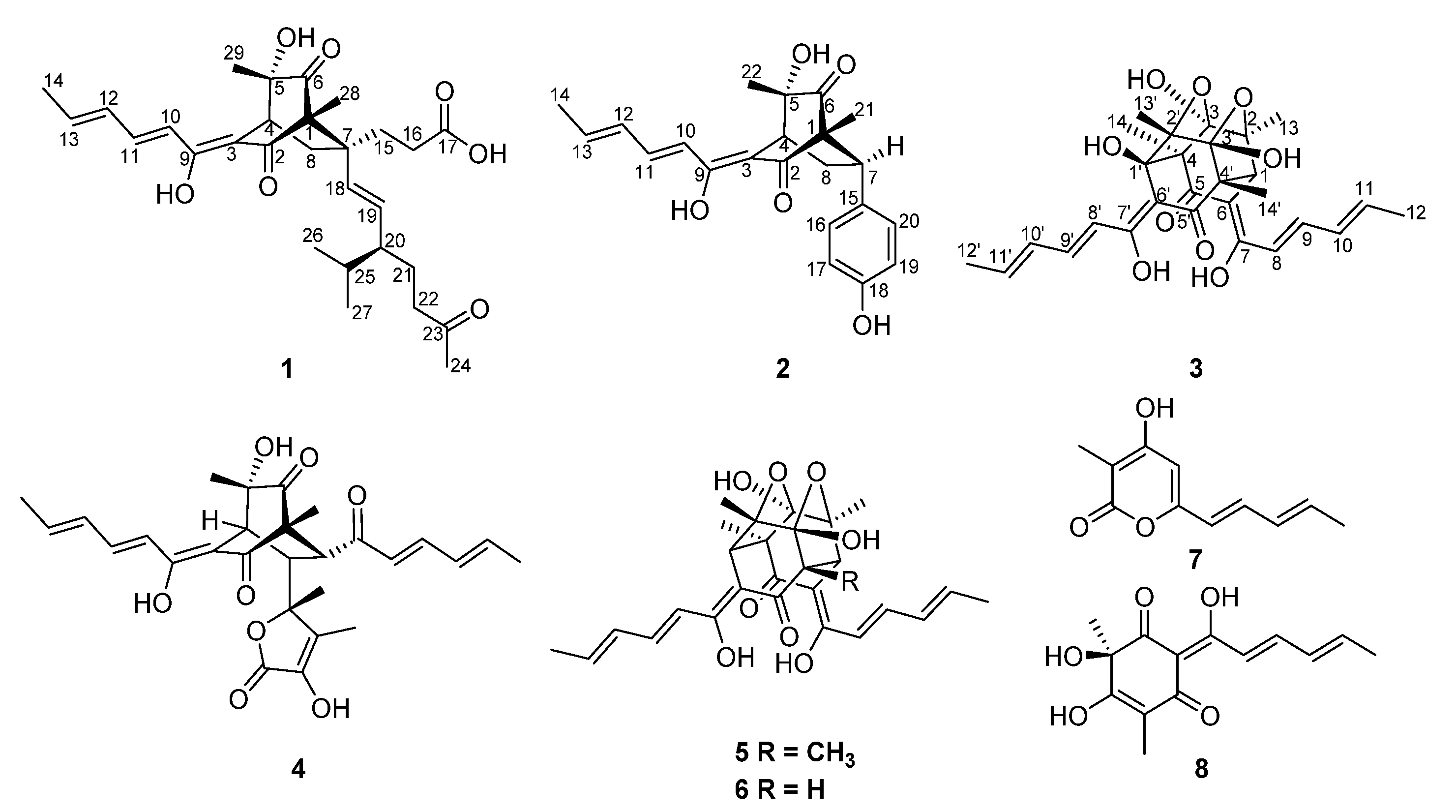
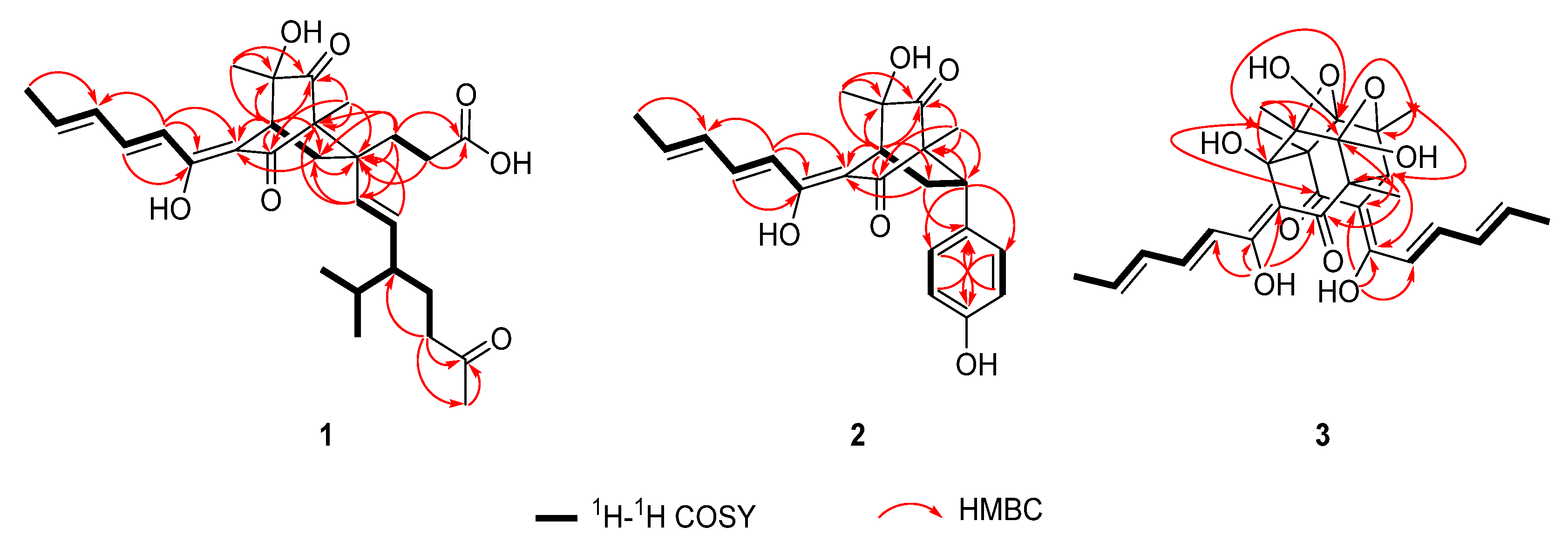
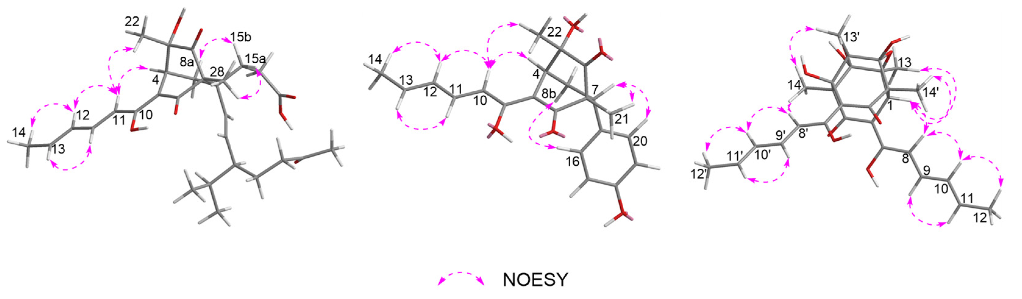
3.1. Isolation and Structure Elucidation

Acresorbicillinol A (1) was obtained as a pale yellow solid, and its molecular formula was established as C29H40O7 based on HRESIMS data at 501.2850 [M + H]+ (calcd for C29H41O7, 501.2847), indicating 10 degrees of unsaturation. The IR spectrum indicated the presence of hydroxy (3399 cm−1) and ketone (1722 cm−1) groups. The 1H NMR data (Table 1 and Figure S1) of 1 showed signals for six methyl signals [δH 2.16 (s, H3-24), 1.89 (d, J = 7.0 Hz, H3-14), 1.16 (s, H3-28), 1.12 (s, H3-29), 0.86 (d, J = 7.0 Hz, H3-26), and 0.81 (d, J = 7.0 Hz, H3-27)], five methylene protons [δH 2.42 (m, H-22a), 2.38 (m, H-8a), 2.30 (m, H-22b), 2.16 (m, H2-16), 1.97 (dd, J = 13.3, 2.8 Hz, H-8b), 1.81 (td, J = 13.2, 4.8 Hz, H-15a), 1.64 (m, H-21a), 1.50 (m, H-15b), and 1.23 (m, H-21b)], three methine protons [δH 3.18 (t, J = 2.8 Hz, H-4), 1.68 (m, H-20), and 1.54 (m, H-25)], six olefinic protons [δH 7.26 (dd, J = 14.6, 10.9 Hz, H-11), 6.42 (d, J = 14.6 Hz, H-10), 6.39 (dd, J = 14.6, 10.9 Hz, H-12), 6.20 (dq, J = 14.6, 7.0 Hz, H-13), 5.18 (d, J = 15.6 Hz, H-18), and 5.13 (dd, J = 15.6, 9.0 Hz, H-19)]. Detailed interpretation of the 13C NMR and HSQC data (Table 1, Figures S2 and S4) of 1 revealed the presence of 29 carbon resonances corresponding to six methyls, five sp3 methylenes, three sp3 methines, six sp2 methines, three sp3 quarternary carbons with one oxygenated, two sp2 non-protonated carbons and four carbonyl carbons (δC 212.4, 212.3, 200.3 and 178.3, respectively). These data accounted for all 1H and 13C NMR resonances of 1 except for three unobserved exchangeable protons, suggesting that 1 was a bicyclic compound. The planar structure of 1 was assigned through detailed analysis of the 1H-1H COSY and HMBC correlations (Figure 2, Figures S3 and S5). The 1H-1H COSY (Figure 2) correlations of H-10/H-11/H-12/H-13/H3-14, combined with the HMBC correlations from H-10 to the olefinic carbons C-3 (δC 112.3) and C-9 (δC 167.6) and from H-11 to C-9, suggested the presence of the enolic sorbyl side chain. The HMBC correlations (Figure 2) from H-4 to C-3, the sp3 quarternary carbon C-5 (δC 75.4) and two ketone carbons C-2 (δC 200.3) and C-6 (δC 212.3), from H3-28 to the sp3 quarternary carbon C-1 (δC 70.3), C-2 and C-6, and from H3-29 to C-4, C-5, and C-6 permitted the completion of the cyclohexandione ring, with the enolic sorbyl unit positioned at C-3 and two methyl groups located at C-1 and C-5, respectively. Meanwhile, the 1H-1H COSY (Figure 2) correlations of H-18/H-19/H-20/H2-21/H2-22 and of H-20/H-25/H3-26/H3-27, as well as the HMBC correlations from H2-22 to the ketone carbon C-23 (δC 212.4) and C-24 (δC 30.0), and from H3-24 to C-22 and C-23, established the 3-isopropyl-6-oxohept-1-en-1-yl (C-18–C-27) subunit. Moreover, the 1H-1H COSY (Figure 2) correlations of H2-15/H2-16, and the HMBC correlations from H2-15 and H2-16 to the carbonyl carbon C-17 (δC 178.3), indicated that carbonyl carbon C-17 was attached to C-16 directly. Additional HMBC correlations from H2-15 to the sp3 quarternary carbon C-7 (δC 47.8) and the olefinic carbon C-18 (δC 135.4), from H2-16 and H-19 to C-7, and from H-18 to C-7 and C-15, indicated that C-7 was located between C-15 and C-18. Key HMBC correlations from H-4, H2-8 and H3-28 to C-7, and from H2-15 and H-18 to C-1 and C-8, along with the 1H-1H COSY correlations of H-4/H2-8 implied that C-1 and C-8 were all connected to C-7, permitting the completion of the bridged bicyclo [2.2.2]octane-2,6-dione core structure. By consideration of the molecular formula and the chemical shifts of C-5 (δC 75.4) and C-17 (δC 178.3), these two carbons should be hydroxylated. Thus, the planar structure of 1 was established as shown (Figure 1).
The relative configuration of 1 was determined by NOESY correlations, coupling constants and HMBC correlations. The NOESY correlation (Figure 3 and Figure S6) of H-10 with H-4 assigned the olefin C-3/C-9 as Z geometry. The geometry of the conjugated diene was assigned as 10E, 12E by the large coupling constants (JH-10/H-11 = 14.6 Hz and JH-12/H-13 = 14.6 Hz) along with the NOESY correlations of H3-14 with H-12 and of H-13 with H-11. The E geometry of the C-18/C-19 double bond was also deduced by the large coupling constant between H-18 and H-19 (15.6 Hz). The NOESY correlations of H-10 with H-4 and H3-29 suggested that these protons were close in space. Moreover, the strong HMBC correlations from H-8a to C-3 and C-15, and from H-8b to C-5, and the weak correlation from H-8a to C-5, as well as the lack of HMBC correlation from H-8b to C-3 and C-15, indicated that H-8a and C-15 were eclipsed and that H-8b and C-3 were gauche [20,29]. Meanwhile, the NOESY correlations of H3-28 with H-15a, and of H-8a with H-15b, assigned the relative configurations of C-1 and C-7. However, the relative configuration for C-20 could not be established by the NOESY data. The absolute configuration for 1 was assigned by a comparison of the experimental and calculated ECD spectra of two pairs of enantiomers, (1R,4S,5S,7R,20S)-1 (1a), (1S,4R,5R,7S,20R)-1 (1b), (1R,4S,5S,7R,20R)-1 (1c), and (1S,4R,5R,7S,20S)-1 (1d). The ECD calculations were conducted using time-dependent density functional theory (TDDFT) at the CAM-B3LYP/6-31G(d) level. The overall calculated ECD spectrum of 1a1d was then generated according to Boltzmann weighting of the conformers (Figure S19). For compound 1 the experimental first positive (200 nm), second negative (228 nm), third negative (270 nm), fourth negative (315 nm) and fifth positive (352 nm) Cotton effects compared well with the calculated ECD curve for (1R,4S,5S,7R,20S)-1 (1a), which showed five corresponding Cotton effects around 200, 222, 270, 315 and 350 nm (Figure 4). Therefore, qualitative analysis of the result allowed the assignment of the absolute configuration of 1 as 1R,4S,5S,7R,20S.
Acresorbicillinol B (2) was obtained as a pale yellow solid. The molecular formula of 2 was assigned as C22H24O5 (11 degrees of unsaturation) based on its HRESIMS data at m/z 369.1696 [M + H]+ (calcd for C22H25O5, 369.1697). The 1H and 13C NMR spectroscopic data (Table 1, Figures S7 and S8), in association with the HSQC spectrum (Figure S10), indicated 22 carbon resonances including 3 methyl groups, 1 sp3 methylenes, 2 sp3 methines, 2 sp3 non-protonated carbons with 1 oxygenated, 12 olefinic or aromatic carbons (8 protonated), and 2 carbonyl carbons (δC 211.4, and 199.7, respectively), which were similar to those of 1. Analysis of the 1H-1H COSY and HMBC data (Figure 2, Figures S9 and S11) of 2 determined the same bicyclo [2.2.2]octane-2,6-dione moiety with the enolic sorbyl substituted at C-3. However, the substitutes at C-7 of 2 were different from those of 1. The HMBC correlations from H2-8 to C-15 (δC 133.9), from H-7 to C-15, C-16 (δC 130.5) and C-20 (δC 130.5), from H-16/H-20 to C-7 (δC 47.5) and C-18 (δC 157.7) and from H-17/H-19 to C-15 completed the para-hydroxyphenyl group located at C-7. On the basis of these data, the planar structure of 2 was established as shown (Figure 1).
The relative stereochemistry of 2 was determined by NOESY correlations and coupling constants as well as by comparison with those of 1 and the known compound sorbicatechol C [30]. The large coupling constants (JH-10/H-11 = 14.6 Hz and JH-12/H-13 = 14.6 Hz), along with NOESY correlations (Figure 3 and Figure S12) of H3-14 with H-12 and of H-13 with H-11 indicated that the geometry of the conjugated diene was 10E, 12E. Furthermore, the NOESY correlation (Figure 3) of H-4 with H-10 implied a Z geometry of the C3/C9 double bond. Other NOESY correlations of H-10 with H-4 and H3-22, and of H-4 with H3-22, placed these protons on the same side. While NOESY correlations of H-8b (δH 1.80, ddd, J = 13.6, 6.1, 2.7 Hz) with H-16 (H-20), and of H-7 with H3-21, combined with the strong HMBC correlations from H-8b to C-5 and C-15, the weak correlation from H-8b to C-3 and lack of HMBC correlation from H-8a to C-5 and C-15 determined the relative stereochemistry of C-7 and C-1 as shown. The absolute configuration of 2 was also determined by a comparison of the experimental and calculated ECD spectra for enantiomers (1R,4S,5S,7R)-2 (2a) and (1S,4R,5R,7S)-2 (2b). As shown in Figure 4, the experimental ECD spectrum of 2 showed good agreement with the calculated ECD spectrum of (1R,4S,5S,7R)-2 (2a), suggesting the absolute configuration of 1R,4S,5S,7R for 2. Thus, the structure of 2 was defined as shown.
Acresorbicillinol C (3) was obtained as a bright yellow solid, and its molecular formula was deduced to be C28H32O9 (13 degrees of unsaturation) on the basis of the HRESIMS data at m/z 513.2116 [M + H]+ (calcd for C28H33O9, 513.2119). The IR absorptions suggested the presence of hydroxy (3420 cm−1) and ketone (1664 cm−1) groups. Its 1H NMR data (Table 2 and Figure S13) revealed signals of eight olefinic protons [δH 6.10–7.49], one methine proton [δH 3.71 (s, H-1)] and six methyls [δH 1.89 (d, J = 6.8 Hz, H3-12’), 1.83 (d, J = 6.8 Hz, H3-12), 1.31 (s, H3-14’), 1.30 (s, H3-13), 1.29 (s, H3-14), 1.17 (s, H3-13’)]. The 13C NMR spectrum (Table 2 and Figure S14) and the HSQC data (Figure S16) displayed a total of 28 carbon resonances, which were assignable to 6 methyl groups, 8 sp2 methines, 1 sp3 methines, 13 non-protonated carbons containing 2 carbonyls (δC 199.3, and 190.9), 4 sp2 non-protonated with two oxygenated, 7 sp3 non-protonated carbon with 5 oxygenated. These signals (Table 2 and Figures S13−S16) were very similar to those of trichodimerol (5) [31,32], except that the proton at the C-1’ position in 5 was changed to a hydroxy moiety in 3. This was evidenced by the HRESIMS data and HMBC correlations (Figure 2 and Figure S17) from H3-13’ and H3-14 to C-1’ (δC 78.3). Therefore, 3 was 1’-hydroxylated analogue of 5.
The relative configuration of 3 was confirmed by NOESY correlations and coupling constants. The NOESY correlations (Figure 3 and Figure S18) of H-9/H-11, of H-8/H-10/H-12, of H-9’/H-11’ and of H-8’/H-10’/H-12’, along with the large coupling constants (JH-8/H-9 = JH-10/H-11 = JH-8/H-9 = JH-10/H-11 = 14.6 Hz) suggested the 8E, 10E, 8’E and 10’E configurations of the conjugated dienes in the sorbyl side chains. Meanwhile, the NOESY correlations of H-1/H-8 and H3-14/H-8’ suggested the Z geometry of C-6/C-7 and C-6’/C-7’ double bonds. Furthermore, the NOESY correlations of H-1/H3-13, of H3-13’/H3-14 and of H3-14’/H-1 inferred that these protons were in close proximity to their related functional groups, respectively. The similar Cotton effects in the ECD spectra of 3 and 5 deduced the absolute configuration of 3 to be the same as that of 5, which was further verified by ECD calculations (Figure 4). The calculated ECD curve of (1S,2S,3R,4R,1’R,2’S,3’R,4’R)-3 (3a) matched well with the experimental data, suggesting the absolute configuration to be 1S,2S,3R,4R,1’R,2’S,3’R,4’R. Thus, the structure of 3 was defined as depicted.
Except for the new compounds 13, the structure of five known sorbicillinoids isolated in this study were confirmed by comparison of the spectroscopic data with those in the literature [20,21,22]. The resulting EtOAc extracts of A. chrysogenum cultivated on the rice were screened by HPLC analysis (Figure S20).

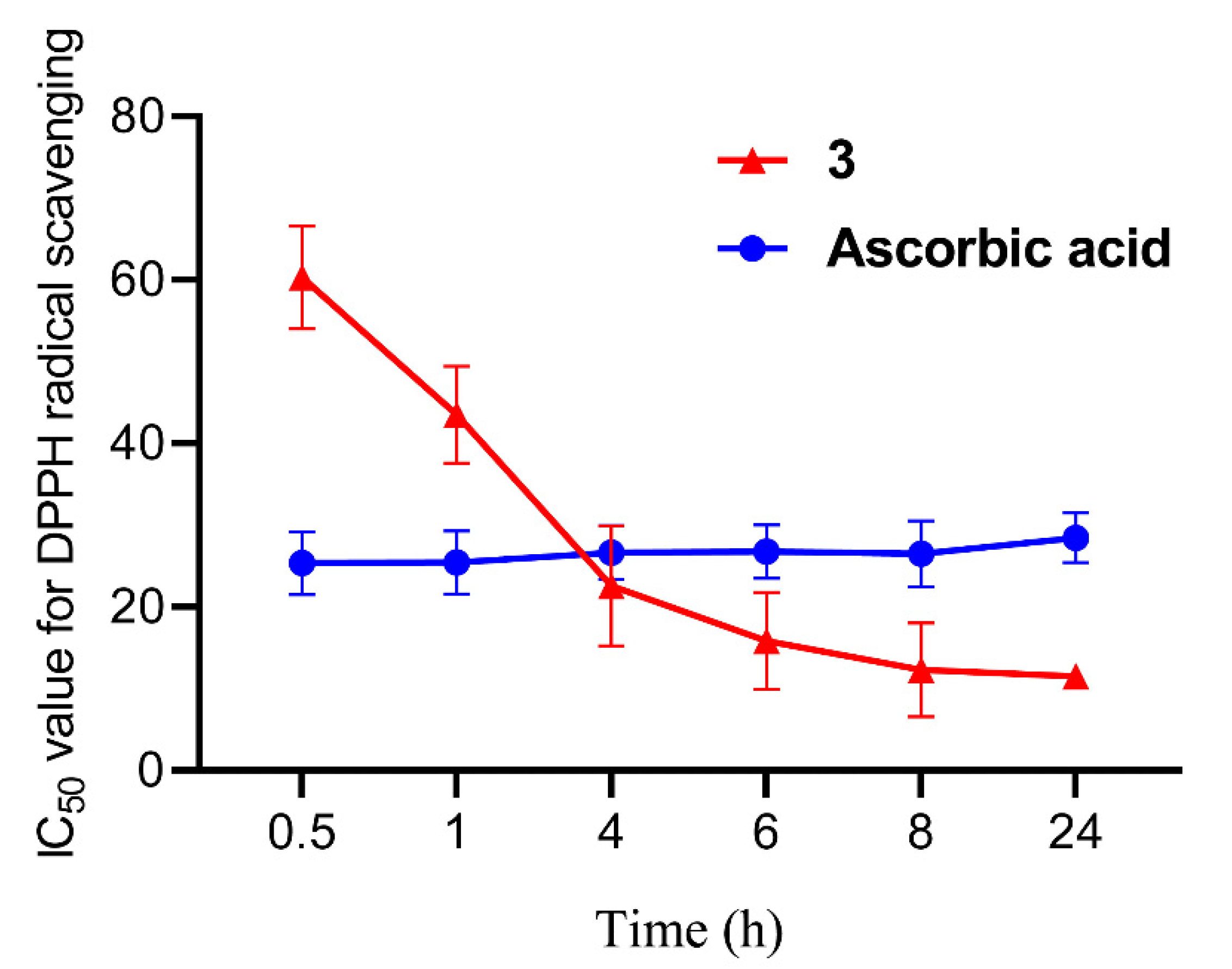
3.2. Biological Activities Evaluation

To explore the bioactivities of compounds 18, their abilities of anti-microorganisms and DPPH radical scavenging were evaluated. The results showed that compounds 2 and 3 exhibited the moderate activities against S. aureus and C. neoformans with the IC50 values of 86.93 ± 1.72 and 69.06 ± 10.50 μM, respectively. However, other compounds did not give IC50 value at a concentration below 100 μM (Table 3). No candidate compounds could significantly inhibit the growth of C. albicans and P. aeruginosa. Compound 3 might function as the β-1,6-glucan inhibitor to inhibit the fungal growth as its structural analogue bisvertinolone [33]. Bisvertinolone also exhibited significant inhibitory activity against S. aureus with the minimal inhibitory concentration (MIC) value of 30 μg/mL [34]. However, only several monomeric sorbicillinoids from Scytalidium album exhibited the weak activity against C. neoformans with the MIC value of over 38 μg/mL [35].
Through the DPPH radical scavenging assay, compound 3 exhibited strong activity with the IC50 value of 60.29 ± 6.28 μM after standing for 0.5 h, and then we continued to record its radical scavenging activity for 24 h (at 1, 4, 6, 8 and 24 h). Compound 3 gave the significant activity with the IC50 values of 43.52 ± 5.93, 22.57 ± 7.34, 15.85 ± 5.94, 12.30 ± 5.74 and 11.53 ± 1.53 μM, respectively, indicating that 3 displays the time-dependent manner for DPPH radical scavenging. Compared with the IC50 value of ascorbic acid as the positive control, which was 25.36 ± 3.82 to 28.45 ± 3.04 μM, compound 3 represents one novel DPPH radical scavenging agent (Figure 5 and Table 4). Compound 8 exhibited the radical scavenging activity with the IC50 values of 155.40 ± 12.42 and 55.36 ± 14.92 μM for 0.5 and 24 h, respectively. Although the IC50 values of 4, 5 and 6 were over 200 μM for 0.5 h, their radical scavenging activity significantly enhanced at 24 h, and the IC50 values were 151.87 ± 15.63, 116.83 ± 3.93 and 102.48 ± 5.04 μM, respectively (Table 4). Compounds 4, 5, 6 and 8 also displayed the time-dependent manner as compound 3. The time-dependent manner of sorbicillinoids for radical scavenging was previously reported, including for oxosorbicillinol, trichotetronine, bisorbicillinolide and methylbisorbibutenolide [22,36,37]. There was a different scavenging values of 4 and 8 between this study and the reports in Hirota’s Lab, and the reaction buffer might be the key determination factor. Additionally, the IC50 values of compounds 1, 2 and 7 exceeded 200 μM, even standing for 24 h, indicating that they did not have DPPH radical scavenging ability (Table 4). DPPH radical scavenging activity of other representative sorbicillinoids has been reported, including for bisorbicillinol, bisvertinolone and bisorbibetanone, which showed ED50 values of 31.4, 44.3 and 62.5 μM, respectively [21,37]. To date, compound 3 displayed the best DPPH radical scavenging activity for 24 h among all reported sorbicillinoids.

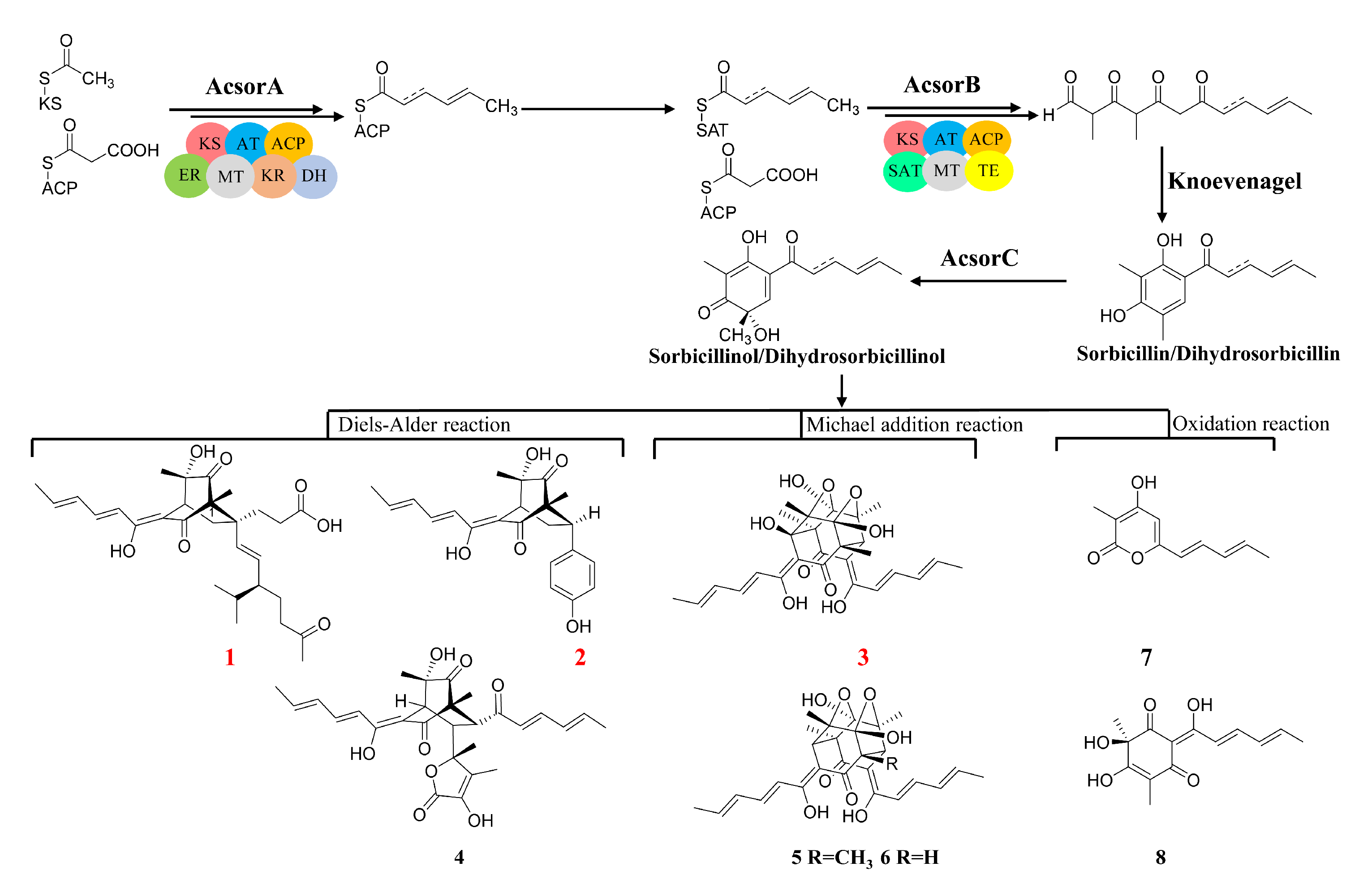
3.3. Determination of Acsor Cluster Boundary and Its Proposed Biosynthetic Pathway of Sorbicillinoid

To confirm the boundary of the sorbicillinoid biosynthetic gene cluster, the total RNA was isolated from A. chrysogenum C10 after incubation in the modified MDFA medium (also producing sorbicillinoids as in the rice solid medium) for 1, 3 and 5 days, and used as a template for real-time RT-PCR, the transcriptions of all 10 genes, including orf2 (ACRE_048080), AcsorD (ACRE_048110), AcsorR2 (ACRE_048120), AcsorT (ACRE_048130), AcsorE (ACRE_048140), AcsorR1 (ACRE_048150), AcsorC (ACRE_048160), AcsorB (ACRE_048170), AcsorA (ACRE_048180) and orf1 (ACRE_048200), were analysed (Figure 6A). Transcriptional results showed that AcsorA, AcsorB, AcsorC, AcsorD, AcsorE, AcsorT, AcsorR1 and AcsorR2 displayed a similar transcriptional pattern. In other words, the transcriptional level gradually increases during the fermentation. However, orf2 was silent during fermentation. Although orf1 was transcribed, the transcriptional trend was significantly different from other genes in the Acsor cluster. Thus, orf1 and orf2 are considered to be situated outside the Acsor cluster (Figure 6B). Combining with the results from bioinformatic analysis, a 35.5 kb Acsor cluster was identified that contains eight genes encoding one high-reducing polyketide synthase AcsorA, one non-reducing PKS AcsorB, two FAD-dependent monooxygenases AcsorC and AcsorD, one major facilitator superfamily transporter AcsorT, two putative regulators AcsorR1 and AcsorR2 and one putative serine hydrolase AcsorE.
Based on the confirmation of Acsor cluster, the biosynthetic pathway of compounds 18 was proposed. Sorbicillinoid biosynthesis starts from the formation of the polyketide backbone via condensation of acetate units catalyzed by AcsorA and AcsorB to generate sorbicillin and dihydrosorbicillin, and then they are oxidative dearomatized by AcsorC to form the common precursor-sorbicillinol and dihydrosorbicillinol. Sorbicillinol and its derivatives can be converted to 1, 2 and 4 by a Diels–Alder reaction. Compounds 3, 5 and 6 were biosynthesized by a Michael addition of sorbicillinol. Compounds 7 and 8 could be formed from sorbicillinol by an oxidation reaction (Figure 7). The structure diversification of sorbicillinoid derivatives was likely due to the multi-functions of AcsorD in A. chrysogenum.

4. Conclusions

In summary, eight sorbicillinoid derivatives including three new ones, acresorbicillinols A–C (13), were isolated from the marine-derived fungus A. chrysogenum. The absolute configurations of compounds 13 were determined by ECD calculations. Compound 3 exhibited strong DPPH radical scavenging, indicating that it can be regarded as one novel DPPH radical scavenging agent. Compounds 2 and 3 exhibited the moderate activities against S. aureus and C. neoformans, respectively. Meanwhile, the boundary of the Acsor cluster was confirmed and the biosynthetic pathway of compounds 18 was also proposed. This study suggests that A. chrysogenum is a potential pool for novel sorbicillinoids and radical scavenging agents.

Supplementary Materials

The following supporting information can be downloaded at: https://0-www-mdpi-com.brum.beds.ac.uk/article/10.3390/jof8050530/s1, Figure S1: 1H NMR spectrum of acresorbicillinol A (1; 500 MHz, CD3OD), Figure S2: 13C NMR spectrum of acresorbicillinol A (1; 125 MHz, CD3OD), Figure S3: 1H-1H COSY spectrum of acresorbicillinol A (1, CD3OD), Figure S4: HSQC spectrum of acresorbicillinol A (1, CD3OD), Figure S5: HMBC spectrum of acresorbicillinol A (1, CD3OD), Figure S6: NOESY spectrum of acresorbicillinol A (1, CD3OD), Figure S7: 1H NMR spectrum of acresorbicillinol B (2; 500 MHz, CD3OD), Figure S8: 13C NMR spectrum of acresorbicillinol B (2; 125 MHz, CD3OD), Figure S9: 1H-1H COSY spectrum of acresorbicillinol B (2, CD3OD), Figure S10: HSQC spectrum of acresorbicillinol B (2, CD3OD), Figure S11: HMBC spectrum of acresorbicillinol B (2, CD3OD), Figure S12. NOESY spectrum of acresorbicillinol B (2, CD3OD) Figure S13: 1H NMR spectrum of acresorbicillinol C (3; 500 MHz, DMSO:CDCl3 = 3:1), Figure S14: 13C NMR spectrum of acresorbicillinol C (3; 125 MHz, DMSO:CDCl3 = 3:1), Figure S15: 1H-1H COSY spectrum of acresorbicillinol C (3, DMSO:CDCl3 = 3:1), Figure S16: HSQC spectrum of acresorbicillinol C (3, DMSO:CDCl3 = 3:1), Figure S17: HMBC spectrum of acresorbicillinol C (3, DMSO:CDCl3 = 3:1), Figure S18: NOESY spectrum of acresorbicillinol C (3, DMSO:CDCl3 = 3:1), Figure S19: ECD conformers of acresorbicillinols A–C (13). Figure S20: HPLC profiles of the extracts from the rice solid medium of A. chrysogenum after 7 days fermentation. Tale S1: Primers used in this study.

Author Contributions

Conceptualization, C.D. and S.W.; methodology, C.D., S.W. and J.R.; software, R.H.; validation, S.W.; formal analysis, E.L., M.W. and R.H.; investigation, C.D. and S.W.; resources, G.L.; data curation, C.D.; writing—original draft preparation, C.D., S.W. and R.H.; writing—review and editing, Y.P., L.L. and G.L.; visualization, R.H.; supervision, Y.P., L.L. and G.L.; project administration, Y.P., L.L. and G.L.; funding acquisition, Y.P. and L.L. All authors have read and agreed to the published version of the manuscript.

Funding

This research was supported by the National Key Research and Development Program of China (2021YFC2300400) and the National Natural Science Foundation of China (31770056, 32022002, 21977113 and 32170067).

Institutional Review Board Statement

Not applicable.

Informed Consent Statement

Not applicable.

Data Availability Statement

All data generated or analyzed in this study are available within the manuscript and are available from the corresponding authors upon request.

Acknowledgments

We thank Francisco Fierro (Universidad Autónoma Metropolitana-Unidad Iztapalapa, Mexico) for providing the A. chrysogenum C10 and Wenzhao Wang (Institute of Microbiology, CAS) for HRESIMS analysis. We also appreciate Guanghua Huang (Fudan University, Shanghai, China) and Linqi Wang (Institute of Microbiology, CAS) providing the strains of Candida albicans SC5314 and Cryptococcus neoformans W1585, respectively.

Conflicts of Interest

The authors declare no conflict of interest.

References

  1. Wang, H.N.; Sun, S.S.; Liu, M.Z.; Yan, M.C.; Liu, Y.F.; Zhu, Z.; Zhang, Z. Natural bioactive compounds from marine fungi (2017–2020). J. Asian Nat. Prod. Res. 2022, 24, 203–230. [Google Scholar] [CrossRef] [PubMed]
  2. Karthikeyan, A.; Joseph, A.; Nair, B.G. Promising bioactive compounds from the marine environment and their potential effects on various diseases. J. Genet. Eng. Biotechnol. 2022, 20, 14. [Google Scholar] [CrossRef] [PubMed]
  3. Julianti, E.; Abrian, I.A.; Wibowo, M.S.; Azhari, M.; Tsurayya, N.; Izzati, F.; Juanssilfero, A.B.; Bayu, A.; Rahmawati, S.I.; Putra, M.Y. Secondary metabolites from marine-derived fungi and actinobacteria as potential sources of novel colorectal cancer drugs. Mar. Drugs 2022, 20, 67. [Google Scholar] [CrossRef] [PubMed]
  4. Zhao, J.C.; Li, X.M.; Gloer, J.B.; Wang, B.G. First total syntheses and antimicrobial evaluation of penicimonoterpene, a marine-derived monoterpenoid, and its various derivatives. Mar. Drugs 2014, 12, 3352–3370. [Google Scholar] [CrossRef] [Green Version]
  5. Terfehr, D.; Dahlmann, T.A.; Kück, U. Transcriptome analysis of the two unrelated fungal β-lactam producers Acremonium chrysogenum and Penicillium chrysogenum: Velvet-regulated genes are major targets during conventional strain improvement programs. BMC Genom. 2017, 18, 272. [Google Scholar] [CrossRef] [Green Version]
  6. Terfehr, D.; Dahlmann, T.A.; Specht, T.; Zadra, I.; Kürnsteiner, H.; Kück, U. Genome sequence and annotation of Acremonium chrysogenum, producer of the β-lactam antibiotic cephalosporin C. Genome Announc. 2014, 2, 5. [Google Scholar] [CrossRef] [Green Version]
  7. Hou, X.; Zhang, X.; Xue, M.; Zhao, Z.; Zhang, H.; Xu, D.; Lai, D.; Zhou, L. Recent advances in sorbicillinoids from fungi and their bioactivities (Covering 2016–2021). J. Fungi 2022, 8, 62. [Google Scholar] [CrossRef]
  8. Harned, A.M.; Volp, K.A. The sorbicillinoid family of natural products: Isolation, biosynthesis, and synthetic studies. Nat. Prod. Rep. 2011, 28, 1790–1810. [Google Scholar] [CrossRef]
  9. Meng, J.; Wang, X.; Xu, D.; Fu, X.; Zhang, X.; Lai, D.; Zhou, L.; Zhang, G. Sorbicillinoids from fungi and their bioactivities. Molecules 2016, 21, 715. [Google Scholar] [CrossRef]
  10. Cram, D.J.; Tishler, M. Mold metabolites; isolation of several compounds from clinical penicillin. J. Am. Chem. Soc. 1948, 70, 4238. [Google Scholar] [CrossRef]
  11. Cram, D.J. Mold metabolites; the structure of sorbicillin, a pigment produced by the mold Penicillium notatum. J. Am. Chem. Soc. 1948, 70, 4240–4243. [Google Scholar] [CrossRef] [PubMed]
  12. Guzmán-Chávez, F.; Salo, O.; Nygård, Y.; Lankhorst, P.P.; Bovenberg, R.A.L.; Driessen, A.J.M. Mechanism and regulation of sorbicillin biosynthesis by Penicillium chrysogenum. Microb. Biotechnol. 2017, 10, 958–968. [Google Scholar] [CrossRef] [PubMed]
  13. Derntl, C.; Rassinger, A.; Srebotnik, E.; Mach, R.L.; Mach-Aigner, A.R. Identification of the main regulator responsible for synthesis of the typical yellow pigment produced by Trichoderma reesei. Appl. Environ. Microbiol. 2016, 82, 6247–6257. [Google Scholar] [CrossRef] [PubMed] [Green Version]
  14. Salo, O.; Guzmán-Chávez, F.; Ries, M.I.; Lankhorst, P.P.; Bovenberg, R.A.L.; Vreeken, R.J.; Driessen, A.J.M. Identification of a polyketide synthase involved in sorbicillin biosynthesis by Penicillium chrysogenum. Appl. Environ. Microbiol. 2016, 82, 3971–3978. [Google Scholar] [CrossRef] [PubMed] [Green Version]
  15. Alfahad, A.; Abood, A.; Fisch, K.M.; Osipow, A.; Davison, J.; Avramović, M.; Butts, C.P.; Piel, J.; Simpson, T.J.; Cox, R.J. Oxidative dearomatisation: The key step of sorbicillinoid biosynthesis. Chem. Sci. 2014, 5, 523–527. [Google Scholar]
  16. Kahlert, L.; Bassiony, E.F.; Cox, R.J.; Skellam, E.J. Diels-Alder reactions during the biosynthesis of sorbicillinoids. Angew. Chem. Int. Ed. Engl. 2020, 59, 5816–5822. [Google Scholar] [CrossRef] [PubMed] [Green Version]
  17. Kahlert, L.; Cox, R.J.; Skellam, E. The same but different: Multiple functions of the fungal flavin dependent monooxygenase SorD from Penicillium chrysogenum. Chem. Commun. 2020, 56, 10934–10937. [Google Scholar] [CrossRef]
  18. Chen, G.; Chu, J. Characterization of two polyketide synthases involved in sorbicillinoid biosynthesis by Acremonium chrysogenum using the CRISPR/Cas9 system. Appl. Biochem. Biotechnol. 2019, 188, 1134–1144. [Google Scholar] [CrossRef]
  19. Krishna, C. Solid-state fermentation systems—An overview. Crit. Rev. Biotechnol. 2005, 25, 1–30. [Google Scholar] [CrossRef]
  20. Ngo, M.T.; Nguyen, M.V.; Han, J.W.; Park, M.S.; Kim, H.; Choi, G.J. In vitro and in vivo antifungal activity of sorbicillinoids produced by Trichoderma longibrachiatum. J. Fungi 2021, 7, 428. [Google Scholar] [CrossRef]
  21. Abe, N.; Murata, T.; Hirota, A. Novel DPPH radical scavengers, bisorbicillinol and demethyltrichodimerol, from a fungus. Biosci. Biotechnol. Biochem. 1998, 62, 661–666. [Google Scholar] [CrossRef] [PubMed] [Green Version]
  22. Washida, K.; Abe, N.; Sugiyama, Y.; Hirota, A. Novel DPPH radical scavengers, demethylbisorbibutenolide and trichopyrone, from a fungus. Biosci. Biotechnol. Biochem. 2007, 71, 1052–1057. [Google Scholar] [CrossRef] [PubMed]
  23. Frisch, M.J.; Trucks, G.W.; Schlegel, H.B.; Scuseria, G.E.; Robb, M.A.; Cheeseman, J.R.; Scalmani, G.; Barone, V.; Mennucci, B.; Petersson, G.A.; et al. Gaussian 09; Gaussian, Inc.: Wallingford, CT, USA, 2009. [Google Scholar]
  24. Fan, W.; Li, E.; Ren, J.; Wang, W.; Liu, X.; Zhang, Y. Cordycepamides A-E and cordyglycoside A, new alkaloidal and glycoside metabolites from the entomopathogenic fungus Cordyceps sp. Fitoterapia 2020, 142, 104525. [Google Scholar] [CrossRef] [PubMed]
  25. Guo, L.; Lin, J.; Niu, S.; Liu, S.; Liu, L. Pestalotiones A-D: Four new secondary metabolites from the plant endophytic fungus Pestalotiopsis Theae. Molecules 2020, 25, 470. [Google Scholar] [CrossRef] [PubMed] [Green Version]
  26. Long, L.K.; Yang, J.; An, Y.; Liu, G. Disruption of a glutathione reductase encoding gene in Acremonium chrysogenum leads to reduction of its growth, cephalosporin production and antioxidative ability which is recovered by exogenous methionine. Fungal Genet. Biol. 2012, 49, 114–122. [Google Scholar] [CrossRef]
  27. Li, J.; Pan, Y.; Liu, G. Disruption of the nitrogen regulatory gene AcareA in Acremonium chrysogenum leads to reduction of cephalosporin production and repression of nitrogen metabolism. Fungal. Genet. Biol. 2013, 61, 69–79. [Google Scholar] [CrossRef]
  28. Li, H.; Gao, W.; Cui, Y.; Pan, Y.; Liu, G. Remarkable enhancement of bleomycin production through precise amplification of its biosynthetic gene cluster in Streptomyces verticillus. Sci. China Life Sci. 2021, 64, 1–9. [Google Scholar] [CrossRef]
  29. Washida, K.; Abe, N.; Sugiyama, Y.; Hirota, A. Novel secondary metabolites, spirosorbicillinols A, B, and C, from a fungus. Biosci. Biotechnol. Biochem. 2009, 73, 1355–1361. [Google Scholar] [CrossRef]
  30. Xie, C.L.; Zhang, D.; Lin, T.; He, Z.H.; Yan, Q.X.; Cai, Q.; Zhang, X.K.; Yang, X.W.; Chen, H.F. Antiproliferative sorbicillinoids from the deep-sea-derived Penicillium allii-sativi. Front. Microbiol. 2020, 11, 636948. [Google Scholar] [CrossRef]
  31. Liu, W.; Gu, Q.; Zhu, W.; Cui, C.; Fan, G. Dihydrotrichodimerol and tetrahydrotrichodimerol, two new bisorbicillinoids, from a marine-derived Penicillium terrestre. J. Antibiot. 2005, 58, 621–624. [Google Scholar] [CrossRef]
  32. Pang, X.; Zhou, X.; Lin, X.; Yang, B.; Tian, X.; Wang, J.; Xu, S.; Liu, Y. Structurally various sorbicillinoids from the deep-sea sediment derived fungus Penicillium sp. SCSIO06871. Bioorg. Chem. 2021, 107, 104600. [Google Scholar] [CrossRef] [PubMed]
  33. Kontani, M.; Sakagami, Y.; Marumo, S. First β-1,6-glucan biosynthesis inhibitor, bisvertinolone isolated from fungus, Acremonium strictum and its absolute stereochemistry. Tetrahedron Lett. 1994, 35, 2577–2580. [Google Scholar] [CrossRef]
  34. Corral, P.; Esposito, F.P.; Tedesco, P.; Falco, A.; Tortorella, E.; Tartaglione, L.; Festa, C.; D’Auria, M.V.; Gnavi, G.; Varese, G.C.; et al. Identification of a sorbicillinoid-producing Aspergillus strain with antimicrobial activity against Staphylococcus aureus: A new polyextremophilic marine fungus from Barents Sea. Mar. Biotechnol. 2018, 20, 502–511. [Google Scholar] [CrossRef] [PubMed]
  35. El-Elimat, T.; Raja, H.A.; Figueroa, M.; Swanson, S.M.; Falkinham, J.O.; Lucas, D.M.; Grever, M.R.; Wani, M.C.; Pearce, C.J.; Oberlies, N.H. Sorbicillinoid analogs with cytotoxic and selective anti-Aspergillus activities from Scytalidium album. J. Antibiot. 2015, 68, 191–196. [Google Scholar] [CrossRef] [PubMed] [Green Version]
  36. Abe, N.; Yamamoto, K.; Hirota, A. Novel fungal metabolites, demethylsorbicillin and oxosorbicillinol, isolated from Trichoderma sp. USF-2690. Biosci. Biotechnol. Biochem. 2000, 64, 620–622. [Google Scholar] [CrossRef] [Green Version]
  37. Abe, N.; Murata, T.; Hirota, A. Novel oxidized sorbicillin dimers with 1,1-diphenyl-2-picrylhydrazyl-radical scavenging activity from a fungus. Biosci. Biotechnol. Biochem. 1998, 62, 2120–2126. [Google Scholar] [CrossRef] [Green Version]
Figure 1. Structures of compounds 18.
Jof 08 00530 g001
Figure 2. Key COSY and HMBC correlations of compounds 13.
Jof 08 00530 g002
Figure 3. Key NOESY correlations of compounds 13.
Jof 08 00530 g003
Figure 4. Calculated and experimental ECD spectra of compounds 13.
Jof 08 00530 g004
Figure 5. DPPH radical scavenging activity of compound 3 and ascorbic acid as the positive control at 0.5, 1, 4, 6, 8 and 24 h.
Jof 08 00530 g005
Figure 6. (A) Organization of the sorbicillinoid biosynthetic gene cluster. FMO, FAD-dependent monooxygenase; PKS, polyketide synthase; TF, transcriptional factor; AM, auxiliary modifier; MFS, major facilitator superfamily transporter. (B) Transcriptional profiles of the Acsor genes during fermentation.
Jof 08 00530 g006
Figure 7. Proposed biosynthetic pathway of compounds 18.
Jof 08 00530 g007
Table 1. 1H NMR (500 MHz) and 13C NMR data (125 MHz) for 1 and 2.
Position1 a2 a
δH (J in Hz)δCδH (J in Hz)δC
1 70.3, qC 66.7, qC
2 200.3, qC 199.7, qC
3 112.3, qC 113.8, qC
43.18, t (2.8)41.5, CH3.30, t (2.7)42.3, CH
5 75.4, qC 75.2, qC
6 212.3, qC 211.4, qC
7 47.8, qC3.09, dd (10.6, 6.1)47.5, CH
8a2.38, m30.6, CH23.00, ddd (13.6, 10.6, 2.7)32.7, CH2
8b1.97, dd (13.3, 2.8) 1.80, ddd (13.6, 6.1, 2.7)
9 167.6, qC 167.7, qC
106.42, d (14.6)119.5, CH6.48, d (14.6)119.6, CH
117.26, dd (14.6, 10.9)142.9, CH7.37, dd (14.6, 11.0)143.3, CH
126.39, dd (14.6, 10.9)132.3, CH6.41, dd (14.6, 11.0)132.3, CH
136.20, dq (14.6, 7.0)140.0, CH6.23, dq (14.6, 7.0)140.1, CH
141.89, d (7.0)18.9, CH31.90, d (7.0)18.9, CH3
15a
15b
1.81, td (13.2, 4.8)
1.50, m
34.1, CH2 133.9, qC
162.16, m31.4, CH26.80, d (8.4)130.5, CH
17 178.3, qC6.67, d (8.4)116.2, CH
185.18, d (15.6)135.4, CH 157.7, qC
195.13, dd (15.6, 9.0)135.9, CH6.67, d (8.4)116.2, CH
201.68, m50.6, CH6.80, d (8.4)130.5, CH
21a
21b
1.64, m
1.23, m
27.2, CH20.80, s11.4, CH3
22a
22b
2.42, m
2.30, m
42.4, CH21.21, s24.0, CH3
23 212.4, qC
242.16, s30.0, CH3
251.54, m33.4, CH
260.86, d (7.0)21.2, CH3
270.81, d (7.0)19.7, CH3
281.16, s7.4, CH3
291.12, s24.5, CH3
a Recorded in CD3OD.
Table 2. 1H NMR (500 MHz) and 13C NMR data (125 MHz) for 3.
Position3 b
δH (J in Hz)δC
13.71, s53.9, CH
2 78.2, qC
3 107.8, qC
4 59.2, qC
5 190.9, qC
6 100.6, qC
7 167.9, qC
86.49, d (14.6)120.6, CH
97.12, dd (14.6, 10.9)137.8, CH
106.38, overlap131.1, CH
116.10, (14.6, 6.8)136.2, CH
121.83, d (6.8)18.4, CH3
131.30, s25.2, CH3
141.29, s18.8, CH3
1’ 78.3, qC
2’ 78.7, qC
3’ 103.5, qC
4’ 59.2, qC
5’ 199.3, qC
6’ 108.0, qC
7’ 185.2, qC
8’7.38, d (14.6)122.4, CH
9’7.48, dd (14.6, 10.9)146.5, CH
10’6.38, overlap131.1, CH
11’6.42, overlap143.4, CH
12’1.89, d (6.8)18.3, CH3
13’1.17, s22.2, CH3
14’1.31, s18.8, CH3
OH-716.38, s
OH-7’18.02, s
b Recorded in DMSO:CDCl3 = 3:1.
Table 3. Anti-microbial inhibitory activities of compounds 18.
CompoundsS. aureusC. neoformans
IC50 (μM)
1>100>100
286.93 ± 1.72>100
3>10069.06 ± 10.50
4>100>100
5>100>100
6>100>100
7>100>100
8>100>100
Ampicillin0.016 ± 0.004
Amphotericin B0.018 ± 0.003
Table 4. DPPH radical scavenging activities of compounds 18.
CompoundsIC50 Value (μM)
0.5 h1 h4 h6 h8 h24 h
1>200>200>200>200>200>200
2>200>200>200>200>200>200
360.29 ± 6.2843.52 ± 5.9322.57 ± 7.3415.85 ± 5.9412.30 ± 5.7411.53 ± 1.53
4>200>200>200>200>200151.87 ± 15.63
5>200>200>200>200>200116.83 ± 3.93
6>200>200>200>200197.73 ± 27.70102.48 ± 5.04
7>200>200>200>200>200>200
8155.40 ± 12.42129.87 ± 12.0988.38 ± 16.2977.20 ± 15.3871.00 ± 14.5655.36 ± 14.92
Ascorbic acid25.36 ± 3.8225.42 ± 3.8526.65 ± 3.2926.77 ± 3.2426.48 ± 4.0328.45 ± 3.04
Publisher’s Note: MDPI stays neutral with regard to jurisdictional claims in published maps and institutional affiliations.

