
Crisaborole Loaded Nanoemulsion Based Chitosan Gel: Formulation, Physicochemical Characterization and Wound Healing Studies


Abstract
:1. Introduction
2. Results and Discussion
2.1. Solubility Studies of CRB in Oils, Surfactants and Co-Surfactants
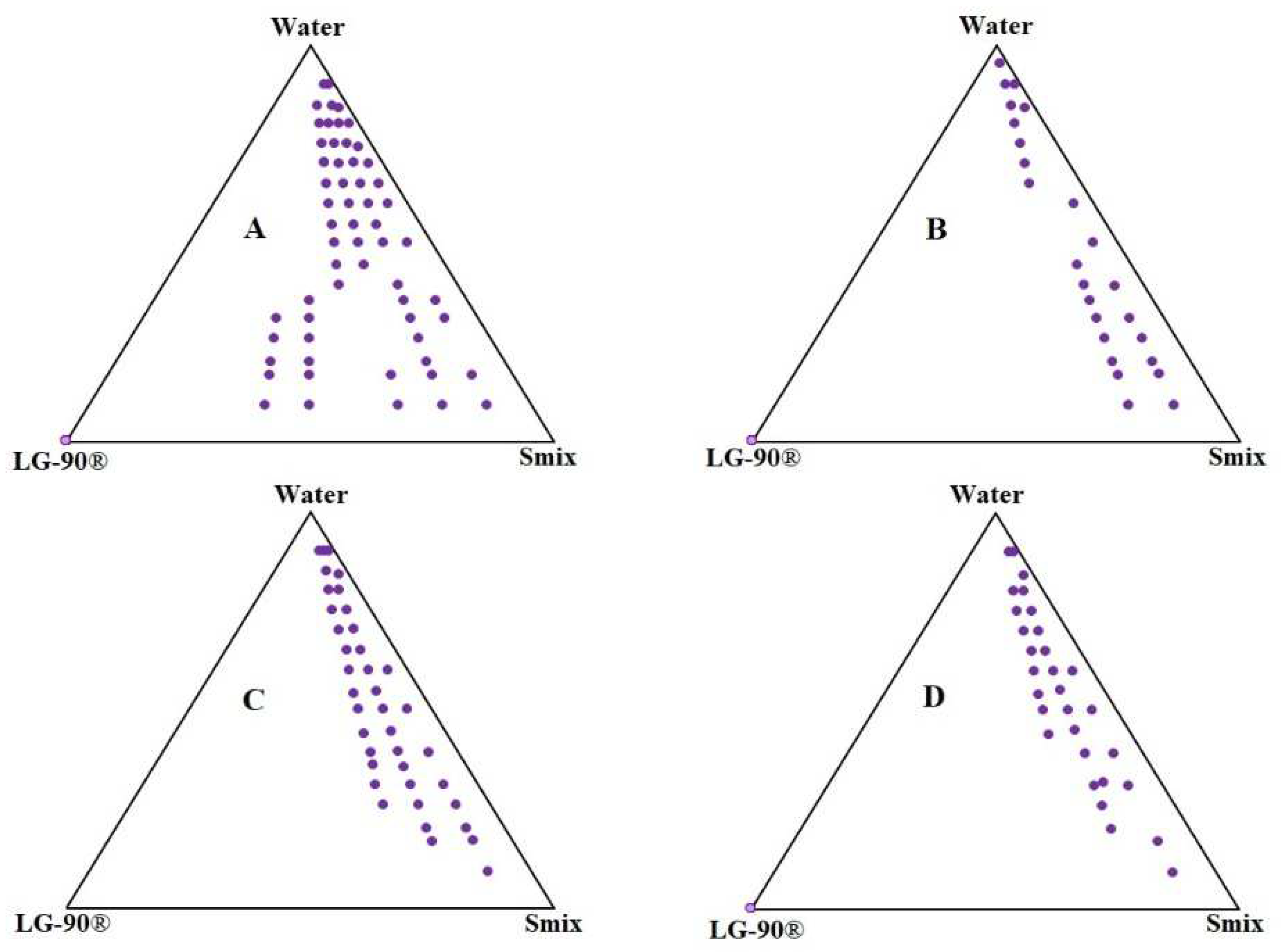
2.2. Pseudo-Ternary Phase Diagram
2.3. Measurement of Physicochemical Properties
2.4. Thermodynamic Stability Studies
2.5. Drug Content Estimation
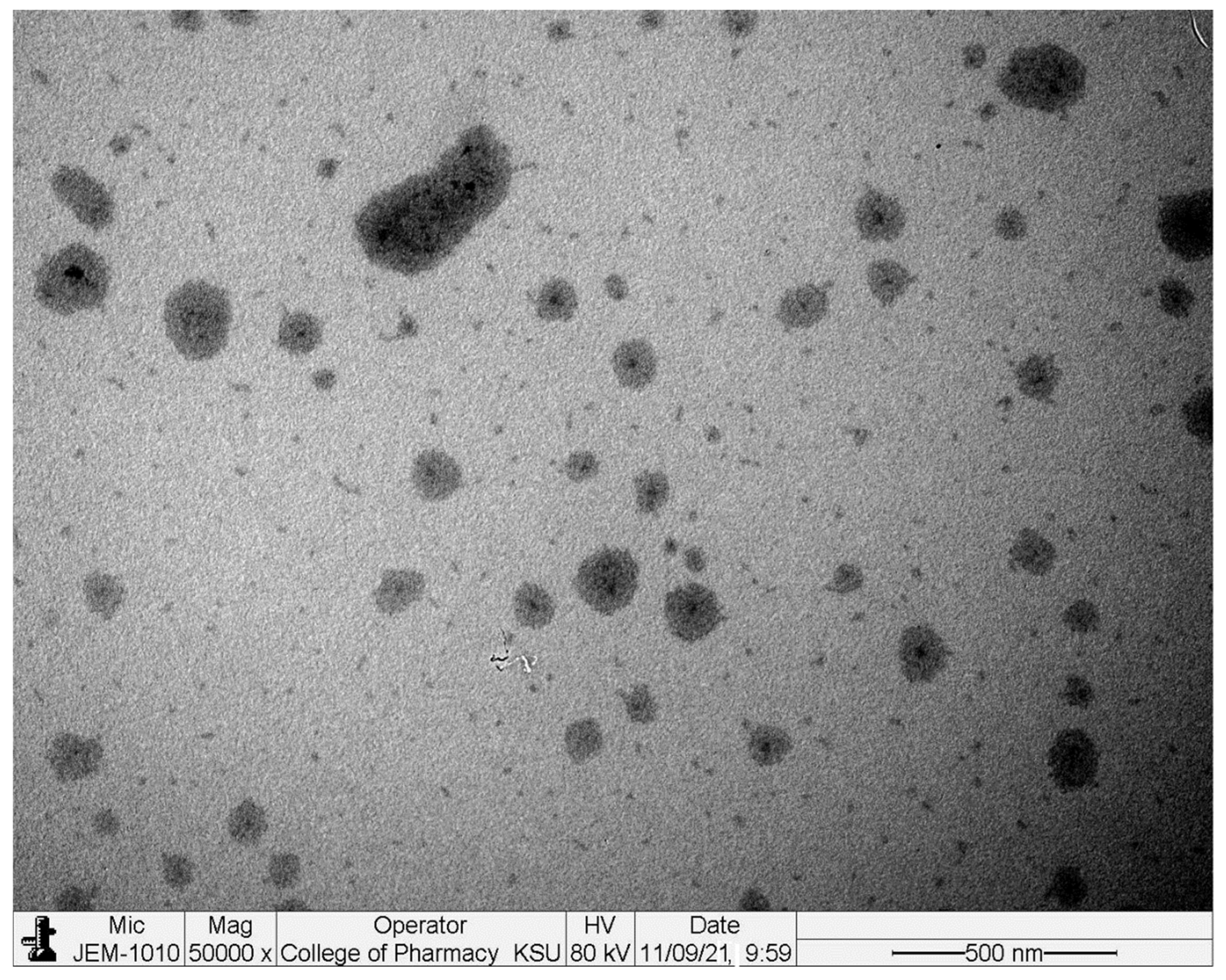
2.6. Transmission Electron Microscopy (TEM)
2.7. Spreadability Studies
2.8. pH
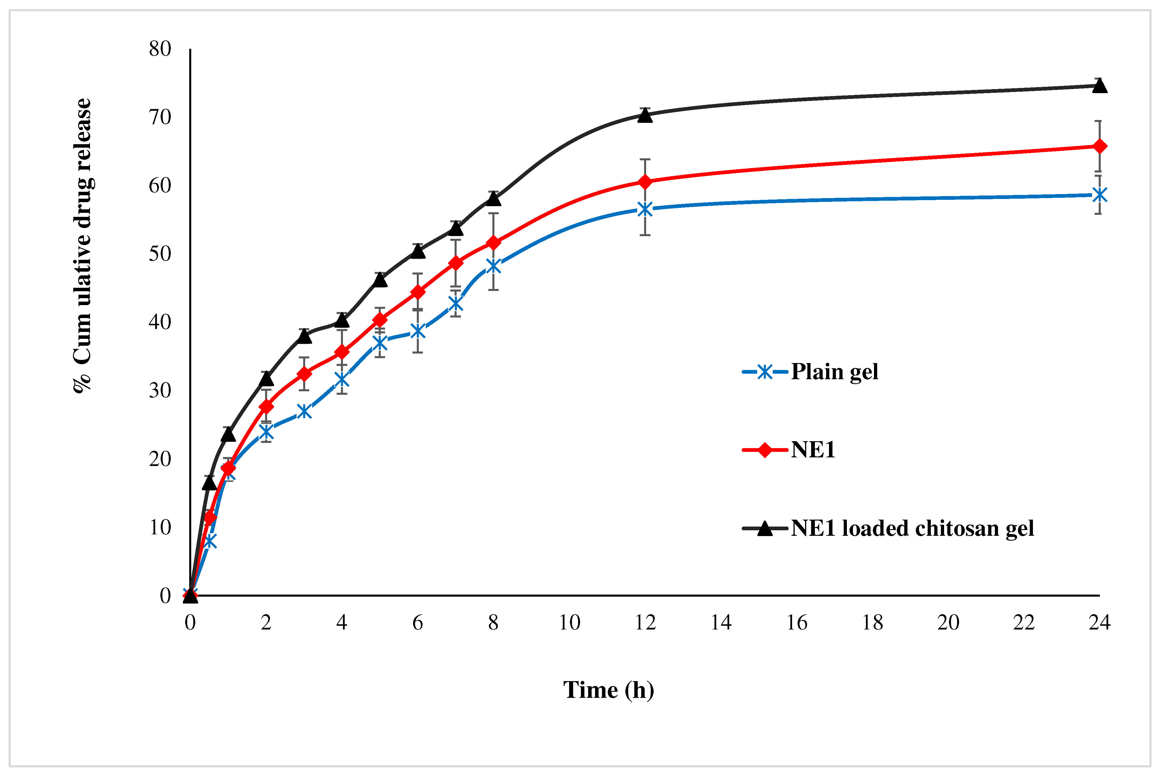
2.9. Drug Release and Kinetics Studies
2.10. Determination of Partition Coefficient, Permeation Coefficient and Flux
2.11. Wound Healing Studies
2.11.1. Acute Dermal Toxicity
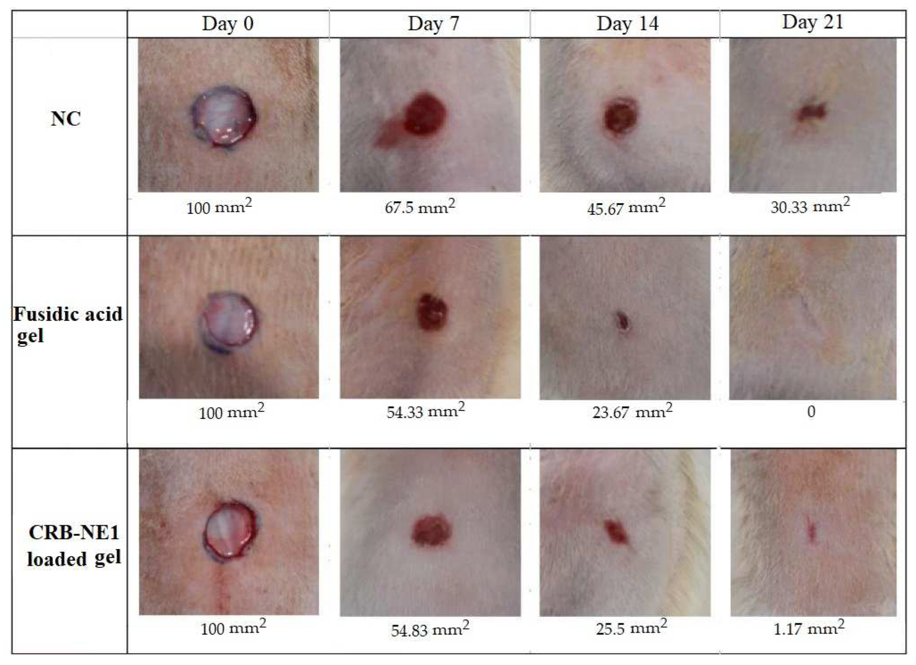
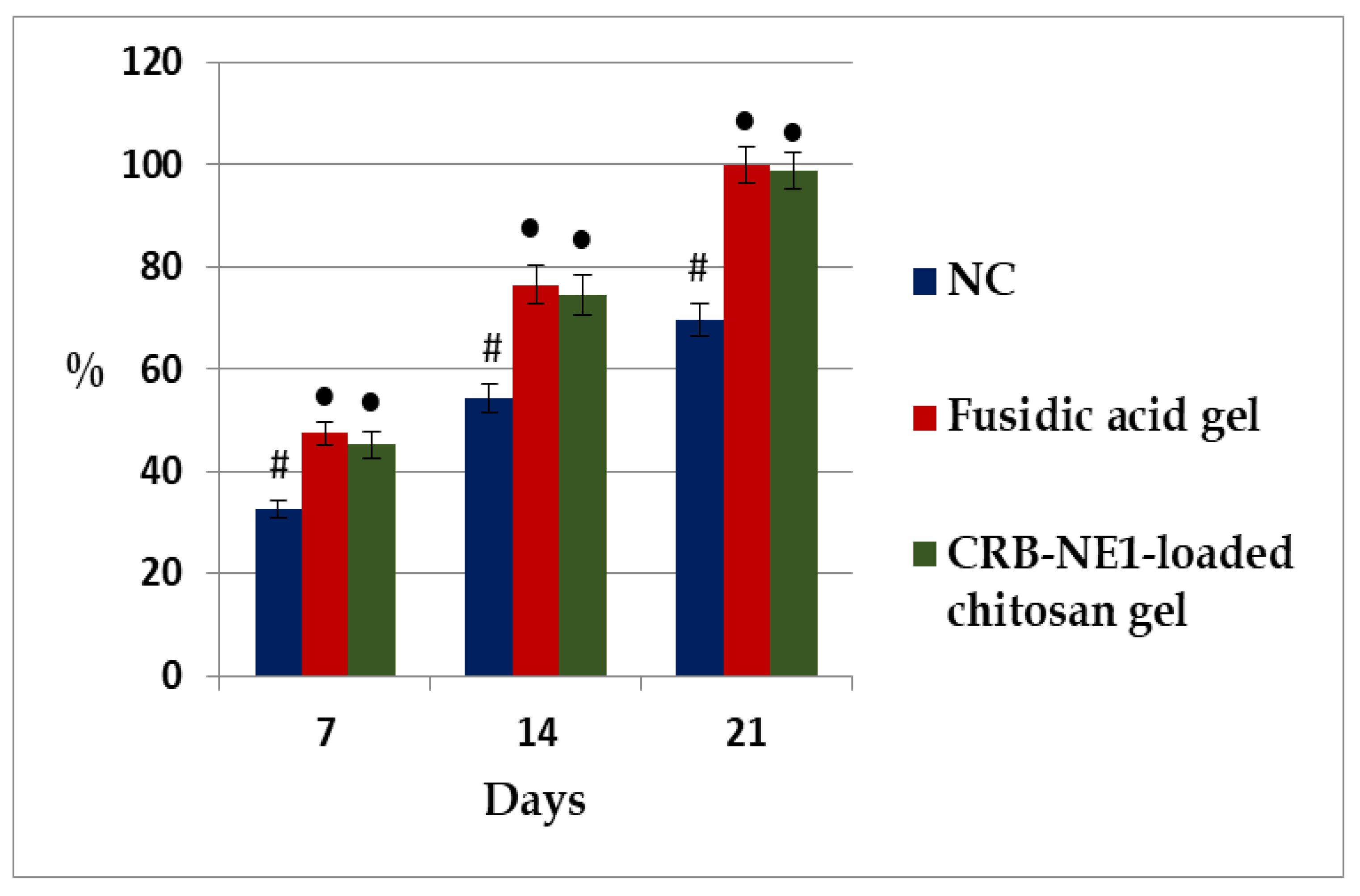
2.11.2. Evaluation of Wound Healing Activity
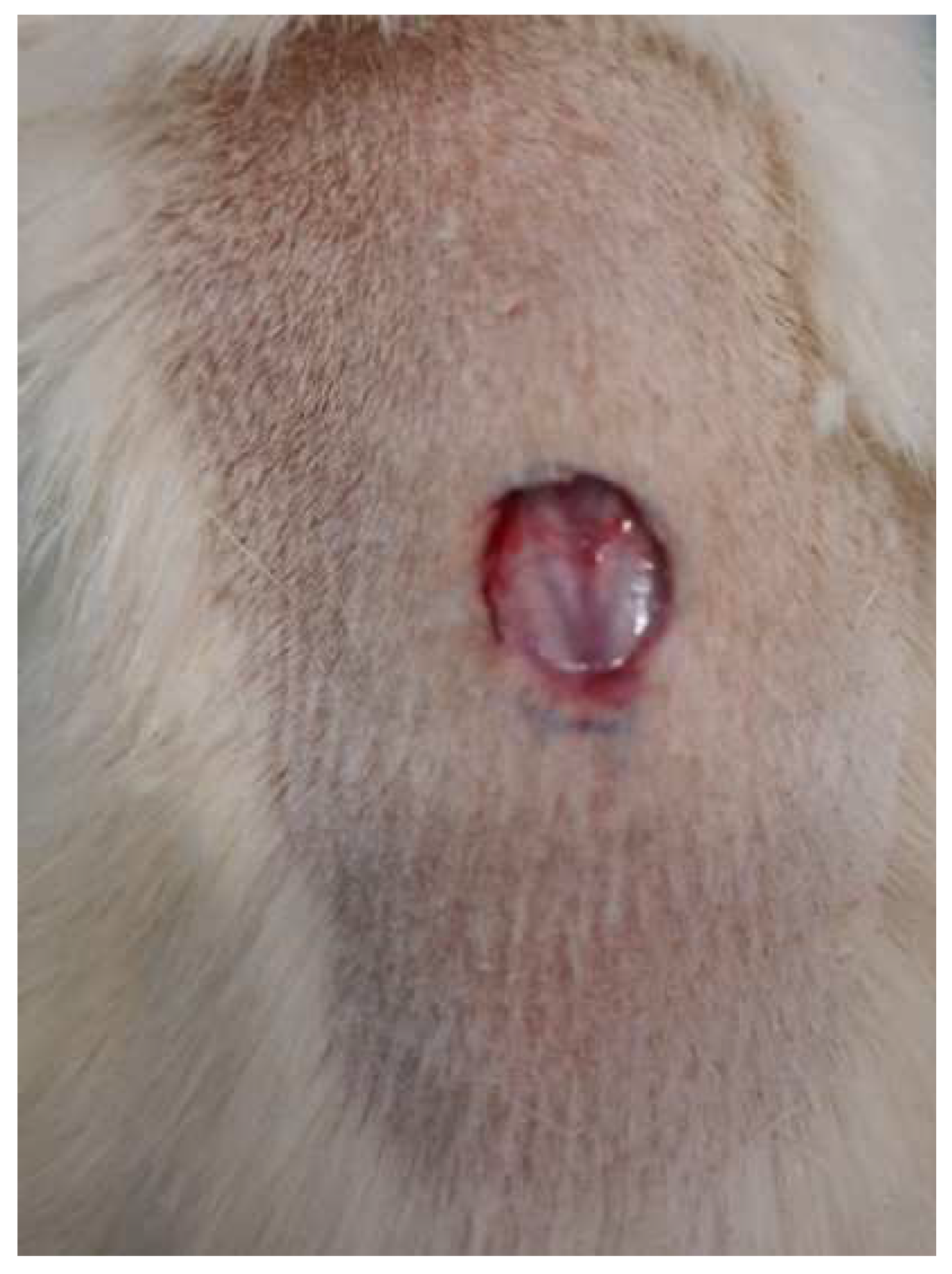
Excision Wound Model
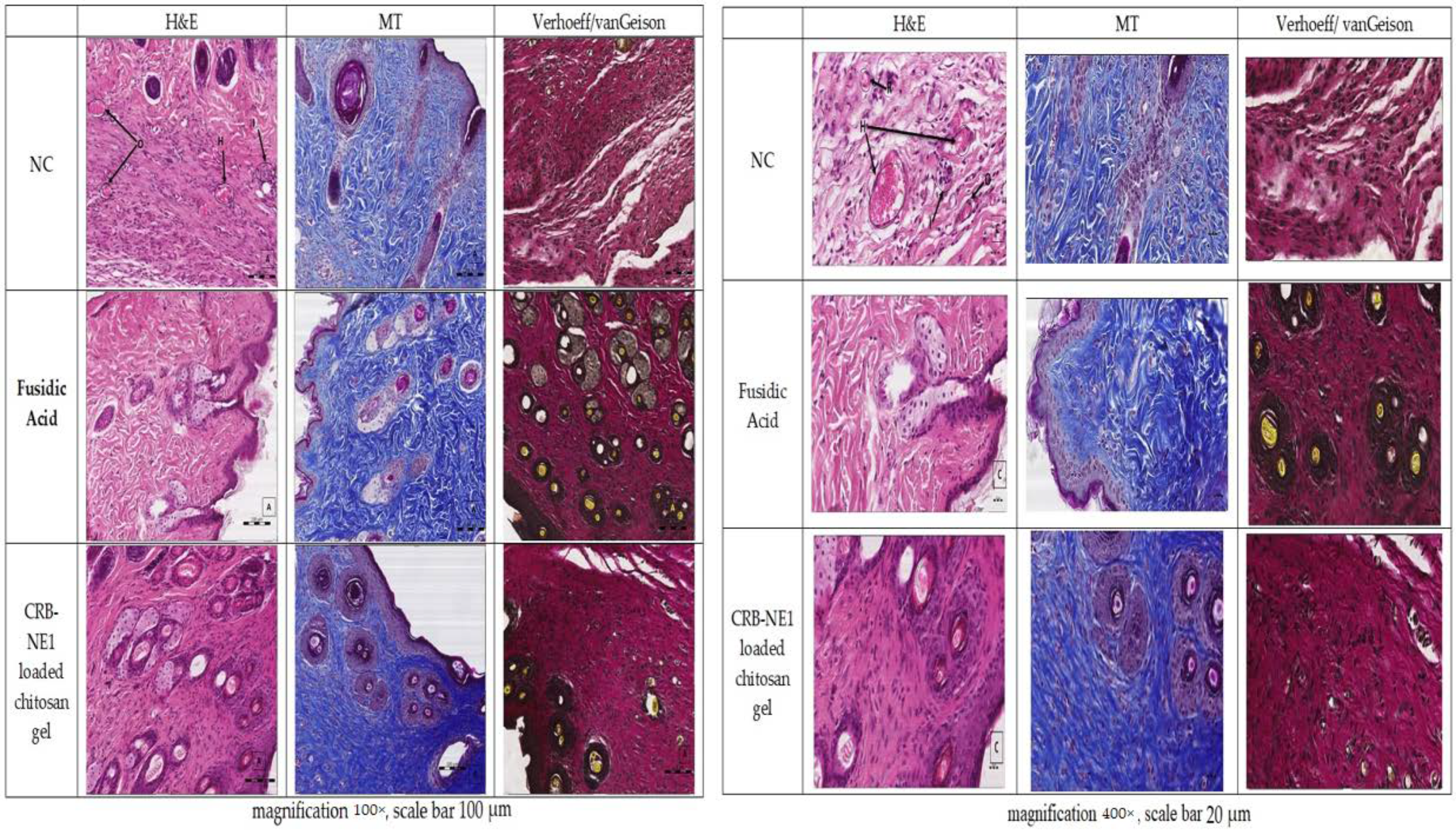
Microscopic Evaluation of the Wound
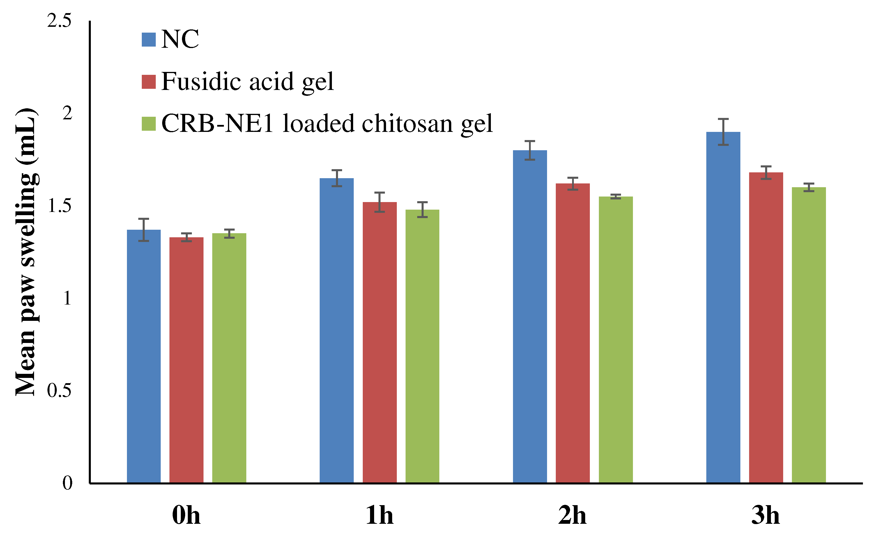
2.12. Evaluation of Anti-Inflammatory
3. Conclusions
4. Materials and Methods
4.1. Materials
4.2. Solubility Studies of CRB in Oils, Surfactants and Co-Surfactants
4.3. Pseudo-Ternary Phase Diagram
4.4. Preparation of Nanoemulsion
4.5. Measurement Physicochemical Properties
4.6. Thermodynamic Stability Studies
4.7. Transmission Electron Microscopy (TEM)
4.8. Preparation of Nanoemulsion Loaded Chitosan Gel
4.9. Drug Content Estimation
4.10. Spreadability Studies
4.11. pH
4.12. Drug Release and Kinetics Studies
4.13. Determination of Partition Coefficient, Permeation Coefficient and Flux
4.14. Wound Healing Studies
Animals
4.15. Acute Dermal Toxicity
4.16. Evaluation of Wound Healing Activity
Excision Wound Model
4.17. Microscopic Evaluation of the Wound
4.18. In Vivo Anti-Inflammatory Activity
4.19. Statistical Analysis
Supplementary Materials
Author Contributions
Funding
Institutional Review Board Statement
Informed Consent Statement
Data Availability Statement
Acknowledgments
Conflicts of Interest
References
- Fuchs, E.; Nowak, J.A. Building Epithelial Tissues from Skin Stem Cells. Cold Spring Harb. Symp. Quant. Biol. 2008, 73, 333–350. [Google Scholar] [CrossRef] [PubMed] [Green Version]
- Landén, N.X.; Li, D.; Ståhle, M. Transition from inflammation to proliferation: A critical step during wound healing. Cell. Mol. Life Sci. 2016, 73, 3861–3885. [Google Scholar] [CrossRef] [PubMed] [Green Version]
- Son, Y.J.; Tse, J.W.; Zhou, Y.; Mao, W.; Yim, E.K.F.; Yoo, H.S. Biomaterials and controlled release strategy for epithelial wound healing. Biomater. Sci. 2019, 7, 4444–4471. [Google Scholar] [CrossRef] [PubMed]
- Strodtbeck, F. Physiology of wound healing. Newborn Infant Nurs. Rev. 2001, 1, 43–52. [Google Scholar] [CrossRef]
- Ponrasu, T.; Madhukumar, K.N.; Ganeshkumar, M.; Iyappan, K.; Sangeethapriya, V.; Gayathri, V.S.; Suguna, L. Efficacy of Acorus calamus on collagen maturation on full thickness cutaneous wounds in rats. Pharmacogn. Mag. 2014, 10 (Suppl. S2), 299–305. [Google Scholar] [CrossRef] [Green Version]
- Baillie, J.K.; Thompson, A.A.; Irving, J.B.; Bates, M.G.; Sutherland, A.; MacNee, W.; Maxwell, S.R.J.; Webb, D.J. Oral antioxidant supplementation does not prevent acute mountain sickness: Double blind, randomized placebo-controlled trial. QJM 2009, 102, 341–348. [Google Scholar] [CrossRef]
- Diegelmann, R.F.; Evans, M.C. Wound healing: An overview of acute, fibrotic and delayed healing. Front. Biosci. 2004, 9, 283–289. [Google Scholar] [CrossRef]
- Upadhyay, G.; Tiwari, N.; Maurya, H.; Upadhyay, J.; Joshi, R.; Ansari, M.N. In vivo wound-healing and antioxidant activity of aqueous extract of Roylea elegans leaves against physically induced burn model in Wistar albino rats. 3 Biotech 2021, 11, 442. [Google Scholar] [CrossRef]
- Akama, T.; Baker, S.J.; Zhang, Y.-K.; Hernandez, V.; Zhou, H.; Sanders, V.; Freund, Y.; Kimura, R.; Maples, K.R.; Plattner, J.J. Discovery and structure–activity study of a novel benzoxaborole anti-inflammatory agent (AN2728) for the potential topical treatment of psoriasis and atopic dermatitis. Bioorg. Med. Chem. Lett. 2009, 19, 2129–2132. [Google Scholar] [CrossRef]
- Jin, S.L.; Ding, S.L.; Lin, S.C. Phosphodiesterase 4 and its inhibitors in inflammatory diseases. Chang Gung Med. J. 2012, 35, 197–210. [Google Scholar] [CrossRef]
- Kailas, A. Crisaborole: A new and effective nonsteroidal topical drug for atopic dermatitis. Dermatol. Ther. 2017, 30, e12533. [Google Scholar] [CrossRef] [PubMed]
- Paton, D.M. Crisaborole: Phosphodiesterase inhibitor for treatment of atopic dermatitis. Drugs Today 2017, 53, 239. [Google Scholar] [CrossRef] [PubMed]
- Cheape, A.C.; Murrell, D.F. 2% Crisaborole topical ointment for the treatment of mild-to-moderate atopic dermatitis. Expert Rev. Clin. Immunol. 2017, 13, 415–423. [Google Scholar] [CrossRef] [PubMed]
- Azmi, N.A.N.; Elgharbawy, A.A.M.; Motlagh, S.R.; Samsudin, N.; Salleh, H.M. Nanoemulsions: Factory for Food, Pharmaceutical and Cosmetics. Processes 2019, 7, 617. [Google Scholar] [CrossRef] [Green Version]
- Pavoni, L.; Perinelli, D.R.; Bonacucina, G.; Cespi, M.; Palmieri, G.F. An Overview of Micro- and Nanoemulsions as Vehicles for Essential Oils: Formulation, Preparation and Stability. Nanomaterials 2020, 10, 135. [Google Scholar] [CrossRef] [Green Version]
- Anwer, M.K.; Iqbal, M.; Aldawsari, M.F.; Alalaiwe, A.; Ahmed, M.M.; Muharram, M.M.; Ezzeldin, E.; Mahmoud, M.A.; Imam, F.; Ali, R. Improved antimicrobial activity and oral bioavailability of Delafloxacin by self-nanoemulsifying drug delivery system (SNEDDS). J. Drug Deliv. Sci. Technol. 2021, 64, 102572. [Google Scholar] [CrossRef]
- Jaiswal, M.; Dudhe, R.; Sharma, S.K. Nanoemulsion: An advanced mode of drug delivery system. 3 Biotech 2015, 5, 123–127. [Google Scholar] [CrossRef] [Green Version]
- Sareen, S.; Joseph, L.; Mathew, G. Improvement in solubility of poor water-soluble drugs by solid dispersion. Int. J. Pharm. Investig. 2012, 2, 12–17. [Google Scholar] [CrossRef] [Green Version]
- Abd, E.; Namjoshi, S.; Mohammed, Y.H.; Roberts, M.S.; Grice, J.E. Synergistic Skin Penetration Enhancer and Nanoemulsion Formulations Promote the Human Epidermal Permeation of Caffeine and Naproxen. J. Pharm. Sci. 2016, 105, 212–220. [Google Scholar] [CrossRef]
- Shakeel, F.; Raish, M.; Anwer, M.K.; Al-Shdefat, R.I. Self-nanoemulsifying drug delivery system of sinapic acid: In vitro and in vivo evaluation. J. Mol. Liq. 2016, 224, 351–358. [Google Scholar] [CrossRef]
- Zainol, S.; Basri, M.; Basri, H.B.; Shamsuddin, A.F.; Gani, S.S.A.; Karjiban, R.A.; Abdul-Malek, E. Formulation Optimization of a Palm-Based Nanoemulsion System Containing Levodopa. Int. J. Mol. Sci. 2012, 13, 13049–13064. [Google Scholar] [CrossRef] [PubMed]
- Shakeel, F.; Haq, N.; El-Badry, M.; Alanazi, F.K.; Alsarra, I.A. Ultra fine super self-nanoemulsifying drug delivery system (SNEDDS) enhanced solubility and dissolution of indomethacin. J. Mol. Liq. 2013, 180, 89–94. [Google Scholar] [CrossRef]
- Shakeel, F.; Haq, N.; Alanazi, F.K.; Alsarra, I.A. Impact of various nonionic surfactants on self-nanoemulsification efficiency of two grades of Capryol (Capryol-90 and Capryol-PGMC). J. Mol. Liq. 2013, 180, 57–63. [Google Scholar] [CrossRef]
- Bello, V.; Mattei, G.; Mazzoldi, P.; Vivenza, N.; Gasco, P.; Idee, J.M.; Robic, C.; Borsella, E. Transmission Electron Microscopy of Lipid Vesicles for Drug Delivery: Comparison between Positive and Negative Staining. Microsc. Microanal. 2010, 16, 456–461. [Google Scholar] [CrossRef]
- Pal, A.P.; Chakraborty, P. Investigation of Corchorus olitorius mucilage as a potential mucoadhesive agent in developing in situ mucoadhesive nasal gel. J. Appl. Pharm. Sci. 2020, 10, 90–98. [Google Scholar] [CrossRef]
- Tao, Q.; Zhong, J.; Wang, R.; Huang, Y. Ionic and Enzymatic Multiple-Crosslinked Nanogels for Drug Delivery. Polymers 2021, 13, 3565. [Google Scholar] [CrossRef]
- Kong, B.J.; Kim, A.; Park, S.N. Properties and in vitro drug release of hyaluronic acid-hydroxyethyl cellulose hydrogels for transdermal delivery of isoliquiritigenin. Carbohydr. Polym. 2016, 147, 473–481. [Google Scholar] [CrossRef]
- Singh, S.; Verma, D.; Mirza, M.A.; Das, A.K.; Dudeja, M.; Anwer, M.K.; Sultana, Y.; Talegaonkar, S.; Iqbal, Z.; Mukharjee, A. Development and optimization of ketoconazole loaded nano-transfersomal gel for vaginal delivery using Box-Behnken design: In vitro, ex vivo characterization and antimicrobial evaluation. J. Drug Deliv. Sci. Technol. 2017, 39, 95–103. [Google Scholar] [CrossRef]
- Sanwal, R.; Chaudhary, A.K. Wound healing and antimicrobial potential of Carissa spinarum Linn. in albino mice. J. Ethnopharmacol. 2011, 135, 792–796. [Google Scholar] [CrossRef]
- Dev, S.K.; Choudhury, P.; Srivastava, R.; Sharma, M. Antimicrobial, anti-inflammatory and wound healing activity of polyherbal formulation. Biomed. Pharmacother. 2019, 111, 555–567. [Google Scholar] [CrossRef]
- Zhang, X.; Kang, X.; Jin, L.; Bai, J.; Liu, W.; Wang, Z. Stimulation of wound healing using bioinspired hydrogels with basic fibroblast growth factor (bFGF). Int. J. Nanomed. 2018, 13, 3897–3906. [Google Scholar] [CrossRef] [PubMed] [Green Version]
- Amadeu, T.P.; Braune, A.S.; Porto, L.C.; Desmoulière, A.; Costa, A.M.A. Fibrillin-1 and elastin are differentially expressed in hypertrophic scars and keloids. Wound Repair Regen. 2004, 12, 169–174. [Google Scholar] [CrossRef] [PubMed]
- Öz, B.E.; Işcan, G.S.; Akkol, E.K.; Süntar, I.; Keleş, H.; Acıkara, B.Ö. Wound healing and anti-inflammatory activity of some Ononis taxons. Biomed. Pharmacother. 2017, 91, 1096–1105. [Google Scholar] [CrossRef]
- Fantini, A.; Demurtas, A.; Nicoli, S.; Padula, C.; Pescina, S.; Santi, P. In Vitro Skin Retention of Crisaborole after Topical Application. Pharmaceutics 2020, 12, 491. [Google Scholar] [CrossRef]
- Taiwan Food and Drug Administration. Assessment Report Trade Name STAQUIS Topical Ointment Active Ingredient: Crisaborole; MOHW-PI 027999; Taiwan Food and Drug Administration: Taipei, Taiwan, 2021.
- Anwer, K.; Jamil, S.; Ibnouf, E.O.; Shakeel, F. Enhanced Antibacterial Effects of Clove Essential Oil by Nanoemulsion. J. Oleo Sci. 2014, 63, 347–354. [Google Scholar] [CrossRef] [Green Version]
- Shakeel, F.; Haq, N.; Alanazi, F.K.; Alsarra, I.A. Polymeric solid self-nanoemulsifying drug delivery system of glibenclamide using coffee husk as a low cost biosorbent. Powder Technol. 2014, 256, 352–360. [Google Scholar] [CrossRef]
- Kalam, M.A.; Raish, M.; Ahmed, A.; Alkharfy, K.M.; Mohsin, K.; Alshamsan, A.; Al-Jenoobi, F.I.; Al-Mohizea, A.M.; Shakeel, F. Oral bioavailability enhancement and hepatoprotective effects of thymoquinone by self-nanoemulsifying drug delivery system. Mater. Sci. Eng. C Mater. Biol. Appl. 2017, 76, 319–329. [Google Scholar] [CrossRef]
- Bhatia, A.; Singh, B.; Raza, K.; Wadhwa, S.; Katare, O.P. Tamoxifen-loaded lecithin organogel (LO) for topical application: Development, optimization and characterization. Int. J. Pharm. 2013, 444, 47–59. [Google Scholar] [CrossRef]
- Abdallah, M.H.; Elsewedy, H.S.; AbuLila, A.S.; Almansour, K.; Unissa, R.; Elghamry, H.A.; Soliman, M.S. Quality by Design for Optimizing a Novel Liposomal Jojoba Oil-Based Emulgel to Ameliorate the Anti-Inflammatory Effect of Brucine. Gels 2021, 7, 219. [Google Scholar] [CrossRef]
- Thomas, L.; Zakir, F.; Mirza, M.A.; Anwer, M.K.; Ahmad, F.J.; Iqbal, Z. Development of Curcumin loaded chitosan polymer based nanoemulsion gel: In vitro, ex vivo evaluation and in vivo wound healing studies. Int. J. Biol. Macromol. 2017, 101, 569–579. [Google Scholar] [CrossRef]
- Ahmed, M.M.; Fatima, F.; Mohammed, A.B. Olive Oil Based Organogels for Effective Topical Delivery of Fluconazole: In-vitro Antifungal Study. JPRI 2020, 32, 29–36. [Google Scholar] [CrossRef]
- Ahmed, M.M.; Fatima, F.; Anwer, K.; Ibnouf, E.O.; Kalam, M.A.; Alshamsan, A.; Aldawsari, M.F.; Alalaiwe, A.; Ansari, M.J. Formulation and in vitro evaluation of topical nanosponge-based gel containing butenafine for the treatment of fungal skin infection. Saudi Pharm. J. 2021, 29, 467–477. [Google Scholar] [CrossRef] [PubMed]
- Anwer, K.; Mohammad, M.; Ezzeldin, E.; Fatima, F.; Alalaiwe, A.; Iqbal, M. Preparation of sustained release apremilast-loaded PLGA nanoparticles: In vitro characterization and in vivo pharmacokinetic study in rats. Int. J. Nanomed. 2019, 14, 1587–1595. [Google Scholar] [CrossRef] [Green Version]
- Mahajan, N.M.; Zode, G.H.; Mahapatra, D.K.; Thakre, S.; Dumore, N.; Gangane, P.S. Formulation development and evaluation of transdermal patch of piroxicam for treating dysmenorrhoea. J. Appl. Pharm. Sci. 2018, 8, 35–41. [Google Scholar] [CrossRef] [Green Version]
- OECD. Guideline for Testing of Chemicals Proposal for a New Draft Guideline 434: Acute Dermal Toxicity-Fixed Dose Procedure; OECD: Paris, France, 2004. [Google Scholar]
- Fieldi, K.J.; White, W.J.; Lang, C.M. Anaesthetic effects of chloral hydrate, pentobarbitone and urethane in adult male rats. Lab. Anim. 1993, 27, 258–269. [Google Scholar] [CrossRef] [PubMed]
- Saeedan, A.S.; Gabr, G.A.; Soliman, G.A.; Fayed, M.H.; Ansari, M.N. The potential anti-inflammatory and wound healing activities of chitosan in rats. Adv. Biores 2016, 7, 1–7. [Google Scholar]
- Ahmad, M.; Ansari, M.N.; Alam, A.; Khan, T.H. Oral Dose of Citrus Peel Extracts Promotes Wound Repair in Diabetic Rats. Pak. J. Biol. Sci. 2013, 16, 1086–1094. [Google Scholar] [CrossRef] [Green Version]
- Hamad, A.M.; Ahmed, H.G. Association of some carbohydrates with estrogen expression in breast lesions among Sudanese females. J. Histotechnol. 2018, 41, 2–9. [Google Scholar] [CrossRef]
- Hamad, A.M.; Ahmed, H.G. Association of Connective Tissue Fibers with Estrogen Expression in Breast Lesions among Sudanese Females. Int. Clin. Pathol. J. 2016, 2, 97–102. [Google Scholar] [CrossRef]
- Bancroft, J.D.; Layton, C. The hematoxylins and eosin. In Bancroft’s Theory and Practice of Histological Techniques, 8th ed.; Suvarna, S.K., Layton, C., Bancroft, J.D., Eds.; Elsevier: Amsterdam, The Netherlands, 2018; p. 131. [Google Scholar]
- Ansari, M.J.; Alshetaili, A.; Aldayel, I.A.; Alablan, F.M.; Alsulays, B.; Alshahrani, S.; Alalaiwe, A.; Ansari, M.N.; Rehman, N.U.; Shakeel, F. Formulation, characterization, in vitro and in vivo evaluations of self-nanoemulsifying drug delivery system of luteolin. J. Taibah Univ. Sci. 2020, 14, 1386–1401. [Google Scholar] [CrossRef]
- Ahmed, M.M.; Anwer, M.K.; Fatima, F.; Alali, A.S.; Kalam, M.A.; Zafar, A.; Alshehri, S.; Ghoneim, M.M. Development of Apremilast Nanoemulsion-Loaded Chitosan Gels: In Vitro Evaluations and Anti-Inflammatory and Wound Healing Studies on a Rat Model. Gels 2022, 8, 253. [Google Scholar] [CrossRef]


| NEs | Droplet Size (nm) | PDI | ZP (mV) | RI | % T |
|---|---|---|---|---|---|
| CRB-NE1 | 64.5 ± 5.3 | 0.202 ± 0.06 | −36.3 ± 4.16 | 1.332 ± 0.03 | 99.8 ± 0.12 |
| CRB-NE2 | 102.7 ± 8.1 | 0.273 ± 0.05 | −28.6 ± 4.23 | 1.334 ± 0.05 | 97.6 ± 0.16 |
| CRB-NE3 | 85.4 ± 7.1 | 0.303 ± 0.01 | −35.6 ± 3.42 | 1.364 ± 0.06 | 98.4 ± 0.14 |
| CRB-NE4 | 123.6 ± 9.8 | 0.295 ± 0.02 | −34.2 ± 3.54 | 1.311 ± 0.04 | 96.5 ± 0.08 |
| Treatments | Epithelialization Period (Days) |
|---|---|
| NC | 28.53 ± 1.75 # |
| Fusidic acid gel | 20.72 ± 1.18 ● |
| CRB-NE1-loaded chitosan gel | 21.45 ± 1.46 ● |
| Groups | Percent Swelling | Percent Inhibition |
|---|---|---|
| NC | 39.68 ± 1.24 # | - |
| Fusidic Acid gel | 25.72 ± 2.01 ● | 35.18 |
| CRB-NE1 loaded chitosan gel | 19.18 ± 0.64 ●# | 51.66 |
| NEs Codes | Compositions (% w/w) | Smix | |||
|---|---|---|---|---|---|
| LG-90® | Tween®-80 | THP® | Water | ||
| CRB-NE1 | 10 | 40 | 40 | 20 | 1:1 |
| CRB-NE2 | 12 | 20 | 20 | 48 | 1:1 |
| CRB-NE3 | 16 | 24 | 24 | 36 | 1:1 |
| CRB-NE4 | 40 | 10 | 10 | 40 | 1:1 |
Publisher’s Note: MDPI stays neutral with regard to jurisdictional claims in published maps and institutional affiliations. |
© 2022 by the authors. Licensee MDPI, Basel, Switzerland. This article is an open access article distributed under the terms and conditions of the Creative Commons Attribution (CC BY) license (https://creativecommons.org/licenses/by/4.0/).
Share and Cite
Ansari, M.N.; Soliman, G.A.; Rehman, N.U.; Anwer, M.K. Crisaborole Loaded Nanoemulsion Based Chitosan Gel: Formulation, Physicochemical Characterization and Wound Healing Studies. Gels 2022, 8, 318. https://0-doi-org.brum.beds.ac.uk/10.3390/gels8050318
Ansari MN, Soliman GA, Rehman NU, Anwer MK. Crisaborole Loaded Nanoemulsion Based Chitosan Gel: Formulation, Physicochemical Characterization and Wound Healing Studies. Gels. 2022; 8(5):318. https://0-doi-org.brum.beds.ac.uk/10.3390/gels8050318
Chicago/Turabian StyleAnsari, Mohd Nazam, Gamal A. Soliman, Najeeb Ur Rehman, and Md. Khalid Anwer. 2022. "Crisaborole Loaded Nanoemulsion Based Chitosan Gel: Formulation, Physicochemical Characterization and Wound Healing Studies" Gels 8, no. 5: 318. https://0-doi-org.brum.beds.ac.uk/10.3390/gels8050318