The Advances in Biomedical Applications of Carbon Nanotubes


Research Institute of Fundamental and Applied Medicine, S.D. Asfendiyarov Kazakh National Medical University, Tole Bi Street 94, Almaty 050000, Kazakhstan
C 2019, 5(2), 29; https://0-doi-org.brum.beds.ac.uk/10.3390/c5020029
Submission received: 16 April 2019
/
Revised: 16 May 2019
/
Accepted: 17 May 2019
/
Published: 23 May 2019
(This article belongs to the Special Issue Biomedical Applications of Carbon Materials)
Abstract
:Unique chemical, physical, and biological features of carbon nanotubes make them an ideal candidate for myriad applications in industry and biomedicine. Carbon nanotubes have excellent electrical and thermal conductivity, high biocompatibility, flexibility, resistance to corrosion, nano-size, and a high surface area, which can be tailored and functionalized on demand. This review discusses the progress and main fields of bio-medical applications of carbon nanotubes based on recently-published reports. It encompasses the synthesis of carbon nanotubes and their application for bio-sensing, cancer treatment, hyperthermia induction, antibacterial therapy, and tissue engineering. Other areas of carbon nanotube applications were out of the scope of this review. Special attention has been paid to the problem of the toxicity of carbon nanotubes.
Keywords:
carbon nanotubes; synthesis; drug delivery; cancer; antibacterial; bio-sensing; hyperthermia; stem cells; toxicity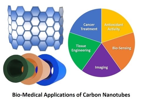
1. Introduction
Carbon nanotubes (CNTs) belong to a new class of nanomaterials that possess unique chemical, physical, and biological properties. Structurally, all CNTs can be considered as carbon allotropes with a cylindrical architecture. Apart from the unique shape and morphology, CNTs demonstrate an excellent capability to conduct electronic and thermal energy. These features have made CNTs an ideal candidate for myriad applications in different fields such as electronics, chemistry, optics, and biomedicine [1,2,3,4,5]. Mainstream CNT use was in the industrial field up until the beginning of the 21st century, when the first reports on the biological application were published [6,7]. After this, there was a boom in scientific interest involving the utilization of CNTS for bio-medicine, specifically bio-sensing, diagnostics, and therapy. This was facilitated by the excellent ability of CNTs to act as carriers of molecules of different origin, including proteins, DNA/RNA, enzymes, and drugs [8]. In addition to their proven delivery capacities, it was also demonstrated that CNTs are capable of effectively by-passing the blood–tissue barrier and penetrating cells [9]. These findings prompted research dedicated to the development of drug delivery platforms based on CNTs [10,11,12]. Another vast area of CNTs applications is in the separation of chiral drugs and various chemicals in pharmaceutical and chemical industries [8,13,14]. This review discusses the progress and main fields of bio-medical use of CNTs based on recently published reports. It encompasses synthesis of CNTs and their application for bio-sensing, cancer treatment, hyperthermia induction, antibacterial therapy, and tissue engineering. Other areas of CNTs biomedical applications were out of the scope of this review. The special chapter highlights the problems with toxicity of CNTs.
2. Structure, Synthesis, and Surface Functionalization
CNTs are nanotubes made of one-atom-thick sheets of graphene, which is a monolayer of carbon atoms incorporated into a two-dimensional honeycomb lattice [15,16]. All CNTs can be sub-classified based on the number of layers into two classes: single-walled carbon nanotubes (SWCNTs) and multi-walled carbon nanotubes (MWCNTs) [8]. There are some differences between SWCNTs and MWCNTs that should be taken into consideration. First, for the synthesis of SWCNTs the chemical catalyst is required, whilst the MWCNTs can be fabricated without the catalyst. Secondly, the process of production of SWCNTs is bulky by itself, and the purity is lower compared to MWCNTs [8]. There are five main methods of fabrication of CNTs: carbon arc-discharge method [17], laser-ablation technique [18], chemical vapor deposition [19,20], flame synthesis method [21,22,23], and spray pyrolysis method [24,25]. Newly-fabricated CNTs are usually being refined from amorphous carbon, fullerenes, and metals by the following methods: acid refluxing, surfactant aided sonication, or air oxidation [8,26,27,28,29]. Table 1 summarizes the main methods of CNTs synthesis.
All types of fabricated CNTs possess an intrinsic problem of poor solubility in any kind of aqueous solution or organic solvents [9]. Apart from insolubility, this hinders the conjunction of CNTs with organic and inorganic molecules. Hence, the functionalization of the surface of CNTs is a crucial step towards bio-medical applications. The surface-functionalization CNTs can be classified into three main methods: covalent functionalization [30,31], noncovalent adsorption of required molecules [32], and the endohedral filling of CNTs inner space [9].
It must be noted that the different methodologies and approaches for surface functionalization may profoundly affect the biocompatibility, toxicity and physical features of CNTs. In fact, the method of functionalization may directly influence of pharmacokinetics and bio-distribution of CNTs [33]. It was found out that non-covalently functionalized CNTs predominately accumulate in liver and spleen tissues, whilst the covalently functionalized CNTs prone to be excreted through urine [34].
It must be also noted that the functionalization usually requires the purification and/or oxidation leading to the changes of the CNTs length and functionality.
Iannazzo et al. showed that surface functionalization led to critical changes of physical, chemical, and biological properties of CNTs, particularly modifications of their antiviral activity [35]. The results of the study indicated the important role of the free carboxylic groups as hydrophilic functionality affecting dispersibility of the CNTs, and their interaction with biological molecular complexes by means of electrostatic or hydrogen bonds. In another study, Baldo et al. developed CNT-based biosensors with high sensitivity for Arginase-1 detection via surface functionalization [36]. It allowed immobilizing the receptor on the sidewalls of CNTs. The proposed nano-system can be potentially employed for detection of Arginase-1 associated with different pathologies such as myeloma and lymphoma.
The solubility and dispersibility of CNTs can be improved through conjugation with polymers. There are two main methods for the polymer modification: noncovalent attachment (polymer wrapping and absorption) and covalent attachment (“grafting”) [37]. There is a range of studies on the effect of polymer modification on the electrochemical, mechanical, thermal, and rheological properties of CNTs [38,39,40,41]. In addition, polymer modification and hybridization with CNTs have been also actively studied with regard to bio-medical applications, particularly for bio-sensing [42,43,44].
In fact, the unique physical and chemical features of the CNTs surface allow combining them with nano-platforms of different origin [16]. These novel multifunctional hybrid nano-constructs can be produced by directly attaching nanoparticles to CNTs’ surface or by wrapping of nanoparticles with CNTs. In this case, the method of synthesis is dictated by the size of nanoparticles. The threshold for nanoparticle size is 100 nm, which allows the direct attachment to the graphene sheet [16]. Otherwise, the sought nanoparticles can be placed in the roll of graphene 2D sheet of graphene [45]. The attachment of required nanoparticles on CNTs’ surface can be conducted using different techniques, such as thermal evaporation [46], pulse laser deposition (PLD) [47], and sputtering [48]. Apart from that, multifunctional hybrid nano-constructs can be fabricated ex situ by mixing the required nanoparticles with graphene sheets or wrapping graphene around them. This provides an opportunity to control morphology, shape, size and functionality of produced hybrids [16,45].
3. Carbon Nanotubes and Bio-Sensing Technology
The main and initial research interest of application of CNTs is bio-sensing. Up to date, about 700 research articles have been published (data of Web of Science) thus reflecting a huge interest from science and industry. The very first publication on this topic can be traced back to 2001, when Kirschner et al. reported on the modification of biosensor surface for detection of the proteins binding fullerenes and carbon nanotubes [49]. In this study, the researchers demonstrated the feasibility of using surface plasmon resonance for real-time protein binding measurements with fullerenes and carbon nanotubes. The followed years are characterized by an outbreak of research and reports in this field. For example, Cai and co-workers were first who fabricated a DNA-biosensor, which has been based on multi-walled carbon nanotubes functionalized with a carboxylic acid group for covalent DNA immobilization and enhanced hybridization detection [50]. Compared to old models of DNA sensors, the carbon nanotube-based system significantly increased the quantity and sensitivity of DNA attachment. It was the first application and demonstration of the ability of carbon nanotubes to serve as electrochemical DNA biosensor for quick and reliable detection of specific hybridization.
Up to now, researchers have been working towards optimization and enhancing of DNA sensitivity of carbon nanotube-based systems. Karimi-Maleh et al. reported on successful fabrication of biosensor equipped with immobilized DNA on a pencil graphite electrode modified with polypyrrole/functionalized multi-walled carbon nanotubes [51]. This system could be potentially employed for detection of anticancer drug 6-mercaptopurine (6-MP) in biological and pharmaceutical samples.
Shahrokhian and co-workers developed a simple platform based on a hairpin oligonucleotide switch and multi-walled carbon nanotubes for the ultrasensitive detection of specific DNA sequences [52]. The authors employed π-stacking interaction of single-strand DNA–MWCNT in order to fabricate electrochemical DNA biosensor. The biosensor showed a good linear correlation range (0.1 μM–10 pM) for the model target DNA, thus making possible the stable, low-cost, and sensitive detection of sequence-specific, non-complementary, and mismatched DNA.
Zribi et al. built up a microfluidic-multiplexed platform with carbon nanotubes-based electrochemical sensors (carbon nanotube/ferrocene) for the detection of pathogenic viral DNA from Hepatitis C and genomic DNA from Mycobacterium tuberculosis [53]. Such a device might be utilized as the point-of-care for multi-target diagnostics of various infections in the hospitals.
DNA-wrapped multi-walled carbon nanotube-modified genosensor for the detection of Escherichia coli (E. coli) was developed by Ozkan-Ariksoysal et al. [54]. For this, DNA-wrapped multi-walled carbon nanotube immobilized graphite sensor was fabricated, and polymerase chain reaction-amplified real samples were tested for the first time. The ssDNA-MWCNTs nano-hybrid system was immobilized on the surface of the PGE for direct electrochemistry of guanine oxidation. This system provided an enhanced signal with high sensitivity.
Another example of hybrid nano-system based on CNTs was fabricated and studied by Chen at el. for rapid and ultrasensitive detection of Mycobacterium tuberculosis [55]. In this study, flower-like carbon nanotubes-polyaniline nanohybrid was developed as a novel redox nano-platform. Functionalized fullerene and enzyme-assisted target recycling were able to enhance the detection signal. The fabricated amperometric DNA biosensor showed high specificity and sensitivity for detection of Mycobacterium tuberculosis in real clinical samples.
Aside from DNA-detection, CNT-based sensing systems have been also actively studied towards other possible biomedical applications, including but not limited to, detection of glucose [56], pesticides [57], cholesterol [58], reactive oxygen species [59], lactate [60], cancer [61], tuberculosis [55], nucleic acids [62], malaria [63], Staphylococcus aureus [64], and anticancer drugs [65].
4. Carbon Nanotubes in Cancer Treatment
The huge surface area and excellent ability to penetrate the cell membrane make CNTs a perfect platform for delivery pharmacological and genetic compounds into the cells. During the last decades, the main focus for utilizing CNTs for drug delivery has been cancer therapy. In fact, the classic approach for cancer treatment is the administration of chemotherapeutic drugs that suffers from systemic toxicity, narrow therapeutic window, drug resistance and low cellular penetration [8]. In this context, CNTs offer an opportunity to shelter anti-cancer drugs thus lowering their toxicity for an organism and enhancing local accumulation in the desired site. Up to date, different types of anti-cancer compounds have been proposed and tested in regards their conjunction and functionality with the CNTs [66,67].
As it can be expected, the major efforts have been done towards the conjugation of standard chemotherapeutics with CNTs that have been widely used for cancer treatment in the clinics such as doxorubicin, flutamide, cisplatin, methotrexate, paclitaxel, etc. In this regard, doxorubicin takes the first place as the main candidate for the coupling with CNTs. For example, Yan et al. developed an active targeting and pH-responsive system for transporting doxorubicin to tumor sites using folic acid-modified multi-walled carbon nanotubes [68]. This novel system showed high drug loading and encapsulation efficiency. At the same time, the results of in vivo experiments demonstrated that the administration of folic acid-modified multi-walled carbon nanotubes enhanced the suppression of tumor growth and decreased the side effects of doxorubicin.
In another work, Le and co-workers proposed a novel pH-responsive drug release system based on doxorubicin conjugated poly(styrene-alt-maleic anhydride) grafted multi-walled carbon nanotubes [69]. The system was prepared using the reversible addition-fragmentation chain transfer polymerization and acid-cleavable hydrazone linkages. The results of in vitro experiments demonstrated successful release of doxorubicin at pH 5.0 (slight release was observed at pH 7.4).
The toxicity of doxorubicin and other chemotherapeutics towards health cells is well known. So that the decreasing doxorubicin free concertation without sacrificing its efficacy has been a topic of research during the last decades. Lee at el. developed a nano-platform based on CNTs that was able to nullify the efflux of cancer cells through the prolonged endolysosome delivery and provide the maximized release of doxorubicin in an acidic hydrolase environment leading to the reduction of the amount of doxorubicin [70]. Pistone et al. reported on the fabrication of pH and temperature controlled drug delivery systems for cancer therapy based on functionalized multi-walled carbon nanotubes [71]. The CNTs were loaded with doxorubicin followed by coverage with the biocompatible polymer polylactide to form hydrogen bonding with PEG and bind doxorubicin inside polymeric chains. The results of tests showed high biocompatibility of nano-systems and ability to deliver doxorubicin to cancer cells.
Doxorubicin in the combination with CNTs was a subject of a range of studies aiming at the treatment of cancer by means of photo-thermal therapy. Wang et al. developed a bi-functional nano-system via a facile method in which poly (N-vinyl pyrrole) was coated on cut multi-walled CNTs [72]. The authors noted that poly-(N-vinyl pyrrole) shell not only is able to significantly increase the photo-thermal effect of CNTs, but can also provide an optimal surface for conjugating anticancer drugs such as doxorubicin. The developed nano-system demonstrated a high drug-loading ratio as well as pH-sensitive unloading capacity for doxorubicin. Oh et al. showed that single-walled CNTs conjugated with doxorubicin have the potential to be employed for combined near-infrared (NIR) cancer photo-thermal and chemotherapy [73]. It was demonstrated that single-walled CNTs provided a slow release and high accumulation of doxorubicin in the cell nucleus of cancer cells (MDA-MB-231 cell line). The combination of near infrared radiation and single-walled CNTs conjugated with doxorubicin-induced death of cancer cells via mitochondrial disruption and generation of reactive oxygen species (ROS).
The photo-thermal effect of single-walled CNTs in combination with doxorubicin for targeting and elimination of breast cancer cells was shown by Jeyamohan et al. [74]. The researchers fabricated single-walled CNTs (conjugated with folic acid), which have been polyethylene glycol bio-functionalized and loaded with doxorubicin. The data of the studies showed that doxorubicin binds at physiological pH (pH 7.4) and is released only at a lower pH, i.e., lysosomal pH (pH 4.0), which is typical for the tumor environment. Under laser irradiation, cancer cell death was observed as a result of thermal ablation caused by CNTs. The authors concluded that the proposed nano-system, combining of laser, drug, and CNTs is a promising therapeutic modality with high treatment efficacy and safety for cancer therapy.
Karimi et al. showed that another anti-cancer drug, methotrexate, can be used in the combination with carboxylated functionalized multi-walled CNTs for the elimination of cancer breast cancer cell by means of photo-thermal therapy [66]. For this, folic acid and methotrexate were conjugated through ethylenediamine to the surface of multi-walled CNTs. The cancer cell death was achieved via thermal ablation induced by CNTs. Znang et al. used the nano-construct made of multi-walled CNTs, gemcitabine (anti-cancer drug), and lentinan (immune-stimulator) to study a possibility of treating cancer by means of chemo and photo-thermal therapy [75]. The proposed nano-system was able to effectively penetrate cancer cells and suppress the viability of cancer cells (upon irradiation by 808 nm laser) comparing to a single use of each component of this system.
The main idea behind the use of CNTs as a platform for drug delivery for cancer treatment is reducing a toxic effect of chemotherapy. For this purpose, Yu et al. fabricated a system based on polyethylene glycol-modified carbon nanotubes with loaded lobaplatin (anti-cancer drug). The CNTs demonstrated a good cell penetrability, stability, pH-controlled release property, and high inhibition rate on liver cancer cells [11]. In another study, Mehrjouei et al. used a computer model to investigate the release of anti-cancer drug cisplatin from CNTs [76]. The data showed that the efficiency of cisplatin release depends on the drug’s adsorption on the outer surface of the nanotube and CNTs composition and their diameter.
As an alternative to traditional chemotherapeutics, some researches proposed to use anticancer compounds of natural origin in the combination with CNTS. It concerns the use of extracts, essences and compounds made from medicinal herbs. One of the most promising anticancer herbal compounds is curcumin. Curcumin is extracted from the rhizome of Curcuma longa (Zingiberaceae). It possesses a proven anticancer activity, and at the same time, it demonstrated cyto-protective potential towards healthy cells [77]. Up to date, there is a dozen published studies on the combination of curcumin and CNTs [78,79]. For example, Singh reported on the successful fabrication of polysaccharide (functionalized with alginate and chitosan) single-walled CNTs to serve as carriers for delivery of curcumin in human lung adenocarcinoma (A549) cells [79]. The study showed pH-dependent drug release potential and anti-cancer activity of CNTs loaded with curcumin. Apart from curcumin, there is a range of studies demonstrating that anticancer herbal compounds can be effectively combined with CNTs: paclitaxel [80,81], docetaxel [82], vinca alkaloids [83], camptothecin [84,85], podophyllotoxin [86], quercetin [87], oridonin [88], etc.
The list of studies on the application of CNTs in cancer treatment is provided in the Table 2.
In fact, CNT-based nano-platforms provide an opportunity for combining imaging and therapeutic activity that can be employed in bio-medicine as ‘theranostic’ modality. In particular, CNTs can be coupled with other nano-systems of different origin to achieve synergetic curative and diagnostic effects [16]. In the context of cancer management, such theranostic hybrid nanoparticles might be utilized for detection, visualization, and treatment of various types of cancer. Moreover, novel hybrid nano-platforms can help to optimize cancer treatment and lower toxic effects of chemotherapeutics. Mashal et al. reported on constructing tissue-mimicking materials with varying concentrations of single-walled CNTs and characterized their dielectric properties and response to the microwave heating [100]. The researchers observed linear temperature increase in mixtures containing CNTs. The results demonstrated the potential of CNTs to be used for microwave detection of breast cancer and selective heating of malignant tissues. In another study, Al Faraj et al. developed doxorubicin-loaded single-walled carbon nanotubes for targeting and monitoring of metastatic regions [101]. The monitoring was provided by free-breathing magnetic resonance imaging (MRI) and bioluminescence imaging. The magnets were positioned over the metastatic tumor sites in the lungs of tumor-bearing mice. The results of the study showed an increase of the treatment efficiency as a result of the combination of non-invasive MRI (to localize sensitively the tumor sites) and magnets that enhance the magnetic targeting of CNTs. In general, CNTs can be effectively coupled with various materials such as metals, polymers and mesoporous silica to be employed as theranostics for cancer management [102].
5. Hyperthermia and Photo-Thermal Therapy
The ability of the CNTs to convert the near infrared radiation (NIR) into the heat is well-known [103]. This unique feature attracted the attention of researchers in terms of its possible implementation in bio-medicine, particularly for the induction of thermal effect on cells and tissues. Mirali et al. proposed to use folate-targeted single-walled CNTs at different concentrations for the ablation of cancer cells under exposure to NIR light irradiation [104]. It was found out that the CNTs concentration as ~13 pg/cell was sufficient to kill 90% of cells, while decreasing the CNTs dosage led to the drop of cell killing efficiency. Moreover, the authors demonstrated that the extent of intercellular internalization was directly correlated to high concentrations of CNTs.
Virani et al. employed single-walled CNTs functionalized with annexin V and phosphatidylserine for photo-thermal ablation of bladder cancer on mice model [92]. CNTs were administrated at a very low dose (0.1 mg per kg), and then, mice (with MB49 murine bladder tumors) were subjected to NIR light for only 30 s exposure. The results showed the full elimination of tumors in the animals exposed to combined treatment (CNTs and NIR). Importantly, the authors reported on the absence of CNTS in the animal organism after 116 days followed by the treatment that indicates the zero toxicity of CNTs.
Apart from the for photo-thermal application, CNTs can be also co-equipped with magnetic particles for diagnostic and therapeutic purposes [97,105]. For example, the functionalization of CNTs with iron and contrast agents enhances visualization of targeted organs (for example with MRI), and the same time it provides an opportunity to induce hyperthermia effect on the tumors. British group from Queen Mary, University of London, fabricated iron-filled multi-walled CNTs loaded with gadolinium (MRI contrast agent) [106]. The CNTs fabrication and modification was achieved through the surface functionalization of CNTs in aqueous GdCl3 solution. The results of the study showed the feasibility of using such hybrid nano-platform for MRI imaging and magnetic hyperthermia for cancer treatment (‘theranostics’). In another work, Zuo et al. proposed to use a novel type of CNTs conjugated with magnetic nanoparticles with low Curie temperature that provides a condition for self-regulating magnetic hyperthermia [107]. The authors investigated the structure, magnetic properties, heat generation ability, and cytotoxicity of the hybrid CNTs on cell culture model (Human epidermal keratinocyte (HaCaT) cells).
Another example of hybridization of CNTs for imaging and synergistic cancer therapy was demonstrated by Zhang et al. [105]. The researchers produced multi-walled CNTs combined with magneto-fluorescent carbon quantum dots/doxorubicin. Such a combination can be utilized for the for dual modal imaging, photo-thermal, and chemo-therapy of cancer. The newly fabricated nano-platform demonstrated a high heat-generating ability, pH and NIR responsive drug delivery, and heat-induced high drug release during in vitro and in vivo experiments (on mice).
Madani et al. functionalized single-walled CNTs with OctaAmmonium-POSS in order to enhance the temperature increase caused by the exposure to NIR laser irradiation (808 nm laser at 1 watt) [94]. In this study, HT-29 colorectal cancer cell line was utilized as a model for photothermal ablation. It was found out that upon exposure to NIR, the group treated by OctaAmmonium-POSS-SWCNT showed a significantly higher temperature increase than the other experimental groups, leading to high rate of cell death of cancer cells compared to the control.
Up to date, the main approach for induction thermal effect has been predominantly based on irradiation of CNTs by laser or infrared light. It must be noted that the application of light is limited to the surfaces of the human body due to the inability of the light to penetrate the skin and reach inner organs affected by cancer or other disorders. As a solution, the light can be delivered via miniaturized sources such as fiber-optic cable. However, it would require the incision of the skin or obtaining direct access to the blood vessels that is traumatic for the patients, and it also bears the risk of bacterial/viral infection. As an alternative to the light irradiation, CNTs can be exposed to external electromagnetic fields in a non-contact and non-invasive manner. In fact, there is a range of reports on achieving thermal effect using the combination of electromagnetic radiation and graphene- or carbon-based nanoparticles [108,109]. These studies indicated the feasibility of exploiting the CNTs for heating deep-seated tumors in hospital environment. Such an approach provides a basis for the development novel theranostic modalities that could assist to detect, visualize and treat malignancies without traumatic consequences for the patients.
6. Antibacterial Therapy
Despite the recent progress on antibacterial therapy, there is a growing problem with the resistance of bacterial strains to antibiotics. This issue was caused by overuse and misuse of antibiotics and delay with the development of new effective antibacterial medicines by the pharmaceutical industry [110,111]. In this regard, the combination of CNTs and antibiotics could be a good solution to overcome such problems, and help to optimize the antibacterial therapy [112,113].
Up to date, there are more than one hundred publications dedicated to the modification and application of CNTs to fight infections of various origin (data of Web of Sciences). The main approach relies on the coating and surface functionalization of CNTs in order to make them suitable for loading antibacterial compounds.
Liu et al. synthesized vancomycin hydrochloride modified multi-walled CNTs [114]. The synthesis was conducted through the reaction of multi-walled CNTs inherent carboxyl group and the vancomycin amide group. The newly fabricated CNTs showed the excellent antibacterial properties which can be potentially harnessed for wound management.
Ciprofloxacin is a well-known antibiotic, and it belongs to the fluoroquinolone class. Ciprofloxacin has been widely used to treat infections caused by Gram-positive and Gram-negative bacteria. However, the resistance to ciprofloxacin has been growing rapidly around the world [115,116]. To overcome this problem, Assali et al. proposed a novel type of functionalized single-walled CNTs with loaded ciprofloxacin [117]. The newly fabricated CNTs have been characterized by using transmission electron microscopy (TEM), Raman spectroscopy, thermos-gravimetric, and microbiological analysis. The results of the study demonstrated that ciprofloxacin loaded-CNTs had a significant increase in the antibacterial activity against the three strains of bacteria compared to ciprofloxacin free drug.
Aside from antibacterial agents, there are also attempts to employ CNTs as a platform for drug delivery of antifungal compounds such as amphotericin B [118,119]. For instance, Pruthi and co-workers developed mannose-conjugated multi-walled CNTs loaded with Amphotericin B for targeted delivery to macrophages using dialysis diffusion method [118]. The data of experiments showed a controllable release of Amphotericin at pH 4, 7.4 and 10, along with enhanced cellular uptake and higher disposition in macrophage-rich organs. The results indicated that this novel nano-system can be utilized for anti-leishmanial therapy in the future.
As an alternative to a classic approach based on the use of antibiotics, some researchers proposed to utilize noble metals such as silver and gold in the combination with CNTs [120]. The noble metals, particularly silver, possess the antibacterial and antifungal activity towards various pathogen microorganisms, including antibiotic-resistant strains [121,122]. Such a unique feature has been harnessed to suppress infections in antibacterial therapy, particularly by the incorporation of silver into wound dressings [123,124].
In fact, CNTs have been also proven to exhibit antibacterial activity towards different bacterial strains [125,126]. Therefore, the coupling antibacterial potential of CNTs and noble metals can be highly beneficial and useful for biomedicine. Up to date, there is a range of reports on the enhancement and optimization of the silver-induced antibacterial effects by using carbon-based platforms for silver deposition, shielding, and site release [127,128]. Seo et al. synthesized multi-walled CNTs and tested their antibacterial activity [129]. For this, CNTs were exposed to the aqueous sulfuric acid solution to form an oxygen-based functional group. In another study, Baek and co-workers also treated multi-walled CNTs with a mixture of acids and conjugated with silver nanoparticles [126]. The antibacterial activity of fabricated CNTs was tested against Methylobacterium spp. and Sphingomonas spp. It was found out that CNTs (at the dosage 30 μg/mL) possess antibacterial activity with minimal cytotoxicity towards both human and animal cells.
The agglomeration of silver nanoparticle remains an issue hampering the research and bio-medical applications. In this case, conjugating of silver particles with CNTs could help to overcome such a problem. Ma et al. utilized polyethyleneimine for conjugating of small-sized silver nano-dots (size of 2.6 nm) to CNTs [130]. The results demonstrated a high antibacterial activity of newly fabricated CNT-hybrids compared to single CNTs, acidified-CNTs and the same amount of silver dots. In another study, Al Aani et al. developed a novel antibacterial hybrid nano-platform made of multi-walled CNTs coated with silver nanoparticles via a facile and rapid method using a microwave treatment [131]. The hybrid CNTs showed an increase in antibacterial activity against two bacterial species: Staphylococcus aureus and Escherichia coli.
Despite the proven antibacterial activity, silver nanoparticles can also exhibit toxicity towards normal cells [132,133]. In this case, the polymer coating of nano-systems can significantly reduce potential cytotoxic effects of silver ions. For example, Nie et al. proposed to use the biopolymer coating in order to shield silver nanoparticles-loaded oxidized CNTs, and to modify their interface properties [134]. The researchers employed two types of dopamine-grafted functional biopolymers, (heparin and chitosan) for the protection of silver ions and synergetic action against bacteria. Later, the same research group reported on fabrication of a self-sterilizing surface with high biocompatibility to efficiently control the potential bacterial infections on the surface of medical devices. For this, the researchers used the mussel-inspired polymer coating strategy to synthesize negatively charged and positively charged nano-dispersions of silver nanoparticles-CNTs via a spray-assisted layer-by-layer assembly [135]. In another study, Fan et al. fabricated a novel type of antibacterial nano-platform consisting of dopamine-conjugated polysaccharide sulfate-anchored and -protected silver–graphene nanocomposites to suppress bacterial infection and contamination [136].
Apart from silver, the other metal nanoparticles have been also successfully conjugated with CNTs such as a copper [137,138]. For instance, Palza et al. fabricated hybrid CNTs by direct impregnation of copper nanostructures into CNTs. The new hybrids were composed of a well-dispersed layered copper hydroxide acetate nanostructures on CNT surface [139]. Such a composition provided a synergetic antibacterial effect against Staphylococcus aureus and Escherichia coli.
7. Carbon Nanotubes and Stem Cells
Stem cells and their application for tissue engineering and medicine attracted the attention of researchers around the world during the last decades. In fact, stem cells have been considered as a magic bullet to treat various pathologies, including oncology, cardiology, dentistry, neurology, and wound management [140,141,142]. There is growing evidence that CNTs could be used as a cell substrate, and could then modulate proliferation and differentiation of various types of stem cells [143].
CNTs have been intensively studied in their application for neural tissue engineering [144,145]. In fact, CNTs provide all requirements for optimal neural regeneration: high binding affinity and excellent electrical conductivity. These unique features make CNTs indispensable for developing neuro-implants designed for growing and repairing the neurons. Singh et al. fabricated plasma-treated chitin carbon nanotube composite scaffolds [145]. The proposed nano-platform demonstrated a very good neuron adhesion, support of the synaptic function of neurons, and improved electrical conductivity. Earlier, Jan et al. demonstrated the feasibility the differentiation mouse embryonic neural stem cells to neurons, astrocytes, and oligodendrocytes with the formation of neurites on layer-by-layer assembled single-walled CNT–polyelectrolyte multilayer thin films [146].
The main application of CNTs for neural engineering, as well as for other types of tissue engineering, is the use of them as a scaffold for the stem cells. Recently, Gupta et al. reported on the fabrication hybrid scaffolds made of multi-walled CNTs/chitosan and graphene nanoplatelets/chitosan [147]. The results of the study demonstrated a good electric conductivity, elasticity, and high cyto-compatibility of hybrid scaffolds. Such a nano-platform can be potentially utilized for the promoting neurite outgrowth and cell differentiation in the close perspective. In another work, the single-walled CNTs have been used to enhance neural differentiation of stem cells from apical papilla [148].
Aside from neural stem cells, CNTs have been successfully combined with embryonic stem cells [149,150]. For example, Brunner et al. proposed a novel method of producing synthetic, non-xenogeneic carbon nanotube-based scaffolds for the culture of human embryonic stem cells [149]. The authors showed that through the modulation of the nanotube processing conditions before film fabrication, it is possible to influence cell adherence, proliferation, and colony morphology. The results indicated the potential for CNT thin films to compete with conventional human embryonic stem cells substrates. Sebaa et al. investigated the potential of graphene and multi-walled CNTs–graphene hybrid nanomaterials for culturing delicate human embryonic stem cells [150]. It was demonstrated that the human embryonic stem cells remained viable and pluripotent, and they proliferated on graphene and multi-walled CNTs–graphene hybrid nano-constructs. Zang and co-workers used fibrous polyethylene terephthalate (PET) matrices in the combination with multi-walled CNTs [151]. Such a combination promoted cell adhesion and proliferation of embryonic stem cells along with maintaining high cell viability.
Another trend in the applications of CNTs is their combination with mesenchymal stem cells for tissue engineering and treatment of various disorders [152,153]. As well as for other types of stem cells, CNTs have been considered as a possible basis for growing and differentiation of mesenchymal stem cells. In fact, the nanoplatforms must meet a range of strict requirements to be suitable for tissue engineering, including good scalability, compatibility, and low cytotoxicity. In this regard, researchers have been working on the fabrication and optimization of CNT-based nano-constructs to serve as a scaffold for mesenchymal stem cells. For example, Lalwani et al. fabricated all-carbon 3-D single- and multiwalled CNT scaffolds as artificial matrices for long-term maintenance and expansion of human adipose-derived human mesenchymal stem cells [154]. The fabricated scaffolds demonstrated a good level of cell viability, attachment, proliferation, and infiltration in CNT scaffolds comparable to poly(lactic-co-glycolic) acid (PLGA) scaffolds.
Pouladzadeh et al. fabricated nanostructured fibrous scaffolds based on flexible thermoplastic polyurethane and surface functionalized multi-walled CNTs to study the impact of the state of electrical conductivity of scaffolds upon neural cells’ proliferation and differentiation of rat mesenchymal stem cells [152]. It was shown that mesenchymal stem cells cultured on multi-walled CNTs possessed enhanced neural differentiation, indicating the importance of high electrical conductivity of the scaffolds for modulation of proliferation and differentiation of stem cells towards the normal neural tissue. Garnica-Gutiérrez et al. studied the effects of acid-functionalized and polycitric acid- (PCA)-polymerized CNTs on mesenchymal stem cells in vitro, including intracellular incorporation, cell proliferation, apoptosis, and cytotoxicity [153]. It was found out that PCA-polymerized CNTs are less cytotoxic and are associated with less apoptotic cell death than the acid-functionalized ones. In addition, it was demonstrated that functionalized undoped CNTs have a strong stimulation potential for cell proliferation, whilst functionalized and polymerized nitrogen-doped CNTs are able of stimulating the apoptosis cascades.
8. Toxicity Studies of Carbon Nanotubes
The main concern over the bio-medical application of CNTs is their toxicity towards healthy cells and tissues. There is a big discussion in the literature whether CNTs should be considered predominantly neutral for the organism or they present a danger and are potentially harmful for the humans and environment. Up to date, there are more than 300 peer-reviewed publications on the toxicity of CNTs (data of Web of Science, 2019).
In fact, the toxicity of CNTs depends on many factors, including but not limited to, CNTs composition, surface morphology, concentration, the way of administration, interaction with chemical and physical components, and predisposition of the organism [160,161]. For example, Zhao et al. studied the possible toxicity of multi-walled CNTs with three different diameters for human umbilical vein endothelial cells [162]. It was revealed that CNTs with the smallest diameter induced a significantly higher level of cytotoxicity than the other CNTs types. It includes the induction of high levels of cytokine release, monocyte adhesion, intracellular reactive oxygen species, and DNA damage. In another study, De Marchi et al. studied the toxicity of carboxylated CNTs to the reef-forming tubeworm Ficopomatus enigmaticus [163]. The results showed that CNTs induced toxicity towards tubeworms, including the reduction of protein and glycogen contents, triggering the oxidative status stress and activation of an antioxidant enzymes defense system. Zhang et al. studied combined toxicities of copper nanoparticles with CNTs on marine microalgae Skeletonema costatum [164]. In the study, it was found out that both types of nano-systems could inhibit the growth of the microalgae; however, the toxicity of CNTs on the microalgae was significantly lower than that of copper nanoparticles.
One of the main fields of studies on the toxicity of CNTs is an impact on the pulmonary system [165,166]. The CNTs belong to the big group of respirable materials, and they potentially can cause the inflammation of lung tissues. In fact, constant inflammation and lung damage might lead to pulmonary fibrosis and respiratory cancer [167]. Some studies demonstrated that CNTs, even in low concentrations, are able to trigger inflammation in the respiratory system of animals [168]. In addition to inflammation, it was revealed that CNTs are also capable of inducing the modulation of proliferation of lung cells as a result of direct injury [169], fibrosis [170], DNA damage [171], and lung cancer [172,173].
Apart from the respiratory tract, it has been shown that CNTs can exhibit toxic effects on the other organs and systems such as reproduction [174,175], nervous system [176,177], liver [178,179], and kidneys [179,180].
However, despite the above-mentioned reports on the harmful impact of CNTs, there is a range of publications indicating the absence or low toxic effects caused by CNTs [181,182]. For instance, Aghar et al. demonstrated that single-walled CNTs and reduced graphene oxide do not affect sperm viability, and the exposure to these nanomaterials does not hinder the sperm sorting process [183]. The results of another study revealed that hydroxylated multi-walled CNTs and carboxylated multi-walled CNTs induced modest toxic effects on human umbilical vein endothelial cells [184]. Lim et al. reported on the absence of teratogenicity of multi-walled CNTs in rats [185]. Earlier, Pantarotto and co-workers demonstrated that the functionalized single-walled CNTs were non-toxic for the human fibroblasts 3T6 and murine 3T3 cells in the concentrations up to 10 mM [186].
Therefore, the discussion about toxicity, carcinogenicity, and teratogenicity of CNTs is still open, and it is far from finalization. In fact, the assessment of carbon-based materials is difficult by itself due to many factors, including CNTs concentration, the method of fabrication, purification, morphology, shape, time of exposure, and the state of cells or organism. So far, the majority of the studies have been limited to in vitro research either on experiments on human or animal cells. There is no data on the systemic application of CNTs on humans. Hence, it is hard to comprehensively analyze the full impact and long-term health effects for the living organisms, and particularly for humans.
9. Conclusions and Future Perspectives
Up to date, there is a wide range of nanomaterials being discovered and studied in the context of applications in the bio-medical field. Among this spectrum of nano-platforms, CNTs demonstrated superior physical, chemical, and biological properties, including excellent mechanical strength, biocompatibility, aspect ratio, resistance to corrosion, and electrical and thermal conductivity. The most important feature of CNTs is a large surface area that provides an opportunity to load and deliver therapeutic molecules to the affected tissues and organs [187]. The unique needle-like shape of CNTs assists drug delivery through enhanced permeability and retention. In addition, CNTs possess a capacity for Raman scattering, photoluminescence, and photoacoustic response. The intrinsic properties of CNTs have been considerably improved through surface functionalization with polymers and other biocompatible materials [188]. Moreover, CNTs morphology allows coupling with different nano-systems and fabrication of hybrid nano-carriers, which can be utilized as theranostics modalities for diagnostics and treating cancer and other pathologies (Figure 1).
The distinctive characteristics of CNTs are a result of the source material (graphene) and nano-size. The size allows reaching remote sites of the organism, passing through cellular membranes and delivering uploaded biologically active substances. The main areas for possible biomedical applications of CNTs are bio-sensing, imaging, cancer treatment, drug and DNA delivery, tissue engineering, immunotherapy, regenerative medicine, and antibacterial therapy (Figure 1).
The main approach for improving and modulating of properties of CNTS is the coating and functionalization of CNTs surface. Such an approach provides longer circulation and stabilization of CNTs by protecting them from undesirable aggregation, degradation, and conglomeration. The functionalization also allows attaching active molecules and ligands making possible to target specific receptors on cancer and other cells for targeted delivery. The last trend is the fabrication of hybrid nano-systems based on CNTs and/or graphene materials. The hybridization provides an opportunity to achieve a synergetic effect originated from two or more components of the nano-system [23]. It would allow a coupling of imaging and treating modalities in one nano-construct (as a theranostic platform). This is a novel approach that can change the standard methods for disease management, particularly for personalized cancer therapy.
In spite of the recent progress and intensive studies, the real application of CNTs in clinical practice has been hampered by a few factors. First of all, the toxicity of CNTs is still a major concern for wide practical implications. There are open questions regarding pharmacokinetics, internalization, and accumulation of CNTs in vital inner organs. In addition, there is a number of reports on the genotoxic potential of CNTs [171,189,190]. However, this topic remains controversial due to limited systemic studies on humans. It must be noted that most of the published research works have been conducted either on cells or animals. So further studies are necessary in order to thoroughly evaluate the toxic impact of CNTs on humans, including possible genotoxic and teratogenic effects.
Funding
This research received no external funding.
Acknowledgments
The author acknowledges the administrative and technical support provided by S.D. Asfendiyarov Kazakh National Medical University (Almaty, Kazakhstan).
Conflicts of Interest
The author declares no conflict of interest.
References
- Ramalingame, R.; Lakshmanan, A.; Muller, F.; Thomas, U.; Kanoun, O. Highly sensitive capacitive pressure sensors for robotic applications based on carbon nanotubes and PDMS polymer nanocomposite. J. Sens. Sens. Syst. 2019, 8, 87–94. [Google Scholar] [CrossRef] [Green Version]
- Lin, J.N.; Yeh, C.Y.; Pan, Y.N.; Lin, M.C.; Fan, F.Y. Effect of carbon nanotubes on in vitro cellular responses for bioglass application. Mater. Lett. 2019, 235, 141–143. [Google Scholar] [CrossRef]
- Filatzikioti, A.; Glezos, N.; Kantarelou, V.; Kyriakis, A.; Pilatos, G.; Romanos, G.; Speliotis, T.; Stathopoulou, D.J. Carbon nanotube Schottky type photodetectors for UV applications. Solid State Electron. 2019, 151, 27–35. [Google Scholar] [CrossRef]
- Muhulet, A.; Miculescu, F.; Voicu, S.I.; Schutt, F.; Thakur, V.K.; Mishra, Y.K. Fundamentals and scopes of doped carbon nanotubes towards energy and biosensing applications. Mater. Today Energy 2018, 9, 154–186. [Google Scholar] [CrossRef]
- Rahman, G.; Najaf, Z.; Mehmood, A.; Bilal, S.; Shah, A.; Mian, S.; Ali, G. An Overview of the Recent Progress in the Synthesis and Applications of Carbon Nanotubes. C 2019, 5, 3. [Google Scholar] [CrossRef]
- Kong, Y.; Cui, D.X.; Ozkan, C.S.; Gao, H.J. Modelling carbon nanotube based bio-nano systems: A molecular dynamics study. Biomicroelectromech. Syst. (Biomems) 2003, 773, 111–116. [Google Scholar]
- Guo, Q.; Shen, X.T.; Li, Y.Y.; Xu, S.Q. Carbon nanotubes-based drug delivery to cancer and brain. Curr. Med. Sci. 2017, 37, 635–641. [Google Scholar] [CrossRef]
- Chou, S.G.; Dresselhaus, M.S.; Humphreys, E.; Chung, S.Y.; VanderSande, J.; Ciang, Y.M.; Swan, A.K.; Unlu, M.S.; Goldberg, B.B. Raman analysis of carbon nanotube bundles for bio-electronic applications. Quantum Confin. Semicond. Nanostruct. 2003, 737, 395–400. [Google Scholar] [CrossRef]
- He, H.; Pham-Huy, L.A.; Dramou, P.; Xiao, D.L.; Zuo, P.L.; Pham-Huy, C. Carbon Nanotubes: Applications in Pharmacy and Medicine. Biomed Res. Int. 2013, 2013, 578290. [Google Scholar] [CrossRef]
- Alshehri, R.; Ilyas, A.M.; Hasan, A.; Arnaout, A.; Ahmed, F.; Memic, A. Carbon Nanotubes in Biomedical Applications: Factors, Mechanisms, and Remedies of Toxicity. J. Med. Chem. 2016, 59, 8149–8167. [Google Scholar] [CrossRef]
- Li, H.P.; Sun, X.W.; Li, Y.J.; Li, B.E.; Liang, C.Y.; Wang, H.S. Preparation and properties of carbon nanotube (Fe)/hydroxyapatite composite as magnetic targeted drug delivery carrier. Mater. Sci. Eng. C 2019, 97, 222–229. [Google Scholar] [CrossRef] [PubMed]
- Yu, S.P.; Li, Q.; Wang, J.L.; Du, J.L.; Gao, Y.D.; Zhang, L.; Chen, L.; Yang, Y.Z.; Liu, X.G. A targeted drug delivery system based on carbon nanotubes loaded with lobaplatin toward liver cancer cells. J. Mater. Res. 2018, 33, 2565–2575. [Google Scholar] [CrossRef]
- Miao, R.; Wu, D.X.; Wang, Q.Y.; Zhao, H.X.; Li, X.; Xiu, Y.; Liu, S.Y. Rapid Separation of Ginsenosides Based on Multi-walled Carbon Nanotubes. Chem. J. Chin. Univ. Chin. 2018, 39, 2178–2184. [Google Scholar]
- Um, J.E.; Song, S.G.; Yoo, P.J.; Song, C.; Kim, W.J. Large-scale separation of single-walled carbon nanotubes by electronic type using click chemistry. Appl. Surf. Sci. 2018, 429, 278–283. [Google Scholar] [CrossRef]
- Geim, A.K.; Novoselov, K.S. The rise of graphene. Nat. Mater. 2007, 6, 183–191. [Google Scholar] [CrossRef] [PubMed]
- Saliev, T.; Akhmetova, A.; Kulsharova, G. Multifunctional hybrid nanoparticles for theranostics. In Core-Shell Nanostructures for Drug Delivery and Theranostics; Elsevier: Amsterdam, The Netherlands, 2018; pp. 177–244. [Google Scholar] [CrossRef]
- Iijima, S. Helical Microtubules of Graphitic Carbon. Nature 1991, 354, 56–58. [Google Scholar] [CrossRef]
- Maser, W.K.; Munoz, E.; Benito, A.M.; Martinez, M.T.; de la Fuente, G.F.; Anglaret, E.; Righi, A.; Sauvajol, J.L. Single-wall carbon nanotubes: Study of production parameters using cw CO2-laser ablation technique. Electron. Prop. Nov. Mater. Mol. Nanostruct. 2000, 544, 213–216. [Google Scholar]
- Fahlman, B.D. Chemical vapor deposition of carbon nanotubes—An experiment in materials chemistry. J. Chem. Educ. 2002, 79, 203–206. [Google Scholar] [CrossRef]
- Okamoto, A.; Kawakubo, T.; Hiraoka, T.; Okazaki, T.; Sugai, T.; Shinohara, H. Diameter control of single-wall carbon nanotubes by the catalytic chemical vapor deposition method. Struct. Electron. Prop. Mol. Nanostruct. 2002, 633, 194–197. [Google Scholar]
- Merchan-Merchan, W.; Saveliev, A.V.; Kennedy, L.; Jimenez, W.C. Combustion synthesis of carbon nanotubes and related nanostructures. Prog. Energy Combust. Sci. 2010, 36, 696–727. [Google Scholar] [CrossRef]
- Xu, Z.W.; Zhao, H.B. Simultaneous measurement of internal and external properties of nanoparticles in flame based on thermophoresis. Combust. Flame 2015, 162, 2200–2213. [Google Scholar] [CrossRef]
- Vander Wal, R.L. Flame synthesis of substrate-supported metal-catalyzed carbon nanotubes. Chem. Phys. Lett. 2000, 324, 217–223. [Google Scholar] [CrossRef]
- Parasuram, B.; Sundaram, S.; Sathiskumar, C.; Karthikeyan, S. Synthesis of multi-walled carbon nanotubes using tire pyrolysis oil as a carbon precursor by spray pyrolysis method. Inorg. Nano Met. Chem. 2018, 48, 103–106. [Google Scholar] [CrossRef]
- Annu, A.; Bhattacharya, B.; Singh, P.K.; Shukla, P.K.; Rhee, H.W. Carbon nanotube using spray pyrolysis: Recent scenario. J. Alloy. Compd. 2017, 691, 970–982. [Google Scholar] [CrossRef]
- Norfazlinayati, O.; Talib, Z.A.; Salleh, N.G.N.; Shaari, A.H.; Hamzah, H.M. Synthesis and Characterization of Polyvinyl alcohol/Polyaniline/Functionalized Multiwalled Carbon Nanotube Composite by Gamma Radiation Method. Int. J. Nanoelectron. Mater. 2018, 11, 435–448. [Google Scholar]
- Ahmad, A.; Razali, M.H.; Kassim, K.; Amin, K.A.M. Synthesis of multiwalled carbon nanotubes supported on M/MCM-41 (M = Ni, Co and Fe) mesoporous catalyst by chemical vapour deposition method. J. Porous Mater. 2018, 25, 433–441. [Google Scholar] [CrossRef]
- Scott, L.T. Methods for the chemical synthesis of carbon nanotubes: An approach based on hemispherical polyarene templates. Pure Appl. Chem. 2017, 89, 809–820. [Google Scholar] [CrossRef]
- Costa, L.; Al-Hashimi, M.; Lansford, K.; Terekhov, A.; Hofmeister, W.; Jeyakumar, R.; Verma, A. Novel Method for the Synthesis of Conjugated Polymer Single-Wall Carbon Nanotube Nanowires. In Proceedings of the 2017 IEEE 17th International Conference on Nanotechnology (IEEE-NANO), Pittsburgh, PA, USA, 25–28 July 2017; pp. 942–945. [Google Scholar]
- Teoh, W.C.; Yeoh, W.M.; Mohamed, A.R. Evaluation of Different Oxidizing Agents on Effective Covalent Functionalization of Multiwalled Carbon Nanotubes. Fuller. Nanotub. Carbon Nanostruct. 2018, 26, 846–850. [Google Scholar] [CrossRef]
- Jafer, A.C.; Veetil, V.T.; Prabhavathi, G.; Yamuna, R. Covalent functionalization and characterization of multi-walled carbon nanotubes using 5,10,15,20-tetra(4-aminophenyl)porphyrinatonickel(II). Fuller. Nanotub. Carbon Nanostruct. 2018, 26, 739–745. [Google Scholar] [CrossRef]
- Zhou, Y.; Fang, Y.; Ramasamy, R.P. Non-Covalent Functionalization of Carbon Nanotubes for Electrochemical Biosensor Development. Sensors 2019, 19, 392. [Google Scholar] [CrossRef]
- Simon, J.; Flahaut, E.; Golzio, M. Overview of Carbon Nanotubes for Biomedical Applications. Materials 2019, 12, 624. [Google Scholar] [CrossRef]
- Ali-Boucetta, H.; Kostarelos, K. Pharmacology of carbon nanotubes: Toxicokinetics, excretion and tissue accumulation. Adv. Drug Deliv. Rev. 2013, 65, 2111–2119. [Google Scholar] [CrossRef]
- Iannazzo, D.; Pistone, A.; Galvagno, S.; Ferro, S.; De Luca, L.; Monforte, A.M.; Da Ros, T.; Hadad, C.; Prato, M.; Pannecouque, C. Synthesis and anti-HIV activity of carboxylated and drug-conjugated multi-walled carbon nanotubes. Carbon 2015, 82, 548–561. [Google Scholar] [CrossRef]
- Baldo, S.; Buccheri, S.; Ballo, A.; Camarda, M.; La Magna, A.; Castagna, M.E.; Romano, A.; Iannazzo, D.; Di Raimondo, F.; Neri, G.; et al. Carbon nanotube-based sensing devices for human Arginase-1 detection. Sens. Bio-Sens. Res. 2016, 7, 168–173. [Google Scholar] [CrossRef]
- Liu, P. Modifications of carbon nanotubes with polymers. Eur. Polym. J. 2005, 41, 2693–2703. [Google Scholar] [CrossRef]
- Gribov, E.N.; Kuznetsov, A.N.; Golovin, V.A.; Krasnikov, D.V.; Kuznetsov, V.L. Effect of modification of multi-walled carbon nanotubes with nitrogen-containing polymers on the electrochemical performance of Pt/CNT catalysts in PEMFC. Mater. Renew. Sustain. Energy 2019, 8, 7. [Google Scholar] [CrossRef] [Green Version]
- Jiang, H.; Lee, E.C. Highly selective, reusable electrochemical impedimetric DNA sensors based on carbon nanotube/polymer composite electrode without surface modification. Biosens. Bioelectron. 2018, 118, 16–22. [Google Scholar] [CrossRef]
- Arrigo, R.; Teresi, R.; Gambarotti, C.; Parisi, F.; Lazzara, G.; Dintcheva, N.T. Sonication-Induced Modification of Carbon Nanotubes: Effect on the Rheological and Thermo-Oxidative Behaviour of Polymer-Based Nanocomposites. Materials 2018, 11, 383. [Google Scholar] [CrossRef]
- Nemeth, K.; Reti, B.; Hernadi, K.; Bata, A.; Adamne, A.M.; Belina, K. Modification of mechanical properties of polymers by SiO2—MgO coated multiwalled carbon nanotubes. In Proceedings of the 2015 IEEE 15th International Conference on Nanotechnology (IEEE-Nano), Rome, Italy, 27–30 July 2015; pp. 296–299. [Google Scholar]
- Buber, E.; Kesik, M.; Soylemez, S.; Toppare, L. A bio-sensing platform utilizing a conjugated polymer, carbon nanotubes and PAMAM combination. J. Electroanal. Chem. 2017, 799, 370–376. [Google Scholar] [CrossRef]
- Del Bonis-O’Donnell, J.T.; Beyene, A.; Chio, L.D.; Demirer, G.; Yang, D.W.; Landry, M.P. Engineering Molecular Recognition with Bio-mimetic Polymers on Single Walled Carbon Nanotubes. JOVE J. Vis. Exp. 2017, e55030. [Google Scholar] [CrossRef]
- Nuzzo, A.; Bilotti, E.; Peijs, T.; Aciern, D.; Filippone, G. Nanoparticle-induced co-continuity in immiscible polymer blends—A comparative study on bio-based PLA-PA11 blends filled with organoclay, sepiolite, and carbon nanotubes. Polymer 2014, 55, 4908–4919. [Google Scholar] [CrossRef]
- Nguyen, K.T.; Zhao, Y.L. Integrated graphene/nanoparticle hybrids for biological and electronic applications. Nanoscale 2014, 6, 6245–6266. [Google Scholar] [CrossRef] [Green Version]
- Zhou, H.Q.; Qiu, C.Y.; Liu, Z.; Yang, H.C.; Hu, L.J.; Liu, J.; Yang, H.F.; Gu, C.Z.; Sun, L.F. Thickness-Dependent Morphologies of Gold on N-Layer Graphenes. J. Am. Chem. Soc. 2010, 132, 944–946. [Google Scholar] [CrossRef]
- Bajpai, R.; Roy, S.; Kulshrestha, N.; Rafiee, J.; Koratkar, N.; Misra, D.S. Graphene supported nickel nanoparticle as a viable replacement for platinum in dye sensitized solar cells. Nanoscale 2012, 4, 926–930. [Google Scholar] [CrossRef]
- Son, J.Y.; Shin, Y.H.; Kim, H.; Jang, H.M. NiO Resistive Random Access Memory Nanocapacitor Array on Graphene. ACS Nano 2010, 4, 2655–2658. [Google Scholar] [CrossRef]
- Kirschner, A.N.; Richardson, C.F.; Wilson, S.R. Biosensor for fullerenes and carbon nanotubes. In Abstracts of Papers of the American Chemical Society; American Chemical Society: Washington, DC, USA, 2001; Volume 221, p. U590. [Google Scholar]
- Cai, H.; Cao, X.N.; Jiang, Y.; He, P.G.; Fang, Y.Z. Carbon nanotube-enhanced electrochemical DNA biosensor for DNA hybridization detection. Anal. Bioanal. Chem. 2003, 375, 287–293. [Google Scholar] [CrossRef]
- Karimi-Maleh, H.; Tahernejad-Javazmi, F.; Atar, N.; Lutfi, M.; Gupta, V.K.; Ensafi, A.A. A Novel DNA Biosensor Based on a Pencil Graphite Electrode Modified with Polypyrrole/Functionalized Multiwalled Carbon Nanotubes for Determination of 6-Mercaptopurine Anticancer Drug. Ind. Eng. Chem. Res. 2015, 54, 3634–3639. [Google Scholar] [CrossRef]
- Shahrokhian, S.; Salimian, R.; Kalhor, H.R. A simple label-free electrochemical DNA biosensor based on carbon nanotube-DNA interaction. RSC Adv. 2016, 6, 15592–15598. [Google Scholar] [CrossRef]
- Zribi, B.; Roy, E.; Pallandre, A.; Chebil, S.; Koubaa, M.; Mejri, N.; Gomez, H.M.; Sola, C.; Korri-Youssoufi, H.; Haghiri-Gosnet, A.M. A microfluidic electrochemical biosensor based on multiwall carbon nanotube/ferrocene for genomic DNA detection of Mycobacterium tuberculosis in clinical isolates. Biomicrofluidics 2016, 10, 014115. [Google Scholar] [CrossRef]
- Ozkan-Ariksoysal, D.; Kayran, Y.U.; Yilmaz, F.F.; Ciucu, A.A.; David, I.G.; David, V.; Hosgor-Limoncu, M.; Ozsoz, M. DNA-wrapped multi-walled carbon nanotube modified electrochemical biosensor for the detection of Escherichia coli from real samples. Talanta 2017, 166, 27–35. [Google Scholar] [CrossRef]
- Chen, Y.H.; Guo, S.L.; Zhao, M.; Zhang, P.; Xin, Z.L.; Tao, J.; Bai, L.J. Amperometric DNA biosensor for Mycobacterium tuberculosis detection using flower-like carbon nanotubes-polyaniline nanohybrid and enzyme-assisted signal amplification strategy. Biosens. Bioelectron. 2018, 119, 215–220. [Google Scholar] [CrossRef]
- Same, S.; Samee, G. Carbon Nanotube Biosensor for Diabetes Disease. Crescent J. Med. Biol. 2018, 5, 1–6. [Google Scholar]
- Deo, R.P.; Wang, J.; Block, I.; Mulchandani, A.; Joshi, K.A.; Trojanowicz, M.; Scholz, F.; Chen, W.; Lin, Y.H. Determination of organophosphate pesticides at a carbon nanotube/organophosphorus hydrolase electrochemical biosensor. Anal. Chim. Acta 2005, 530, 185–189. [Google Scholar] [CrossRef]
- Shi, Q.C.; Peng, T.Z.; Cheng, J.Y. A cholesterol biosensor based on cholesterol oxidase immobilized in a sol-gel on a platinum-decorated carbon nanotubes modified electrode. Chin. J. Anal. Chem. 2005, 33, 329–332. [Google Scholar]
- Mai, Z.B.; Tan, X.C.; Zou, X.Y. A hydrogen peroxide amperometric biosensor based on carbon nanotubes. Chin. J. Anal. Chem. 2006, 34, 801–804. [Google Scholar]
- Pereira, A.C.; Aguiar, M.R.; Kisner, A.; Macedo, D.V.; Kubota, L.T. Amperometric biosensor for lactate based on lactate dehydrogenase and Meldola Blue coimmobilized on multi-wall carbon-nanotube. Sens. Actuators B Chem. 2007, 124, 269–276. [Google Scholar] [CrossRef]
- Shobha, B.N.; Muniraj, N.J.R. Design, modeling and performance analysis of carbon nanotube with DNA strands as biosensor for prostate cancer. Microsyst. Technol. 2015, 21, 791–800. [Google Scholar] [CrossRef]
- Ghrera, A.S.; Pandey, C.M.; Malhotra, B.D. Multiwalled carbon nanotube modified microfluidic-based biosensor chip for nucleic acid detection. Sens. Actuators. B Chem. 2018, 266, 329–336. [Google Scholar] [CrossRef]
- Paul, K.B.; Panigrahi, A.K.; Singh, V.; Singh, S.G. A multi-walled carbon nanotube-zinc oxide nanofiber based flexible chemiresistive biosensor for malaria biomarker detection. Analyst 2017, 142, 2128–2135. [Google Scholar] [CrossRef]
- Choi, H.K.; Lee, J.; Park, M.K.; Oh, J.H. Development of Single-Walled Carbon Nanotube-Based Biosensor for the Detection of Staphylococcus aureus. J. Food Qual. 2017, 2017, 5239487. [Google Scholar] [CrossRef]
- Taei, M.; Salavati, H.; Hasanpour, F.; Shafiei, A. Biosensor Based on ds-DNA-Decorated Fe2O3/SnO2-Chitosan Modified Multiwalled Carbon Nanotubes for Biodetection of Doxorubicin. IEEE Sens. J. 2016, 16, 24–31. [Google Scholar] [CrossRef]
- Karimi, A.; Erfan, M.; Mortazavi, S.A.; Ghorbani-Bidkorbeh, F.; Kobarfard, F.; Shirazi, F.H. Functionalisation of carbon nanotubes by methotrexate and study of synchronous photothermal effect of carbon nanotube and anticancer drug on cancer cell death. IET Nanobiotechnol. 2019, 13, 52–57. [Google Scholar] [CrossRef]
- Sheikh, A.H.; Khalid, A.; Khan, F.; Begum, A. Fluorescent Gadolinium(III)-Oligopeptide Complexes and Carbon Nanotube Composite as Dual Modality Anticancer Agents. Chemistryselect 2019, 4, 228–235. [Google Scholar] [CrossRef]
- Yan, Y.; Wang, R.Z.; Hu, Y.; Sun, R.Y.; Song, T.; Shi, X.Y.; Yin, S.M. Stacking of doxorubicin on folic acid-targeted multiwalled carbon nanotubes for in vivo chemotherapy of tumors. Drug Deliv. 2018, 25, 1607–1616. [Google Scholar] [CrossRef]
- Le, C.M.Q.; Cao, X.T.; Kim, D.W.; Ban, U.H.; Lee, S.H.; Lim, K.T. Preparation of poly(styrene-alt-maleic anhydride) grafted multi-walled carbon nanotubes for pH-responsive release of doxorubicin. Mol. Cryst. Liq. Cryst. 2017, 654, 181–189. [Google Scholar] [CrossRef]
- Lee, Y.K.; Choi, J.; Wang, W.; Lee, S.; Nam, T.H.; Choi, W.S.; Kim, C.J.; Lee, J.K.; Kim, S.H.; Kang, S.S.; et al. Nullifying tumor efflux by prolonged endolysosome vesicles: Development of low dose anticancer-carbon nanotube drug. ACS Nano 2013, 7, 8484–8497. [Google Scholar] [CrossRef]
- Pistone, A.; Iannazzo, D.; Ansari, S.; Milone, C.; Salamo, M.; Galvagno, S.; Cirmi, S.; Navarra, M. Tunable doxorubicin release from polymer-gated multiwalled carbon nanotubes. Int. J. Pharm. 2016, 515, 30–36. [Google Scholar] [CrossRef]
- Wang, D.Q.; Ren, Y.B.; Shao, Y.P.; Yu, D.M.; Meng, L.J. Facile Preparation of Doxorubicin-Loaded and Folic Acid-Conjugated Carbon Nanotubes@Poly(N-vinyl pyrrole) for Targeted Synergistic Chemo Photothermal Cancer Treatment. Bioconjug. Chem. 2017, 28, 2815–2822. [Google Scholar] [CrossRef]
- Oh, Y.; Jin, J.O.; Oh, J. Photothermal-triggered control of subcellular drug accumulation using doxorubicin-loaded single-walled carbon nanotubes for the effective killing of human breast cancer cells. Nanotechnology 2017, 28, 125101. [Google Scholar] [CrossRef]
- Jeyamohan, P.; Hasumura, T.; Nagaoka, Y.; Yoshida, Y.; Maekawa, T.; Kumar, D.S. Accelerated killing of cancer cells using a multifunctional single-walled carbon nanotube-based system for targeted drug delivery in combination with photothermal therapy. Int. J. Nanomed. 2013, 8, 2653–2667. [Google Scholar] [Green Version]
- Zhang, P.; Yi, W.H.; Hou, J.; Yoo, S.; Jin, W.Q.; Yang, Q.S. A carbon nanotube-gemcitabine-lentinan three-component composite for chemo-photothermal synergistic therapy of cancer. Int. J. Nanomed. 2018, 13, 3069–3080. [Google Scholar] [CrossRef]
- Mehrjouei, E.; Akbarzadeh, H.; Shamichali, A.N.; Abbaspour, M.; Salemi, S.; Abdi, P. Delivery of Cisplatin Anti-Cancer Drug from Carbon, Boron Nitride, and Silicon Carbide Nanotubes Forced by Ag-Nanowire: A Comprehensive Molecular Dynamics Study. Mol. Pharm. 2017, 14, 2273–2284. [Google Scholar] [CrossRef]
- Jogi, H.; Maheshwari, R.; Raval, N.; Kuche, K.; Tambe, V.; Mak, K.K.; Pichika, M.R.; Tekade, R.K. Carbon nanotubes in the delivery of anticancer herbal drugs. Nanomedicine 2018, 13, 1187–1220. [Google Scholar] [CrossRef]
- Francis, A.P.; Devasena, T.; Ganapathy, S.; Palla, V.R.; Murthy, P.B.; Ramaprabhu, S. Multi-walled carbon nanotube-induced inhalation toxicity: Recognizing nano bis-demethoxy curcumin analog as an ameliorating candidate. Nanomed. Nanotechnol. 2018, 14, 1809–1822. [Google Scholar] [CrossRef]
- Singh, N.; Sachdev, A.; Gopinath, P. Polysaccharide Functionalized Single Walled Carbon Nanotubes as Nanocarriers for Delivery of Curcumin in Lung Cancer Cells. J. Nanosci. Nanotechnol. 2018, 18, 1534–1541. [Google Scholar] [CrossRef]
- Rathod, V.; Tripathi, R.; Joshi, P.; Jha, P.K.; Bahadur, P.; Tiwari, S. Paclitaxel Encapsulation into Dual-Functionalized Multi-Walled Carbon Nanotubes. AAPS PharmSciTech 2019, 20, 51. [Google Scholar] [CrossRef]
- Karnati, K.R.; Wang, Y.X. Understanding the co-loading and releasing of doxorubicin and paclitaxel using chitosan functionalized single-walled carbon nanotubes by molecular dynamics simulations. Phys. Chem. Chem. Phys. 2018, 20, 9389–9400. [Google Scholar] [CrossRef]
- Panahi, F.H.; Peighambardoust, S.J.; Davaran, S.; Salehi, R. Development and characterization of PLA-mPEG copolymer containing iron nanoparticle-coated carbon nanotubes for controlled delivery of Docetaxel. Polymer 2017, 117, 117–131. [Google Scholar] [CrossRef]
- Garcia-Hevia, L.; Fernandez, F.; Gravalos, C.; Garcia, A.; Villegas, J.C.; Fanarraga, M.L. Nanotube interactions with microtubules: Implications for cancer medicine. Nanomedicine 2014, 9, 1581–1588. [Google Scholar] [CrossRef]
- Ren, Q.Q.; Wu, J.; Zhang, W.C.; Wang, C.; Qin, X.; Liu, G.C.; Li, Z.X.; Yu, Y. Real-time in vitro detection of cellular H2O2 under camptothecin stress using horseradish peroxidase, ionic liquid, and carbon nanotube-modified carbon fiber ultramicroelectrode. Sens. Actuators B Chem. 2017, 245, 615–621. [Google Scholar] [CrossRef]
- Permana, B.; Ohba, T.; Itoh, T.; Kanoh, H. Systematic sorption studies of camptothecin on oxidized single-walled carbon nanotubes. Colloids Surf. A 2016, 490, 121–132. [Google Scholar] [CrossRef]
- Dar, R.A.; Brahman, P.K.; Tiwari, S.; Pitre, K.S. Adsorptive stripping voltammetric determination of podophyllotoxin, an antitumour herbal drug, at multi-walled carbon nanotube paste electrode. J. Appl. Electrochem. 2011, 41, 1311–1321. [Google Scholar] [CrossRef]
- Daneshmehr, S. Carbon Nanotubes for Delivery of Quercetin as Anticancer Drug: Theoretical Study. Proc. Mater. Sci. 2015, 11, 131–136. [Google Scholar] [CrossRef] [Green Version]
- Wang, C.J.; Li, A. Preparation, Characterization, and In Vitro and Vivo Antitumor Activity of Oridonin-Conjugated Multiwalled Carbon Nanotubes Functionalized with Carboxylic Group. J. Nanomater. 2016, 2016, 3439419. [Google Scholar] [CrossRef]
- Oskoueian, A.; Matori, K.A.; Bayat, S.; Oskoueian, E.; Ostovan, F.; Toozandehjani, M. Fabrication, Characterization, and Functionalization of Single-Walled Carbon Nanotube Conjugated with Tamoxifen and Its Anticancer Potential against Human Breast Cancer Cells. J. Nanomater. 2018, 2018, 8417016. [Google Scholar] [CrossRef]
- Yuan, S.P.; Zeng, L.Z.; Zhuang, Y.Y.; Hou, Q.; Song, M.Y. Functionalized single-walled carbon nanotubes for the improved solubilization and delivery of curcumin. Fuller. Nanotub. Carbon Nanostruct. 2016, 24, 13–19. [Google Scholar] [CrossRef]
- Li, H.X.; Zhang, N.; Hao, Y.W.; Wang, Y.L.; Jia, S.S.; Zhang, H.L.; Zhang, Y.; Zhang, Z.Z. Formulation of curcumin delivery with functionalized single-walled carbon nanotubes: Characteristics and anticancer effects in vitro. Drug Deliv. 2014, 21, 379–387. [Google Scholar] [CrossRef]
- Virani, N.A.; Davis, C.; McKernan, P.; Hauser, P.; Hurst, R.E.; Slaton, J.; Silvy, R.P.; Resasco, D.E.; Harrison, R.G. Phosphatidylserine targeted single-walled carbon nanotubes for photothermal ablation of bladder cancer. Nanotechnology 2018, 29, 035101. [Google Scholar] [CrossRef]
- Al Faraj, A.; Shaik, A.P.; Shaik, A.S. Magnetic single-walled carbon nanotubes as efficient drug delivery nanocarriers in breast cancer murine model: Noninvasive monitoring using diffusion-weighted magnetic resonance imaging as sensitive imaging biomarker. Int. J. Nanomed. 2015, 10, 157–168. [Google Scholar] [CrossRef] [PubMed]
- Madani, S.Y.; Tan, A.; Naderi, N.; Seifalian, A.M. Application of OctaAmmonium-POSS functionalized single walled carbon nanotubes for thermal treatment of cancer. J. Nanosci. Nanotechnol. 2012, 12, 9018–9028. [Google Scholar] [CrossRef]
- Raza, K.; Kumar, D.; Kiran, C.; Kumar, M.; Guru, S.K.; Kumar, P.; Arora, S.; Sharma, G.; Bhushan, S.; Katare, O.P. Conjugation of Docetaxel with Multiwalled Carbon Nanotubes and Codelivery with Piperine: Implications on Pharmacokinetic Profile and Anticancer Activity. Mol. Pharm. 2016, 13, 2423–2432. [Google Scholar] [CrossRef]
- Tian, Z.; Yin, M.; Ma, H.M.; Zhu, L.Z.; Shen, H.B.; Jia, N.Q. Supramolecular Assembly and Antitumor Activity of Multiwalled Carbon Nanotube-Camptothecin Complexes. J. Nanosci. Nanotechnol. 2011, 11, 953–958. [Google Scholar] [CrossRef] [PubMed]
- Suo, N.; Wang, M.W.; Jin, Y.; Ding, J.; Gao, X.P.; Sun, X.L.; Zhang, H.Y.; Cui, M.; Zheng, J.L.; Li, N.L.; et al. Magnetic multiwalled carbon nanotubes with controlled release of epirubicin: An intravesical instillation system for bladder cancer. Int. J. Nanomed. 2019, 14, 1241–1254. [Google Scholar] [CrossRef] [PubMed]
- Ji, J.; Liu, M.F.; Meng, Y.; Liu, R.Q.; Yan, Y.; Dong, J.Y.; Guo, Z.Z.; Ye, C.S. Experimental Study of Magnetic Multi-Walled Carbon Nanotube-Doxorubicin Conjugate in a Lymph Node Metastatic Model of Breast Cancer. Med. Sci. Monit. 2016, 22, 2363–2373. [Google Scholar] [CrossRef] [Green Version]
- Lu, Y.J.; Wei, K.C.; Ma, C.C.M.; Yang, S.Y.; Chen, J.P. Dual targeted delivery of doxorubicin to cancer cells using folate-conjugated magnetic multi-walled carbon nanotubes. Colloids Surf. B 2012, 89, 1–9. [Google Scholar] [CrossRef] [PubMed]
- Mashal, A.; Sitharaman, B.; Li, X.; Avti, P.K.; Sahakian, A.V.; Booske, J.H.; Hagness, S.C. Toward Carbon-Nanotube-Based Theranostic Agents for Microwave Detection and Treatment of Breast Cancer: Enhanced Dielectric and Heating Response of Tissue-Mimicking Materials. IEEE Trans Biomed. Med. Eng. 2010, 57, 1831–1834. [Google Scholar] [CrossRef] [Green Version]
- Al Faraj, A.; Shaik, A.S.; Halwani, R.; Alfuraih, A. Magnetic Targeting and Delivery of Drug-Loaded SWCNTs Theranostic Nanoprobes to Lung Metastasis in Breast Cancer Animal Model: Noninvasive Monitoring Using Magnetic Resonance Imaging. Mol. Imaging Biol. 2016, 18, 315–324. [Google Scholar] [CrossRef] [PubMed]
- Wang, L.; Shi, J.J.; Hao, Y.W.; Zhang, P.P.; Zhao, Y.L.; Meng, D.H.; Li, D.; Chang, J.B.; Zhang, Z.Z. Magnetic Multi-Walled Carbon Nanotubes for Tumor Theranostics. J. Biomed. Nanotechnol. 2015, 11, 1653–1661. [Google Scholar] [CrossRef] [PubMed]
- Singh, R.; Torti, S.V. Carbon nanotubes in hyperthermia therapy. Adv. Drug Deliv. Rev. 2013, 65, 2045–2060. [Google Scholar] [CrossRef] [PubMed] [Green Version]
- Murali, V.S.; Wang, R.H.; Mikoryak, C.A.; Pantano, P.; Draper, R.K. The impact of subcellular location on the near infrared-mediated thermal ablation of cells by targeted carbon nanotubes. Nanotechnology 2016, 27, 425102. [Google Scholar] [CrossRef]
- Zhang, M.; Wang, W.T.; Wu, F.; Yuan, P.; Chi, C.; Zhou, N.L. Magnetic and fluorescent carbon nanotubes for dual modal imaging and photothermal and chemo-therapy of cancer cells in living mice. Carbon 2017, 123, 70–83. [Google Scholar] [CrossRef]
- Peci, T.; Dennis, T.J.S.; Baxendale, M. Iron-filled multiwalled carbon nanotubes surface-functionalized with paramagnetic Gd(III): A candidate dual-functioning MRI contrast agent and magnetic hyperthermia structure. Carbon 2015, 87, 226–232. [Google Scholar] [CrossRef]
- Zuo, X.D.; Wu, C.W.; Zhang, W.; Gao, W. Magnetic carbon nanotubes for self-regulating temperature hyperthermia. RSC Adv. 2018, 8, 11997–12003. [Google Scholar] [CrossRef] [Green Version]
- Shiue, R.J.; Gao, Y.D.; Tan, C.; Peng, C.; Zheng, J.B.; Efetov, D.K.; Kim, Y.D.; Hone, J.; Englund, D. Thermal radiation control from hot graphene electrons coupled to a photonic crystal nanocavity. Nat. Commun. 2019, 10, 109. [Google Scholar] [CrossRef]
- Saliev, T.; Baiskhanova, D.M.; Akhmetova, A.; Begimbetova, D.A.; Akishev, M.; Kulsharova, G.; Molkenov, A.; Nurgozhin, T.; Alekseyeva, T.; Mikhalovsky, S. Impact of electromagnetic fields on in vitro toxicity of silver and graphene nanoparticles. Electromagn. Biol. Med. 2019, 38, 21–31. [Google Scholar] [CrossRef]
- Aslam, B.; Wang, W.; Arshad, M.I.; Khurshid, M.; Muzammil, S.; Rasool, M.H.; Nisar, M.A.; Alvi, R.F.; Aslam, M.A.; Qamar, M.U.; et al. Antibiotic resistance: A rundown of a global crisis. Infect. Drug Resist. 2018, 11, 1645–1658. [Google Scholar] [CrossRef] [PubMed]
- Van Puyvelde, S.; Deborggraeve, S.; Jacobs, J. Why the antibiotic resistance crisis requires a One Health approach. Lancet Infect. Dis. 2018, 18, 132–134. [Google Scholar] [CrossRef]
- Carver, J.; McCampbell, N.; Simpson, A.; Ellison, M. Interaction of antibiotic-functionalized carbon nanotubes with antibiotic-resistant bacteria. In Abstracts of Papers of the American Chemical Society; American Chemical Society: Washington, DC, USA, 2018; Volume 256. [Google Scholar]
- Rathi, R.; Maley, C.; Ellison, M. Single-walled carbon nanotubes as a delivery agent to antibiotic-resistant bacteria. In Abstracts of Papers of the American Chemical Society; American Chemical Society: Washington, DC, USA, 2017; Volume 254. [Google Scholar]
- Liu, C.M.; Shi, H.C.; Yang, H.W.; Yan, S.J.; Luan, S.F.; Li, Y.C.; Teng, M.Y.; Khan, A.F.; Yin, J.H. Fabrication of antibacterial electrospun nanofibers with vancomycin-carbon nanotube via ultrasonication assistance. Mater. Des. 2017, 120, 128–134. [Google Scholar] [CrossRef]
- Rehman, A.; Patrick, W.M.; Lamont, I.L. Mechanisms of ciprofloxacin resistance in Pseudomonas aeruginosa: New approaches to an old problem. J. Med. Microbiol. 2019, 68, 1–10. [Google Scholar] [CrossRef]
- Kuang, D.; Zhang, J.; Xu, X.; Shi, W.; Chen, S.; Yang, X.; Su, X.; Shi, X.; Meng, J. Emerging high-level ciprofloxacin resistance and molecular basis of resistance in Salmonella enterica from humans, food and animals. Int. J. Food Microbiol. 2018, 280, 1–9. [Google Scholar] [CrossRef]
- Assali, M.; Zaid, A.N.; Abdallah, F.; Almasri, M.; Khayyat, R. Single-walled carbon nanotubes-ciprofloxacin nanoantibiotic: Strategy to improve ciprofloxacin antibacterial activity. Int. J. Nanomed. 2017, 12, 6647–6659. [Google Scholar] [CrossRef] [PubMed]
- Pruthi, J.; Mehra, N.K.; Jain, N.K. Macrophages targeting of amphotericin B through mannosylated multiwalled carbon nanotubes. J. Drug Target. 2012, 20, 593–604. [Google Scholar] [CrossRef]
- Prajapati, V.K.; Awasthi, K.; Yadav, T.P.; Rai, M.; Srivastava, O.N.; Sundar, S. An oral formulation of amphotericin B attached to functionalized carbon nanotubes is an effective treatment for experimental visceral leishmaniasis. J. Infect. Dis. 2012, 205, 333–336. [Google Scholar] [CrossRef] [PubMed]
- Chaudhari, A.A.; Joshi, S.; Vig, K.; Sahu, R.; Dixit, S.; Baganizi, R.; Dennis, V.A.; Singh, S.R.; Pillai, S. A three-dimensional human skin model to evaluate the inhibition of Staphylococcus aureus by antimicrobial peptide-functionalized silver carbon nanotubes. J. Biomater. Appl. 2019, 33, 924–934. [Google Scholar] [CrossRef] [PubMed]
- Feng, A.; Cao, J.; Wei, J.; Chang, F.; Yang, Y.; Xiao, Z. Facile Synthesis of Silver Nanoparticles with High Antibacterial Activity. Materials 2018, 11, 2498. [Google Scholar] [CrossRef] [PubMed]
- Tang, S.; Zheng, J. Antibacterial Activity of Silver Nanoparticles: Structural Effects. Adv. Healthc. Mater. 2018, 7, e1701503. [Google Scholar] [CrossRef]
- Mogrovejo-Valdivia, A.; Rahmouni, O.; Tabary, N.; Maton, M.; Neut, C.; Martel, B.; Blanchemain, N. In vitro evaluation of drug release and antibacterial activity of a silver-loaded wound dressing coated with a multilayer system. Int. J. Pharm. 2019, 556, 301–310. [Google Scholar] [CrossRef] [PubMed]
- Gao, D.; Zhou, X.; Gao, Z.; Shi, X.; Wang, Z.; Wang, Y.; Zhang, P. Preparation and Characterization of Silver Sulfadiazine-Loaded Polyvinyl Alcohol Hydrogels as an Antibacterial Wound Dressing. J. Pharm. Sci. 2018, 107, 2377–2384. [Google Scholar] [CrossRef]
- Bellingeri, R.; Mulko, L.; Molina, M.; Picco, N.; Alustiza, F.; Grosso, C.; Vivas, A.; Acevedo, D.F.; Barbero, C.A. Nanocomposites based on pH-sensitive hydrogels and chitosan decorated carbon nanotubes with antibacterial properties. Mater. Sci. Eng. C 2018, 90, 461–467. [Google Scholar] [CrossRef] [Green Version]
- Baek, S.; Joo, S.H.; Su, C.M. Antibacterial effect of multi walled-carbon nanotubes-based nanohybrids. In Abstracts of Papers of the American Chemical Society; American Chemical Society: Washington, DC, USA, 2018; Volume 256. [Google Scholar]
- Liu, M.; Liu, T.; Chen, X.; Yang, J.; Deng, J.; He, W.; Zhang, X.; Lei, Q.; Hu, X.; Luo, G.; et al. Nano-silver-incorporated biomimetic polydopamine coating on a thermoplastic polyurethane porous nanocomposite as an efficient antibacterial wound dressing. J. Nanobiotechnol. 2018, 16, 89. [Google Scholar] [CrossRef]
- Archana, D.; Singh, B.K.; Dutta, J.; Dutta, P.K. Chitosan-PVP-nano silver oxide wound dressing: In vitro and in vivo evaluation. Int. J. Biol. Macromol. 2015, 73, 49–57. [Google Scholar] [CrossRef] [PubMed]
- Seo, Y.; Park, C.; Son, J.; Lee, K.; Hwang, J.; Jo, Y.; Lee, D.; Khan, M.S.; Chavan, S.G.; Choi, Y.; et al. Synthesis of Multi-walled Carbon Nanotubes Modified with Silver Nanoparticles and Evaluation of Their Antibacterial Activities and Cytotoxic Properties. JOVE J. Vis. Exp. 2018, 135, e57384. [Google Scholar] [CrossRef]
- Ma, Y.J.; Liu, J.L.; Yin, H.; Xu, X.L.; Xie, Y.; Chen, D.; Wang, Z.Y.; Hui, D.; Zhou, Z.W. Remarkably Improvement in Antibacterial Activity of Carbon Nanotubes by Hybridizing with Silver Nanodots. J. Nanosci. Nanotechnol. 2018, 18, 5704–5710. [Google Scholar] [CrossRef] [PubMed]
- Al Aani, S.; Gomez, V.; Wright, C.J.; Hilal, N. Fabrication of antibacterial mixed matrix nanocomposite membranes using hybrid nanostructure of silver coated multi-walled carbon nanotubes. Chem. Eng. J. 2017, 326, 721–736. [Google Scholar] [CrossRef] [Green Version]
- Zhang, L.Q.; Wang, W.X. Dominant Role of Silver Ions in Silver Nanoparticle Toxicity to a Unicellular Alga: Evidence from Luminogen Imaging. Environ. Sci. Technol. 2019, 53, 494–502. [Google Scholar] [CrossRef]
- Wang, Y.J.; Lee, Y.H. The applicability of zebra fish animal models for acute toxicity testing of silver nanoparticle. Toxicol. Lett. 2018, 295, S204. [Google Scholar] [CrossRef]
- Nie, C.X.; Cheng, C.; Peng, Z.H.; Ma, L.; He, C.; Xia, Y.; Zhao, C.S. Mussel-inspired coatings on Ag nanoparticle-conjugated carbon nanotubes: Bactericidal activity and mammal cell toxicity. J. Mater. Chem. B 2016, 4, 2749–2756. [Google Scholar] [CrossRef]
- Nie, C.X.; Yang, Y.; Cheng, C.; Ma, L.; Deng, J.; Wang, L.R.; Zhao, C.S. Bioinspired and biocompatible carbon nanotube-Ag nanohybrid coatings for robust antibacterial applications. Acta Biomater. 2017, 51, 479–494. [Google Scholar] [CrossRef] [PubMed]
- Fan, X.; Yang, F.; Nie, C.X.; Yang, Y.; Ji, H.F.; He, C.; Cheng, C.; Zhao, C.S. Mussel-Inspired Synthesis of NIR-Responsive and Biocompatible Ag-Graphene 2D Nanoagents for Versatile Bacterial Disinfections. ACS Appl. Mater. Interfaces 2018, 10, 296–307. [Google Scholar] [CrossRef] [PubMed]
- Karthikeyan, A.; Coulombe, S.; Kietzig, A.M.; Stein, R.S.; van de Ven, T. Interaction of oxygen functionalized multi-walled carbon nanotube nanofluids with copper. Carbon 2018, 140, 201–209. [Google Scholar] [CrossRef]
- Abu-Elala, N.M.; AbuBakr, H.O.; Khattab, M.S.; Mohamed, S.H.; El-hady, M.A.; Ghandour, R.A.; Morsi, R.E. Aquatic environmental risk assessment of chitosan/silver, copper and carbon nanotube nanocomposites as antimicrobial agents. Int. J. Biol. Macromol. 2018, 113, 1105–1115. [Google Scholar] [CrossRef]
- Palza, H.; Saldias, N.; Arriagada, P.; Palma, P.; Sanchez, J. Antibacterial Carbon Nanotubes by Impregnation with Copper Nanostructures. JOM 2017, 69, 1319–1324. [Google Scholar] [CrossRef]
- Francis, E.; Kearney, L.; Clover, J. The effects of stem cells on burn wounds: A review. Int. J. Burns Trauma 2019, 9, 1–12. [Google Scholar]
- Ramotowski, C.; Qu, X.; Villa-Diaz, L.G. Progress in the Use of Induced Pluripotent Stem Cell-Derived Neural Cells for Traumatic Spinal Cord Injuries in Animal Populations: Meta-Analysis and Review. Stem Cells Transl. Med. 2019. [Google Scholar] [CrossRef]
- Crupi, M.J.F.; Bell, J.C.; Singaravelu, R. Concise Review: Targeting cancer stem cells and their supporting niche using oncolytic viruses. Stem Cells 2019. [Google Scholar] [CrossRef]
- Lee, J.R.; Ryu, S.; Kim, S.; Kim, B.S. Behaviors of stem cells on carbon nanotube. Biomater. Res. 2015, 19, 3. [Google Scholar] [CrossRef]
- Choi, G.; Lee, J.W.; Cha, J.Y.; Kim, Y.J.; Choi, Y.S.; Schulz, M.J.; Moon, C.K.; Lim, K.T.; Kim, S.Y.; Kang, I. A Spray-On Carbon Nanotube Artificial Neuron Strain Sensor for Composite Structural Health Monitoring. Sensors 2016, 16, 1171. [Google Scholar] [CrossRef] [PubMed]
- Singh, N.; Chen, J.; Koziol, K.K.; Hallam, K.R.; Janas, D.; Patil, A.J.; Strachan, A.; Hanley, J.G.; Rahatekar, S.S. Chitin and carbon nanotube composites as biocompatible scaffolds for neuron growth. Nanoscale 2016, 8, 8288–8299. [Google Scholar] [CrossRef] [PubMed] [Green Version]
- Jan, E.; Kotov, N.A. Successful differentiation of mouse neural stem cells on layer-by-layer assembled single-walled carbon nanotube composite. Nano. Lett. 2007, 7, 1123–1128. [Google Scholar] [CrossRef] [PubMed]
- Gupta, P.; Agrawal, A.; Murali, K.; Varshney, R.; Beniwal, S.; Manhas, S.; Roy, P.; Lahiri, D. Differential neural cell adhesion and neurite outgrowth on carbon nanotube and graphene reinforced polymeric scaffolds. Mater. Sci. Eng. C 2019, 97, 539–551. [Google Scholar] [CrossRef]
- Simonovic, J.; Toljic, B.; Nikolic, N.; Peric, M.; Vujin, J.; Panajotovic, R.; Gajic, R.; Bekyarova, E.; Cataldi, A.; Parpura, V.; et al. Differentiation of stem cells from apical papilla into neural lineage using graphene dispersion and single walled carbon nanotubes. J. Biomed. Mater. Res. Part A 2018, 106, 2653–2661. [Google Scholar] [CrossRef]
- Brunner, E.W.; Jurewicz, I.; Heister, E.; Fahimi, A.; Bo, C.; Sear, R.P.; Donovan, P.J.; Dalton, A.B. Growth and Proliferation of Human Embryonic Stem Cells on Fully Synthetic Scaffolds Based on Carbon Nanotubes. ACS Appl. Mater. Interfaces 2014, 6, 2598–2603. [Google Scholar] [CrossRef] [PubMed]
- Sebaa, M.; Nguyen, T.Y.; Paul, R.K.; Mulchandani, A.; Liu, H.N. Graphene and carbon nanotube-graphene hybrid nanomaterials for human embryonic stem cell culture. Mater. Lett. 2013, 92, 122–125. [Google Scholar] [CrossRef]
- Zang, R.; Yang, S.T. Multiwalled carbon nanotube-coated polyethylene terephthalate fibrous matrices for enhanced neuronal differentiation of mouse embryonic stem cells. J. Mater. Chem. B 2013, 1, 646–653. [Google Scholar] [CrossRef]
- Pouladzadeh, F.; Katbab, A.A.; Haghighipour, N.; Kashi, E. Carbon nanotube loaded electrospun scaffolds based on thermoplastic urethane (TPU) with enhanced proliferation and neural differentiation of rat mesenchymal stem cells: The role of state of electrical conductivity. Eur. Polym. J. 2018, 105, 286–296. [Google Scholar] [CrossRef]
- Garnica-Gutierrez, R.L.; Lara-Martinez, L.A.; Palacios, E.; Masso, F.; Contreras, A.; Hernandez-Gutierrez, S.; Cervantes-Sodi, F. Effect of Functionalized Carbon Nanotubes and their Citric Acid Polymerization on Mesenchymal Stem Cells In Vitro. J. Nanomater. 2018, 2018. [Google Scholar] [CrossRef]
- Lalwani, G.; D’agati, M.; Gopalan, A.; Patel, S.C.; Talukdar, Y.; Sitharaman, B. Three-dimensional carbon nanotube scaffolds for long-term maintenance and expansion of human mesenchymal stem cells. J. Biomed. Mater. Res. Part A 2017, 105, 1927–1939. [Google Scholar] [CrossRef] [Green Version]
- Xu, B.Y.; Ju, Y.; Cui, Y.B.; Song, G.B. Carbon nanotube array inducing osteogenic differentiation of human mesenchymal stem cells. Mater. Sci. Eng. C 2015, 51, 182–188. [Google Scholar] [CrossRef]
- Lee, J.H.; Lee, J.Y.; Yang, S.H.; Lee, E.J.; Kim, H.W. Carbon nanotube-collagen three-dimensional culture of mesenchymal stem cells promotes expression of neural phenotypes and secretion of neurotrophic factors. Acta Biomater. 2014, 10, 4425–4436. [Google Scholar] [CrossRef]
- Park, S.Y.; Kang, B.S.; Hong, S. Improved neural differentiation of human mesenchymal stem cells interfaced with carbon nanotube scaffolds. Nanomedicine 2013, 8, 715–723. [Google Scholar] [CrossRef]
- Yan, X.X.; Yang, W.; Shao, Z.W.; Yang, S.H.; Liu, X.Z. Graphene/single-walled carbon nanotube hybrids promoting osteogenic differentiation of mesenchymal stem cells by activating p38 signaling pathway. Int. J. Nanomed. 2016, 11, 5473–5484. [Google Scholar] [CrossRef] [PubMed]
- Wang, M.; Castro, N.J.; Li, J.; Keidar, M.; Zhang, L.G. Greater Osteoblast and Mesenchymal Stem Cell Adhesion and Proliferation on Titanium with Hydrothermally Treated Nanocrystalline Hydroxyapatite/Magnetically Treated Carbon Nanotubes. J. Nanosci. Nanotechnol. 2012, 12, 7692–7702. [Google Scholar] [CrossRef] [PubMed]
- Qi, W.; Tian, L.L.; An, W.Z.; Wu, Q.; Liu, J.L.; Jiang, C.; Yang, J.; Tang, B.; Zhang, Y.F.; Xie, K.J.; et al. Curing the Toxicity of Multi-Walled Carbon Nanotubes through Native Small-molecule Drugs. Sci. Rep. 2017, 7, 2815. [Google Scholar] [CrossRef]
- Francis, A.P.; Devasena, T. Toxicity of carbon nanotubes: A review. Toxicol. Ind. Health 2018, 34, 200–210. [Google Scholar] [CrossRef]
- Zhao, X.Q.; Chang, S.W.; Long, J.M.; Li, J.; Li, X.Q.; Cao, Y. The toxicity of multi-walled carbon nanotubes (MWCNTs) to human endothelial cells: The influence of diameters of MWCNTs. Food Chem. Toxicol. 2019, 126, 169–177. [Google Scholar] [CrossRef] [PubMed]
- De Marchi, L.; Oliva, M.; Freitas, R.; Neto, V.; Figueira, E.; Chiellini, F.; Morelli, A.; Soares, A.M.V.M.; Pretti, C. Toxicity evaluation of carboxylated carbon nanotubes to the reef-forming tubeworm Ficopomatus enigmaticus (Fauvel, 1923). Mar. Environ. Res. 2019, 143, 1–9. [Google Scholar] [CrossRef]
- Zhang, C.; Chen, X.H.; Tan, L.J.; Wang, J.T. Combined toxicities of copper nanoparticles with carbon nanotubes on marine microalgae Skeletonema costatum. Environ. Sci. Pollut. Res. 2018, 25, 13127–13133. [Google Scholar] [CrossRef]
- Knudsen, K.B.; Berthing, T.; Jackson, P.; Poulsen, S.S.; Mortensen, A.; Jacobsen, N.R.; Skaug, V.; Szarek, J.; Hougaard, K.S.; Wolff, H.; et al. Physicochemical predictors of Multi-Walled Carbon Nanotube-induced pulmonary histopathology and toxicity one year after pulmonary deposition of 11 different Multi-Walled Carbon Nanotubes in mice. Basic Clin. Pharm. 2019, 124, 211–227. [Google Scholar] [CrossRef]
- Gernand, J.M.; Casman, E.A. A meta-analysis of carbon nanotube pulmonary toxicity studies--how physical dimensions and impurities affect the toxicity of carbon nanotubes. Risk Anal. Off. Publ. Soc. Risk Anal. 2014, 34, 583–597. [Google Scholar] [CrossRef] [PubMed]
- Kobayashi, N.; Izumi, H.; Morimoto, Y. Review of toxicity studies of carbon nanotubes. J. Occup. Health 2017, 59, 394–407. [Google Scholar] [CrossRef] [Green Version]
- Kasai, T.; Umeda, Y.; Ohnishi, M.; Kondo, H.; Takeuchi, T.; Aiso, S.; Nishizawa, T.; Matsumoto, M.; Fukushima, S. Thirteen-week study of toxicity of fiber-like multi-walled carbon nanotubes with whole-body inhalation exposure in rats. Nanotoxicology 2015, 9, 413–422. [Google Scholar] [CrossRef] [PubMed]
- Frank, E.A.; Carreira, V.S.; Birch, M.E.; Yadav, J.S. Carbon Nanotube and Asbestos Exposures Induce Overlapping but Distinct Profiles of Lung Pathology in Non-Swiss Albino CF-1 Mice. Toxicol. Pathol. 2016, 44, 211–225. [Google Scholar] [CrossRef] [Green Version]
- Qin, Y.; Li, S.; Zhao, G.; Fu, X.; Xie, X.; Huang, Y.; Cheng, X.; Wei, J.; Liu, H.; Lai, Z. Long-term intravenous administration of carboxylated single-walled carbon nanotubes induces persistent accumulation in the lungs and pulmonary fibrosis via the nuclear factor-kappa B pathway. Int. J. Nanomed. 2017, 12, 263–277. [Google Scholar] [CrossRef] [PubMed]
- Catalan, J.; Siivola, K.M.; Nymark, P.; Lindberg, H.; Suhonen, S.; Jarventaus, H.; Koivisto, A.J.; Moreno, C.; Vanhala, E.; Wolff, H.; et al. In vitro and in vivo genotoxic effects of straight versus tangled multi-walled carbon nanotubes. Nanotoxicology 2016, 10, 794–806. [Google Scholar] [CrossRef]
- Kasai, T.; Umeda, Y.; Ohnishi, M.; Mine, T.; Kondo, H.; Takeuchi, T.; Matsumoto, M.; Fukushima, S. Lung carcinogenicity of inhaled multi-walled carbon nanotube in rats. Part. Fibre Toxicol. 2016, 13, 53. [Google Scholar] [CrossRef]
- Sargent, L.M.; Porter, D.W.; Staska, L.M.; Hubbs, A.F.; Lowry, D.T.; Battelli, L.; Siegrist, K.J.; Kashon, M.L.; Mercer, R.R.; Bauer, A.K.; et al. Promotion of lung adenocarcinoma following inhalation exposure to multi-walled carbon nanotubes. Part. Fibre Toxicol. 2014, 11, 3. [Google Scholar] [CrossRef]
- Johansson, H.K.L.; Hansen, J.S.; Elfving, B.; Lund, S.P.; Kyjovska, Z.O.; Loft, S.; Barfod, K.K.; Jackson, P.; Vogel, U.; Hougaard, K.S. Airway exposure to multi-walled carbon nanotubes disrupts the female reproductive cycle without affecting pregnancy outcomes in mice. Part. Fibre Toxicol. 2017, 14, 17. [Google Scholar] [CrossRef] [Green Version]
- Xu, C.; Liu, Q.; Liu, H.; Zhang, C.L.; Shao, W.T.; Gu, A.H. Toxicological assessment of multi-walled carbon nanotubes in vitro: Potential mitochondria effects on male reproductive cells. Oncotarget 2016, 7, 39270–39278. [Google Scholar] [CrossRef]
- Facciola, A.; Visalli, G.; La Maestra, S.; Ceccarelli, M.; D’Aleo, F.; Nunnari, G.; Pellicano, G.F.; Di Pietro, A. Carbon nanotubes and central nervous system: Environmental risks, toxicological aspects and future perspectives. Environ. Toxicol. Pharm. 2019, 65, 23–30. [Google Scholar] [CrossRef]
- Ma, X.W.; Zhong, L.; Guo, H.B.; Wang, Y.F.; Gong, N.Q.; Wang, Y.Q.; Cai, J.; Liang, X.J. Multiwalled Carbon Nanotubes Induced Hypotension by Regulating the Central Nervous System. Adv. Funct. Mater. 2018, 28, 1705479. [Google Scholar] [CrossRef]
- Zhang, H.Y.; Chen, R.L.; Shao, Y.; Wang, H.L.; Liu, Z.G. Effects of exposure of adult mice to multi-walled carbon nanotubes on the liver lipid metabolism of their offspring. Toxicol. Res. UK 2018, 7, 809–816. [Google Scholar] [CrossRef] [Green Version]
- Yu, S.P.; Su, X.D.; Du, J.L.; Wang, J.L.; Gao, Y.D.; Zhang, L.; Chen, L.; Yang, Y.Z.; Liu, X.G. The cytotoxicity of water-soluble carbon nanotubes on human embryonic kidney and liver cancer cells. New Carbon Mater. 2018, 33, 36–46. [Google Scholar] [CrossRef]
- Goornavar, V.; Biradar, S.; Pradhan, A.K.; Ramesh, G.T. Biological impact of as-produced and purified Single Wall Carbon Nanotube in Kidney epithelial cells. FASEB J. 2013, 27. [Google Scholar]
- Adeli, M.; Mirab, N.; Alavidjeh, M.S.; Sobhani, Z.; Atyabi, F. Carbon nanotubes-graft-polyglycerol: Biocompatible hybrid materials for nanomedicine. Polymer 2009, 50, 3528–3536. [Google Scholar] [CrossRef]
- McDevitt, M.R.; Chattopadhyay, D.; Jaggi, J.S.; Finn, R.D.; Zanzonico, P.B.; Villa, C.; Rey, D.; Mendenhall, J.; Batt, C.A.; Njardarson, J.T.; et al. PET Imaging of Soluble Yttrium-86-Labeled Carbon Nanotubes in Mice. PLoS ONE 2007, 2, e907. [Google Scholar] [CrossRef] [PubMed]
- Asghar, W.; Shafiee, H.; Velasco, V.; Sah, V.R.; Guo, S.R.; El Assal, R.; Inci, F.; Rajagopalan, A.; Jahangir, M.; Anchan, R.M.; et al. Toxicology Study of Single-walled Carbon Nanotubes and Reduced Graphene Oxide in Human Sperm. Sci. Rep. 2016, 6, 30270. [Google Scholar] [CrossRef]
- Li, Z.; Liu, T.; Long, J.M.; Wu, Y.; Yan, B.; Ma, P.; Cao, Y. The toxicity of hydroxylated and carboxylated multi-walled carbon nanotubes to human endothelial cells was not exacerbated by ER stress inducer. Chin. Chem. Lett. 2019, 30, 582–586. [Google Scholar] [CrossRef]
- Lim, J.H.; Kim, S.H.; Lee, I.C.; Moon, C.; Shin, D.H.; Kim, H.C.; Kim, J.C. Evaluation of Maternal Toxicity in Rats Exposed to Multi-Wall Carbon Nanotubes during Pregnancy. Environ. Health Toxicol. 2011, 26, e2011006. [Google Scholar] [CrossRef]
- Pantarotto, D.; Briand, J.P.; Prato, M.; Bianco, A. Translocation of bioactive peptides across cell membranes by carbon nanotubes. Chem. Commun. 2004, 1, 16–17. [Google Scholar] [CrossRef]
- Al Faraj, A. SWCNTs as novel theranostic nanocarriers for cancer diagnosis and therapy: Towards safe translation to the clinics. Nanomedicine 2016, 11, 1431–1445. [Google Scholar] [CrossRef]
- Gao, F.Q.; Xie, S.X.; Sitharaman, B.; Booske, J.H.; Hagness, S.C. Functionalized Carbon Nanotube Theranostic Agents for Microwave Diagnostic Imaging and Thermal Therapy of Tumors. In Proceedings of the 8th European Conference on Antennas and Propagation (EuCAP 2014), The Hague, The Netherlands, 6–11 April 2014; pp. 691–693. [Google Scholar]
- Demirci, O.; Hasimi, N.; Kilinc, E.; Tolan, V. Evaluation of Genotoxic Effects of C-60 Fullerene-gamma-Fe2O3 and Multi-Wall Carbon Nanotubes-gamma-Fe2O3 Nanoparticles. Kafkas Univ. Vet. Fak. 2018, 24, 799–805. [Google Scholar]
- Moller, P.; Jacobsen, N.R. Weight of evidence analysis for assessing the genotoxic potential of carbon nanotubes. Crit. Rev. Toxicol. 2017, 47, 871–888. [Google Scholar] [CrossRef] [PubMed]
Figure 1.
Scheme of main bio-medical applications of carbon nanotubes.


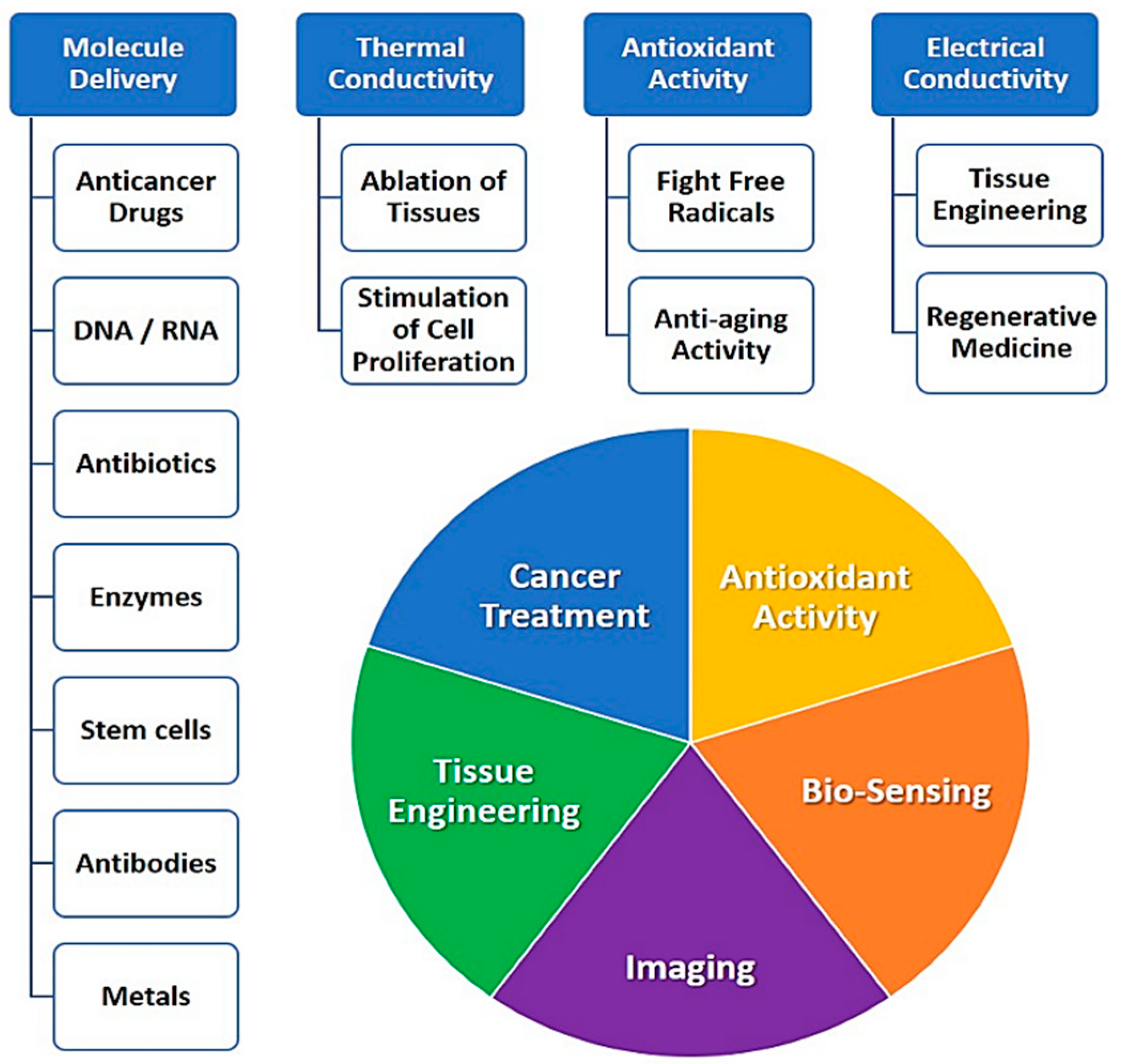
Table 1.
Main methods of carbon nanotubes (CNTs) synthesis.
| Method of CNTs Synthesis | Details | Type of CNTs | Ref. |
|---|---|---|---|
| Carbon arc-discharge method | CNTs growth on the negative end of the carbon electrode in an argon-filled vessel | Single-walled CNTs | [17] |
| Laser-ablation technique | Fabrication of CNTs using continuous wave 10.6 μm CO2-laser in the presence of metals (Ni and Co) | Single-walled CNTs | [18] |
| Chemical vapor deposition | CNTs growth was achieved at temperature 1000 C in the presence of iron-doped alumina catalyst particles | Single-walled and double-walled CNTs | [19] |
| Flame synthesis method | Co-flow diffusion flame type. Catalysts: metal nitrate + TiO2 | Multi-walled CNTs | [23] |
| Spray pyrolysis method | Synthesis of CNTs using tire pyrolysis oil as a carbon precursor with ferrocene as a catalyst at 950 °C | Multi-walled CNTs | [24] |
Table 2.
Examples of the studies on the applications of carbon nanotubes in cancer treatment.
| Type of CNTs | Model/Cell Line | CNTs Dosage | Results | References |
|---|---|---|---|---|
| Single-Walled CNTs | human breast cancer cells (MCF-7) | 12.67 and 5.49 μg/mL | Higher cytotoxic action towards cancer cells | [89] |
| Single-Walled CNTs | MDA-MB-231 human breast cancer cells | 3.125, 6.25, 12.5, and 25 μg/mL | Induction of death of cancer cells under NIR-irradiation | [73] |
| Single-Walled CNTs | A549 and NIH 3T3 cells | 4, 8, 12, 16, and 20 μg/mL | Delivery of Curcumin to cancer cells; induction death and apoptosis in cancer cells | [79] |
| Single-Walled CNTs | splenic lymphocytes | 36.8 and 123 μg/L | Delivery of Curcumin to splenic lymphocytes | [90] |
| Single-Walled CNTs | PC-3 tumor cells | 5, 15, 40 μM | Enhancement of curcumin anti-tumor activity; inhibition of cancer cells | [91] |
| Single-Walled CNTs | mice with bladder cancer | 0.1 mg SWCNT per kg body weight | Effective photothermal ablation of bladder cancer | [92] |
| Single-Walled CNTs | murine breast cancer cells | 2.5, 5, 7.5, and 10 μg/mL | Delivery of Doxorubicin to cancer cells | [93] |
| Single-Walled CNTs | human colorectal cancer cells (HT-29) | 1 mg/mL | Destruction of cancer cells under NIR-irradiation | [94] |
| Multi-Walled CNTs | HeLa cells/mice | 5 mg/kg | In vitro and in vivo cancer cell inhibition | [68] |
| Multi-Walled CNTs | MCF-7 and MDA-MB-231 human breast cancer cells/rats | 0.1, 0.2, 0.4, 0.6, 0.8, and 1 mg/mL | Effective delivery of Docetaxel and its release | [95] |
| Multi-Walled CNTs | HeLa cells | 1.67 μg/mL | Enhancement of antitumor activity of Camptothecin | [96] |
| Multi-Walled CNTs | HepG-2 cells | 1.875, 3.75, 6.25, 12.5, 25, 50, 100, and 200 μg/mL | Enhancement of antitumor activity of Oridonin | [88] |
| Multi-Walled CNTs | bladder cancer 5637 and T24 cells | 2.5, 40 µg/mL | Enhancing cytotoxicity of epirubicin and inhibiting proliferation in vitro and in vivo | [97] |
| Multi-Walled CNTs | breast cancer cells of EMT-6 mice | 2.5, 25 µg/mL | Inhibition in lymph node metastases to EMT-6 breast cancer cells | [98] |
| Multi-Walled CNTs | U87 human glioblastoma cells | 0.001–1000 μg/mL | Targeted delivery of Doxorubicin | [99] |
© 2019 by the author. Licensee MDPI, Basel, Switzerland. This article is an open access article distributed under the terms and conditions of the Creative Commons Attribution (CC BY) license (http://creativecommons.org/licenses/by/4.0/).
Share and Cite
MDPI and ACS Style
Saliev, T. The Advances in Biomedical Applications of Carbon Nanotubes. C 2019, 5, 29. https://0-doi-org.brum.beds.ac.uk/10.3390/c5020029
AMA Style
Saliev T. The Advances in Biomedical Applications of Carbon Nanotubes. C. 2019; 5(2):29. https://0-doi-org.brum.beds.ac.uk/10.3390/c5020029
Chicago/Turabian StyleSaliev, Timur. 2019. "The Advances in Biomedical Applications of Carbon Nanotubes" C 5, no. 2: 29. https://0-doi-org.brum.beds.ac.uk/10.3390/c5020029
Note that from the first issue of 2016, this journal uses article numbers instead of page numbers. See further details here.