Assessment of Antioxidant Activity of Pure Graphene Oxide (GO) and ZnO-Decorated Reduced Graphene Oxide (rGO) Using DPPH Radical and H2O2 Scavenging Assays

 , , and
Abstract
:
1. Introduction
2. Materials and Methods
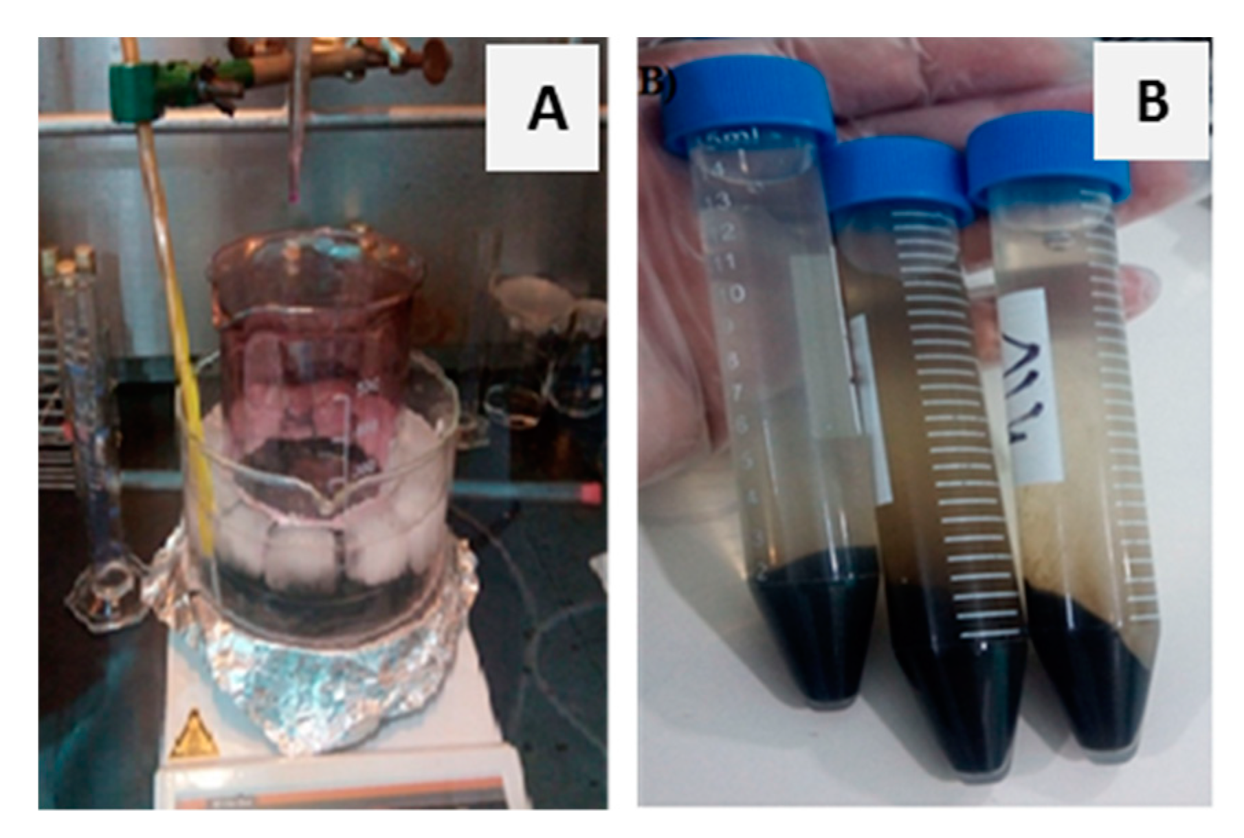
2.1. GO and ZnO–rGO Nanocomposites Synthesis and Characterization
2.2. GO and ZnO–rGO Nanocomposites Antioxidant Activity
3. Results and Discussion
3.1. Structural Characteriwations
3.1.1. XRD
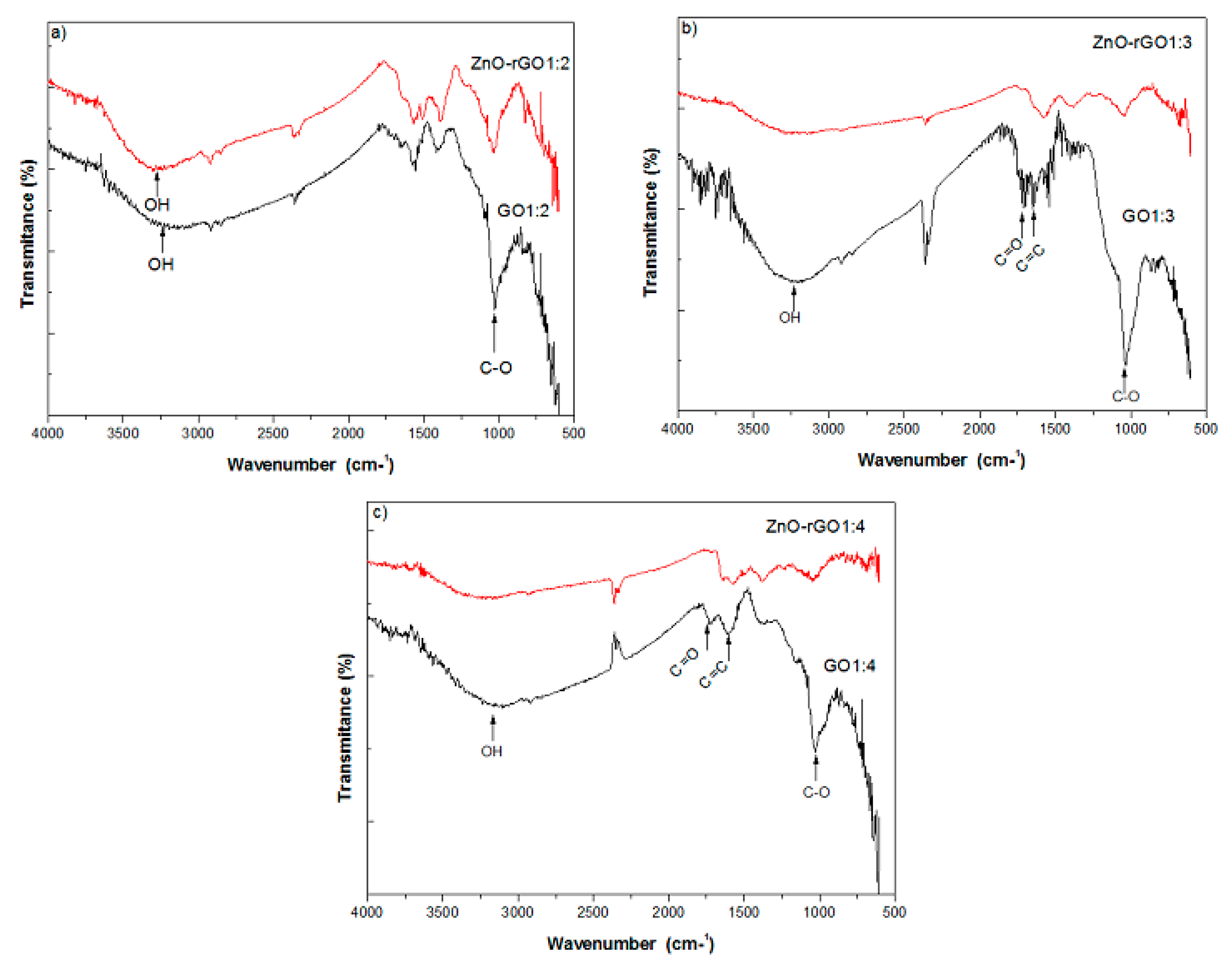
3.1.2. FTIR
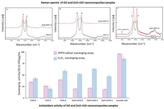
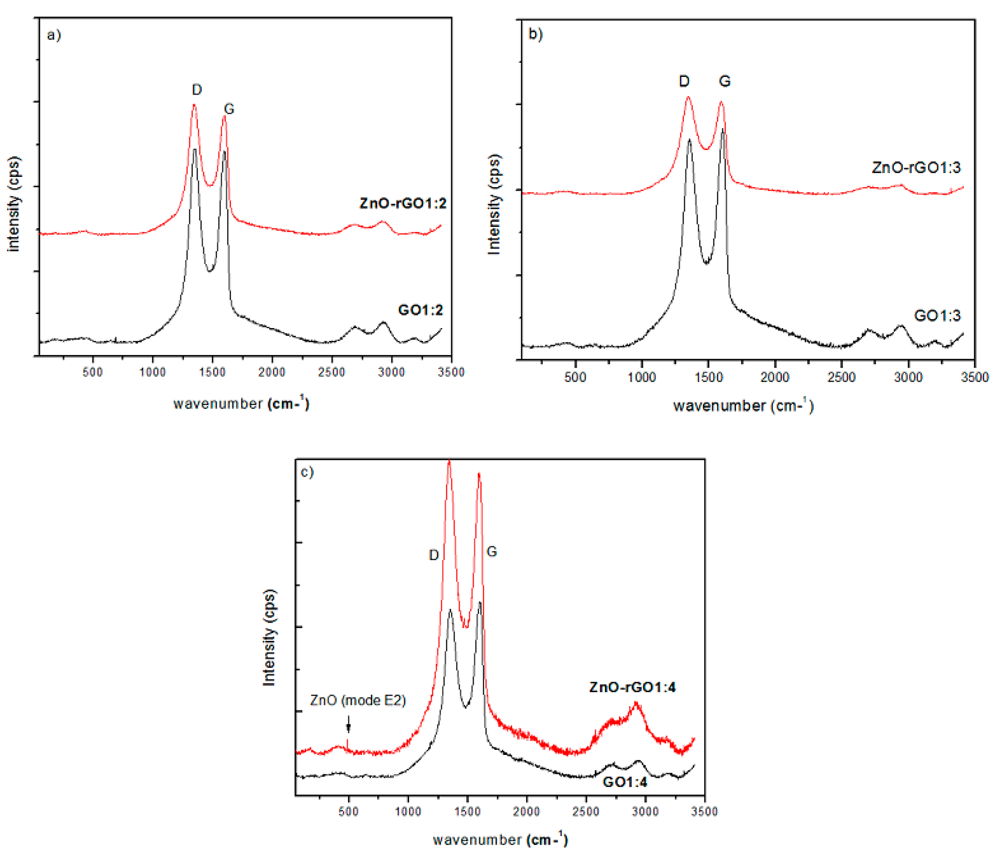
3.1.3. Raman
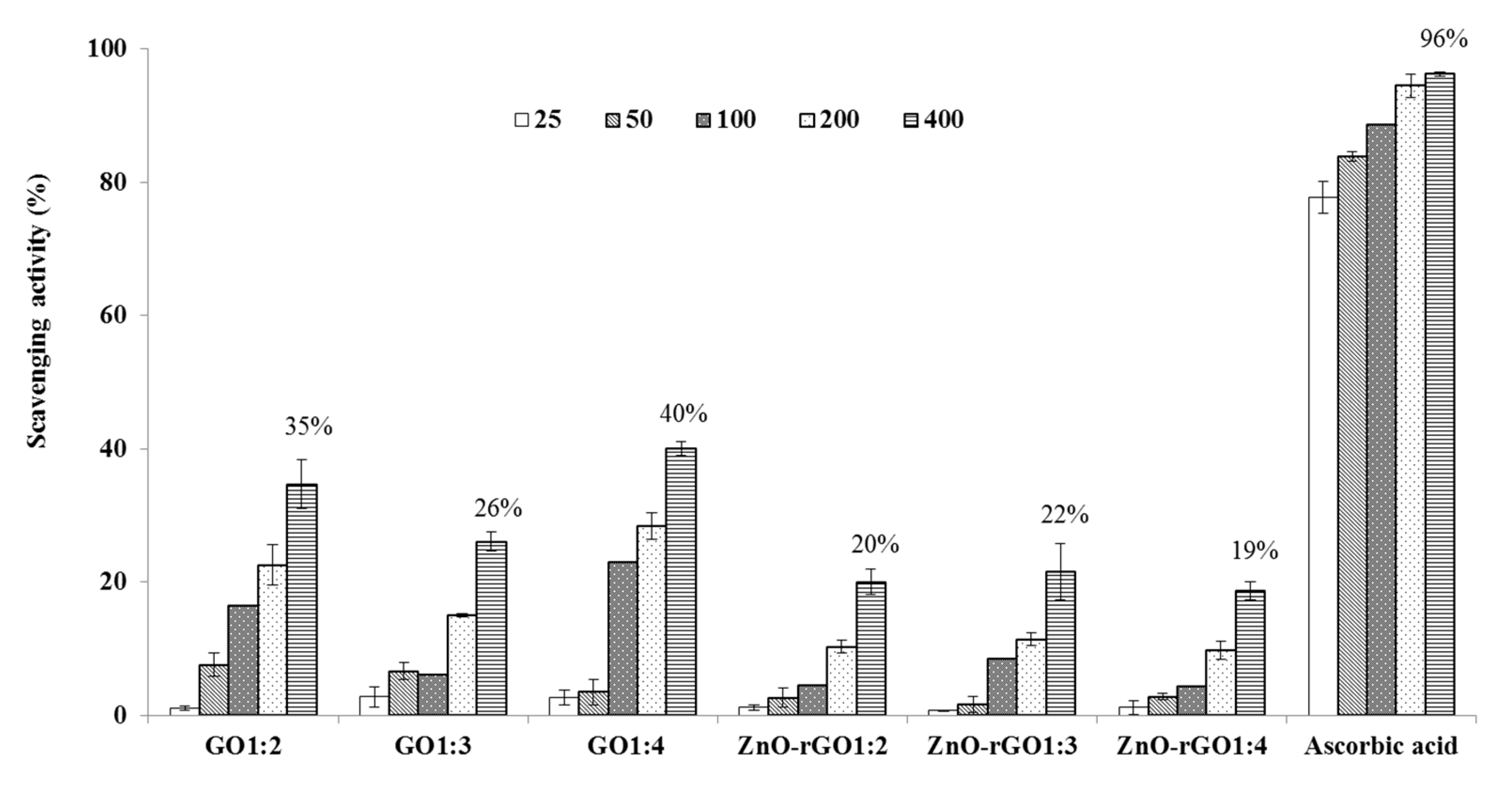
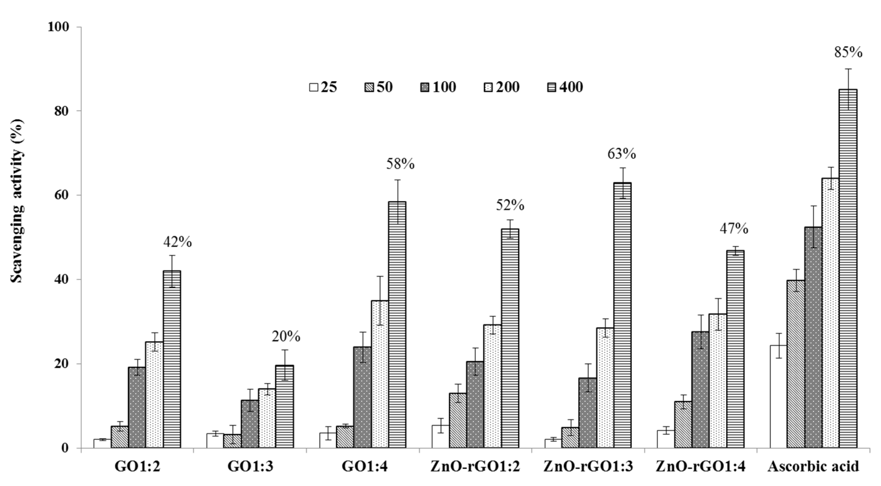
3.2. Antioxidant Assays
4. Conclusions
Author Contributions
Funding
Acknowledgments
Conflicts of Interest
References
- Novoselov, K.S.; Geim, A.K.; Morozov, S.V.; Jiang, D.; Zhang, Y.; Dubonos, S.V.; Grigorieva, I.V.; Firsov, A.A. Electric field effect in atomically thin carbon films. Science 2004, 306, 666–669. [Google Scholar] [CrossRef] [PubMed]
- Khan, M.M.; Cho, M.H. Green synthesis, photocatalytic and photoelectrochemical performance of an Au–graphene nanocomposite. RSC Adv. 2015, 5, 26897–26904. [Google Scholar] [CrossRef]
- Ali, I.; Basheer, A.A.; Mbianda, X.Y.; Burakov, A.; Galunin, E.; Burakova, I.; Mkrtchyan, E.; Tkachev, A.; Grachev, V. Graphene based adsorbents for remediation of noxious pollutants from wastewater. Environ. Int. 2019, 127, 160–180. [Google Scholar] [CrossRef] [PubMed]
- Khan, M.E.; Khan, M.M.; Cho, M.H. Recent progress of metal–graphene nanostructures in photocatalysis. Nanoscale 2018, 10, 9427–9440. [Google Scholar] [CrossRef] [PubMed]
- Tadyszak, K.; Wychowaniec, J.K.; Litowczenko, J. Biomedical applications of graphene-based structures. Nanomaterials 2018, 8, 944. [Google Scholar] [CrossRef]
- Alam, S.N.; Sharma, N.; Kumar, L. Synthesis of graphene oxide by modified Hummers method and its thermal reduction to obtain reduced graphene oxide. Graphene 2017, 6, 1–18. [Google Scholar] [CrossRef]
- Marcano, D.C.; Kosynkin, D.V.; Berlin, J.M.; Sinitskii, A.; Sun, Z.; Slesarev, A.; Alemany, L.B.; Lu, W.; Tour, J.M. Improved synthesis of graphene oxide. ACS Nano. 2010, 4, 4806–4814. [Google Scholar] [CrossRef]
- Omar, F.S.; Ming, H.N.; Hafiz, S.M.; Ngee, L.H. Microwave synthesis of zinc oxide/reduced graphene oxide hybrid for adsorption-photocatalysis application. Int J. Photoenergy 2014, 2014, 176835. [Google Scholar] [CrossRef]
- Parnianchi, F.; Nazari, M.; Maleki, J.; Mohebi, M. Combination of graphene and graphene oxide with metal and metal oxide nanoparticles in fabrication of electrochemical enzymatic biosensors. Int. Nano Lett. 2018, 8, 229–239. [Google Scholar] [CrossRef]
- Papageorgiou, D.G.; Kinloch, I.A.; Young, R.J. Mechanical properties of graphene and graphene-based nanocomposites. Prog. Mater. Sci. 2017, 90, 75–127. [Google Scholar] [CrossRef]
- Khan, M.E.; Khan, M.M.; Cho, M.H. Biogenic, synthesis of Ag–graphene nanocomposite with efficient photocatalytic degradation, electrical conductivity and photoelectrochemical performance. New J. Chem. 2015, 39, 8121–8129. [Google Scholar] [CrossRef]
- Siddeswara, D.M.K.; Vishnu Mahesh, K.R.; Sharma, S.C.; Mylarappa, M.; Nagabhushana, H.; Ananthraju, K.S.; Nagaswarupa, H.P.; Prashantha, S.C.; Raghavendra, N. ZnO decorated graphene nanosheets: An advanced material for the electrochemical performance and photocatalytic degradation of organic dyes. Nanosyst. Phys. Chem. Math. 2016, 7, 678–682. [Google Scholar] [CrossRef]
- Durmus, Z.; Kurt, B.Z.; Durmus, A. Synthesis and characterization of graphene oxide/zinc oxide (GO/ZnO) nanocomposite and its utilization for photocatalytic degradation of basic Fuchsin dye. ChemistrySelect 2019, 4, 271–278. [Google Scholar] [CrossRef]
- Shadmehri, A.A.; Namvar, F.; Miri, H.; Yaghmaei, P.; Moghaddam, M.N. Assessment of antioxidant and antibacterial activities of zinc oxide nanoparticles, graphene and graphene decorated by zinc oxide nanoparticles. Int. J. Nano Dimens. 2019, 10, 395–403. [Google Scholar]
- Blois, M.S. Antioxidant determinations by the use of a stable free radical. Nature 1958, 181, 1199–1200. [Google Scholar] [CrossRef]
- Ruch, R.J.; Cheng, S.J.; Klaunig, E. Prevention of cytotoxicity and inhibition of intercellular communication by antioxidant catechins isolated from Chinese green tea. Carcinogenesis 1989, 10, 1003–1008. [Google Scholar] [CrossRef]
- Farhanini, Y.; Khing, N.T.; Hao, C.C.; Sang, L.P.; Basyirah, M.N.; Noorashikin, M.S. The electrochemical behavior of zinc oxide/reduced graphene oxide composite electrode in dopamine. Malays. J. Anal. Sci. 2018, 22, 227–237. [Google Scholar]
- Moon, I.K.; Lee, J.; Ruoff, R.S.; Lee, H. Reduced graphene oxide by chemical graphitization. Nat. Commun. 2010, 1, 73. [Google Scholar] [CrossRef]
- Kim, K.S.; Rhee, K.Y.; Park, S.J. Influence of multi-walled carbon nanotubes on the electrochemical performance of transparent graphene electrodes. Mater. Res. Bull. 2011, 46, 1301–1306. [Google Scholar] [CrossRef]
- Zhong, X.; Jin, J.; Li, S.; Niu, Z.; Hu, W.; Li, R.; Ma, J. Aryne cycloaddition: Highly efficient chemical modification of graphene. Chem. Commun. 2010, 46, 7340–7734. [Google Scholar] [CrossRef]
- Lo, S.S.; Huang, D.; Tu, C.H.; Jan, D.J. Formation and Raman scattering of seed-like ZnO nanostructure. J. Raman Spectrosc. 2009, 40, 1694–1697. [Google Scholar] [CrossRef]
- Qiu, Y.; Wang, Z.; Owens, A.C.; Kulaots, I.; Chen, Y.; Kane, A.B.; Hurt, R.H. Antioxidant chemistry of graphene-based materials and its role in oxidation protection technology. Nanoscale 2014, 6, 11744–11755. [Google Scholar] [CrossRef] [PubMed]
- Lee, Y.M.; Yoon, Y.; Yoon, H.; Song, S.; Park, H.M.; Lee, Y.Y.; Shin, H.; Hwang, S.W.; Yeum, K.J. Enhanced antioxidant activity of bioactives in colored grains by nano-carriers in human lens epithelial cells. Molecules 2018, 23, 1327. [Google Scholar] [CrossRef] [PubMed]
- Rajeswari, R.; Prabu, H.G. Synthesis characterization, antimicrobial, antioxidant, and cytotoxic activities of ZnO nanorods on reduced graphene oxide. J. Inorg. Organomet. Polym. 2018, 28, 679–693. [Google Scholar] [CrossRef]
- Suresha, D.; Pavan Kumarb, M.A.; Nagabhushan, H.; Sharma, S.H. Cinnamon supported facile green reduction of grapheme oxide, its elimination and antioxidant activities. Mater. Lett. 2015, 151, 93–95. [Google Scholar] [CrossRef]


| Samples | GO1:2 | GO1:3 | GO1:4 |
|---|---|---|---|
| 2θ (°) | 11.92 | 11.47 | 11.18 |
| d (Å) | 07.40 | 07.70 | 07.90 |
© 2019 by the authors. Licensee MDPI, Basel, Switzerland. This article is an open access article distributed under the terms and conditions of the Creative Commons Attribution (CC BY) license (http://creativecommons.org/licenses/by/4.0/).
Share and Cite
Baali, N.; Khecha, A.; Bensouici, A.; Speranza, G.; Hamdouni, N. Assessment of Antioxidant Activity of Pure Graphene Oxide (GO) and ZnO-Decorated Reduced Graphene Oxide (rGO) Using DPPH Radical and H2O2 Scavenging Assays. C 2019, 5, 75. https://0-doi-org.brum.beds.ac.uk/10.3390/c5040075
Baali N, Khecha A, Bensouici A, Speranza G, Hamdouni N. Assessment of Antioxidant Activity of Pure Graphene Oxide (GO) and ZnO-Decorated Reduced Graphene Oxide (rGO) Using DPPH Radical and H2O2 Scavenging Assays. C. 2019; 5(4):75. https://0-doi-org.brum.beds.ac.uk/10.3390/c5040075
Chicago/Turabian StyleBaali, Nacera, Assia Khecha, Aicha Bensouici, Giorgio Speranza, and Noudjoud Hamdouni. 2019. "Assessment of Antioxidant Activity of Pure Graphene Oxide (GO) and ZnO-Decorated Reduced Graphene Oxide (rGO) Using DPPH Radical and H2O2 Scavenging Assays" C 5, no. 4: 75. https://0-doi-org.brum.beds.ac.uk/10.3390/c5040075