Article

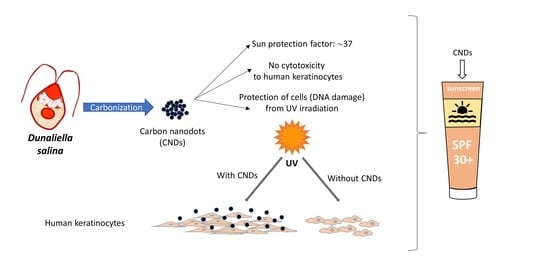
Carbon Nanodots Synthesized from Dunaliella salina as Sun Protection Filters

by
Theodoros G. Chatzimitakos
1,
Athanasia Kasouni
2,
Anastassios Troganis
2,
Ioannis Leonardos
2,
Ioannis Tzovenis
3,
Alexandros Ntzouvaras
3 and
Constantine Stalikas
1,*
1
Laboratory of Analytical Chemistry, Department of Chemistry, University of Ioannina, 45110 Ioannina, Greece
2
Department of Biological Applications and Technologies, University of Ioannina, 45110 Ioannina, Greece
3
Department of Ecology and Systematics, Faculty of Biology, National and Kapodistrian University of Athens, Panepistimiopolis Zografou, 15701 Athens, Greece
*
Author to whom correspondence should be addressed.
Submission received: 29 September 2020 / Revised: 24 October 2020 / Accepted: 27 October 2020 / Published: 30 October 2020
(This article belongs to the Collection Feature Papers in the Science and Engineering of Carbons)

Abstract

:
Carbon nanodots (CNDs) are more and more being exploited for various applications including biological ones. To this end, they have been thoroughly studied for their potential as antibacterial, wound healing, and bioimaging agents. In this study, we examined the sun protection properties of CNDs. Dunaliella salina was selected as a promising precursor for the synthesis of CNDs which were compared with those produced by citric acid, a widely used precursor for such materials. The CNDs were examined spectrophotometrically, and the sun protection factors were calculated. Additionally, in vitro experiments were carried out to evaluate their UV protection properties and to obtain better insight into whether CNDs are suitable to be used as filters for the development of new sunscreens. The results were conclusive that both CNDs possess favorable properties that potentiate their use for the development of sunscreens. However, the CNDs from Dunaliella salina were found to be superior to those derived from citric acid. Therefore, they can further be exploited as sun protection filters.

Graphical Abstract

1. Introduction

It is well known that exposure to sunlight has many benefits for human health. However, overexposure has been associated with various skin problems, such as sunburn, accelerated skin aging [1], even cancer [2]. This is the reason that the World Health Organization has classified ultraviolet light as carcinogenic [3]. Although the most damaging type of irradiation for the skin is UV-C (280–200 nm), it poses no threat, since it is filtered from the ozone layer [4]. Among the other two types, UV-B (320–280 nm) is the most dangerous, since it has almost 40 times higher energy than the UV-A (400–320nm) type and thus, its erythemal power is a thousand times as high as that of UV-A [1,5].
The use of sunscreens has been proved to protect the skin from UV damages. Many compounds are being synthesized and examined in terms of their UV absorbance properties. These substances fall between two main categories: inorganic photoactive compounds and organic UV absorbers [6]. The organic compounds are often toxic and exhibit low photostability, while the inorganic analogs have a higher agglomeration tendency at concentrations needed to achieve sufficient protection. Therefore, the demand for new compounds that can be used in sunscreens is increasing. Such compounds must have good UV absorption properties and photostability, negligible toxicity, and must be environmentally friendly, while, ideally, they should combine more than one beneficial activity. To this end, many efforts have been made (and their number still increases) to develop new sunscreens, using compounds from natural resources, such as plant extracts [1,5,7,8]. This is due to many activities that plant extracts exhibit, including antioxidant, antibacterial, etc.
Carbon nanodots (CNDs) are a type of nanomaterial that can potentially combine all the above favorable properties and present unique optical properties, among the carbon-based nanomaterials. Many studies have been carried out in the last few years, demonstrating their fluorescent properties (emission is generated, usually, after excitation at the UV region), photostability, negligible toxicity towards eukaryotic cells, and environmental friendliness [9,10,11]. Additionally, CNDs have been found to possess other non-fluorescent properties, such as antibacterial, anticancer, wound healing etc. [12,13]. In our laboratory, CNDs from human fingernails have been used to produce CNDs that exhibit wound healing properties [14]. In another study, we proved clearly that nitrogen-doped and nitrogen and sulfur co-doped CNDs exhibit antibacterial properties [15]. Based on the above, CNDs exhibit fluorescence, by absorbing UV light, and have many other favorable properties. Altogether, that can be put to good use to produce multifunctional sunscreens.
This study aims to explore the potential in vitro sun protection properties of CNDs. To this end, CNDs from two different sources were synthesized and compared. For the one kind of CNDs, a well-established synthesis based on the hydrothermal treatment of citric acid was employed. The other kind of CNDs was produced using a carbonization procedure and Dunaliella salina as the carbon-based biomass. Strains of the green flagellate Dunaliella salina (Dunal) Teodoresco 1905 (Chlorophyta, Chlamydomonadales) are usually found in hypersaline environments [16]). This organism is very efficiently adapted to such extreme environmental conditions rendering it a valuable species for producing high added value biomolecules. It accumulates glycerol as an osmoregulator at high salinity, and β-carotene at high UV radiation to protect its photosynthetic machinery from stress [17]. These molecules not only are in abundance in Dunaliella salina but also exhibit better biological activities, compared to their synthetic analogs [18]. Moreover, the mix of components present in the microalgae results in superior activities, compared with single components, highlighting the potential of microalgae for the development of novel cosmetics for skin [19,20]. The sun protection factors of the as-synthesized CNDs were calculated and compared to obtain a better insight into whether the CNDs possess sun protection properties, or the properties are dependent on the precursor material. The suitability of the CNDs was further examined by an in vitro toxicity and a UV protection study.

2. Materials and Methods

2.1. Chemicals

Crystal violet was obtained from Merck (Darmstadt, Germany). Fetal bovine serum (FBS), Dulbecco’s Modified Eagle’s medium (DMEM), antibiotics (50 units mL−1 penicillin and 50 μg mL−1 streptomycin), phosphate buffer saline (PBS), formaldehyde solution (37% w/v), Ribonuclease type A, trypsin solution 0.05%, were obtained from Sigma Aldrich (Steinheim, Germany). Propidium iodide staining solution (1.0 mg mL−1 in water) was obtained from Biotium (Fremont, California). Double distilled water (DDW) was used throughout the experiments. The solvents used were of HPLC grade. The crystal violet solution used for the estimation of cell number consisted of 0.4% w/v crystal violet, 5 mL of 37% w/v formaldehyde solution, 0.25 g sodium chloride, 50 mL methanol, and 45 mL DDW. N-replete Conway medium (1 L) consisted of: Na2EDTA 0.045g; H3BO3 0.0336g; KNO3 0.116g; NaH2PO4.2H2O 0.02g; MnCl2.4H2O 0.00036g; FeCl3.6H2O 0.0013g; ZnCl2 0.0021g; CoCl2.6H2O 0.002g; (NH4)6Mo7O2.4H2O 0.0009g; CuSO4.5H2O 0.002g and concentrated HCl 0.01mL; Conway medium vitamin stock consisted of 200 mg thiamine hydrochloride and 10 mg cyanocobalamin in 100 mL of deionized water [21].

2.2. Instruments

A Perkin Elmer Spectrum (Two FTIR PerkinElmer, Waltham, MA, USA) was used to record FTIR spectra (of powdered samples), using an attenuated total reflectance accessory. High resolution-transmission electron microscopy (HR-TEM) images were obtained with a JEOL JEM-2100 microscope (Jeol SAS, Croissy-Sur-Seine, France) operated at 200 kV equipped with LaB6 filament. Samples for HR-TEM measurements were prepared by depositing a drop of CNDs dispersion on carbon-coated copper grids and drying, at room temperature. The UV-Vis absorption spectra of CNDs solutions were recorded on a Lambda 35 UV-Vis spectrometer (Perkin Elmer, Rodgau, Germany). All fluorescence measurements and photostability experiments of CNDs solutions were performed on an RF 5301 PC spectrofluorometer (Shimadzu, Kyoto, Japan) in a quartz cell, with 1-cm pathlength and excitation and emission slits set at 5 nm bandpass. Lyophilization of the samples was carried out by an Alpha 1-4 LD freeze-dryer (Christ, Osterode, Germany). Digital images were captured with an Infinity 2-2 digital camera (Lumenera, Danville, Canada), mounted on an Olympus BX43 microscope.

2.3. Dunaliella Salina Culture Conditions

The strain of Dunaliella salina used was Duna 32 (Athual, strain bank, University of Athens, Athens, Greece), isolated from the Greek salt works of Megalo Embolo (Aegean Sea, Angellochori, North Greece). It was cultivated in quadruplicate batches of 5 L volume of mildly aerated (0.5 vvm) synthetic seawater (Tropical Marine™ salts dissolved in deionized water, (Wartenberg, Germany) of 120 ppt salinity at a temperature of 26 ± 1 °C, a photoperiod of 12:12 h L: D, 100 μmol photons m−2·s−1. The seawater was enriched with 1mL N-replete Conway medium and 0.1 mL Conway medium vitamin stock [21]. The strain was cultivated until senescence (at least 20 days).

2.4. Synthesis of CNDs

For the synthesis of CNDs from Dunaliella salina (CNDs-DS), 30 mg dried algal biomass was placed into a crucible and heated into a pre-heated oven for 2 h at 250 °C. After cooling down to room temperature, water was added to the black powder and the mixture was ultrasonicated for 5 min. Then, the mixture was centrifuged at 3500 rpm for 5 min and the supernatant was collected. The procedure was repeated until the collected supernatant exhibited no fluorescence. The collected supernatants were filtered through 0.20 μm syringe filters and then freeze-dried. The obtained powder (~12.8 mg; synthetic yield: 38.7%) was stored in the dark, at room temperature.
The citric acid-based CNDs (CNDs-CA) were synthesized by placing 0.20 g of citric acid in a stainless-steel Teflon-lined autoclave and heating in an oven at 200 °C, for 3 h. After cooling to room temperature, double-distilled water was added, and the solution was centrifuged at 8000 rpm for 10 min. The supernatant was collected, filtered through 0.20 μm syringe filters, and then freeze-dried. The obtained powder (~114 mg; synthetic yield: 57%) was stored in the dark, at room temperature.

2.5. Sun Protection Factor (SPF) Calculation

To determine the SPF of the CNDs, the UV spectrum of the CNDs was recorded between 290 and 320 nm. SPFs were calculated using Mansur’s Equation (1), based on previous studies [1,2].
S P F = C F × 290 320 E E ( λ ) × I ( λ ) × A B S ( λ )
where CF is the correction factor (=10), EE(λ) is the erythemogenic effect of radiation at wavelength λ, I(λ) is the solar light intensity at wavelength λ and ABS(λ) is the absorbance of the tested solution at a wavelength λ. The EE(λ) × I(λ) values are constant according to Sayre et. al. [22].

2.6. In Vitro Toxicity Study

The toxicity study was carried out, according to our previous publications [14,23,24]. In brief, HaCaT cells were seeded into 24-well plates and incubated overnight. Different concentrations of CNDs (100, 200, 400, 600, and 800 μg mL−1) were added to each well. Cells incubated in the absence of CNDs were taken as control. In all cases, three parallel samples were prepared for each tested concentration. After incubating for 24 h, the culture supernatant was removed, and cells were washed with PBS. Cell viability was assessed using the crystal violet assay. The optical absorbance was measured at 570 nm and the % cell viability was calculated using the formula:
% cell viability = (ODtreated/ODnon-treated) ∗ 100
where % cell viability represents the number of alive cells, ODnon-treated represents the absorbance of control cells and ODtreated represents the absorbance of the cells treated with CNDs.

2.7. In Vitro UV Protection

HaCaT cells were seeded in 24-well plates and incubated overnight. After complete adhesion of cells in the plate surface, 200 μg mL−1 CNDs were added and cells were incubated overnight. Next, the medium was removed, and cells were washed twice with PBS. A small amount of PBS was added so that a thin layer of PBS was above cells, and cells were irradiated using a UV lamp in the range 280–340 nm (peak emission at 314 nm). The dose was 60 mj/cm2. Next, the PBS was replaced with fresh medium, and cells were left to incubate for 24 h. After incubation, the cells were counted following the crystal violet assay, according to our previous study [25].

2.8. Cell Cycle Analysis

HaCaT cells were treated with CNDs as described in Section 2.7. After irradiation, cells were harvested, and their DNA content was analyzed by flow cytometry according to our previous study [14].

2.9. Statistical Analysis

All assays were replicated three times and p values lower than 0.05 were considered to be statistically significant. Results were expressed as mean ± standard deviation and the levels of significance between samples were compared by one-way ANOVA using Student’s t-test, as a post-hoc test for comparison of means. Statistically significant differences at p < 0.05, p < 0.01 and p <0.001 are denoted in Figures with *, ** and ***, respectively.

3. Results and Discussions

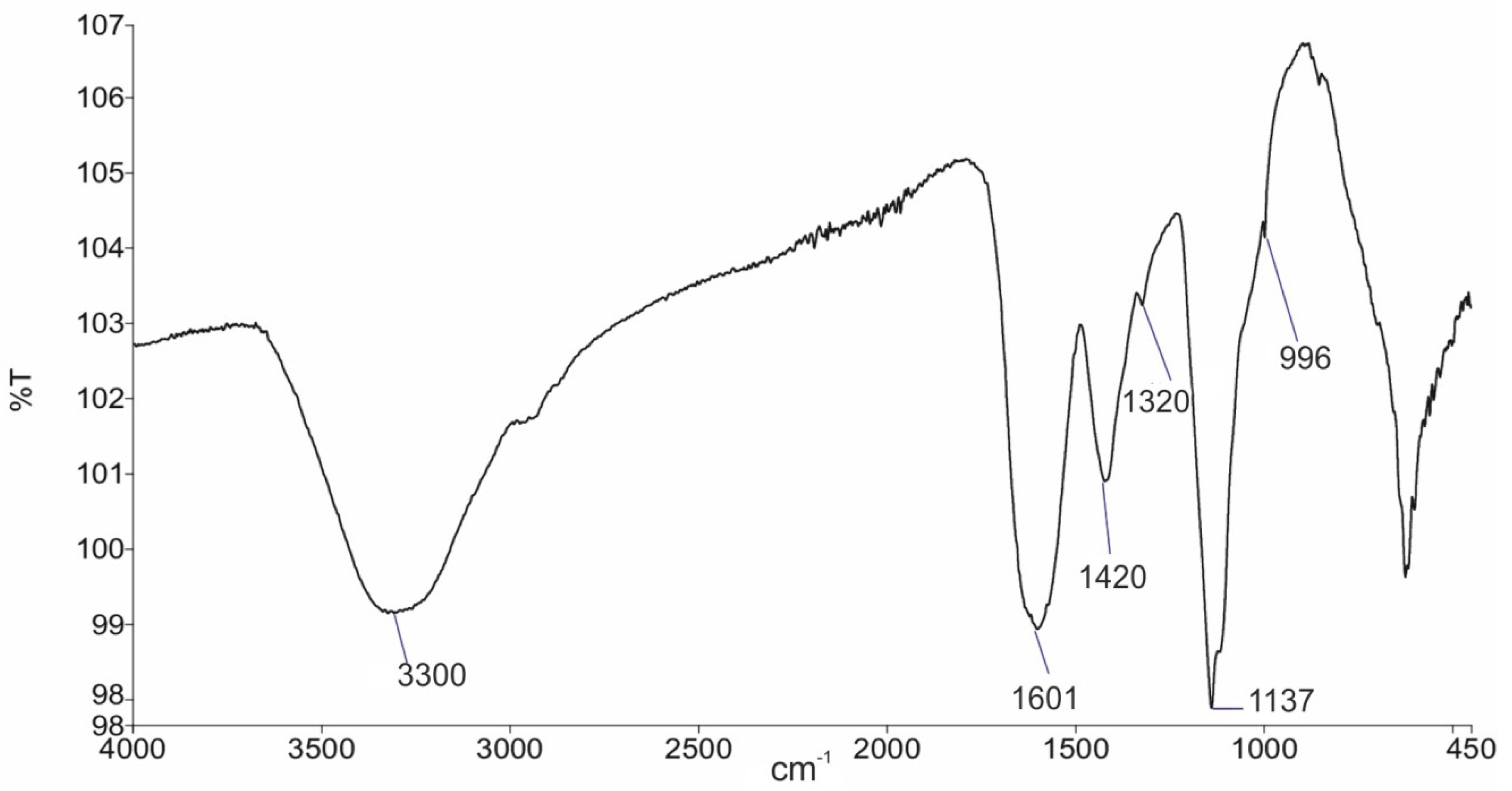
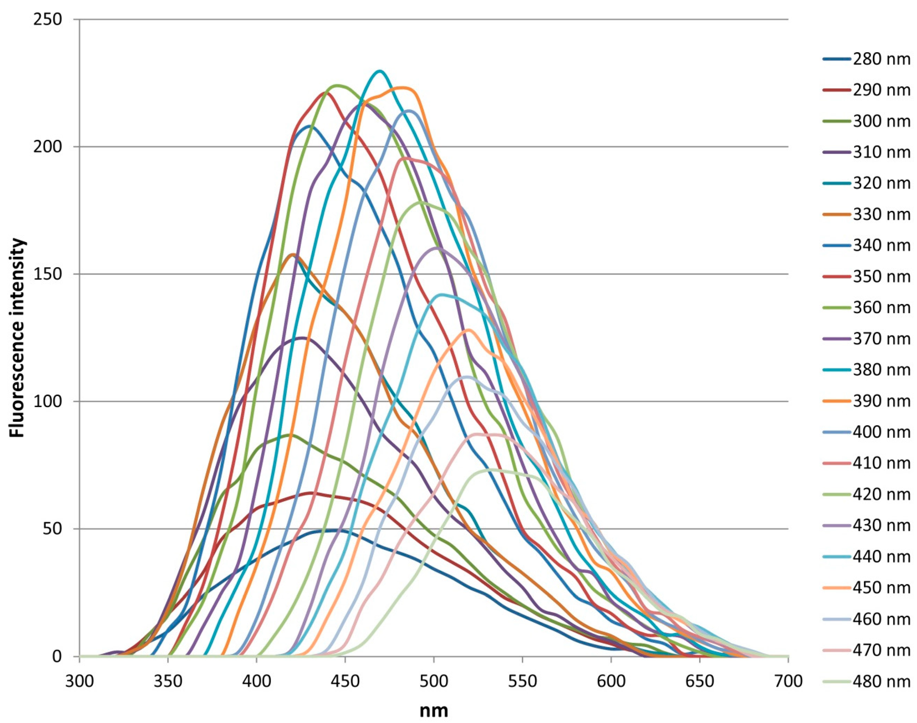
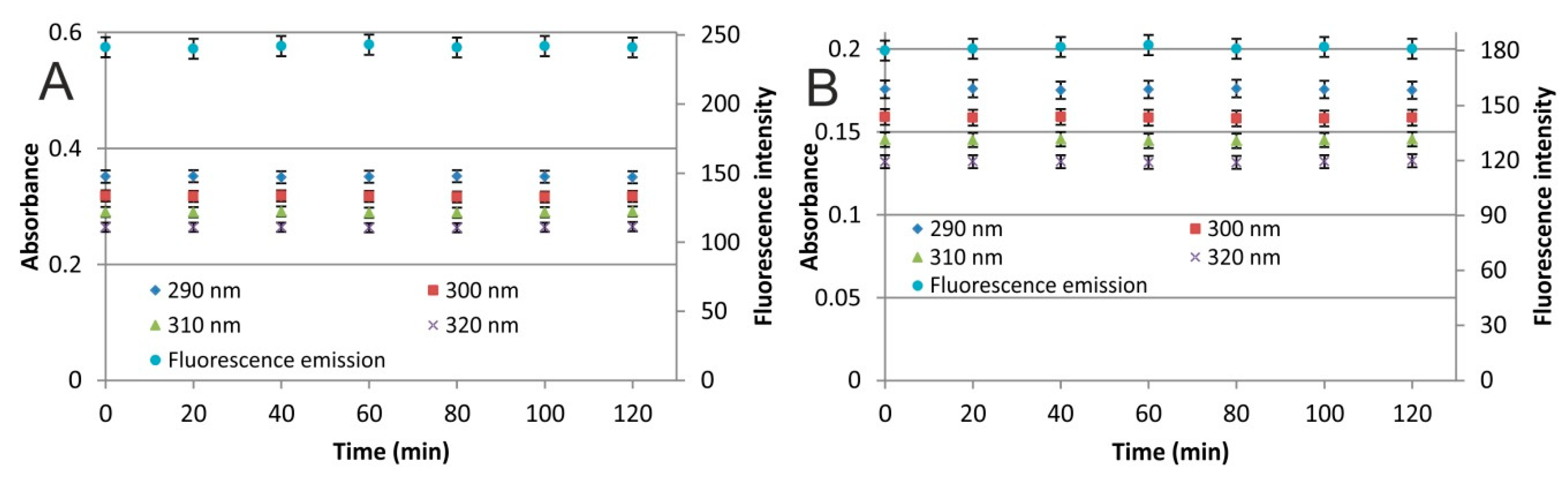
3.1. CNDs Characterization and Optical Properties
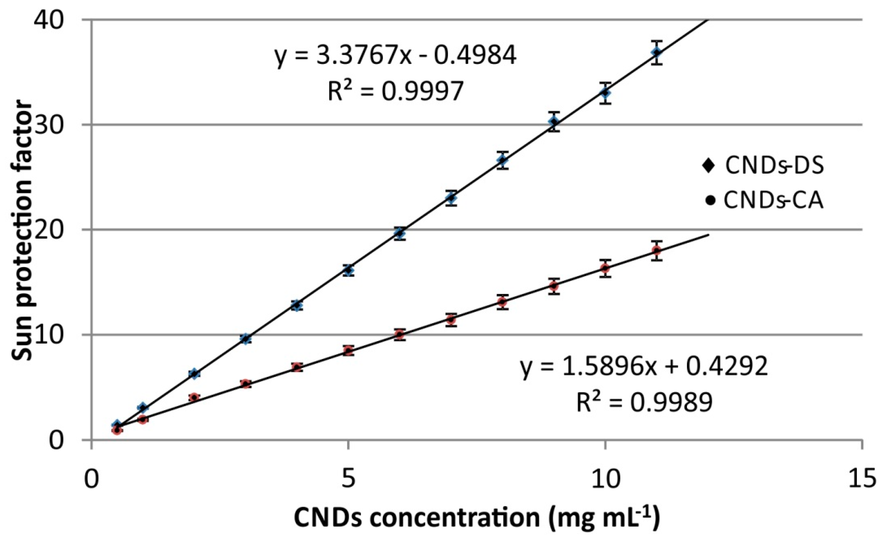
Detailed characterization data of the citric acid-based CNDs can be found in our previous study [15]. As regards the Dunaliella salina-based CNDs, Figure 1 depicts their FTIR spectrum. The broad absorption band around 3300 cm−1 is characteristic of O–H stretching vibrations. The broad peak at 2930 cm−1 is attributed to the stretching vibration of sp3 C–H moieties. Peaks attributed to C=C stretching and –COO– asymmetric and symmetric stretching can be seen at 1600 and 1420 cm−1, respectively [24,26,27]. The peaks at 1137 and the shoulder at 996 cm−1 can be ascribed to C–O, and C=O stretching vibrations [28,29]. As can be seen in Figure 2, the average size of CNDs-DS is between 3.0 and 3.5 nm, which is typical of CNDs structure.
Figure 3 shows the UV-Vis spectrum of the CNDs-DS. A weak shoulder can be seen at ~270 nm. This can be attributed to the n-π* and π-π* transitions of the –C-O bonds, from carboxyl groups, or to the π-π* transitions or aromatic –C=C bonds [23,24,30,31]. The fluorescence emission spectra of the CNDs-DS at excitation wavelengths between 280 and 480 nm are illustrated in Figure 4. Maximum fluorescence emission was recorded at 470 nm after excitation at 380 nm. The fluorescence emission was found to be excitation dependent, since emission shifts from 400 to 550 nm, as the excitation wavelength increases. The photostability of CNDs was examined by recording the UV-Vis absorbance and the fluorescence emission intensity (for excitation at 300 nm) after 2 h of irradiation with a 150W Xenon lamp. The photostability was examined in the UV-B region (290–320 nm) because as potential sun protection filters they should be stable after irradiation in this region. The CNDs were found to be stable with respect to their optical properties rendering them good candidates for use in sunscreens (Figure 5).

3.2. SPF of CNDs

To quantitatively assess the effectiveness of a sunscreen formulation, the SPF factor was calculated. The absorbance values of CNDs solutions (concentrations between 0.5 and 11 mg mL−1) for both CNDs species were recorded between 290 and 320 nm (with an increment of 5 nm). Using the Mansur equation, the SPFs were calculated and can be seen in Figure 6. According to the results, a solution with an SPF of ~37 can be prepared using 11 mg mL−1 (1.1% w/v) CNDs-DS. A solution containing the same concentration of CNDs-CA has an SPF of 18. As can be seen from Figure 6, in all tested concentrations the CNDs-CA exhibit nearly two times lower SPF compared to CNDs-DS. This signifies that although both CNDs have sun protection properties, their properties are dependent on other parameters, such as the precursors employed and the synthesis procedure. Since better results were obtained from the CNDs-DS, further experiments were carried out using this material. Until now, there have been only two reports dealing with the use of CNDs to enhance the UV protection properties of other materials. In the first study, the authors examined various CNDs as an additive to PVA films and found that they can enhance their UV properties [32]. The CNDs were synthesized from citric acid, ethylene glycol, and N,N’-bis(2-aminoethyl)-1,3-propanediamine, and a 0.7% w/w addition of CNDs in the PVA films resulted in an SPF of 22. In the second study, authors functionalized cotton fabrics with CNDs from citric acid, ethylenediamine, and borax [33] and the resulting fabrics exhibited an SPF of 28 (the bare fabrics exhibited an SPF of 9).
As stated in the introduction, many natural resources are also being studied to extract compounds with UV protection properties. Each of them has its pros and cons. For instance, the ink from sepia exhibits much higher SPF compared with the examined CNDs. However, the formulations containing eumelanin (deriving from melanin from sepia ink) have not been put on the market, due to aesthetic reasons (sunscreens have an unpleasant dark color) [20]. The CNDs-DS solution exhibits a pale-yellow color, as can be seen from the UV-Vis spectrum, and this could be aesthetically more pleasant, compared with a dark-colored product. Hence, CNDs are considered as a propitious material for sun protection applications. However, the use of Dunaliella salina is more favorable since it is a low-cost, renewable material. Taking into consideration that most sunscreens have a content of 10% w/v in nanoparticles, the superiority of CNDs-DS as sunscreen additives is further strengthened since a 10 times lower content of CNDs-DS can be used to produce sunscreens with high SPF. The reduction in the mass of nanoparticles used in sunscreens can have many benefits, such as reduction of the cost and avoidance of potential side effects (caused by a high concentration of nanomaterials).

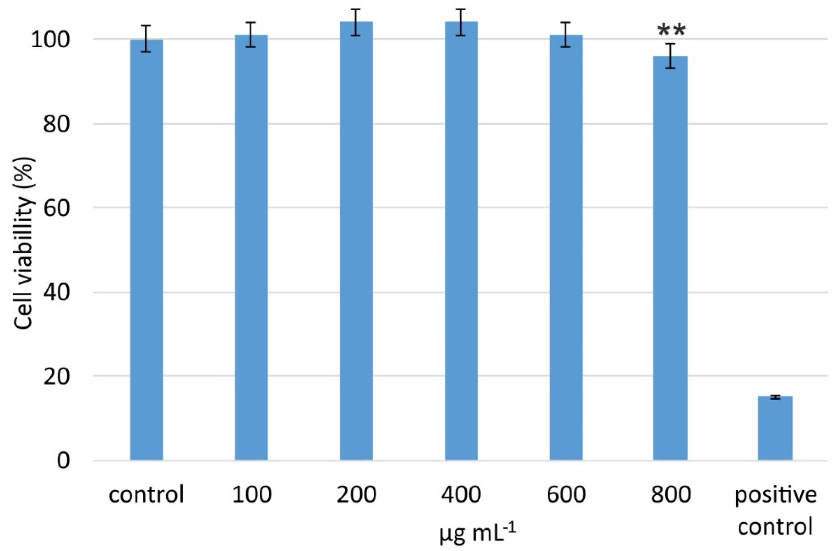
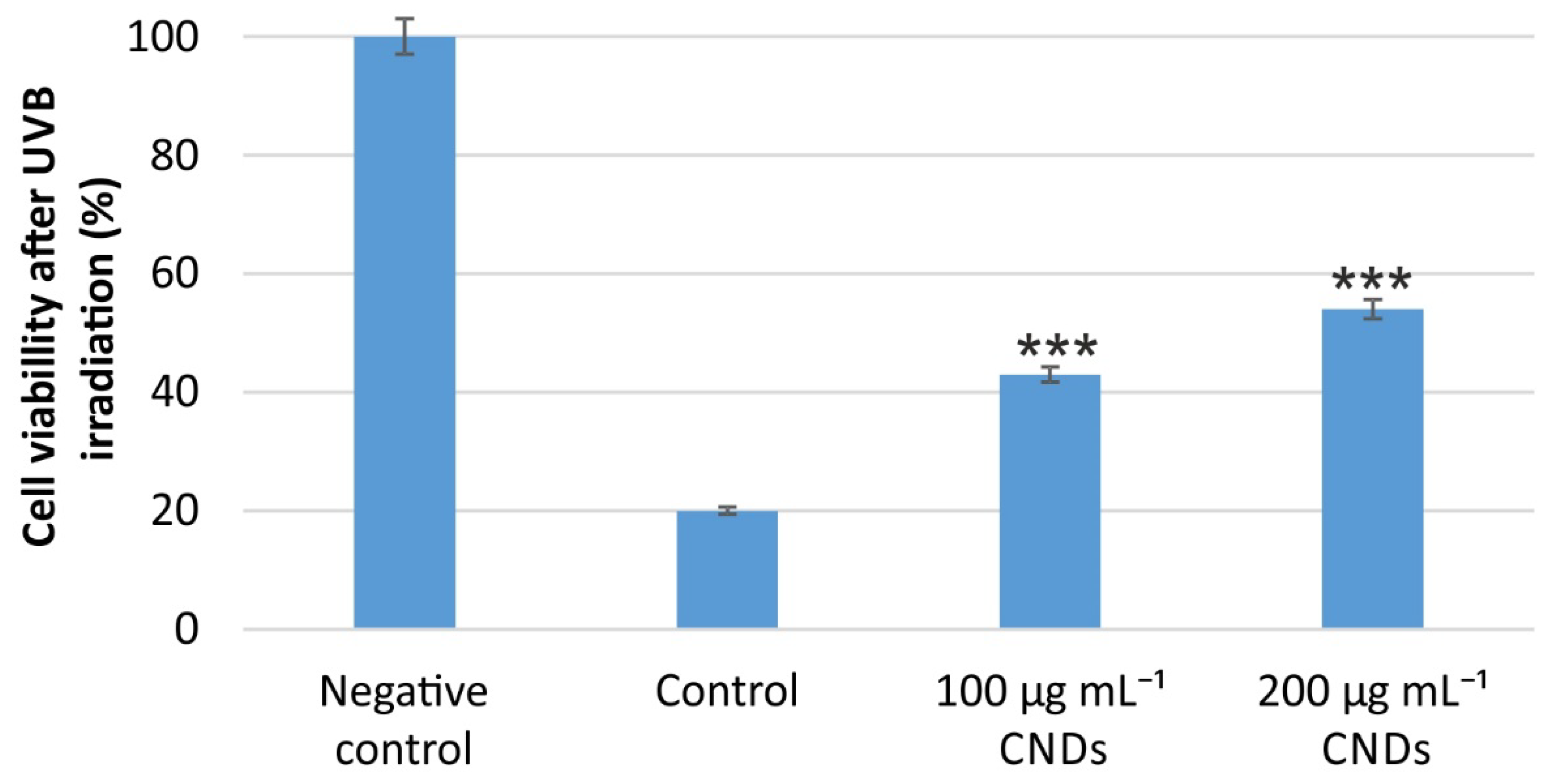
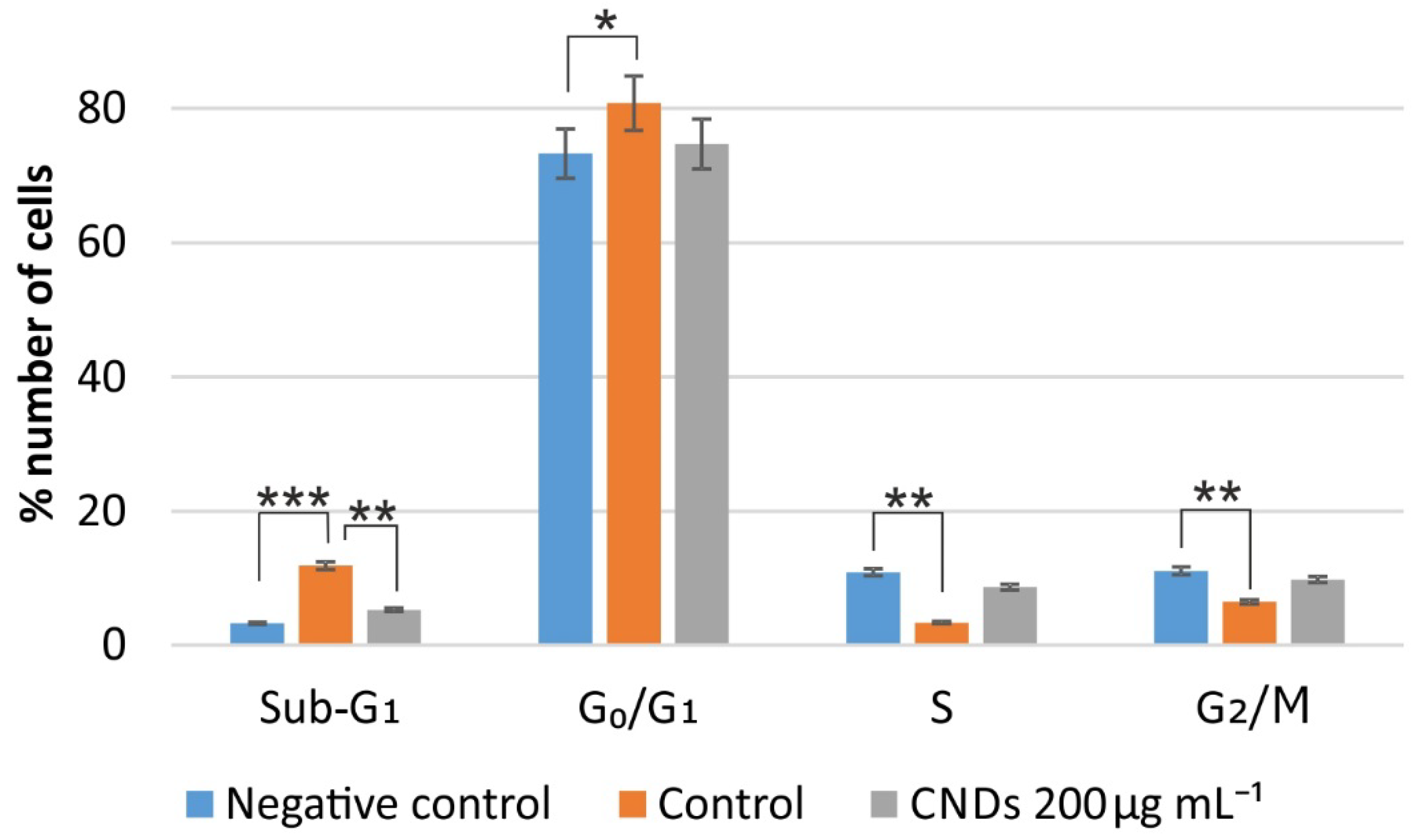
3.3. In Vitro Assessment of CNDs Toxicity, UV Protection, and Cell Cycle

Despite their favorable UV absorption properties, CNDs will be of no use if they are toxic towards eukaryotic cells. In this context, we examined the cell viability of HaCaT keratinocyte cells, after treatment with different concentrations of CNDs-DS, as can be seen in Figure 7. Although the use of normal human epidermal keratinocytes over HaCaT cells (spontaneously immortalized aneuploid human keratinocytes) is still controversial, the use of the latter is highly accepted, as they offer more reproducible results, compared with the former [34,35]. It is obvious that CNDs-DS are non toxic to the cells at concentrations in the range of 100–600 μg mL−1.
Next, we evaluated the potency of CNDs-DS to protect cells from UV irradiation. Although the spectrophotometrically calculated SPF was promising, it is of high importance to evaluate the in vitro effect of the CNDs. For this reason, cells were firstly treated with the CNDs-DS, so that they could enter cell cytoplasm and then they were irradiated with UV light. The cell viability was measured and compared with that of control cells, which were irradiated without any pre-treatment with CNDs-DS. As can be seen in Figure 8, a pre-treatment with 100 μg mL−1 of CNDs-DS can increase the viability of cells by 23% (compared with control), while a pre-treatment with an amount of CNDs-DS twice as much, increases cell viability by 34%, compared with control cells.
It is known that UVB radiation can cause damage to cell DNA. Therefore, obtaining information about the proliferation state of cells is also important. The DNA damage has been associated with abnormal cell cycle progression. On the other hand, the physiological cell cycle progression is of high importance to maintain normal cell function. To gain a better insight into the UV protection properties of the CNDs-DS, we examined the cell cycle progression of cells subjected to irradiation, previously treated with CNDs-DS, and non-treated. As can be seen in Figure 9, exposure of cells to UVB irradiation increased the number of sub-G1 cells by four times, compared with the non-irradiated cells (statistically significant at p < 0.001). When cells were pre-treated with CNDs-DS, the number of cells in the sub-G1 phase was 50% lower compared with the control cells (statistically significant for p < 0.01). Additionally, it can be seen that the UVB irradiation arrested cells in the G0/G1 phase (increasing the percentage of cells in this phase) (statistically significant for p < 0.05), while it decreased the number of cells in the S and the G2/M phases, compared with the negative control (statistically significant at p < 0.01). Interestingly, cells pre-treated with the CNDs-DS exhibit a cell cycle profile which is more similar to that of the negative control cells, signifying that CNDs-DS hinder the arrest of cells in the G0/G1 phase (no statistically significant differences were observed).

4. Conclusions

In this study, the in vitro UV protection properties of CNDs from Dunaliella salina and CNDs from citric acid were examined. Our results demonstrate that both CNDs species exhibit sun protection properties, however, to a different degree. The CNDs deriving from Dunaliella salina exhibit an enhanced SPF, compared to CNDs from citric acid. The CNDs-DS exhibit negligible cytotoxicity towards eukaryotic cells, while they were found to protect cells from UV irradiation. This was further validated by the cell cycle analysis, which confirmed that the percentage of cells treated with CNDs-DS in the cell cycle phases were similar to non-irradiated cells. Despite that this is the first step trying to shed light on another potential application of CNDs, the results corroborate the real prospect of CNDs being used in sunscreen formulations to enhance their SPFs. Since CNDs can combine other beneficial properties (bestowed by heteroatom doping) they can be the basis to develop multifunctional sunscreens. More research should be carried out on examining the UV protection of CNDs in sunscreen formulations. Additionally, further studies are needed to strengthen the biocompatibility and their other biological properties.

Author Contributions

Conceptualization, T.G.C., A.K., and C.S.; methodology, T.G.C., A.K. and C.S.; resources, A.T., I.L., I.T., and A.N.; writing—original draft preparation, T.G.C., A.K. and C.S.; writing—review and editing, T.G.C., A.K. A.T., I.L., I.T., A.N. and C.S.; supervision, C.S.; project administration, C.S. All authors have read and agreed to the published version of the manuscript.

Funding

This research was funded by the Greek Operational Programme “FISHERIES AND MARITIME 2014–2020”.

Acknowledgments

Authors want to thank G. Thyphronitis for providing access to the facilities at the Biological Applications and Technologies Department of the University of Ioannina as well as A. Avgeropoulos for obtaining the TEM images.

Conflicts of Interest

The authors declare no conflict of interest.

References

  1. Priyanka, S.; Inala, M.S.R.; Nandini, H.; Kutty, A.; Kiranmayee, P. A pilot study on sun protection factor of plant extracts: An observational study. Asian J. Pharm. Clin. Res. 2018, 11, 67–71. [Google Scholar]
  2. Nicoara, A.I.; Ene, V.L.; Voicu, B.B.; Bucur, M.A.; Neacsu, I.A.; Vasile, B.S.; Iordache, F. Biocompatible ag/fe-enhanced Tio2 nanoparticles as an effective compound in sunscreens. Nanomaterials 2020, 10, 570. [Google Scholar] [CrossRef] [PubMed] [Green Version]
  3. World Health Organization. Environmental Health Criteria (EHC) 160: Ultraviolet Radiation; WHO: Geneva, Switzerland, 1994; Volume 1. [Google Scholar]
  4. Kaur, C.D.; Saraf, S. In vitro sun protection factor determination of herbal oils used in cosmetics. Pharmacogn. Res. 2010, 2, 22–25. [Google Scholar]
  5. Malsawmtluangi, C.; Nath, D.K.; Jamatia, I.; Lianhimgthangi, E.Z.; Pachuau, L. Determination of Sun Protection Factor (SPF) number of some aqueous herbal extracts. J. Appl. Pharm. Sci. 2013, 3, 150–151. [Google Scholar]
  6. Hess, S.C.; Permatasari, F.A.; Fukazawa, H.; Schneider, E.M.; Balgis, R.; Ogi, T.; Okuyama, K.; Stark, W.J. Direct synthesis of carbon quantum dots in aqueous polymer solution: One-pot reaction and preparation of transparent UV-blocking films. J. Mater. Chem. A 2017, 5, 5187–5194. [Google Scholar] [CrossRef]
  7. Costa, C.; Detoni, C.B.; Branco, C.; Botura, M.B.; Branco, A. In vitro photoprotective effects of Marcetia taxifolia ethanolic extract and its potential for sunscreen formulations. Rev. Bras. Farm. 2015, 25, 413–418. [Google Scholar] [CrossRef] [Green Version]
  8. Ebrahimzadeh, M.A.; Enayatifard, R.; Khalili, M.; Ghaffarloo, M.; Saeedi, M.; Charati, J.Y. Correlation between sun protection factor and antioxidant activity, phenol and flavonoid contents of some medicinal plants. Iran. J. Pharm. Res. 2014, 13, 1041–1048. [Google Scholar]
  9. Chatzimitakos, T.; Stalikas, C. Antimicrobial properties of carbon quantum dots. In Nanotoxicity, 1st ed.; Rajendran, S., Mukherjee, A., Nguyen, T., Godugu, C., Shuklapp, R.K., Eds.; Elsevier GmbH: Munich, Germany, 2020; pp. 301–315. [Google Scholar]
  10. Kasouni, A.; Chatzimitakos, T.; Stalikas, C. Bioimaging Applications of Carbon Nanodots: A Review. J. Carbon Res. 2019, 5, 19. [Google Scholar] [CrossRef] [Green Version]
  11. Li, H.; Kang, Z.; Liu, Y.; Lee, S.T. Carbon nanodots: Synthesis, properties and applications. J. Mater. Chem. 2012, 22, 24230–24253. [Google Scholar] [CrossRef]
  12. Wang, J.; Qiu, J. A review of carbon dots in biological applications. J. Mater. Sci. 2016, 51, 4728–4738. [Google Scholar] [CrossRef]
  13. Zeng, Q.; Shao, D.; He, X.; Ren, Z.; Ji, W.; Shan, C.; Qu, S.; Li, J.; Chen, L.; Li, Q. Carbon dots as a trackable drug delivery carrier for localized cancer therapy: In vivo. J. Mater. Chem. B 2016, 4, 5119–5126. [Google Scholar] [CrossRef]
  14. Chatzimitakos, T.G.; Kasouni, A.I.; Troganis, A.N.; Stalikas, C.D. Carbonization of Human Fingernails: Toward the Sustainable Production of Multifunctional Nitrogen and Sulfur Codoped Carbon Nanodots with Highly Luminescent Probing and Cell Proliferative/Migration Properties. ACS Appl. Mater. Interfaces 2018, 10, 16024–16032. [Google Scholar] [CrossRef]
  15. Chatzimitakos, T.G.; Kasouni, A.I.; Troganis, A.N.; Stalikas, C.D. Exploring the antibacterial potential and unraveling the mechanism of action of non-doped and heteroatom-doped carbon nanodots. J. Nanopart. Res. 2020, 22, 36. [Google Scholar] [CrossRef]
  16. Turner, M.F. Dunaliella: Physiology, biochemistry, and biotechnology. J. Exp. Mar. Bio. Ecol. 1993, 171, 142–144. [Google Scholar] [CrossRef]
  17. Ben-Amotz, A.; Avron, M. The biotechnology of mass culturing Dunaliella for products of commercial interest. In Algal and Cyanobacterial Biotechnology; Cresswell, R.C., Rees, T.A.V., Shah, N., Eds.; Longman Scientific and Technical: New York, NY, USA, 1989; pp. 91–114. [Google Scholar]
  18. Rocio Gomez, J.O. Apoptosis comparison effects between synthetic and natural β-carotene from dunaliella salina on MDA-MB-231 Brest Cancer Cells. J. Microb. Biochem. Technol. 2015, 7, 51–56. [Google Scholar] [CrossRef] [Green Version]
  19. Magarelli, M.; Passamonti, P.; Renieri, C. Purification, characterization and analysis of sepia melanin from Purification, characterization and analysis of sepia melanin from. Rev. Ces Med. Vet. y Zootec. 2010, 5, 18–29. [Google Scholar]
  20. Solano, F. Photoprotection and skin pigmentation: Melanin-related molecules and some other new agents obtained from natural sources. Molecules 2020, 25, 1537. [Google Scholar] [CrossRef] [PubMed] [Green Version]
  21. Walne, P.R. Studies on the Food Value of Nineteen Genera of Algae to Juvenile Bivalves of the Genera Ostrea, Crassostrea, Mercenaria and Mytilus. Fish. Invest. Ser. 1970, 26, 62p. [Google Scholar]
  22. Sayre, R.M.; Agin, P.P.; LeVee, G.J.; Marlowe, E. A comparison of in vivo and in vitro testing of sunscreening formulas. Photochem. Photobiol. 1979, 29, 559–566. [Google Scholar] [CrossRef] [PubMed]
  23. Chatzimarkou, A.; Chatzimitakos, T.G.; Kasouni, A.; Sygellou, L.; Avgeropoulos, A.; Stalikas, C.D. Selective FRET-based sensing of 4-nitrophenol and cell imaging capitalizing on the fluorescent properties of carbon nanodots from apple seeds. Sens. Actuators B Chem. 2018, 258, 1152–1160. [Google Scholar] [CrossRef]
  24. Chatzimitakos, T.; Kasouni, A.; Sygellou, L.; Avgeropoulos, A.; Troganis, A.; Stalikas, C. Two of a kind but different: Luminescent carbon quantum dots from Citrus peels for iron and tartrazine sensing and cell imaging. Talanta 2017, 175, 305–312. [Google Scholar] [CrossRef] [PubMed]
  25. Chatzimitakos, T.; Kasouni, A.; Sygellou, L.; Leonardos, I.; Troganis, A.; Stalikas, C. Human fingernails as an intriguing precursor for the synthesis of nitrogen and sulfur-doped carbon dots with strong fluorescent properties: Analytical and bioimaging applications. Sens. Actuators B Chem. 2018, 267, 494–501. [Google Scholar] [CrossRef]
  26. Prasannan, A.; Imae, T. One-pot synthesis of fluorescent carbon dots from orange waste peels. Ind. Eng. Chem. Res. 2013, 52, 15673–15678. [Google Scholar] [CrossRef]
  27. Edison, T.N.J.I.; Atchudan, R.; Sethuraman, M.G.; Shim, J.J.; Lee, Y.R. Microwave assisted green synthesis of fluorescent N-doped carbon dots: Cytotoxicity and bio-imaging applications. J. Photochem. Photobiol. B Biol. 2016, 161, 154–161. [Google Scholar] [CrossRef]
  28. Wang, H.; Lu, Q.; Hou, Y.; Liu, Y.; Zhang, Y. High fluorescence S, N co-doped carbon dots as an ultra-sensitive fluorescent probe for the determination of uric acid. Talanta 2016, 155, 62–69. [Google Scholar] [CrossRef]
  29. Pudza, M.Y.; Abidin, Z.Z.; Rashid, S.A.; Yasin, F.M.; Noor, A.S.M.; Issa, M.A. Eco-friendly sustainable fluorescent carbon dots for the adsorption of heavy metal ions in aqueous environment. Nanomaterials 2020, 10, 315. [Google Scholar] [CrossRef] [PubMed] [Green Version]
  30. Shi, W.; Fan, H.; Ai, S.; Zhu, L. Preparation of fluorescent graphene quantum dots from humic acid for bioimaging application. New J. Chem. 2015, 39, 7054–7059. [Google Scholar] [CrossRef]
  31. Shokry, A.; Khalil, M.M.A.; Ibrahim, H.; Soliman, M.; Ebrahim, S. Highly Luminescent Ternary Nanocomposite of Polyaniline, Silver Nanoparticles and Graphene Oxide Quantum Dots. Sci. Rep. 2019, 9, 16984. [Google Scholar] [CrossRef] [Green Version]
  32. Hu, G.; Lei, B.; Jiao, X.; Wu, S.; Zhang, X.; Zhuang, J.; Liu, X.; Hu, C.; Liu, Y. Synthesis of modified carbon dots with performance of ultraviolet absorption used in sunscreen. Opt. Express 2019, 27, 7629. [Google Scholar] [CrossRef]
  33. Zuo, D.; Liang, N.; Xu, J.; Chen, D.; Zhang, H. UV protection from cotton fabrics finished with boron and nitrogen co-doped carbon dots. Cellulose 2019, 26, 4205–4212. [Google Scholar] [CrossRef]
  34. Khalil, C.; Shebaby, W. UVB damage onset and progression 24 h post exposure in human-derived skin cells. Toxicol. Rep. 2017, 4, 441–449. [Google Scholar] [CrossRef]
  35. Smolinska, E.; Moskot, M.; Jakóbkiewicz-Banecka, J.; Wegrzyn, G.; Banecki, B.; Szczerkowska-Dobosz, A.; Purzycka-Bohdan, D.; Gabig-Ciminska, M. Molecular action of isoflavone genistein in the human epithelial cell line HaCaT. PLoS ONE 2018, 13, e0192297. [Google Scholar] [CrossRef] [PubMed]
Figure 1. FTIR spectrum of the synthesized carbon nanodots of Dunaliella salina (CNDs-DS).
Carbon 06 00069 g001
Figure 2. (A) TEM image and (B) size distribution of the synthesized CNDs-DS.
Carbon 06 00069 g002
Figure 3. UV-Vis spectrum of the synthesized CNDs-DS.
Carbon 06 00069 g003
Figure 4. Fluorescence emission spectra of the synthesized CNDs-DS at excitation wavelengths between 280 and 480 nm.
Carbon 06 00069 g004
Figure 5. Absorbance (at 290, 300, 310, and 320 nm) and fluorescence values of (A) CNDs-DS and (B) CNDs-CA solutions, irradiated with a 150W Xenon lamp; data are presented as means ± standard deviation; n = 3.
Carbon 06 00069 g005
Figure 6. Sun protection factors of CNDs solutions at different concentrations; data are presented as means ± standard deviation; n = 3.
Carbon 06 00069 g006
Figure 7. % Viability of HaCaT cells after incubation with CNDs-DS for 24 h; No statistically significant differences were recorded between control and cells treated with concentrations 100–600 μg mL−1, while between control cells and cells treated with 800 μg mL−1 there is a statistically significant difference at p < 0.01 (**); positive control: treatment with H2O2 100 μΜ.
Carbon 06 00069 g007
Figure 8. % Viability of HaCaT cells: (1) negative control (cells not irradiated), (2) control (irradiated cells, without CNDs-DS), (3) cells pre-treated with 100 μg mL−1 CNDs-DS, and (4) cells pre-treated with 200 μg mL−1 CNDs-DS. The samples treated with the CNDs were found to be statistically different from the control sample at p < 0.001 (***).
Carbon 06 00069 g008
Figure 9. % number of cells in the four phases of cell cycle; negative control: cells not irradiated, control cells: irradiated cells, without CNDs-DS treatment, sample: cells pre-treated with 200 μg mL−1 CNDs-DS; * denotes statistically significant differences at p < 0.05, ** at p < 0.01 and *** at p < 0.001.
Carbon 06 00069 g009
Publisher’s Note: MDPI stays neutral with regard to jurisdictional claims in published maps and institutional affiliations.

