Article

Exopolysaccharides Production by Cultivating a Bacterial Isolate from the Hypersaline Environment of Salar de Uyuni (Bolivia) in Pretreatment Liquids of Steam-Exploded Quinoa Stalks and Enzymatic Hydrolysates of Curupaú Sawdust

by
Diego Chambi
1,†,
Luis Romero-Soto
1,2,†,
Roxana Villca
1,
Felipe Orozco-Gutiérrez
3,
José Vega-Baudrit
3,
Jorge Quillaguamán
4,
Rajni Hatti-Kaul
2,
Carlos Martín
5,* and
Cristhian Carrasco
1,*
1
Instituto de Investigación y Desarrollo de Procesos Químicos, Facultad de Ingeniería, Universidad Mayor de San Andrés, P.O. Box 12958 La Paz, Bolivia
2
Division of Biotechnology, Faculty of Engineering, Lund University, P.O. Box 124, SE-221 00 Lund, Sweden
3
National Nanotechnology Laboratory, National Center for High Technology, San José 10109, Costa Rica
4
Planta Piloto de Bioprocesos, Facultad de Ciencias y Tecnología, Universidad Mayor de San Simón, Cochabamba, Bolivia
5
Department of Chemistry, Umeå University, P.O. Box 901 87 Umeå, Sweden
*
Authors to whom correspondence should be addressed.
First authorship shared by D.C. and L.R.-S.
Submission received: 19 January 2021 / Revised: 22 February 2021 / Accepted: 24 February 2021 / Published: 28 February 2021
(This article belongs to the Special Issue Food Wastes: Feedstock for Value-Added Products: 2nd Edition)

Abstract

:
The halotolerant bacterial strain BU-4, isolated from a hypersaline environment, was identified as an exopolysaccharide (EPS) producer. Pretreatment liquids of steam-exploded quinoa stalks and enzymatic hydrolysates of Curupaú sawdust were evaluated as carbon sources for EPS production with the BU-4 strain, and the produced EPS was characterized using FTIR, TGA, and SEM. Cultivation was performed at 30 °C for 48 h, and the cells were separated from the culture broth by centrifugation. EPS was isolated from the cell pellets by ethanol precipitation, and purified by trichloroacetic acid treatment, followed by centrifugation, dialysis, and freeze-drying. EPS production from quinoa stalks- and Curupaú sawdust-based substrates was 2.73 and 0.89 g L−1, respectively, while 2.34 g L−1 was produced when cultivation was performed on glucose. FTIR analysis of the EPS revealed signals typical for polysaccharides, as well as ester carbonyl groups and sulfate groups. High thermal stability, water retention capacity and gel-forming ability were inferred from SEM and TGA. The capability of the halotolerant isolate for producing EPS from pretreatment liquids and hydrolysates was demonstrated, and characterization of the EPS revealed their broad application potential. The study shows a way for producing value-added products from waste materials using a bacterium from a unique Bolivian ecosystem.

1. Introduction

During the past several decades, a variety of extremophiles, microorganisms isolated from different extreme conditions, such as very high or very low temperatures, low pH, or very high saline concentrations, have been investigated concerning their adaptation to relevant environments, and their biotechnological potential [1,2,3,4]. Halophilic microorganisms belong to a group of extremophiles capable of surviving in salt-rich environments, e.g., saline lakes and soils [5,6,7]. They are distinguished into slight, moderate and extreme halophiles. Slight halophiles, including halotolerant bacteria, do not require NaCl for growth but tolerate salinity levels between 3 and 5% (w/v) NaCl, while moderate and extreme halophiles can grow in NaCl concentration ranges of 5–20 and 20–30% (w/v), respectively [8,9].
Halophilic bacteria produce and secrete a variety of biopolymers, including exopolysaccharides (EPS), lectins and polyhydroxyalkanoates (PHA), which provide protection against adverse environmental conditions [10,11,12]. The secreted EPS and other biopolymers allow the producing halophiles to adhere to different surfaces and to form a protective sheath against the environmental stress [13,14]. The structure and unique properties of EPS allow their applications as additives enhancing rheological properties and texture of food products [15]. They can also be used in the pharmaceutical industry and for biomedical applications, as enhancers of immunomodulatory, antitumor, antiviral, anti-inflammatory or antioxidant activities [16]; especially sulfated EPS have medical interest due to their modulating activity over some tumor-cells [11,17]. Since EPS can trap heavy metals [18], they are viscous and have the capability to form emulsions, they can be used as emulsifiers. EPS can also be used in wastewater treatment [19,20]. Some EPS can be a source of oligosaccharides for use in cosmetics [11].
A number of halophilic bacteria has been isolated from locations at high altitudes (between 3000 and 4500 m above sea level) in the southwestern Andean region of Bolivia, where the salt concentrations can be as high as 103 g L−1 as result of the scarcity of precipitations [21,22,23,24]. Those microbes are also exposed to a high dose of natural UV radiation, which induces the production of compounds such as EPS for their self-protection [25]. Salar de Uyuni, located at 3656 m above sea level, with temperatures ranging between −10 and 20 °C, is an interesting environment for exploring new bacterial species with biotechnological potential. Earlier studies have reported the isolation of biopolymer-producing halophilic bacteria, such as Halomonas boliviensis [12] and Halomonas andesensis [22] from Laguna Colorada, a saline lake at 4300 m above sea level not far from Salar de Uyuni.
Quinoa (Chenopodium quinoa Willd.) is a highly-nutritive, widely-cultivated crop with origins in the Andean region, including Bolivia, with annual local production of 60,000 tons between 2010 and 2018 [26,27]. Its non-edible parts, e.g., stalks, constitute a very interesting, yet unused, renewable raw material for the production of value-added materials [28,29]. Curupaú (Anadenanthera colubrina) is a hardwood species widely available in South America and used in timber production. Curupaú sawdust, which is generated in large amounts by local sawmills, is a potential raw material for valorization using different biotechnological platforms [30]. Bioconversion of lignocellulosic materials, such as quinoa stalks and Curupaú sawdust, by pretreatment and enzymatic hydrolysis of their polysaccharides is a way of generating sugars that can be converted to different value-added products by microbial fermentation [31]. In the present study, a halotolerant bacterial strain, hereafter referred to as BU-4 isolate, producing high amounts of EPS with interesting characteristics, was isolated from a hypersaline environment at Salar de Uyuni. The paper describes the production of EPS by cultivation of BU-4 in glucose-based medium, and in pretreatment liquids of quinoa stalks and enzymatic hydrolysates of Curupaú sawdust, respectively. The characterization of the produced EPS regarding morphology, thermal properties, and functional groups is also presented.

2. Materials and Methods

2.1. Microorganism Isolation and Culture Conditions

The bacterial strain BU-4 was isolated from salt crystals obtained from Salar de Uyuni [21]. It is a Gram-positive bacterium with elongated rod-shaped cells that interact with one another to generate a significant quantity of biofilm as an aggregate. Based on morphological (Appendix A), physico-chemical (Appendix B), and biochemical (Appendix C) tests, the bacterium, which had spore-forming ability and facultative anaerobic metabolism, was classified within the genus Bacillus, and it may further be catalogued as either B. subtilis or B. atrophaeus [32,33]. Next-generation sequencing will be conducted to complete the identification. The strain was maintained on a solid medium containing (%, w/v) NaCl (4.45), MgSO4.7H2O (0.025), CaCl2.2H2O (0.009), KCl (0.05), NaBr (0.006), peptone (0.5), yeast extract (1.0), glucose (0.1), and granulated agar (2.0) [21]. The pH was adjusted to 7.5 using 1 mol L−1 NaOH, and incubation was carried out at 30 °C for 20 h. The used chemicals were purchased from Sigma Aldrich and Amresco.

2.2. Lignocellulosic Feedstocks

Stalks of white royal quinoa (C. quinoa Willd.) were collected from different locations (Huancani, −18.709615°, −66.874864°; Santiago de Quillacas, −19.261023°, −6.820049°; Huari, −19.137280°, −66.771995°; Sevarullo, −19.366673°, −66.866977°, and Orinoca, −18.974068°, −67.241646°) in the department of Oruro, and Salinas de Garci-Mendoza (−19.889402°, −67.589835°) in the department of Potosí. These regions are among the main producers and exporters of royal quinoa, and the sampling was performed right at the end of the harvest season, in May 2017. The stalks, which were approximately 80 cm tall on average, were first dried and then hammer-milled and sieved to a 1–1.7 mm-particle size using a portable sieve shaker. The milled stalks were extensively washed with hot water in a concrete mixer to remove as much as possible saponins and other residues, and then dewatered to a moisture content of approximately 74% using a hydraulic press. The prepared feedstock was stored at 4 °C until pretreatment.
Sawdust of Curupaú wood from the north of La Paz and Santa Cruz departments was supplied by MARSA SRL sawmill (La Paz, Bolivia). The sawdust was size-homogenized using a blade mill and then sieved to obtain a particle size of 1.8 mm. The sawdust was washed with distilled water at 50 °C with stirring at 500 rpm. This operation was performed for one hour, and it was repeated until the output water was clear, which, on average, was after the sixth rinse. The methods used for preparation of the materials are based on previously described protocols [30].

2.3. Pretreatment of Lignocellulosic Feedstocks

Acid impregnation and steam pretreatment of quinoa stalks were performed according to the procedures described in previous research [34]. Batches of 200 g stalks were impregnated with 2% (v/v) phosphoric acid solution at a solid-liquid ratio of 1:5 at room temperature for about 8 h. Subsequently, the remaining liquid was separated, and the sample with an average moisture content of 76% was treated at 190 °C and 12.5 bar for 5 min, followed by a sudden decompression.
Curupaú sawdust was impregnated with 1.5% (v/v) sulfuric acid solution for 8 h, followed by dewatering by vacuum filtration until the final moisture content was 60%. After that, steam pretreatment was performed at 200 °C and 17 bar for 10 min according to Carrasco et al. (2018) [30]. For both materials, the slurry resulting after pretreatment was separated by vacuum filtration, and the pretreated solids and pretreatment liquids were stored at 4 °C.

2.4. Enzymatic Hydrolysis

Enzymatic hydrolysis (EH) of the pretreated solids was performed using the cellulase preparation Celluclast 1.5 L and the β-glucosidase preparation Novozym 188, both provided by Novozymes A/S (Bagsværd, Denmark). Several hydrolysis batches were performed, and the reaction mixture consisted of 1.5 g water insoluble solids (WIS) suspended in enough volume of 0.1 M sodium acetate buffer (pH 4.8) as to reach a total weight of 150 g. The hydrolysis was performed in 300-mL vessels equipped with stirrers and operating at 120 rpm. The load of Celluclast 1.5 L was 20 FPU g−1 WIS, and the amount of Novozym 188 corresponded to a β-glucosidase activity of 23.7 IU g−1 WIS. The hydrolysis vessels were kept in a water bath at 40 °C for 96 h. The hydrolysates were separated by filtration and subsequently stored at −18 °C awaiting analysis.

2.5. Cultivation of the Bacterial Isolate BU-4

For assessing the effect of salt concentration on cell growth and EPS production by the halophilic bacterial isolate BU-4, complex medium composed of (%, w/v) MgSO4 × 7H2O (0.025), CaCl2 × 2H2O (0.009), KCl (0.05), NaBr (0.006), peptone (0.5), yeast extract (0.5), and glucose (2.0), and containing between 10 and 70 g L−1 of NaCl, was used. For investigating the effect of different medium components on cell growth and EPS production, the concentrations of yeast extract and peptone were varied between 1 and 5 g L−1, and that of glucose between 10 and 30 g L−1, while NaCl concentration was kept constant at 50 g L−1. A factorial design 23 was used to analyze the effect of the components (Table 1). All the cultivations were performed in 50-mL bottles containing 22 mL of the medium and inoculated with 3 mL of a cell suspension with initial OD620 of 2.1. The bottles were sealed with rubber caps, and the cultivation was run at 30 °C and 100 rpm for 48 h. Samples were withdrawn at regular intervals for analyses of cell growth and EPS production as previously reported [35,36]. All experiments were carried out in duplicates.
The halotolerant bacterium was adapted to lignocellulosic media by sequentially replacing 10, 30, 60, 80, and 100% of glucose in solid media with corresponding amounts of either pretreatment liquid of steam-exploded quinoa stalks or enzymatic hydrolysate of Curupaú sawdust. The remaining composition of the culture medium was kept constant. After that, cultivations on the pretreatment liquid and the hydrolysate were performed for producing EPS. In that set of experiments, the medium contained 50 g L−1 NaCl, 10 g L−1 of sugars, 3 g L−1 yeast extract and 5 g L−1 peptone. Sampling and analyses were carried out using the same procedures as those used for the glucose-based medium.

2.6. EPS Extraction

After culturing the bacterial isolate BU-4 in different media for 120–140 h, the culture broth was centrifuged at 15,200 × g for 15 min at 4 °C (SL 40 FR Centrifuge, Thermo Scientific, Waltham, MA, USA). After separating the cell pellets, three volumes of absolute ethanol were added to the supernatant, and the mixture was left to stand for 12 h at 4 °C to ensure complete precipitation. The suspension was then centrifuged again to separate the precipitate, which was then dissolved in four volumes of HPLC-grade distilled water, filled in dialysis bags (MW 10, 20 and 50 kDa), and dialyzed for 48 h each at 150 rpm and 4 °C in an orbital shaker. The product was then freeze-dried for 48 h at −80 °C for removing ethanol. The freeze-dried sample was dissolved in 10% (v/v) trichloroacetic acid (TCA) and left to stand for 30 min to ensure the removal of residual proteins and salts. Afterwards, centrifugation at 5900× g for 15 min was carried out and the sample was dialyzed again for 48 h, followed by a second freeze-drying for 72 h. The used protocol is based on methodologies typically applied for isolation and purification of EPSs of bacterial origin [36]. In some experiments longer dialysis time was used in order to improve mass transfer and to achieve higher purity. The purified EPS was quantified gravimetrically using an analytical balance.

2.7. Analyses

Cell growth was determined by measuring the optical density (OD) at 620 nm with a spectrophotometer (UV 1800, Shimadzu, Japan) as previously reported [35]. Culture samples (500 µL) were centrifuged at 16,900× g for 5 min, diluted with 4.5 mL of acidified water (with H2SO4), and stirred for 10 s. Each sample was filtered through a 20 µm filter, and sugar consumption was analyzed by HPLC using an Aminex HPX-87H column (Bio-Rad, Hercules, CA, USA) coupled to a Prominence HPLC system (Shimadzu Corp., Kyoto, Japan), equipped with a CBM-20A system controller, an isocratic pump (LC-30AD), a refractive index detector (RID-20A), a column oven (CTO-10 ASVP), a degasser (DGU-20A), and an autosampler (SIL-30AC). Glucose, xylose, arabinose, and cellobiose were quantified in the cultivations in lignocellulose-based media, and glucose was quantified in those in glucose-based medium. The HPLC analysis was performed in duplicates using 20 μL samples and a mobile phase of 5 mM H2SO4 at a flow rate of 0.6 mL min−1 at 65 °C.

2.7.1. Monosaccharide Analysis in the EPS

Monosaccharides present in the produced EPS were determined by acid hydrolysis of the biopolymer followed by chromatographic analysis [37]. Ten milligram aliquots of the freeze-dried EPS samples were mixed with 175 μL of 72% (w/w) sulfuric acid in glass tubes, and then incubated for 60 min in a water bath at 30 °C. Afterwards, water was added for diluting sulfuric acid to 4% (w/w), and the samples were heated to 100 °C for three hours. Finally, the tubes were cooled, vortexed, and centrifuged for 1 min at 9400× g at room temperature. The supernatant (1 mL) was transferred to a small tube, and pH was adjusted to 5 using 0.1 M Ba (OH)2. Samples were then centrifuged for 5 min at 2700 × g and filtered through a 0.2 μm membrane filter. The released monosaccharides were quantitated with an HPAEC-PAD (ICS-5000, Dionex, Sunnyvale, CA, USA) using a CarboPac PA20 column (150 mm × 3 mm, 6.5 µm) fitted with a guard column (30 mm × 3 mm). The mobile phase was 0.75 mM NaOH (Merck, Solna, Sweden) at 0.5 mL min−1. Standards of glucose, xylose and arabinose were used for calibration.

2.7.2. Determination of Total Protein

Protein content in EPS and cultivation samples was carried out using a standard Biuret method as described elsewhere [38,39]. All measurements were performed in duplicates.

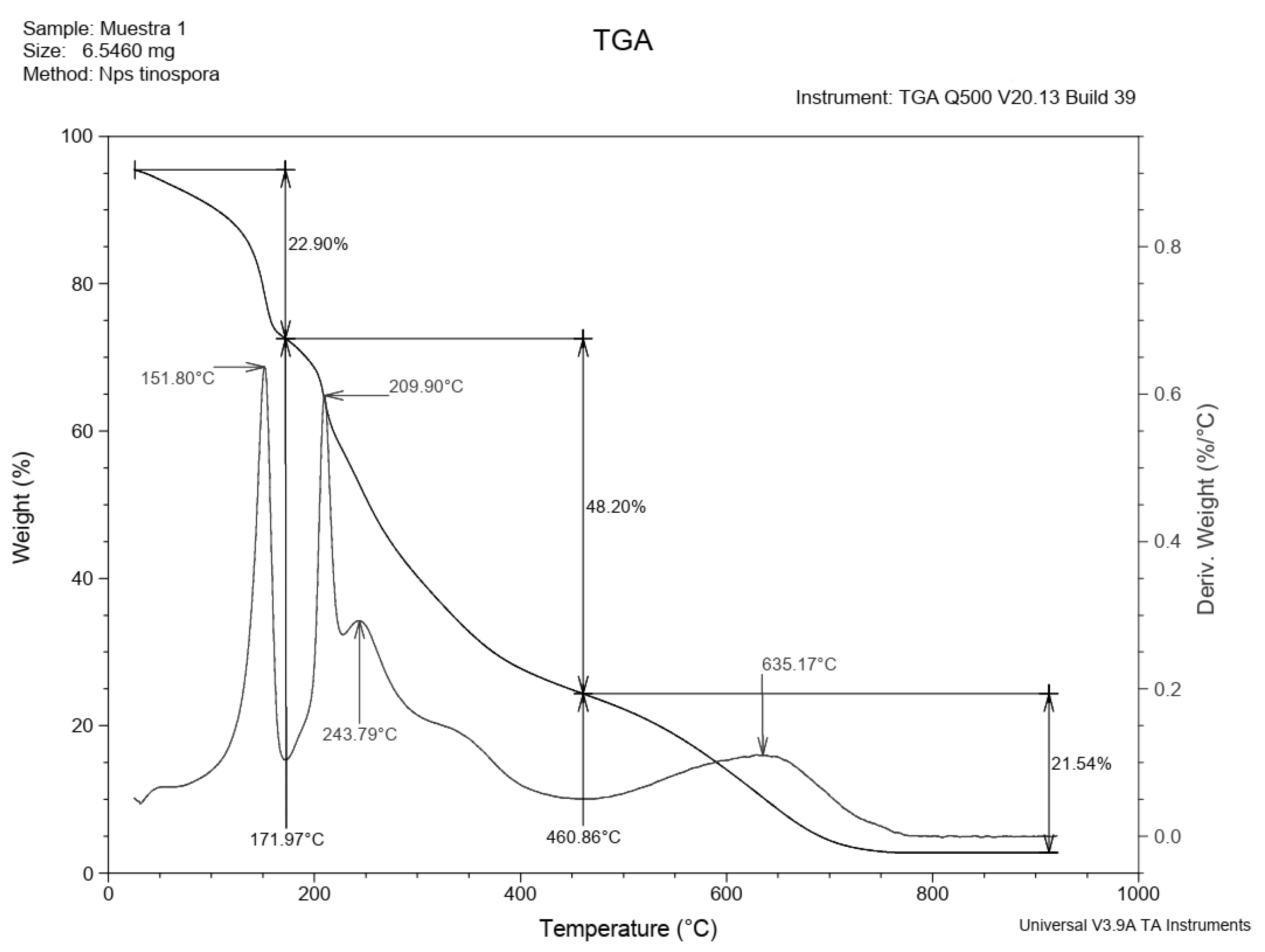
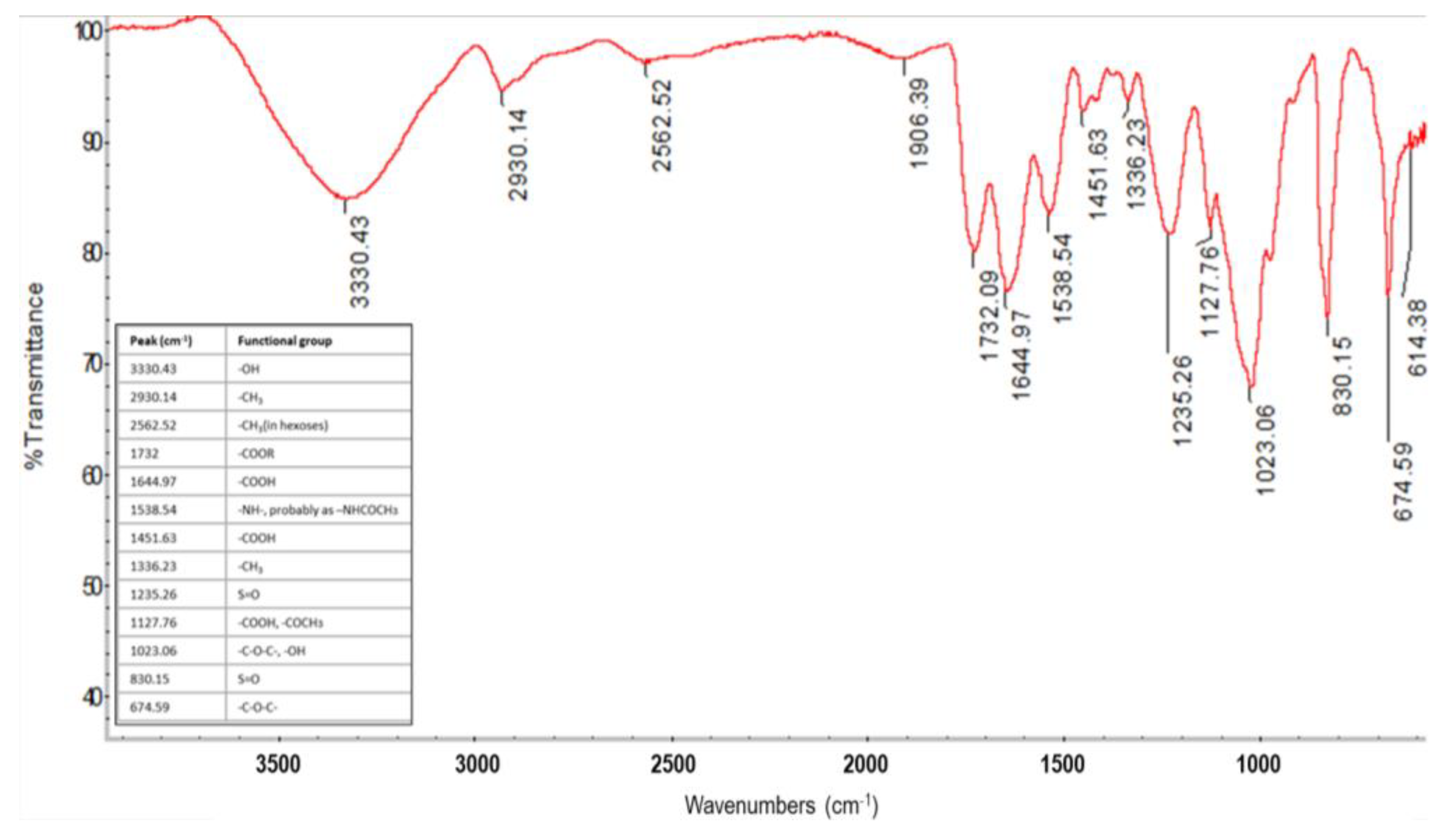
2.7.3. FTIR and TGA Analysis of EPS

FTIR of the EPS samples was carried out in a Nicolet 6700 spectrometer (Thermo Fischer Scientific, Waltham, MA, USA). Dried samples (100 mg) of EPS were scanned between 500 and 4000 cm−1 to identify the most representative peaks. Thermo Gravimetric Analysis (TGA) was performed on a TGA Q500 (Thermal Analysis Instruments, New Castle, DE, USA) instrument using 654 mg of the sample. The inner chamber was conditioned with nitrogen gas and heated at 10 °C min−1 up to 1000 °C. The thermal treatment (heating speed) was adjusted depending on the mass variation measured by the detector [40].

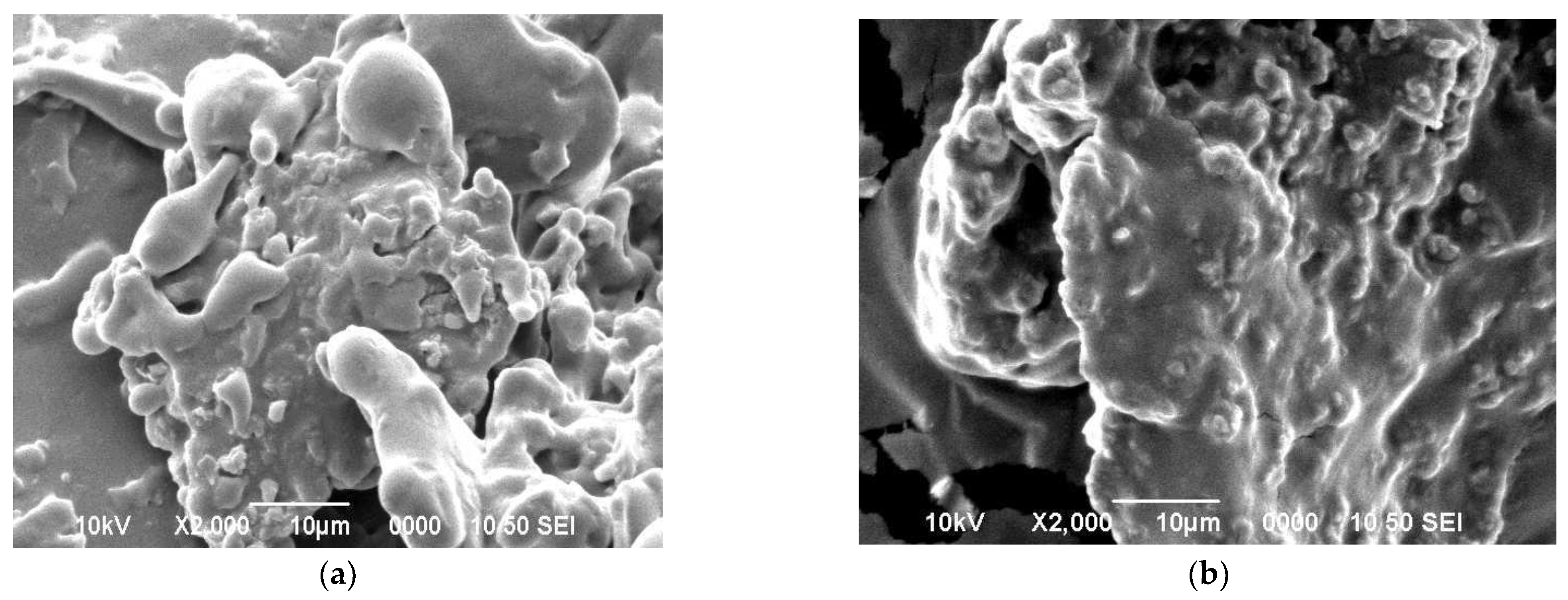
2.7.4. Scanning Electron Microscopy

The samples of EPS were analyzed using a scanning electron microscope JSM-5900 LV (JEOL, Tokyo, Japan), at a voltage of 10–20 kV, and a pressure of 1E−4 Pa. Prior to the analysis, an EPS sample was mixed with water until a homogeneous solution was obtained, and a second sample was analyzed directly without solubilizing it in water. Samples were spread over carbon paper for analysis.

3. Results

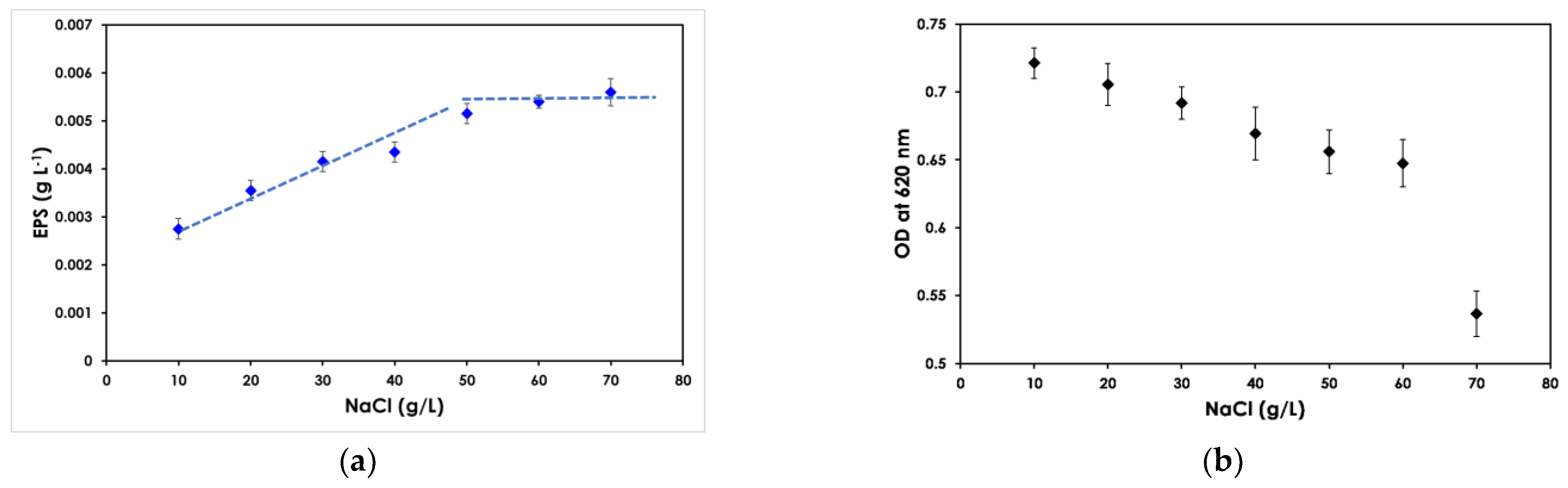
3.1. Effect of Salinity on EPS Production and Cell Growth

The effect of varying salinity on cell growth and EPS production by BU-4 isolate was screened in a glucose-based medium containing different NaCl amounts. Increasing the salinity in the medium (from 10 to 50 g L−1 NaCl), resulted in a directly proportional increase of the EPS production (Figure 1a). The highest EPS concentration (5 mg L−1) was achieved at NaCl concentration of 50 g L−1, above which no further increase was observed. On the other hand, cell growth, measured as absorbance at 620 nm, decreased with the increase in salinity (Figure 1b). In the NaCl concentration range between 10 and 60 g L−1 NaCl, the cell growth decreased moderately but steadily, while a sharp decrease was observed with increase in NaCl concentration from 60 to 70 g L−1. Based on these results, NaCl concentration of 50 g L−1 was chosen for further experiments within this study.

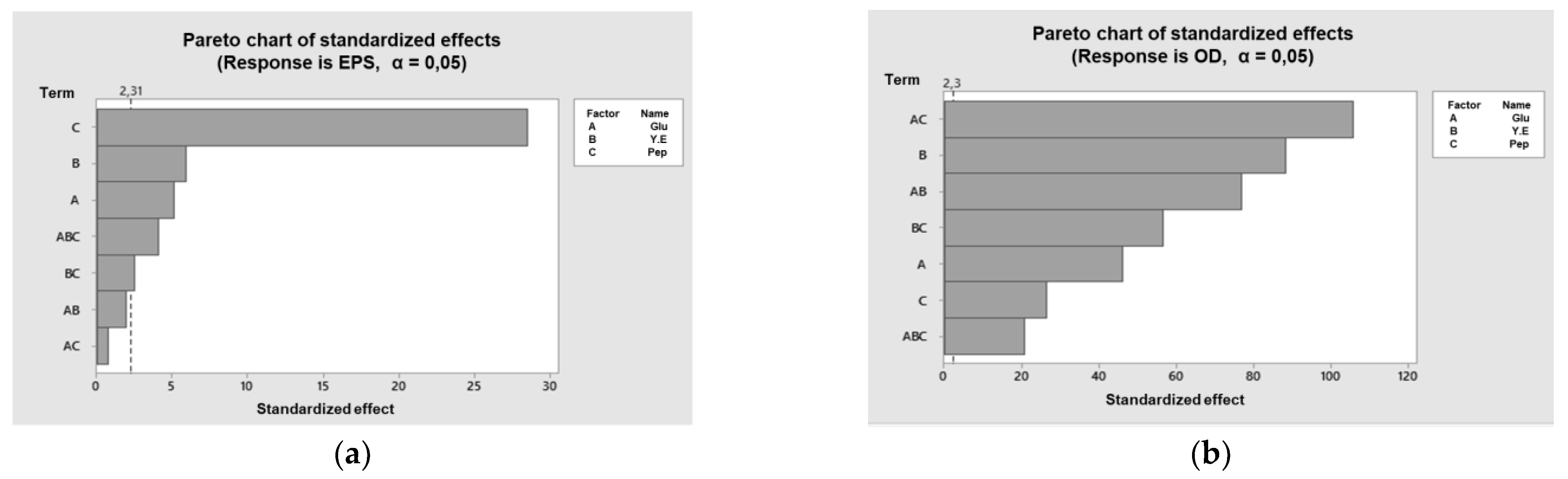
3.2. Effect of the Medium Components on Cell Growth and EPS Production

The effect of yeast extract, peptone and glucose on BU-4 cell growth and EPS production was evaluated by varying their concentrations individually and making different combinations according to a factorial design (Table 1).
The highest EPS production (2.36 g L−1) was achieved in the experimental run with concentrations of peptone, yeast extract and glucose of 3, 5 and 10 g L−1, respectively. As shown in the Pareto chart of standardized effects, peptone concentration was the factor exerting the strongest effect on EPS production, while glucose and yeast extract and different interactions exerted relatively minor influence (Figure 2a). On the other hand, for cell growth, the interaction between the concentrations of glucose and peptone exerted the strongest influence, followed closely by yeast extract concentration and by the interaction between the concentrations of glucose and yeast extract (Figure 2b).

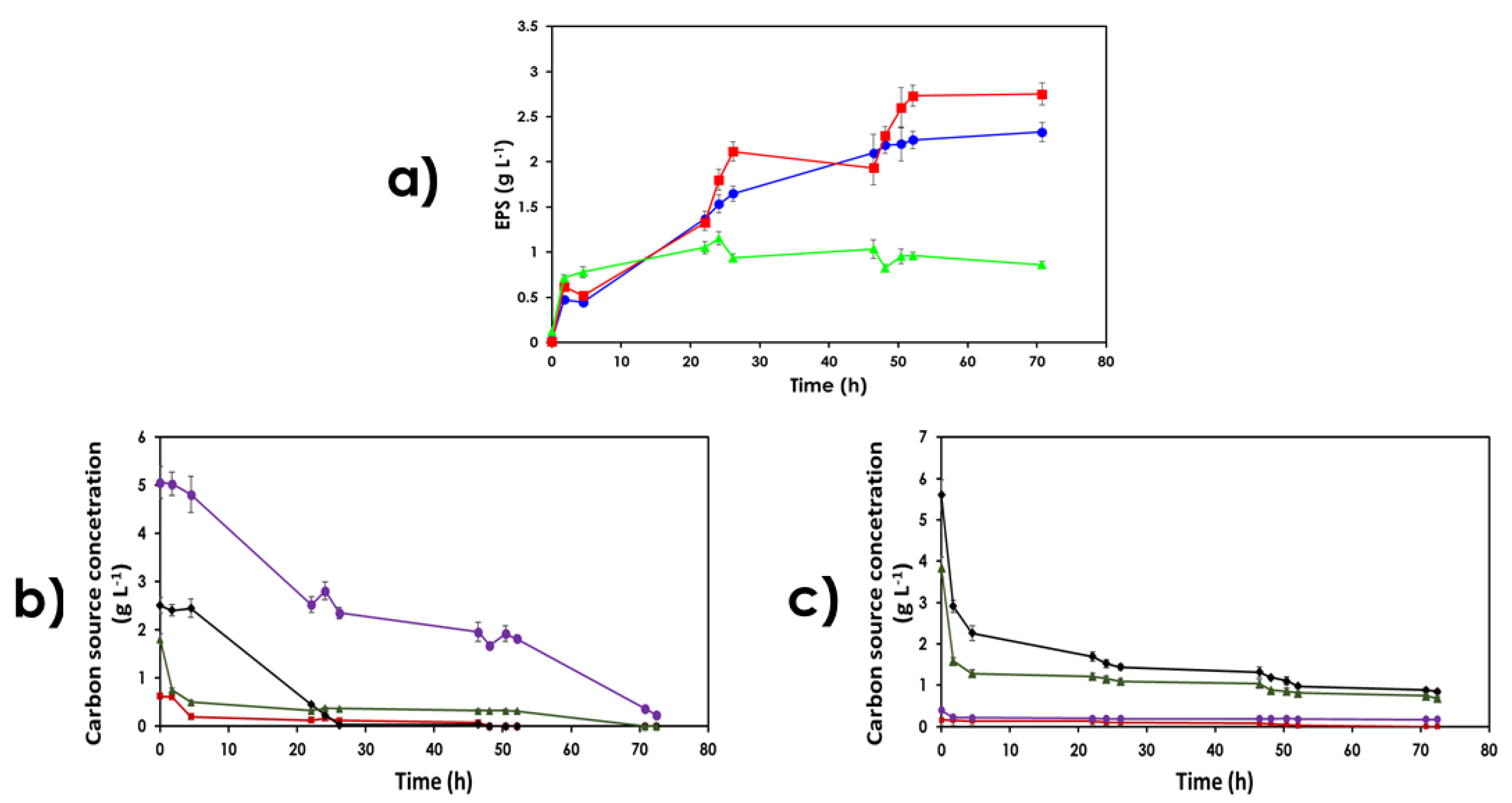
3.3. Use of Lignocellulose-Based Substrates for EPS Production

After elucidating the effects of salinity and medium components on cell growth and EPS production, a new experimental series directed to evaluating the use of lignocellulose-based media as carbon sources was performed. As a first step, the ability of BU-4 strain to use lignocellulose-based substrates was screened by cultivating it in solid media, where a part of glucose had been replaced by an equivalent amount of either pretreatment liquid of steam-exploded quinoa stalks or enzymatic hydrolysate of Curupaú sawdust and keeping the remaining components of the culture medium unchanged.
Once it was established that the BU-4 isolate can grow on lignocellulosic media, EPS production was investigated by cultivation using pretreatment liquids of quinoa stalks and enzymatic hydrolysate of Curupaú sawdust, respectively, as the only carbon sources. A parallel cultivation on glucose-based medium (10 g L−1 of glucose) as carbon source was run as reference. Both lignocellulose-based substrates contained approximately the same amount of glucose (around 4.5 g L−1), but their content of other sugars was different (Table 2). The pretreatment liquid of steam-exploded quinoa stalks contained a high concentration of xylose (9.03 g L−1) and relevant amounts of other sugars, such as glucose (4.51 g L−1), galactose (3.28 g L−1), arabinose (3.24 g L−1) and cellobiose (1.13 g L−1). On the other hand, the hydrolysate of Curupaú sawdust, besides glucose, contained only arabinose (3.84 g L−1) and very low amounts of xylose (0.39 g L−1) and cellobiose (0.16 g L−1). The pretreated liquid of quinoa stalks was diluted, and the hydrolysate of Curupaú sawdust was spiked with glucose so that the concentration of total sugars was always 10 g L−1.
Different cultivation patterns were observed when growing the BU-4 isolate in lignocellulose-based media (Figure 3). A better growth and higher EPS production was observed during cultivation in the pretreatment liquid of steam-exploded quinoa stalks than when the hydrolysate of Curupaú sawdust was used as substrate. As during the cultivation in glucose-based medium, the EPS production in quinoa stalks pretreatment liquid increased along the cultivation time (Figure 3a). On the other hand, the cultivation in the hydrolysate of Curupaú sawdust resulted in clear EPS formation only during the first hours, with a plateau-like curve for the rest of the process, and with some minor decreases after 24 and 48 h. It should be noted, however, that in spite of the better performance in the pretreatment liquid of quinoa stalks, a decrease in EPS production was observed between 26 and 46 h, and that the production rate decreased during the last 20 h. The sugar consumption dynamics during cultivation (Figure 3b,c) is in agreement with the EPS production. Most of the available sugars were consumed during cultivation in the lignocellulose-based media, with a more complete utilization in the quinoa stalk pretreatment liquid (Figure 3b) than in the Curupaú hydrolysate (Figure 3c). On the other hand, the consumption of proteins was in the order of glucose-based medium (84%) > quinoa stalks (82%) > Curupaú sawdust (72%) (data not shown).

3.4. Characterization of the Produced EPS

The EPS was extracted from the supernatant of BU-4 grown in glucose, and subjected to scanning electron microscopy (SEM) directly or after dissolving it in water (Figure 4). The SEM images of the undissolved EPS revealed rounded agglomerations, which can be interpreted as areas accumulating high quantities of water (Figure 4a). That might be an indication of the porosity of the surface as reported in previous studies [41]. The images of the dissolved EPS show a polymer with some agglomerations (Figure 4b), which is characteristic of polymers having glycosidic bonds and with high tendency to form gels [42].
The analysis of the monomeric composition was performed for the EPS produced by BU-4 in the glucose-based medium and for the one produced in the pretreatment liquid of quinoa stalks. The analysis revealed that glucose represented over 90% of the EPS produced in the glucose-based medium, whereas the EPS obtained from the pretreatment liquid of quinoa stalks was a heteropolysaccharide with xylose and glucose accounting together for almost 80% of the dry weight, and with arabinose as a minor fraction (Table 3). In spite of the TCA treatment performed for deproteinization, the EPS samples still contained some amount of protein (Data not shown).
The FTIR analysis of the purified EPS revealed several carbohydrate-related peaks including hydroxyl groups, methyl groups of linear glycans and carbonyl ester bonds (Figure 5) [36,43,44]. For instance, the signal at 2562 cm−1 can be assigned to the C-H stretching of methyl or methylene groups of hexoses or deoxyhexoses [36]. A peak in the region of 1644 cm−1 related to carboxylic acids, can be associated with the higher water retention capacity of the polysaccharide [35,45]. The band around 1127 cm−1 is a characteristic peak of uronic or acetyl esters in polysaccharides [46]. Being a polysaccharide, it is more likely that this peak reflects the presence of uronic acids. The absorbance band at 1023 cm−1 can be assigned to C-O stretching vibrations in glycosidic linkages of oligo- or polysaccharides or in connection to hydroxyl groups [23]. The peak at 830 cm−1 is characteristic of polysaccharides with sulfate groups attached to the monosaccharide components [46,47]. Vibration bands at 830 and 845 cm−1 have previously been associated with 4-O- and 2-O-sulfate groups in agar-type polysaccharides [46]. This suggests the presence of a sulfate group in the polysaccharide chain. Although the presence of sulfate groups is a very unusual phenomenon in bacterial polysaccharides, it has already been reported for EPS produced by the halophilic bacteria Halomonas ventosae and Halomonas anticariensis [47]. Polysaccharides containing sulfate groups are polymers having a number of bioactive properties that are of potential interest in medicine [11].
TGA analysis of the EPS produced by the BU-4 isolate grown in glucose-based medium revealed the first weight loss between 30 and 172 °C with a weight reduction of 22.9% (Figure 6), which is due to water desorption because of the degradation of hydrogen bonds. The second weight loss was of the order of 48.2%, occurring between 172 and 461 °C and is related to the disintegration phase or pyrolysis. Finally, the last weight loss was 21.5% between 461 and 920 °C, and can be attributed to the reconfiguration of morphological structures to their more stable configuration [48].
The maximal weight reduction rate was observed at 151.8 °C in the first stage, which is higher than the normal temperature at which other polysaccharides lose water. That might confirm the hydrophilic potential of the exopolysaccharides [25,49], which is a consequence of its high-water retention capacity, and it is usually linked to the presence of hydroxyl groups [5,50]. In the second stage, maximum degradation was observed at 209.9 and 243.8 °C. The second stage degradation occurs at a higher rate than the other two stages (a reduction of approximately 30% in weight), at a temperature that is lower than that of other polysaccharides such as gellan gum (250 °C) and dextran (320 °C), which might indicate a shorter polymer chain of the BU-4 EPS. Finally, the third stage shows a very high disintegration temperature (635 °C), which might suggest that the produced EPS has high thermal stability, an attribute of polysaccharides with heterogeneous molecular structure and with presence of a significant number of sulfate groups or uronic groups, both of which are difficult to break [2,51].

4. Discussion

EPS production is largely considered to be one of the response strategies used by microorganisms for withstanding environmental stress, such as extremes of temperature, pH and salinity [52]. High salt-concentration environments, like the one in Salar de Uyuni, from where the bacterial strain used in this study was isolated, have been reported to induce EPS production by previously described halophilic and halotolerant microorganisms [53,54,55,56]. Salar de Uyuni is characterized by high salinity, with salt concentration values in the range of 132–177 g L−1 [57], which coupled with relatively low pH values (4.6–5.8) results in an extremely harsh habitat for living organisms. For surviving in that environment, endemic microorganisms develop self-protection strategies including the production of extracellular biopolymers [19,24,58]. Additionally, it has been theorized that the presence of metallic species in the form of salts contribute largely to both the microbial diversity found in Salar de Uyuni and to its biotechnological potential [56]. It was of our interest to investigate how the halotolerance and the EPS-building ability of a bacterial strain isolated from a high-salinity environment is manifested in cultivations under controlled laboratory conditions. In this study, EPS production by the halotolerant bacterial strain BU-4 isolated from the Salar de Uyuni was investigated, and the potential for using lignocellulosic feedstocks from the Bolivian Altiplano as substrate for bacterial cultivation was evaluated.
The screening of the effect of different salinity conditions on the cultivation of BU-4 isolate revealed that under controlled laboratory cultivation the bacterial strain retains its halotolerance and its ability to produce EPS (Figure 1). Furthermore, the NaCl concentration resulting in the highest EPS production was identified, and that NaCl concentration was used in further cultivation experiments using pretreatment liquids and hydrolysates of lignocellulosic materials. The investigation of the effect of different components of the glucose-based medium provided useful input for having a valid reference for comparison with the cultivation in the lignocellulose-based media.
An interesting—and impactful—result of this investigation is the observation that cultivating the bacterial isolate BU-4 in a pretreatment liquid of quinoa stalks, with xylose as the main sugar constituent, is not only possible, but it also can lead to a better EPS production than that achieved in cultivation using synthetic carbon sources with only glucose (Figure 3a). Furthermore, it was found that the pretreatment liquid of quinoa stalks is a better substrate than the hydrolysate of Curupaú sawdust, whose main sugar was glucose, for cultivation of the BU-4 isolate. BU-4 isolate displayed the ability to use most of the sugars contained in both lignocellulose-based media (Figure 3b–c) for growing and producing EPS.
Although EPS production during cultivation in quinoa stalks pretreatment liquid was not as stable as in the glucose-based medium, the general pattern was comparable, and EPS formation increased steadily during most of cultivation time, whereas in the hydrolysate of Curupaú sawdust, EPS was formed only at the beginning (Figure 3a). Such difference might be attributed to the presence of inhibitors in the latter medium [59], and that might be related to both the pretreatment conditions and the chemical composition of the feedstock (Table 4). In this work, Curupaú sawdust was acid-pretreated at 200 °C for 10 min, while quinoa stalks were pretreated by acid-assisted steam explosion at 190 °C for 5 min. The higher temperature and longer time used for Curupaú sawdust result in higher pretreatment severity, and that leads to higher degree of sugar degradation and formation of bioconversion inhibitors. We have shown previously that the relatively harsh conditions used for pretreatment of Curupaú sawdust result in formation of inhibitors, such as acetic acid, furfural and 5-hydroxymethylfurfural [30]. This is the likely cause for the poorer cultivation parameters observed when using the hydrolysate of Curupaú sawdust compared to those of cultivation in the pretreatment liquid of steam-pretreated quinoa stalks. Removal of the inhibitors by detoxification of the hydrolysate is an approach to be considered in future research [59]. The effect of potential nutrients contained in hydrolysates of different lignocellulosic materials also deserves future attention. It has been shown previously that in media with different nutritional supplements, the range of nutrient sources affects the production of EPS and the cell growth [60,61]. Although in this study supplementation with yeast extract and soy peptone resulted in good growth and high EPS production, optimizing their use based on the actual requirements of each hydrolysate is an issue of economical significance.
The observed periodical decreases in EPS production during cultivation in both lignocellulose-based media was noteworthy. This might be attributed to the bacterial strain being periodically forced to break down a certain amount of EPS as a self-protection mechanism to maintain its growth as it typically happens with other halotolerant organisms [62]. Perhaps, some lignocellulose-derived compounds make the bacterial strain more susceptible to the high salt concentration than that in glucose-based medium. Further detailed studies of cultivation of the BU-4 in NaCl-supplemented lignocellulosic media would be required for confirming this hypothesis.
The monosaccharide composition of the resulting EPS seems to be connected with the composition of the substrate used. The use of the pretreatment liquid of quinoa stalks, a xylose-rich substrate (Table 2), as the carbon source, resulted in the production of an EPS, which contained xylose as the main monosaccharide component (Table 3). On the other hand, growing the BU-4 isolate in a glucose-based synthetic substrate resulted in an EPS composed predominantly of glucose. This reflects the ability of the bacterial isolate to not only grow using a variety of carbon sources, but also using those carbon and nitrogen sources for effectively building up EPS [63]. The remaining protein in the EPS suggests that the purification method still requires optimization.
SEM images of the dissolved EPS revealed the formation of some agglomerations and irregular porosity, which is an indicator of the existence of enough capillary force to hold fluids, and hence of high water retention capacity [37]. Furthermore, TGA analysis revealed that the EPS experienced a significant weight loss at a rather high temperature (151.8 °C) compared to other polysaccharides of vegetable origin whose dehydration is initiated below 100 °C [64], or with chitosan, which gets dehydrated at around 90 °C [65]. This is a first indicator that the EPS is somewhat better at retaining its physically absorbed water (hydrophilic properties), which is a feature that is usually related to good gel-formation capacities. The final disintegration temperature of 635 °C suggests a significant presence of uronic acid moieties, which possess a rather high affinity for water molecules, again confirming the possibility of the BU-4 EPS from glucose-based medium to be good at gel formation [66,67]. Both the high water retention capacity and gel-forming ability are desired characteristics of EPS for medical and pharmaceutical applications [68,69,70,71,72].

5. Conclusions

This study demonstrated the potential of the halophilic Bacillus sp. isolate BU-4 from the Bolivian Altiplano for producing exopolysaccharides with broad and interesting application possibilities. This potential may be a direct consequence of the environmental conditions pushing the endemic organisms to develop physiological adaptive strategies to thrive in such a habitat.
The feasibility of using pretreatment liquids of a local agricultural residue, quinoa stalks, as cultivation medium for BU-4 was demonstrated. It was revealed that higher EPS production can be achieved in pretreatment liquid of steam-pretreated quinoa stalks than in glucose-based medium, which is a useful feature in making the EPS production economically feasible.
Characteristics of the produced EPS suggesting possible uses in the food and pharmaceutical industries were elucidated, but further studies are still needed to explore their full application possibilities, and to clarify the biotechnological and industrial potential of this halophilic strain. In that direction, the genetic characterization of the BU-4 isolate, further insights into its ability to produce EPS from different carbon sources, and new inputs on EPS characterization are investigations underway.

Author Contributions

Conceptualization: C.C. and L.R.-S.; methodology: C.C., L.R.-S., J.Q., J.V.-B., R.H.-K., and C.M.; formal analysis: L.R.-S., and D.C.; investigation: D.C., L.R.-S., R.V., F.O.-G., J.V.-B., and J.Q.; resources: C.C. and C.M.; writing—original draft preparation: L.R.-S., and D.C.; writing—review and editing: C.C., C.M., J.V.-B., J.Q., and R.H.-K.; visualization: L.R.-S.; supervision: C.C., and R.H.-K.; project administration: C.C., and C.M.; funding acquisition: C.C., and C.M. All authors have read and agreed to the published version of the manuscript.

Funding

This research was funded by SIDA (Swedish International Development Cooperation Agency) and the Swedish Research Council (2016-05822).

Institutional Review Board Statement

Not applicable.

Informed Consent Statement

Not applicable.

Acknowledgments

The administrative and technical support provided by Instituto de Investigación y Desarrollo de Procesos Químicos is acknowledged. Jaime Moreno-García is thanked for critical reading of the manuscript.

Conflicts of Interest

The authors declare no conflict of interest.

Appendix A. Morphological Characterization

Under microscopic examination, the morphological characteristics such as color, type of surface, and shape of the colonies of the bacterial isolate BU-4 were identified. The pure culture was composed of rod-shaped motile cells.

Appendix B. Physico-Chemical Characterization

Salinity: From the pure culture, the bacterial isolate BU-4 was seeded in Petri dishes with the solid medium described in 2.1, but with varying NaCl concentrations (0, 0.5, 1, 3, 5, 7, 10, 13, 15, 20% (w/v)) at 35 °C and pH 7.5.
Optimal pH: From the pure culture, the bacterial isolate BU-4 was seeded in Petri dishes with the solid medium, but with varying pH (4, 5, 6, 7, 8, 9, 10, 11) maintaining a temperature of 35 °C and the optimal salt concentration.

Appendix C. Biochemical Tests

Acidification: The bacterial isolate BU-4 was seeded in Petri dishes with the solid medium described in 2.1 with and without glucose using methyl red, a pH indicator. That allows determining if the final product of the metabolism is an acid.
Exoenzymes—amylases: The bacterial isolate BU-4 was incubated in the solid medium with 0.5% (w/v) starch added, at 35 °C and pH 7.5 for 24 h. Starch degradation halo was observed by adding 2 mL Lugol solution on the surface of the culture medium. After 10 min, excess Lugol solution was discarded and amylase activity was detected based on the formation of a clear halo surrounded by a blue zone.
Exoenzymes—Proteases: The bacterial isolate BU-4 was incubated in the solid medium with 0.5% (w/v) powdered milk added, at 35 °C and pH 7.5 for 24 h. The activity was determined if a transparent halo forms around the colonies.
Exoenzymes—Urease: The bacterial isolate BU-4 was seeded in solid commercial medium Urea Agar Base OXOID CM0053, at 35 °C and for 24 h. The activity indicator test is positive, when phenol red changes the medium color from pale-yellow to pink-red around the colony in an alkaline environment.
Exoenzymes—DNAse: The bacterial isolate BU-4 was seeded in solid commercial medium DNA-SE Agar OXOID CM0321, at 35 °C and for 24 h. The activity was determined by flooding the Petri dishes with 1 N HCl. The test is positive if a transparent halo forms around the colonies.
Assimilation of carbon sources: The strain was evaluated for the ability to use difference carbon sources using a BIOLOG GN2 kit.
Inhibition test: Antibiotic inhibition tests were performed with the help of discs impregnated with different antibiotics. The bacterial isolate BU-4 was seeded in the solid medium described in 2.1, then the impregnated disks were placed on top of the medium with microorganism. The plates were incubated for seven days at 28 °C. After this period, the presence or absence of an inhibition halo was observed, indicating sensitivity or resistance, respectively.

References

  1. Rampelotto, P.H. Extremophiles and extreme environments. Life (Basel) 2013, 3, 482–485. [Google Scholar] [CrossRef]
  2. Nicolaus, B.; Kambourova, M.; Oner, E.T. Exopolysaccharides from extremophiles: From fundamentals to Biotechnology. Environ. Technol. 2010, 31, 1145–1158. [Google Scholar] [CrossRef]
  3. Coker, J.A. Extremophiles and Biotechnology: Current Uses and Prospects. F1000Res 2016, 5. [Google Scholar] [CrossRef]
  4. Merino, N.; Aronson, H.S.; Bojanova, D.P.; Feyhl-Buska, J.; Wong, M.L.; Zhang, S.; Giovannelli, D. Living at the extremes: Extremophiles and the limits of life in a planetary context. Fron. Microbiol. 2019, 10, 780. [Google Scholar] [CrossRef] [PubMed] [Green Version]
  5. Galinski, E.; Truper, H. Microbial behavior in salt-stressed ecosystems. FEMS Microbiol. Rev. 1994, 15, 95–108. [Google Scholar] [CrossRef]
  6. Galinski, E. Osmoadaptation in bacteria. Adv. Microb. Physiol. 1995, 37, 273–328. [Google Scholar]
  7. Barghini, P.; Silvi, S.; Aquilanti, A.; Marcelli, M.; Fenice, M. Bacteria from marine salterns as a model of microorganisms adapted to high environmental variations. J. Environ. Prot. Ecol. 2014, 75, 897–906. [Google Scholar]
  8. Ollivier, B.; Caumette, P.; Garcia, J.L.; Mah, R.A. Anaerobic bacteria from hypersaline environments. Microbiol. Rev. 1994, 58, 27–38. [Google Scholar] [CrossRef]
  9. Margesin, R.; Schinner, F. Potential of halotolerant and halophilic microorganisms for Biotechnology. Extremophiles 2001, 5, 73–83. [Google Scholar] [CrossRef]
  10. Quillaguamán, J.; Hatti-Kaul, R.; Mattiason, B.; Alvarez, M.; Delgado, O. Halomonas boliviensis sp. nov., an alkalitolerant, moderate halophile isolated from soil around a Bolivian hypersaline lake. Int. J. Syst. Evol. Microbiol. 2004, 54, 721–725. [Google Scholar] [CrossRef]
  11. Mata, J. Caracterización de los exopolisacáridos producidos por microorganismos halófilos pertenecientes a los géneros Halomonas, Alteromonas, Idiomarina, Palleronia y Salipiger. Ph.D. Thesis, Universidad de Granada, Granada, Spain, 2006. [Google Scholar]
  12. Guzmán, H.; Van-Thuoc, D.; Martín, J.; Hatti-Kaul, R.; Quillaguamán, J. A process for the production of ectoine and poly(3-hydroxybutyrate) by Halomonas boliviensis. Appl. Microbiol. Biotechnol. 2009, 84, 1069–1077. [Google Scholar] [CrossRef]
  13. Czaczyk, K.; Myszka, K. Biosynthesis of extracellular polymeric substances (EPS) and its role in microbial biofilm formation. Pol. J. Environ. Stud. 2007, 16, 799–806. [Google Scholar]
  14. Orgad, O.; Oren, Y.; Walker, S.L.; Herzberg, M. The role of alginate in Pseudomonas aeruginosa EPS adherence, viscoelastic properties and cell attachment. Biofouling 2011, 27, 787–798. [Google Scholar] [CrossRef] [PubMed]
  15. De Vuyst, L.; de Vin, F. Exopolysaccharides from lactic acid bacteria. In Comprehensive Glycoscience; Kamerling, J.P., Ed.; Elsevier: Amsterdam, The Netherlands; volume 2, pp. 477–519.
  16. Prasanna, P.H.P.; Grandison, A.S.; Charalampopoulos, D. Bifidobacteria in milk products: An overview of physiological and biochemical properties, exopolysaccharide production, selection criteria of milk products and health benefits. Food Res. Int. 2014, 55, 247–262. [Google Scholar] [CrossRef]
  17. Raposo, M.F.D.J.; de Morais, R.M.S.C.; de Morais, B.; Miranda, A.M. Bioactivity and applications of sulphated polysaccharides from marine microalgae. Mar. drugs 2013, 11, 233–252. [Google Scholar] [CrossRef] [PubMed] [Green Version]
  18. Chen, J.; Czajka, D.; Lion, L.; Shuler, M.; Ghiorse, W. Trace metal mobilization in soil by bacterial polymers. Environ. Health Perspec. 1995, 103, 53–58. [Google Scholar]
  19. Sheng, G.P.; Yu, H.Q.; Li, X.Y. Extracellular polymeric substances (EPS) of microbial aggregates in biological wastewater treatment systems: A review. Biotechnol. Adv. 2010, 28, 882–894. [Google Scholar] [CrossRef] [PubMed]
  20. Ding, Z.; Bourven, I.; Guibaud, G.; van Hullebusch, E.D.; Panico, A.; Pirozzi, F.; Esposito, G. Role of extracellular polymeric substances (EPS) Production in bioaggregation: Application to wastewater treatment. Appl. Microbiol. Biotechnol. 2015, 99, 9883–9905. [Google Scholar] [CrossRef]
  21. Quillaguamán, J.; Delgado, O.; Mattiasson, B.; Hatti-Kaul, R. Chromohalobacter sarecensis sp. nov., a psychrotolerant moderate halophile bacterium isolated from the saline Andean region of Bolivia. Int. J. Syst. Evol. Microbiol 2004, 54, 1921–1926. [Google Scholar]
  22. Guzmán, D.; Quillaguamán, J.; Munoz, M.; Hatti-Kaul, R. Halomonas andesensis sp. nov., a moderate halophile isolated from the saline lake Laguna Colorada in Bolivia. Int. J. Syst. Evol. Microbiol. 2010, 60, 749–753. [Google Scholar] [CrossRef] [Green Version]
  23. Rodriguez-Contreras, A.; Koller, M.; de Sousa Dias, M.M.; Calafell, M.; Braunegg, G.; Marqués-Calvo, M.S. Novel poly[(R)-3-hydroxybutyrate]-producing bacterium isolated from a Bolivian hypersaline lake. Food Technol. Biotechnol. 2013, 51, 123–130. [Google Scholar]
  24. Haferburg, G.; Gröning, J.A.; Schmidt, N.; Kummer, N.A.; Erquicia, J.C.; Schlömann, M. Microbial diversity of the hypersaline and lithium-rich Salar de Uyuni, Bolivia. Microbiol. Res. 2017, 199, 19–28. [Google Scholar] [CrossRef] [PubMed]
  25. Ehling-Schulz, M.; Bilger, W.; Scherer, S. UV-B-induced synthesis of photoprotective pigments and extracellular polysaccharides in the terrestrial cyanobacterium Nostoc commune. J. Bacteriol. 1997, 1940–1945. [Google Scholar] [CrossRef] [PubMed] [Green Version]
  26. Alandia, G.; Rodriguez, J.P.; Jacobsen, S.E.; Bazile, D.; Condori, B. Global expansion of quinoa and challenges for the Andean region. Glob. Food Sec. 2020, 26, 100429. [Google Scholar] [CrossRef]
  27. Canales, N.; Gomez, J.; Fielding, M.; Dugarte, M. The potential of quinoa in Bolivia’s bioeconomy. In SEI Report; Swedish Environment Institute: Stockholm, Sweden, 2020; p. 14. [Google Scholar]
  28. Carrasco, C. Lignocellulosic Production Studies on Sugarcane Bagasse, Paja Brava, Wheat Straw, Quinoa Stalks and Curupaú. Ph.D. Thesis, Department of Chemical Engineering, Lund University, Lund, Sweden, 2013. [Google Scholar]
  29. Salas-Veizaga, D.M.; Villagomez, R.; Linares-Pastén, J.A.; Carrasco, C.; Álvarez, M.T.; Adlercreutz, P.; Nordberg Karlsson, E. Extraction of glucuronoarabinoxylan from quinoa stalks (Chenopodium Quinoa Willd.) and evaluation of xylooligosaccharides produced by GH10 and GH11 xylanases. J. Agric. Food Chem. 2017, 65, 8663–8673. [Google Scholar] [CrossRef] [PubMed]
  30. Carrasco, C.; Quispe, L.; Lidén, G. Study of sulphuric acid-catalyzed steam pretreatment of the hardwood Anadenanthera colubrina. ENERLAC 2018, 2, 1, 54–68. [Google Scholar]
  31. Gandla, M.L.; Martín, C.; Jönsson, L.J. Analytical enzymatic saccharification of lignocellulosic biomass for conversion to biofuels and bio-based chemicals. Energies 2018, 11, 2936. [Google Scholar] [CrossRef] [Green Version]
  32. Sella, S.R.; Vandenberghe, L.P.; Soccol, C.R. Bacillus atrophaeus: Main characteristics and biotechnological applications-a review. Crit. Rev. Biotechnol. 2015, 35, 533–545. [Google Scholar] [CrossRef] [PubMed]
  33. Zhu, W.; Wang, Y.Z.; Yan, F.; Song, R.T.; Li, Z.J.; Li, Y.Q.; Song, B. Physical and chemical properties, percutaneous absorption-promoting effects of exopolysaccharide produced by Bacillus atrophaeus WYZ strain. Carbohydr. Polym. 2018, 192, 52–60. [Google Scholar] [CrossRef]
  34. Carrasco, C.; Cuno, D.; Carlqvist, K.; Galbe, M.; Lidén, G. SO2-catalysed steam pretreatment of quinoa stalks. J. Chem. Technol. Biotechnol. 2015, 90, 64–71. [Google Scholar] [CrossRef]
  35. Romero-Soto, A.; Thabet, H.; Maghembe, R.; Gameiro, D.; Van-Thuoc, D.; Dishisha, T.; Hatti-Kaul, R. Metabolic potential of the moderate halophile Yangia sp. ND199 for co-production of polyhydroxybutyrate and exopolysaccharides. MicrobiologyOpen 2021, 10, e1160. [Google Scholar] [CrossRef]
  36. Kanamarlapudi, S.L.R.K.; Muddada, S. Characterization of exopolysaccharide produced by Streptococcus thermophilus CC30. Biomed. Res. Int. 2017, 4201809. [Google Scholar] [CrossRef] [PubMed] [Green Version]
  37. Mota, R.; Guimarães, R.; Buttel, Z.; Rossi, F.; Colica, G.; Silva, C.; Santos, C.; Gales, L.; Zille, A.; Philippis, R.; et al. Production and characterization of extracellular carbohydrate polymer from Cyanothece sp. CCY 0110. Carbohydr. Polym. 2013, 92, 1408–1415. [Google Scholar] [CrossRef]
  38. Shrivastaw, K.P.; Jhamb, S.S.; Kumar, A. Quantitation of the protein content of diphtheria and tetanus toxoids by the Biuret method during production of combined vaccines. Biologicals 1995, 23, 61–63. [Google Scholar] [CrossRef]
  39. Liu, Z.L.; Pan, J.H. A practical method for extending the biuret assay to protein determination of corn-based products. Food Chem. 2017, 224, 289–293. [Google Scholar] [CrossRef]
  40. Mora-Murillo, L.D.; Orozco-Gutiérrez, F.; Vega-Baudrit, J.; González-Paz, R.J. Thermal-mechanical characterization of polyurethane rigid foams: Effect of modifying bio-polyol content in isocyanate prepolymers. J. Renew. Mater. 2017, 5, 220–230. [Google Scholar] [CrossRef]
  41. Rani, R.P.; Anandharaj, M.; Sabhapathy, P.; Ravindran, A.D. Physiochemical and biological characterization of novel exopolysaccharide produced by Bacillus tequilensis FR9 isolated from chicken. Int. J. Biol. Macromol. 2017, 96, 1–10. [Google Scholar] [CrossRef]
  42. Sutherland, I.W. Structure-function relationships in microbial exopolysaccharide. Biotechnol. Adv. 1994, 12, 393–448. [Google Scholar] [CrossRef]
  43. Rasulov, B.A.; Rozi, P.; Pattaeva, M.A.; Yili, A.; Aisa, H.A. Exopolysaccharide-based bioflocculant matrix of Azotobacter chroococcum XU1 for synthesis of AgCl nanoparticles and its application as a novel biocidal nanobiomaterial. Materials 2016, 9, 528. [Google Scholar] [CrossRef] [Green Version]
  44. Kielak, A.; Castellane, T.; Campanharo, J.; Colnago, L.A.; Costa, O.Y.A.; Corradi da Silva, M.; van Veen, J.A.; Lemos, E.G.M.; Kuramae, E.E. Characterization of novel Acidobacteria exopolysaccharides with potential industrial and ecological applications. Sci. Rep. 2017, 7, 41193. [Google Scholar] [CrossRef]
  45. Copikova, J.; Barros, A.S.; Šmídová, I.; Černá, M.; Teixeira, D.H.; Delgadillo, I.; Synytsya, A.; Coimbra, M.A. Influence of hydration of food additive polysaccharides on FT-IR spectra distinction. Carbohydr. Polym. 2006, 63, 355–359. [Google Scholar] [CrossRef]
  46. Barros, F.C.; Silva, D.C.; Sombra, V.G.; Maciel, J.S.; Feitosa, J.P.; Freitas, A.L.; Paula, R.C. Structural characterization of polysaccharide obtained from red seaweed Gracilaria caudata (J Agardh). Carbohydr. Polym. 2013, 92, 598–603. [Google Scholar] [CrossRef] [Green Version]
  47. Mata, J.A.; Béjar, V.; Llamas, I.; Arias, S.; Bressollier, P.; Tallon, R.; Urdaci, M.C.; Quesada, E. Exopolysaccharides produced by the recently described halophilic bacteria Halomonas ventosae and Halomonas anticariensis. Res. Microbiol. 2006, 157, 827–835. [Google Scholar] [CrossRef]
  48. Daoub, R.; Elmurabak, A.; Misran, M.; Hassan, E.; Osman, M. Characterization and functional properties of some natural acacia gums. J. Saudi Soc. Agric. Sci. 2016, 17, 241–249. [Google Scholar] [CrossRef] [Green Version]
  49. Dinić, M.; Pecikoza, U.; Djokić, J.; Stepanović-Petrović, R.; Milenković, M.; Stevanović, M.; Filipović, N.; Begović, J.; Golić, N.; Lukić, J. Exopolysaccharide produced by probiotic strain Lactobacillus paraplantarum BGCG11 reduces inflammatory hyperalgesia in rats. Front Pharmacol. 2018, 9, 1. [Google Scholar] [CrossRef] [PubMed]
  50. Faria, S.; Petkowicz, C.; Morais, S.; Terrones, M.; Resende, M.; França, F.; Cardoso, V. Characterization of xanthan gum produced from sugar cane broth. Carbohydr. Polym. 2011, 86, 469–476. [Google Scholar] [CrossRef] [Green Version]
  51. Suja, L.D.; Summers, S.; Gutierrez, T. Role of EPS, dispersant and nutrients on the microbial response and MOS formation in the Subarctic Northeast Atlantic. Fron. Microbiol. 2017, 8, 676. [Google Scholar] [CrossRef] [PubMed] [Green Version]
  52. Yin, W.; Wang, Y.; Liu, L.; He, J. Biofilms: The microbial “protective clothing” in extreme environments. Int. J. Mol. Sci. 2019, 20, 3423. [Google Scholar] [CrossRef] [Green Version]
  53. Bouchotroch, S.; Quesada, E.; Izquierdo, I.; Rodriguez, M.; Béjar, V. Bacterial exopolysaccharides produced by newly discovered bacteria belonging to the genus Halomonas, isolated from hypersaline habitats in Morocco. J. Ind. Microbiol. Biotechnol. 2000, 24, 374–378. [Google Scholar] [CrossRef]
  54. Martínez-Cánovas, M.J.; Quesada, E.; Martínez-Checa, F.; del Moral, A.; Bejar, V. Salipiger mucescens gen. nov., sp. nov., a moderately halophilic, exopolysaccharide-producing bacterium isolated from hypersaline soil, belonging to the α-proteobacteria. Int. J. Syst. Evol. Microbiol 2004, 54, 1735–1740. [Google Scholar] [CrossRef] [PubMed] [Green Version]
  55. Biswas, J.; Paul, A.K. Diversity and production of extracellular polysaccharide by halophilic microorganisms. Biodiversity Int. J. 2017, 1, 32–39. [Google Scholar]
  56. Pérez-Llano, Y.; Rodríguez-Pupo, E.C.; Druzhinina, I.S.; Chenthamara, K.; Cai, F.; Gunde-Cimerman, N.; Batista-García, R.A. Stress reshapes the physiological response of halophile fungi to salinity. Cells 2020, 9, 525. [Google Scholar] [CrossRef] [Green Version]
  57. Schmidt, N. Hydrogeological and hydrochemical investigations at the Salar de Uyuni (Bolivia) with regard to the extraction of lithium. FOG 2010, 26, 1–131. [Google Scholar]
  58. Pérez-Fernández, C.A.; Iriarte, M.; Rivera-Pérez, J.; Tremblay, R.L.; Toranzos, G.A. Microbiota dispersion in the Uyuni salt flat (Bolivia) as determined by community structure analyses. Int. Microbiol. 2019, 22, 325–336. [Google Scholar] [CrossRef]
  59. Jönsson, L.J.; Martín, C. Pretreatment of lignocellulose: Formation of inhibitory by-products and strategies for minimizing their effects. Bioresour. Technol. 2016, 199, 103–112. [Google Scholar] [CrossRef] [Green Version]
  60. Degeest, B.; De Vuyst, L. Indication that the nitrogen source influences both amount and size of exopolysaccharides produced by Streptococcus thermophilus LY03 and modelling of the bacterial growth and exopolysaccharide production in a complex medium. Appl. Environ. Microbiol. 1999, 65, 2863–2870. [Google Scholar] [CrossRef] [PubMed] [Green Version]
  61. Zisu, B.; Shah, N.P. Effects of pH, temperature, supplementation with whey protein concentrate, and adjunct cultures on the production of exopolysaccharides by Streptococcus thermophiles 1275. J. Dairy Sci. 2003, 86, 3405–3415. [Google Scholar] [CrossRef] [Green Version]
  62. Ewert, M.; Deming, J.W. Sea ice microorganisms: Environmental constraints and extracellular responses. Biology (Basel) 2013, 2, 603–628. [Google Scholar] [CrossRef] [PubMed]
  63. Peng, L.; Li, J.; Liu, Y.; Xu, Z.; Wu, J.Y.; Ding, Z.; Shi, G. Effects of mixed carbon sources on galactose and mannose content of exopolysaccharides and related enzyme activities in Ganoderma lucidum. RSC Adv. 2016, 6, 39284–39291. [Google Scholar] [CrossRef]
  64. Savi, A.; Calegari, G.; Queiroz, V.; Pereira, E.; Teixeira, S. Chemical characterization and antioxidant of polysaccharide extracted from Dioscorea bulbifera. J. King Saud Univ. Sci. 2020, 32, 636–642. [Google Scholar] [CrossRef]
  65. Georgieva, V.; Zvezdova, D.; Vlaev, L. Non-isothermal kinetics of thermal degradation of chitosan. Chem. Cent. J. 2012, 6, 81. [Google Scholar] [CrossRef] [Green Version]
  66. Kumar, C.G.; Joo, H.S.; Choi, J.W.; Koo, Y.M.; Chang, C.S. Purification and characterization of an extracellular polysaccharide from haloalkalophilic Bacillus sp. I-450. Enzyme Microb. Technol. 2004, 34, 673–681. [Google Scholar] [CrossRef]
  67. Joulak, I.; Azabou, S.; Finore, I.; Poli, A.; Nicolaus, B.; Donato, P.D. and Attia, H. Structural characterization and functional properties of novel exopolysaccharide from the extremely halotolerant Halomonas elongata S6. Int. J. Biol. Macromol. 2020, 164, 95–104. [Google Scholar] [CrossRef] [PubMed]
  68. Nicolaus, B.; Lama, L.; Panico, A.; Schiano Moriello, V.; Romano, I.; Gambacorta, A. Production and characterization of exopolysaccharides excreted by thermophilic bacteria from shallow, marine hydrothermal vents of Flegrean areas (Italy). Syst. Appl. Microbiol. 2002, 25, 319–325. [Google Scholar] [CrossRef] [PubMed]
  69. Arena, A.; Maugeri, T.L.; Pavone, B.; Iannello, D.; Gugliandolo, C.; Bisignano, G. Antiviral and Immunoregulatory Effect of a novel Exopolysaccharide from a Marine Thermotolerant Bacillus licheniformis. Int. Immunopharmacol. 2006, 6, 8–13. [Google Scholar] [CrossRef] [PubMed]
  70. Kianpour, S.; Ebrahiminezhad, A.; Mohkam, M.; Tamaddon, A.M.; Dehshahri, A.; Heidari, R.; Ghasemi, Y. Physicochemical and biological characteristics of the nanostructured polysaccharide-iron hydrogel produced by microorganism Klebsiella oxytoca. J. Basic Microbiol. 2017, 57, 132–140. [Google Scholar] [CrossRef]
  71. Ching, S.H.; Bansal, N.; Bhandari, B. Alginate gel particles–a review of production techniques and physical properties. Crit. Rev. Food Sci. Nutr. 2017, 57, 1133–1152. [Google Scholar] [CrossRef]
  72. Saroia, J.; Yanen, W.; Wei, Q.; Zhang, K.; Lu, T.; Zhang, B. A review on biocompatibility nature of hydrogels with 3D printing techniques, tissue engineering application and its future prospective. Bio-Des. Manuf. 2018, 1, 265–279. [Google Scholar] [CrossRef]
Figure 1. Effect of salinity on EPS production (a) and cell growth (measured as OD620) (b) during cultivation of the BU-4 strain in glucose-based medium.
Fermentation 07 00033 g001
Figure 2. Pareto charts of standardized effects of glucose (A), yeast extract (B) and peptone (C) on EPS production (a) and cell growth (b) during cultivation of the BU-4 strain in synthetic media.
Fermentation 07 00033 g002
Figure 3. (a) EPS production by cultivating the BU-4 strain in a glucose-based medium (), and in pretreatment liquids of quinoa stalks (), and Curupaú sawdust (); (b) Carbon source consumption for quinoa stalks pretreatment liquid: xylose (), glucose (♦), arabinose (), and cellobiose (); (c) Carbon source consumption for Curupaú hydrolysates: glucose (♦), arabinose (), xylose (), and cellobiose ().
Fermentation 07 00033 g003
Figure 4. SEM of the EPS produced by the BU-4 isolate grown in glucose. (a) Undissolved EPS; (b) dissolved EPS.
Fermentation 07 00033 g004
Figure 5. FTIR spectrum of the EPS produced by the BU-4 isolate in glucose-based medium. The inner table gives the peak assignment.
Fermentation 07 00033 g005
Figure 6. TGA of the EPS produced by the BU-4 isolate in glucose-based medium.
Fermentation 07 00033 g006
Table 1. Concentrations (g L−1) of peptone, yeast extract and glucose used in the cultivation of BU-4 isolate in glucose-based medium. Production of EPS (g L−1) is also presented.
No.PeptoneYeast ExtractGlucoseEPS Production
111100.508
211300.586
315101.090
415300.361
551102.008
651301.684
755102.358
855302.242
Table 2. Sugar composition of the pretreatment liquid of steam-exploded quinoa stalks (PL-QS) and the enzymatic hydrolysate of Curupaú sawdust (EH-CS).
Lignocellulose-Based SubstratesConcentration (g L−1)
CellobioseGlucoseXyloseGalactoseArabinose
PL-QS1.134.519.033.283.24
EH-CS0.164.440.39-3.84
Table 3. Content of monosaccharides in the produced EPS, % (w/w).
GlucoseXyloseArabinose
EPS from glucose-based medium90.5--
EPS from quinoa stalks30.248.716.3
Table 4. Main components of the used lignocellulosic feedstocks, % (w/w).
FeedstockCelluloseHemicelluloseLigninAsh
Quinoa stalks35.715.421.94.2
Curupaú sawdust43.315.620.014.0
Publisher’s Note: MDPI stays neutral with regard to jurisdictional claims in published maps and institutional affiliations.

