Article

Regulation of β-Disaccharide Accumulation by β-Glucosidase Inhibitors to Enhance Cellulase Production in Trichoderma reesei

Chongqing Key Laboratory of Industrial Fermentation Microorganism, School of Chemistry and Chemical Engineering, Chongqing University of Science and Technology, Chongqing 401331, China
*
Author to whom correspondence should be addressed.
These authors contributed equally to this work.
Submission received: 26 March 2022 / Revised: 4 May 2022 / Accepted: 16 May 2022 / Published: 17 May 2022
(This article belongs to the Special Issue Applied Microorganisms and Industrial/Food Enzymes)

Abstract

:
Trichoderma reesei is a high-yield producer of cellulase for applications in lignocellulosic biomass conversion, but its cellulase production requires induction. A mixture of glucose and β-disaccharide has been demonstrated to achieve high-level cellulase production. However, as inducers, β-disaccharides are prone to be hydrolyzed by β-glucosidase (BGL) during fermentation, therefore β-disaccharides need to be supplemented through feeding to overcome this problem. Here, miglitol, an α-glucosidase inhibitor, was investigated as a BGL inhibitor, and exhibited an IC50 value of 2.93 μg/mL. The cellulase titer was more than two-fold when miglitol was added to the fermentation medium of T. reesei. This method was similar to the prokaryotic expression system using unmetabolized isopropyl-β-D-thiogalactopyranoside (IPTG) as the inducer instead of lactose to continuously induce gene expression. However, cellulase activity was not enhanced with BGL inhibition when lactose or cellulose was used as an inducer, which demonstrated that the transglycosidase activity of BGL is important for the inducible activity of lactose and cellulose. This novel method demonstrates potential in stimulating cellulase production and provides a promising system for T. reesei protein expression.

1. Introduction

Excessive exploitation and utilization of petroleum, coals, natural gas and other fossil fuels have resulted in a series of problems, including diminishing reserves, increasingly severe pollution, climate changes and the frequent occurrence of natural disasters. These problems largely restrict socioeconomic sustainability [1]. Lignocellulosic biorefineries, which involve the production of biofuels and biochemicals from forestry and agricultural residues, are important alternatives for addressing the energy crisis and sustainable economic development [2,3,4]. However, the robust supramolecular structures of lignocellulosic biomasses can only be hydrolyzed into fermentable sugars by efficient cellulase [5,6]. High-yield cellulase strains mainly originate from Trichoderma reesei, but the biosynthesis of the cellulases must be induced [7,8].
Sophorose is the most efficient inducer for T. reesei cellulase synthesis, and its inducing ability is more than 2500 times higher than that of cellobiose [9]. Unfortunately, sophorose is so expensive that it has never been used as a sole inducer for cellulase production. According to the cellulase production process developed by NREL for evaluating the biochemical conversion of lignocellulosic biomass to ethanol, the mixture glucose-sophorose has been used as an inducer of T. reesei. When grown on this substrate, T. reesei has been shown to productively secrete cellulase [10]. In preliminary studies, a mixture of glucose and β-disaccharide (MGD) was prepared from glucose through the transglycosylation reaction catalyzed by β-glucosidase (BGL) and used as an inducer for the efficient synthesis of cellulases [11,12,13,14]. A maximum cellulase titer of 102.63 IU/mL was achieved by engineered T. reesei, using MGD as an inducer [15]. Moreover, transcriptomic and iTRAQ proteomic analyses showed that the secretory volumes of major cellulases are higher than those of other commonly used inducers [16,17], but the BGL was low. To overcome this disadvantage, aabgl1 encoding BGL in Aspergillus aculeatus was heterologously expressed in T. reesei Rut C30. The BGL activity of recombinant T. reesei increased 71-fold compared to the parent strain [16], however, we found that the cellulase activity was significantly reduced.
It is speculated that the BGL expressed by T. reesei could degrade sophorose into glucose; consequently, sophorose does not maintain induction concentrations in cells or is completely degraded, thus losing its inducing ability. Hence, this sophorose containing inducer must be supplemented through continual feeding. Although the transglycosylation activity of BGL in T. reesei has been confirmed to be irreplaceable during cellulase fermentation when cellulose or lactose is adopted as the inducer [18,19], it is no longer needed when sophorose is directly used. We assume that if the BGLs are inhibited, an appropriate intracellular concentration of sophorose for induction can be theoretically maintained, which decreases the dosage and consumption of inducers.
Although BGL is pivotal in the degradation of cellulose components into glucose [20], research generally indicates that the BGL secreted by T. reesei is insufficient, and the efficient hydrolysis of cellulose components is achieved only when enough BGL is added [21,22,23,24,25]. Therefore, the actions of the background BGL are generally not considered. Hence, the inhibition or knockout of BGL genes does not significantly affect the quality of T. reesei cellulase.
In the present study, miglitol, an α-glucosidase inhibitor [26], was used to inhibit BGL in T. reesei fermentation, and we investigated whether intracellular β-disaccharide can be maintained at an appropriate induction concentration to stabilize cellulase production. This is an alternative strategy is to induce the synthesis of cellulase, providing a viable method for further efficient cellulase production by fed-batch fermentation.

2. Materials and Methods

2.1. Materials and Microorganisms

Trichoderma reesei Rut C30 was a gift from the USDA ARS Culture Collection. Recombinant T. reesei PB-3 and PB-4 were developed by overexpressing aabgl1 (GenBank: D64088.1) using the pdc1 promoter (encoding pyruvate decarboxylase) in T. reesei RUT C30, which had four and three gene copies, respectively [16]. The spores were preserved in 50% glycerol at −80 °C. β-Glucosidase SUNSON® was purchased from Sunson Industry Group Co., Ltd. (Yinchuan, Ningxia, China).
The mixture of glucose and β-disaccharides (MGD) was synthesized from glucose by β-glucosidase (BGL) through a transglycosylation reaction [11]. In detail, BGL was added to a substrate containing 600 g/L glucose at 20 IU/g (glucose), and the transglycosylation reaction was performed at 65 °C and pH 4.8 for 72 h, after which the BGL was deactivated by incubating the mixture at 100 °C for 5 min. Finally, the MGD composition was 410.20 g/L glucose, 60.56 g/L gentiobiose, 9.34 g/L cellobiose and 13.66 g/L sophorose.

2.2. Production of Cellulase and β-Glucosidase from T. reesei C30, PB3 and PB4

T. reesei Rut C30, PB-3 and PB-4 were applied separately in the production of cellulase and BGL, using MGD, lactose or cellulose as an inducer. Lignocellulase production in shake flasks was performed, according to a previous work [11]. T. reesei was cultured on malt extract agar plates (3% malt extract and 1.5% agar) for 7 d. Spores were then harvested and inoculated into 250-mL Erlenmeyer flasks containing 50 mL of a medium that was composed of 4 g/L glucose and 10 g/L corn steep liquor. After 24 h, mycelium was inoculated at 8% (v/v) into 250-mL flasks containing 50 mL of a fermentation medium for cellulase and BGL production. The fermentation medium contained 10 g/L carbon source (MGD, lactose or cellulose) supplemented with 1 g/L peptone, 0.3 g/L urea, 0.8 g/L CaCl2, 0.5 mL/L Tween 80, 4 g/L KH2PO4, 0.6 g/L MgSO4·7H2O, 2.8 g/L (NH4)2SO4, 10 mg/L FeSO4·7H2O, 3.4 mg/L MnSO4·H2O, 2.8 mg/L ZnSO4·7H2O, 4 mg/L CoCl2 and 500 mL/L 0.2 M Na2HPO4-citric acid (pH 5.0) was formulated, as described previously. Each flask was incubated at 28 °C under shaking at 150 rpm. Triplicate was applied. Time-courses for the production of cellulase and BGL were monitored at 24, 36, 48 and 60 h.

2.3. Development and Validation of β-Glucosidase Inhibitors

Miglitol inhibitor with a concentration gradient (0–5 mg/mL) was added to the reaction mixtures, which contained 200 μL of 15 mM cellobiose as a substrate and 200 μL properly diluted enzyme solution. The reaction mixtures were incubated at 50 °C for 30 min and then boiled for 2 min to stop the reaction. BGL activity was determined by the same method described in Section 2.9. GraphPad Prism software (version 8; San Diego, CA, USA) software was used to draw the IC50 curves, and the IC50 of miglitol on BGL inhibition was obtained.
Aesculin was used to identify β-glucosidase activity in a fermentation broth (24, 36, 48, 60 and 72 h) from T. reesei Rut C30 supplemented with 1 g/L miglitol using 10 g/L MGD as the carbon source [27], and the nitrogen source and inorganic salts were the same as the fermentation medium described in Section 2.2. Then, 0.5 g/L aesculin and 1.0 g/L FeSO4·7H2O were dissolved in 1 mL of a 0.2 M HAC-NaAC buffer. The fermentation broth (200 μL) was dropped into 1 mL of the test solution and incubated at 50 °C for 1 h. Finally, 200 μL of the mixture was extracted and placed into a 96-well plate for imaging. The time-course of BGL production was monitored at 24, 36, 48, 60 and 72 h, with water added as a negative control.

2.4. Molecular Docking

Two crystal structures of β-glucosidase (BGL) were downloaded from the RCSB PDB. The PDB IDs of BGL were named 3ZYZ and 3AHY. The structures of miglitol, sophorose and cellobiose were exported to PubChem.
All the water molecules, co-crystal ligands and heteroatoms were deleted from the receptor using AutoDock. Hydrogen atoms and Gasteiger charges were added. The output files were saved in the format of pdbqt. The ligands were energy-minimized before they were converted to the format of pdbqt. Then, the active site of the molecular docking was determined by referring to the original ligand coordinates in the target protein, and a 10 × 10 × 10 (Å3) molecular pocket was set, according to the BGL protein active pocket, as an effective structure domain for docking with ligands. AutoDock was used to perform molecular docking and analyze the best binding modes for receptor–ligand interactions. The conformers with the lowest Gibbs free binding energy (estimated as ∆G in kcal/mol) were selected for PostDock analysis. Miglitol, sophorose and cellobiose were used as ligands and BGL was used as a receptor. Finally, the docking results were visualized on PyMOL [28].

2.5. Effects of Miglitol on the Growth and Cellulase Induction of T. reesei

To verify whether miglitol can be used as a carbon source for T. reesei, the spores were inoculated into plates (35 mm in diameter) containing 10 g/L miglitol as the sole carbon source and the rest of the ingredients were the same as the fermentation medium described in Section 2.2, but the peptone was removed. Glucose as the sole carbon source was used as a control. The plates were cultured at 28 °C for 48 h.
To test the effect of miglitol on the cellulase activity of T. reesei Rut C30, we measured cellulase activity in the fermentation medium with or without miglitol (1 g/L), supplemented using 10 g/L glucose as the carbon source. Enzyme activity was detected after 48 h of shake flask incubation. The fermentation broth was centrifuged at 5000 rpm for 4 min to obtain the supernatant for the cellulase activity assay. The method for cellulase production in shake flasks was described in Section 2.2, and the enzyme activity assay method was described in Section 2.9.
To detect the effect of miglitol on T. reesei growth, we inoculated 1 µL of spores in the plates with fermentation medium containing different concentrations of miglitol (0, 0.125, 0.25, 0.5, 1 and 2 g/L) and cultured them at 28 °C for 96 h. The nitrogen source and inorganic salts were the same as the fermentation medium described in Section 2.2, but 10 g/L MGD was used as the carbon source. Meanwhile, three groups were tested in parallel, and mycelium growth was measured at 12 h intervals.

2.6. Enzyme Production Induced by Different Inducers with/without Miglitol Supplemented

For cellulase and β-glucosidase production after the addition of miglitol, the spores of T. reesei Rut C30 were inoculated into 250-mL Erlenmeyer flasks containing 50 mL medium composed of 5 g/L glucose and 10 g/L corn steep liquor. After 24 h of cultivation at 28 °C and 150 rpm on a rotary shaker, mycelium was inoculated at 8% (v/v) into 250-mL Erlenmeyer flasks containing 50 mL fermentation medium for cellulase production. The fermentation medium was formulated based on the recipe developed in method 2.2, but 10 g/L MGD, 10 g/L lactose or 10 g/L cellulose was used as the sole carbon source. In the groups with different carbon sources, 1 g/L miglitol was added to one group, and the other group without the miglitol addition was used as a control. Cellulase activity and β-glucosidase activity were measured at 12 h intervals until after 96 h of culture. Each experiment was conducted in triplicate.

2.7. qPCR

Total RNA was extracted using a fungal total RNA isolation kit (Sangon Biotech, Shanghai, China), according to the manufacturer’s instructions. Reverse transcription was carried out using a PrimeScript RT reagent kit (Takara, Tokyo, Japan), according to the manufacturer’s instructions. Quantitative PCR was performed on a Bio-Rad myIQ2 thermocycler (Bio-Rad, Richmond, CA, USA). Amplification reactions were performed using TB Green® Premix Ex Taq™ II (Tli RNaseH Plus) (Takara), according to the manufacturer’s instructions. The sar1 gene was used as a normalized control, and the primers are listed in Table S1. The expression of genes was calibrated by the 2−ΔCt method, and at least three biological replicates were carried out for each experiment.

2.8. Protein Analysis

The fermentation broth (prepared according to Section 2.6) was centrifuged at 4 °C and 5000 rpm for 4 min to remove hyphal pellets, and the supernatant was transferred to a new tube for further analysis. Then, 8 µL of the supernatant was mixed with 2 µL of 5× native sample loading buffer and boiled for 10 min for denaturation. After that, the samples and a PageRulerTM prestained protein ladder (10 to 180 kDa) were loaded onto 10% SDS-polyacrylamide separating gels. Then, the gels were pre-run at 80 V for 30 min and run at 120 V for 100 min in Tris-glycine buffer. Finally, clear bands were obtained after staining and destaining.

2.9. Analysis Methods

Specifically, 2 mL of a mixture of shake flask fermentation was sampled. Then, the sample was centrifuged at 4 °C and 5000 rpm for 4 min to remove hyphal pellets, and the supernatant was transferred to another tube for further analysis. The activities of the cellulase and β-glucosidase (BGL) were determined using standard protocols [29]. In brief, 50 mg of filter paper (Whatman No. 1, GE healthcare, Sheffield, UK) was added to 1 mL of a 0.1 M HAC-NaAC buffer at pH 4.8. Then, 500 μL of an enzyme solution diluted to the appropriate concentration was added to the mixture and incubated at 50 °C for 60 min. The reaction was stopped by adding 2 mL of alkaline 3,5-dinitrosalicylic (DNS) and boiling for 5 min, followed by immediate ice incubation. Then, the mixture was diluted fourfold, and the absorbance at 540 nm was detected for correction of cellulase activity. One unit of cellulase activity was defined as the amount of the enzyme needed to release 1 μmol of reducing sugar per minute.
BGL activity was measured using 15 mM cellobiose (Sigma, St. Louis, MO, USA) as a substrate [29]. Each assay was carried out in 400 μL of a reaction mixture containing 200 μL of the enzyme solution diluted to the appropriate concentration and 200 μL of the respective substrate and incubated at 50 °C for 30 min. One unit of β-glucosidase activity was defined as 1 mL of the enzyme solution needed to hydrolyze cellobiose to produce 1 μmol of glucose per minute.
Cellobiohydrolase activity was assayed according to the methods described elsewhere, which used p-nitrophenol-D-cellbioside (Sigma–Aldrich, St. Louis, MO, USA) as a substrate [30]. The assays were carried out in 150 μL of a reaction mixture containing 100 μL of the culture supernatant and 50 μL of 1 mg/mL pNPC with incubation at 50 °C for 30 min. Then, 150 µL of 10% Na2CO3 was added to terminate the reaction. Afterwards, the mixture was diluted fourfold, and the absorbance at 415 nm was detected for correction of cellobiohydrolase activity. One unit of cellobiohydrolase activity was defined as the amount of the enzyme required to release 1 μg of pNP from the substrate per minute under the standard assay conditions.
For xylanase activity determination [31], 180 μL of 1% oat spelt xylan (TCL, Tokyo, Japan) in 50 mM sodium citrate buffer at pH 4.8 was mixed with 20 μL of the diluted enzyme and incubated for 5 min. The following steps were similar to the cellulase activity analysis. One unit of xylanase activity was defined as the amount of the enzyme needed to release 1 μmol of reducing sugar per minute.
Glucose was determined using the biological sensor S-1 (Shenzhen Sieman Technology Co., Ltd., Shenzhen, China).
All results are presented as the mean of triplicates and replications, and are from three independent experiments with standard deviation significance set as p < 0.05.

3. Results and Discussion

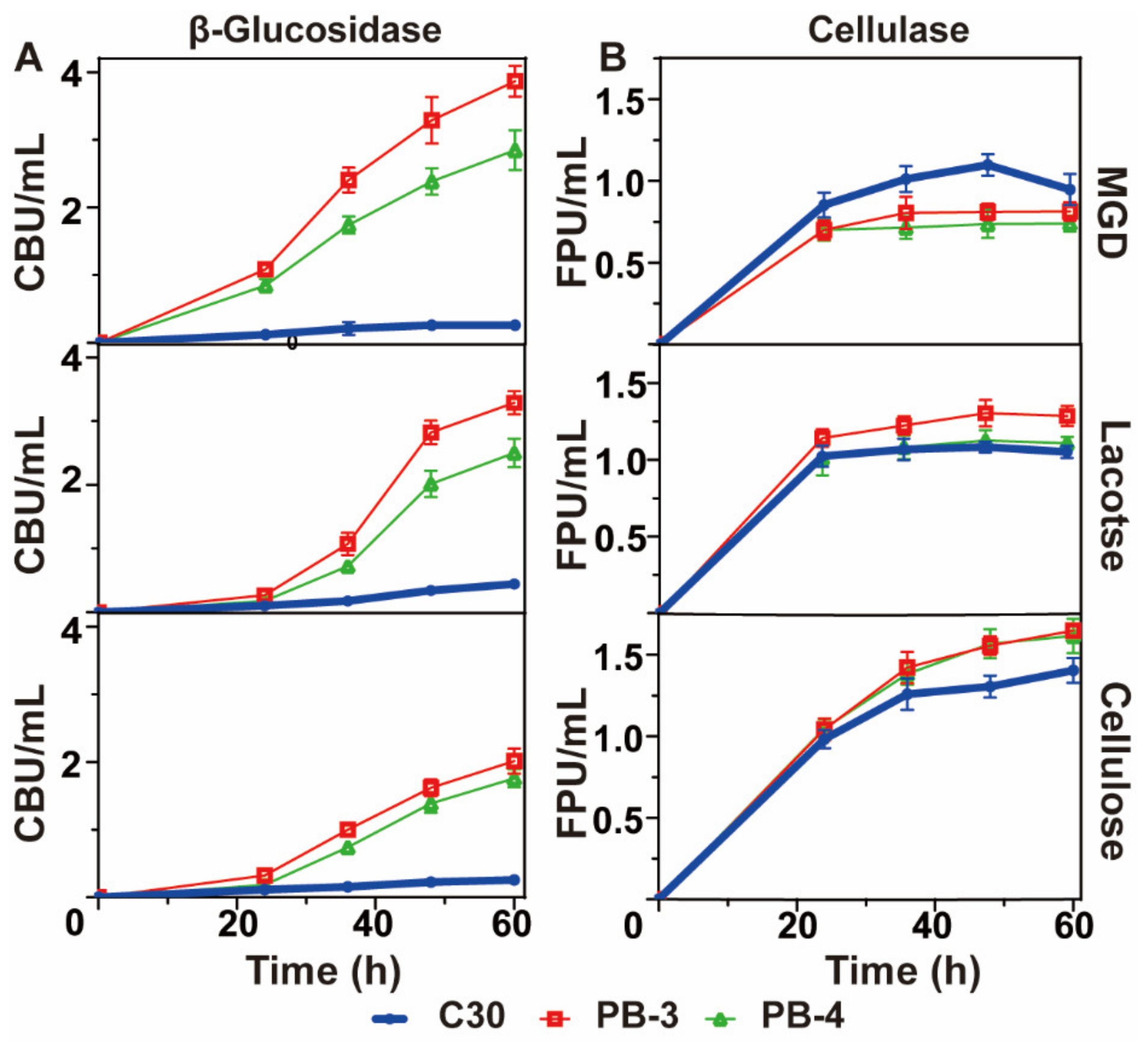
3.1. Effect of Different Inducers on the Synthesis of Cellulase from T. reesei Rut C30, PB3 and PB4

T. reesei, as an industrial fermentation strain of cellulase, has a low level of β-glucosidase (BGL) expression. Hence, BGL must be supplemented through commercial enzymes or through genetic engineering to overcome the lack of BGL [32]. In our previous research, aabgl1 encoding β-glucosidase 1 from A. aculeatus with high specific activity was used as the target gene to achieve higher BGL activity in T. reesei Rut C30, forming a series of genetically engineered T. reesei strains. Of these, four gene copies in T. reesei PB3 were integrated into the genome, enhancing the BGL activity by 73 times. Recombinant T. reesei PB4 integrated three copies, resulting in lower BGL activity than that of T. reesei PB3. However, we found that the cellulase activity of T. reesei PB3 and PB4 was significantly reduced when MGD was used as an inducer [16]. It was speculated that the sophorose in MGD was degraded into glucose, thus losing its inducing ability, so that the cellulase and BGL were evaluated using T. reesei Rut C30 and the two recombinant T. reesei with different inducers (MGD, lactose or cellulose) (Figure 1).
The BGL activities of both T. reesei PB3 and PB4 were significantly improved, regardless of the inducer (Figure 1A). Compared to T. reesei Rut C30, the BGL activity of T. reesei PB3 under the induction of MGD, lactose and cellulose was improved by 14.88, 7.45 and 7.73 times, respectively, and the BGL activity of T. reesei PB4 was enhanced by 10.96, 5.68 and 6.77 times, respectively.
Improving the BGL level relieves the inhibitory effects of cellobiose on cellobiohydrolase and endoglucanase, thereby significantly improving the cellulase activity [33]. In the present study, cellulase activity was improved under induction by cellulose or lactose, but was weakened when MGD was used as an inducer (Figure 1B). Under induction by cellulose or lactose, the cellulase activity of PB3 relative to that of T. reesei Rut C30 increased by 17.86% and 22.86%, respectively, and the cellulase activity of T. reesei PB4 relative to that of T. reesei Rut C30 increased by 15.71% and 3.7%, respectively. Unexpectedly, the cellulase activity of T. reesei PB3 and T. reesei PB4 relative to T. reesei Rut C30 under induction by MGD decreased by 26.36% and 32.73%, respectively.
Sophorose is the strongest inducer for cellulase production by T. reesei known thus far, but BGL degrades sophorose into glucose, which makes it lose its inducing ability and ultimately leads to a decrease in the expression levels of the cellulase gene. With cellulose as the inducer, cellobiohydrolase and endoglucanase synergistically degrade cellulose into cellobiose, which can enter cells. The inducing ability of cellobiose is 2000 times lower than that of sophorose, but BGL has the ability to catalyze cellobiose into sophorose by a transglycosidation reaction to efficiently induce cellulase production [19]. Thus, when cellulose is used as an inducer, the transglycosylation activity of BGL is needed. The induction process of lactose is similar to that of cellulose [18]. Hence, when cellulose or lactose is used as an inducer, the transglycosylation activity of BGL is indispensable for maintaining its inducing ability. When MGD is used, the transglycosylation activity of BGL is not needed, rather, its hydrolytic activity degrades sophorose into glucose and loses its inducibility. Moreover, the sophorose as an inducer was needed at a very low concentration, and extra sophorose tended to be hydrolysed into glucose by β-glucosidase, which is used as a carbon source [14].
Thus, we propose an innovative method of cellulase production. Namely, inhibition of the BGL of T. reesei deprives the sophorose metabolizing ability of T. reesei, which allows BGL-deficient cellulase to be continually produced with MGD as the inducer. This process would not affect the use of cellulase because the cellulase produced by T. reesei sufficiently supplements BGL during practical cellulase applications. Moreover, during BGL supplementation, the background activity of BGL can be ignored, indicating that the innovative method of cellulase production does not affect the quality of cellulase. This idea is similar to the prokaryotic expression system using isopropyl-β-D-thiogalactopyranoside (IPTG), which cannot be metabolized to replace lactose as the inducer, resulting in the persistent induction of the gene expression without the addition of continual flow inducers; therefore, this new method demonstrates potential in stimulating cellulase production and a promising system of T. reesei protein expression.

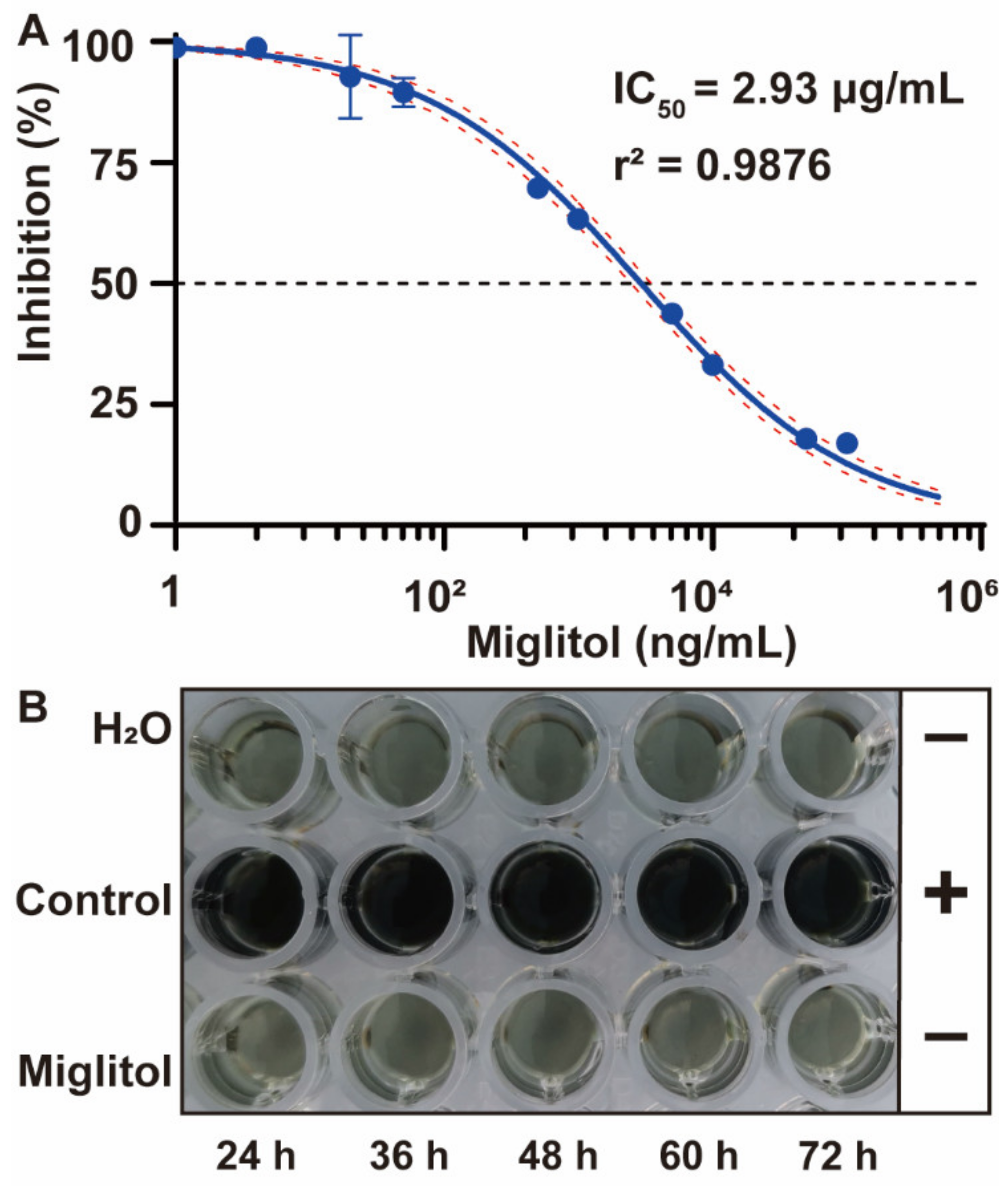
3.2. Development and Validation of β-Glucosidase Inhibitors

To rapidly validate the feasibility of the above-mentioned method for producing cellulase, we tried BGL inhibitor and added this inhibitor to test whether MGD improves the induction of cellulase production by T. reesei. Although there is no commercial β-glucosidase (BGL) inhibitor, α-glucosidase inhibitors are commonly used to decrease oral blood sugar to treat type II diabetes. Commercially available drugs include miglitol, acarbose and voglibose. We preliminarily tested the inhibitory effects of these three compounds on BGL and found that miglitol had the best inhibitory effects on BGL.
A miglitol concentration gradient (0–5 mg/mL) was used with cellobiose as the substrate, and the inhibition rates on BGL were detected under the optimal enzyme reaction conditions. Miglitol inhibited extracellular BGL activity with an IC50 value of 2.93 μg/mL (Figure 2A). Furthermore, a quick screening method was employed to identify the BGL activity of fermentation broth from T. reesei Rut C30 using MGD as an inducer at 24, 36, 48, 60 or 72 h. Aesculin solutions that turned black indicated the presence of BGL. We found that the enzyme solution detected by the aesculin solution turned black in the miglitol-free medium, however, it did not turn black when miglitol was supplemented (Figure 2B). The color changes were consistent with those in the negative control, suggesting that miglitol efficiently suppresses the β-glucosidase activity of T. reesei.

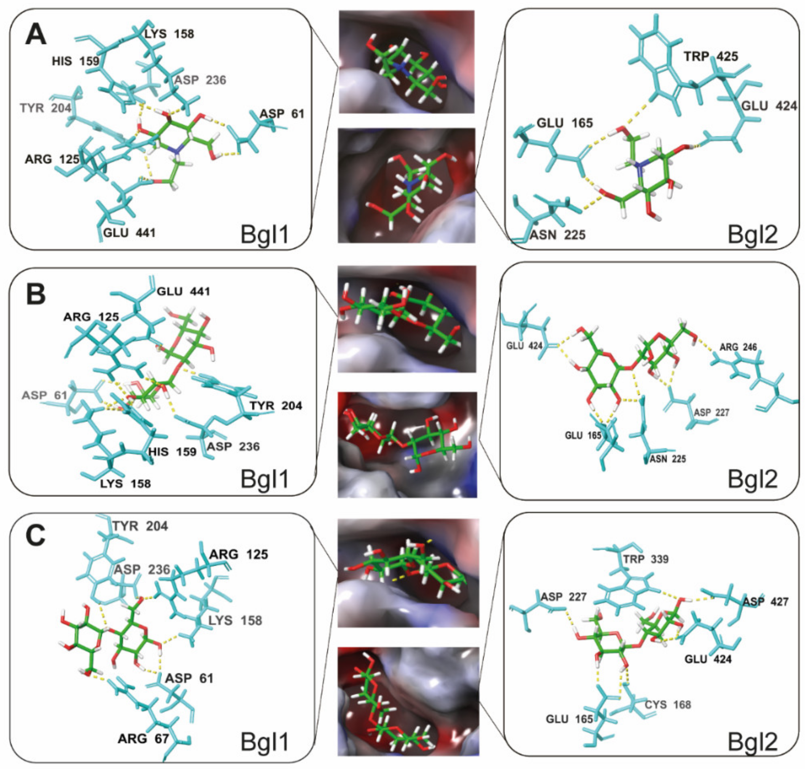
3.3. Molecular Mechanism by which Miglitol Inhibits β-Glucosidase

To analyze the molecular mechanism by which miglitol inhibits BGL, we molecularly docked miglitol to two BGLs (Bgl1 and Bgl2) previously reported in T. reesei and set the substrates (cellobiose and sophorose) as control tests. Bgl1 and Bgl2 are the most important extracellular and intracellular BGLs in T. reesei, respectively. Figure 3A shows the associations of miglitol with the two BGLs. Figure 3B,C illustrate the docking between the substrates (sophorose and cellobiose) and the two BGLs. The results showed that the binding sites between miglitol and the two BGLs are located at the active sites and are competitive inhibitors.
Molecular docking experiments showed that miglitol binds with several key active residues (aspartic acid, glutamate, arginine and tyrosine) in the active site of BGL to form hydrogen bonds. The binding sites between miglitol and BGL were magnified, showing that miglitol binds with seven key amino acid residues of Bgl1 and four key amino acid residues of Bgl2. Notably, the main interactive affinity is attributed to the presence of hydroxyl groups, and it is the key to anchoring the substrate to the site catalytic pocket [34,35]. The formation of more types and larger quantities of hydrogen bonds indicates that the ligand- and protein-binding ability is stronger. Molecular docking experiments indicated that the quantity of bonds in Bgl1 was greater than that in Bgl2, suggesting that the affinity of miglitol and Bgl1 was stronger with more stable docking and a higher matching degree. Overall, miglitol matched well with the two types of BGLs, indicating that miglitol is an efficient BGL inhibitor and an excellent experimental material with intracellular functionality.
Based on the molecular docking experiments, we predicted that miglitol is a potential BGL competitive inhibitor. The substrates (cellobiose and sophorose) of the two BGLs were docked with Bgl1 and Bgl2, and the active site between the different ligands and proteins were the same (Table 1).
The docking results showed that the binding energy values of Bgl1 or Bgl2 with miglitol were less than −6 kcal/mol, which indicated that the binding force between the two BGLs and miglitol was stronger. In addition, the binding energy of miglitol was significantly higher than those of the two substrates, suggesting that the miglitol and Bgl1 affinity is the strongest. For Bgl2, the scores of miglitol were similar to those of sophorose, and they were both better than those of cellobiose, indicating that the affinity between cellobiose and Bgl2 is the lowest. Hence, the binding energy of miglitol with both BGLs was higher than that with the two substrates (cellobiose and sophorose). The above analyses indicated that the miglitol and BGL interactive force is strong enough to inhibit BGL. With the coexistence of miglitol, sophorose and cellobiose, the affinity of miglitol for Bgl1 or Bgl2 was stronger and competitively inhibited enzyme and substrate binding, further inhibiting the activity of BGL and the decomposition of sophorose. As a result, the intracellular sophorose concentration, which continually induces cellulase gene expression, was maintained.

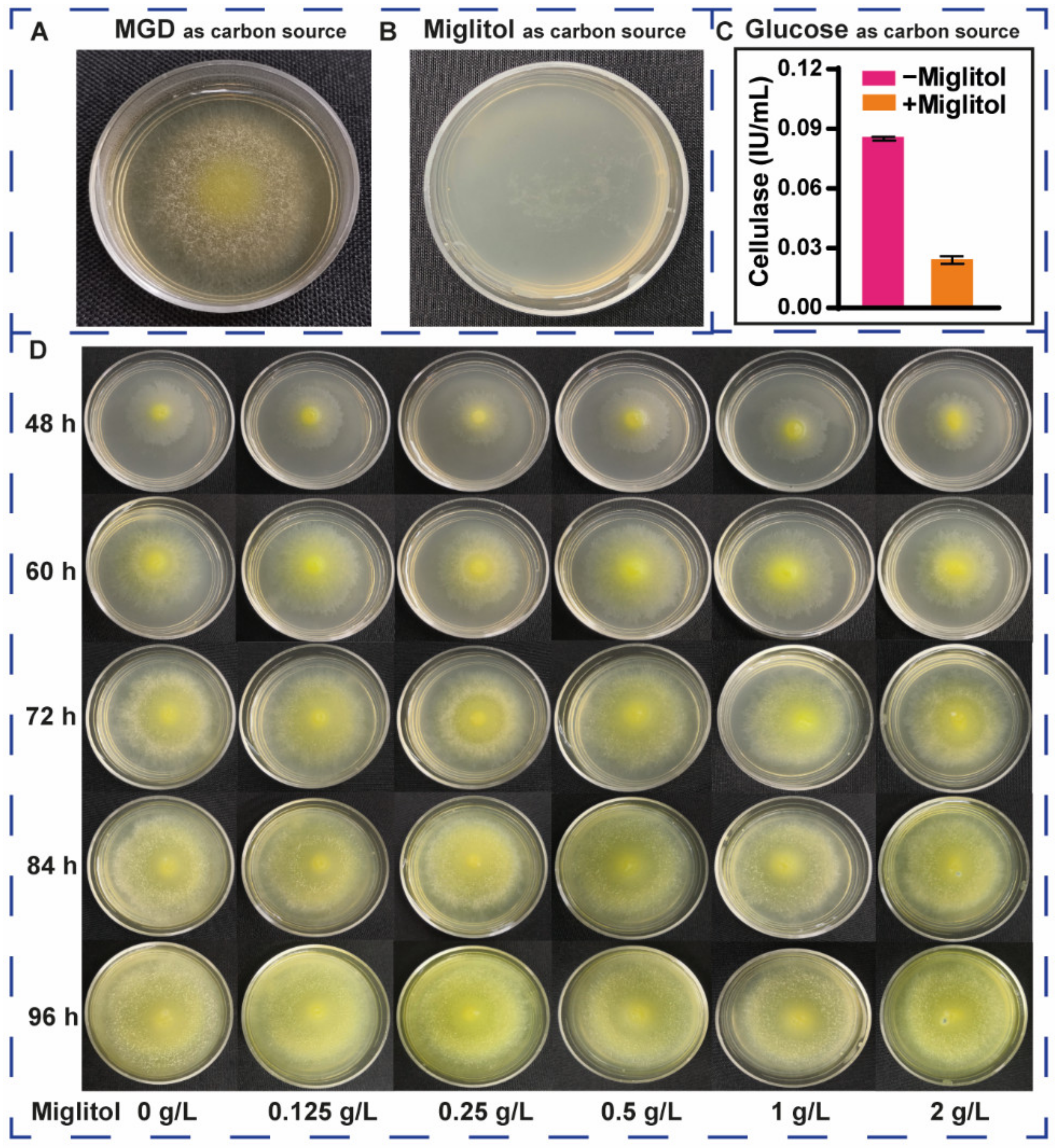
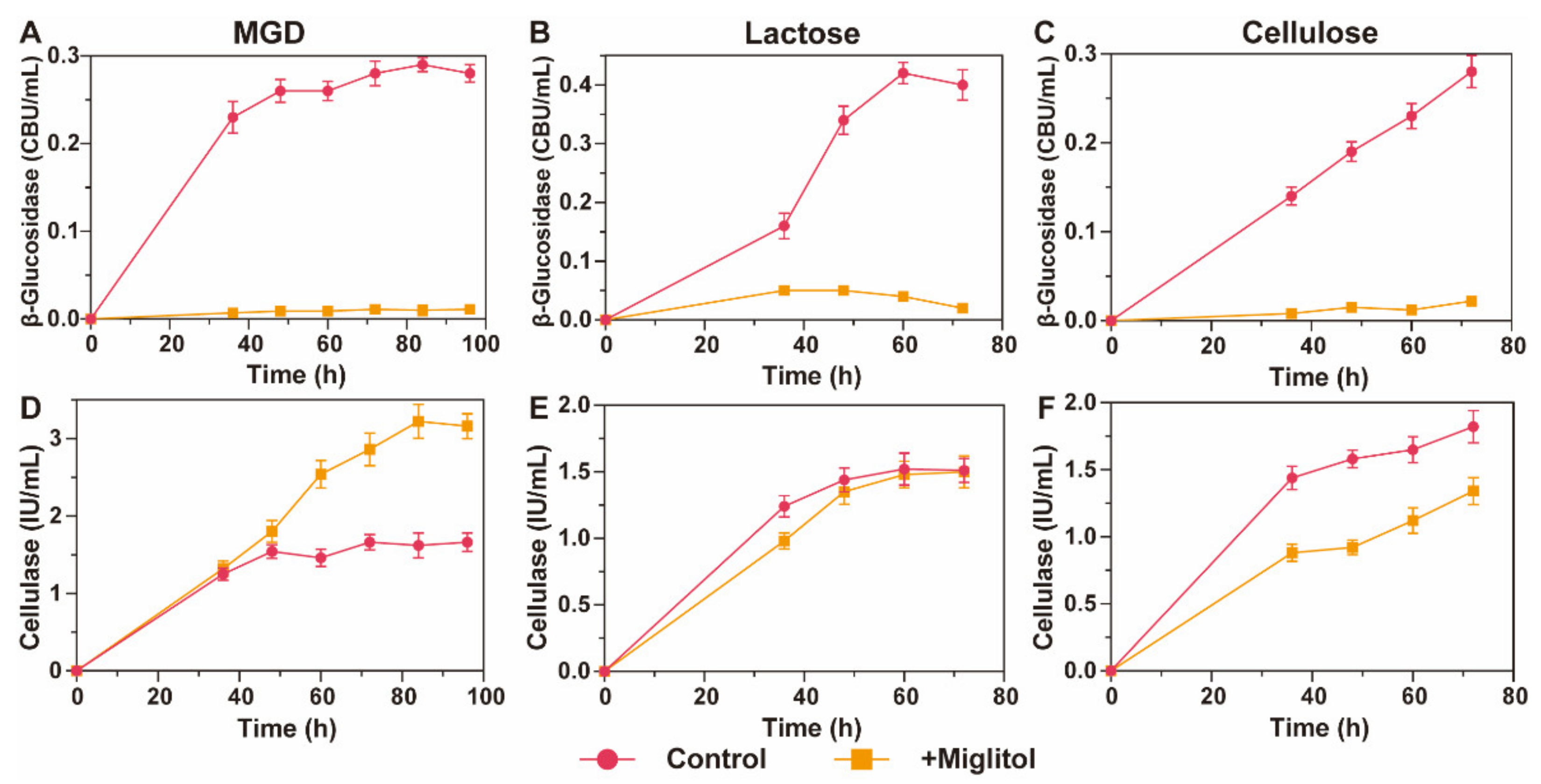
3.4. Effects of Miglitol on the Growth and Induction of T. reesei

To validate whether T. reesei uses miglitol as the carbon source, we conducted plate growth experiments using MGD or miglitol as the sole carbon source, with the other medium components and culture conditions being completely consistent (Figure 4). The strains grew well in MGD (Figure 4A), however, when miglitol was used as the sole carbon source, the strains did not grow (Figure 4B), indicating that T. reesei cannot metabolize miglitol as the carbon source. To further validate whether miglitol acts as an inducer of cellulase synthesis, 1 g/L miglitol was added to the fermentation medium, with 10 g/L glucose as a carbon source. In the absence of the inducer, a small amount of cellulase was detected with an activity of 0.085 IU/mL, and the cellulase activity was then further reduced to 0.024 IU/mL when miglitol was added to the fermentation medium (Figure 4C). The reason was that miglitol could not induce T. reesei to generate cellulase, but instead inhibited β-glucosidase and thereby inactivated cellulase. The results suggested that miglitol cannot be used by T. reesei as a carbon source or as an inducer of cellulase synthesis.
To further verify whether miglitol affects the growth of strains, we added different concentrations of miglitol to solid plates with MGD as the carbon source. At the same culture time, the concentration of miglitol, even up to 2 g/L, did not significantly affect the growth diameter of the strains (Figure 4D). Although the above results suggested that miglitol does not affect the growth of T. reesei or the induced synthesis of cellulase, it significantly inhibits the activity of BGL. Moreover, the activity of BGL produced by T. reesei was low. The results of the half maximal inhibitory concentration (IC50) showed that trace miglitol inactivated BGL. However, to severely inactivate BGL, we added 1 g/L miglitol in subsequent experiments to validate that the intracellular β-disaccharide concentration of T. reesei can be stabilized, which continually induces the synthesis of cellulase.

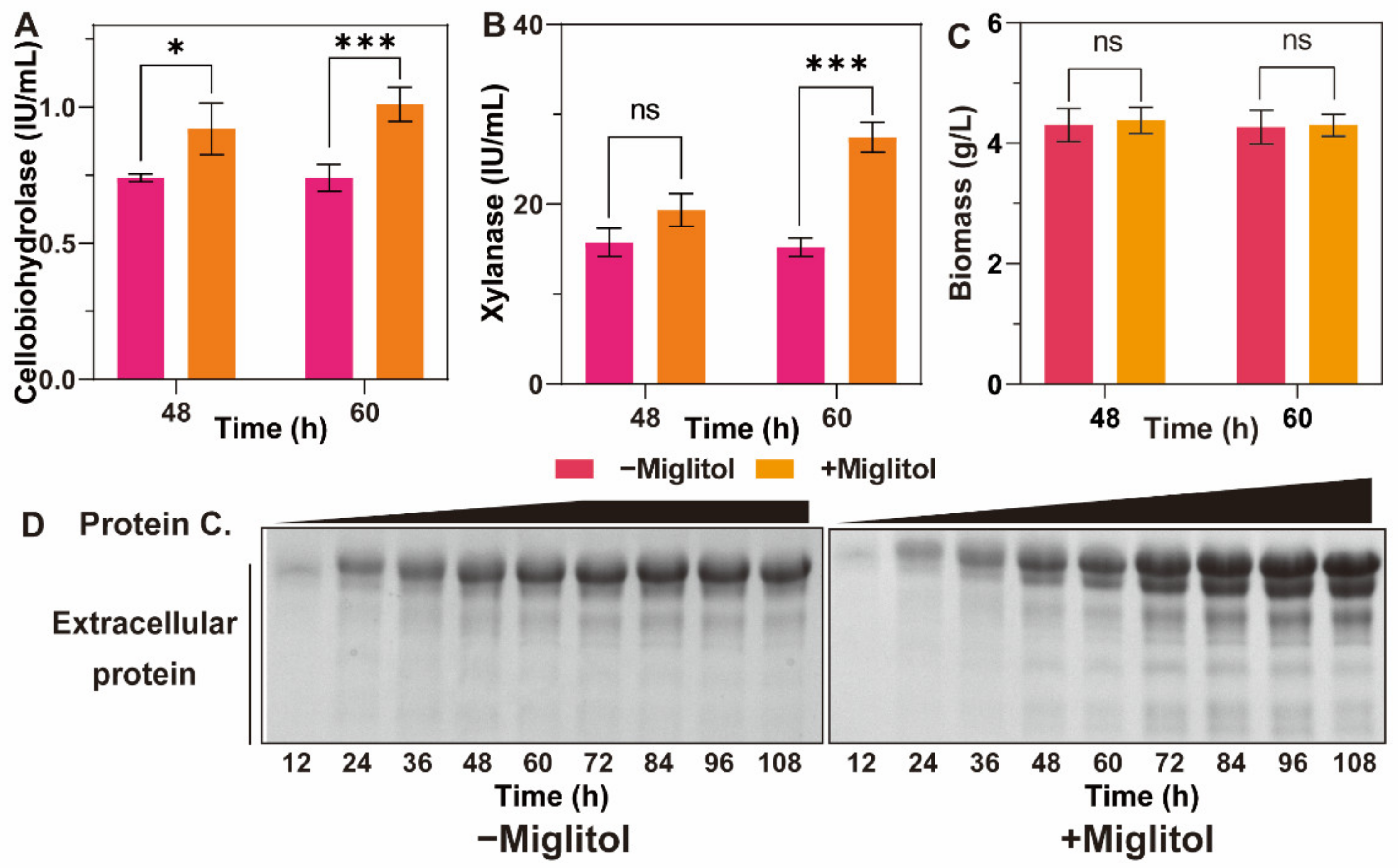
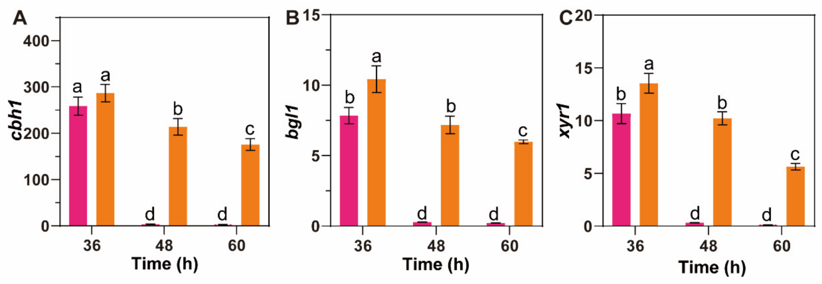
3.5. β-Glucosidase Suppression Promotes MGD to Persistently Induce Cellulase Synthesis in T. reesei

Figure 5 illustrates the production of cellulase and BGL by T. reesei cultured in fermentation medium with MGD, lactose or cellulose as the sole inducer. The activity of BGL in the presence of miglitol was effectively inhibited regardless of the inducer, which was consistent with the results above (Figure 5A–C). We then analyzed whether inducer degradation can be prevented so that the cellulase synthesis by T. reesei can be continually stimulated after the suppression of BGL.
Figure 5D shows cellulase production induced by MGD with the addition of miglitol. Interestingly, cellulase activity was persistently enhanced. In the absence of miglitol, the cellulase activity was maximized to 1.54 IU/mL at 48 h, then decreased after 60 h and increased sometime thereafter, which was due to the extracellular release of intracellular cellulase after cell death. In contrast, the cellulase activity after miglitol addition reached 1.8 IU/mL at 48 h, but further increased afterwards. The maximum cellulase activity achieved 3.22 IU/mL at 84 h, which was 2.09 times (p < 0.01) higher than that without the addition of miglitol. This result suggested that the addition of miglitol inactivated BGL, which prevented the degradation of the inducers in MGD (e.g., sophorose), allowing continual induction of cellulase synthesis in T. reesei. This is equivalent to endowing T. reesei with a phenotype that cannot metabolize sophorose but does not lose its ability to synthesize sophorose-induced cellulase. The above result provides a new clue for cellulase production in T. reesei. In the future, genetically engineered T. reesei can be developed to avoid the use of miglitol or other similar inhibitors. Moreover, the BGL activity of T. reesei is extremely low, and extra supplementation is needed during the hydrolysis of straw biomass. Hence, this method of cellulase production does not affect the enzyme quality.
However, the results of cellulase production with lactose or cellulose as the inducer were opposite to the result with MGD (Figure 5D–F). Although BGL activity was also inhibited after the addition of miglitol (Figure 5B,C), the cellulase activity was not further enhanced (Figure 5E,F), which was consistent with the results obtained without the addition of miglitol, and the enzymatic activity was even reduced. The cellulase activity was not significantly decreased after the addition of miglitol compared to the lactose control (Figure 5E); however, the activity was significantly weakened by 26% in the presence of miglitol when cellulose was used as an inducer (Figure 5F). These results indicated that BGL is indispensable when cellulose or lactose act as an inducer. Reportedly, the transglycosylation activity of BGL is critical for the inducibility of lactose or cellulose [18,19]. The reason is that a strong cellulase inducer, sophorose, can be produced in T. reesei, which was directly demonstrated by our previous findings. MGD contains a certain amount of sophorose and does not require the transglycosylation activity of BGL, which can induce T. reesei to persistently synthesize cellulase after BGL inhibition.
Overall, the cellulase activity reached its highest level at 48 h and started to decrease after 60 h without miglitol addition, indicating that the carbon source and inducer in the medium were already metabolized by T. reesei, leading to the retarded growth of the strain. To further explore whether the activities of cellobiohydrolase and xylanase were also enhanced after the addition of miglitol, we measured the activities of these two enzymes at the two tested time points (Figure 6A,B). The results showed that the changing trends were consistent with that of cellulase activity, as the two enzymatic activities without the addition of miglitol started to decrease after 60 h. After the addition of miglitol, however, the activities of cellobiohydrolase and xylanase at 60 h were 1.36 and 1.80 times higher than the corresponding results without miglitol, respectively (p < 0.01), indicating that the enzyme would be more efficient for hydrolyzing lignocellulosic biomass pretreated by alkali, in which the hemicelluloses and xylan remain hydrolyzed together with the cellulose component [36]. This comparison further validates that the inducing activity of MGD can be maintained after BGL inhibition by miglitol. The experimental results to test whether the addition of miglitol affects the biomass of the strain at the two tested time points (Figure 6C) were consistent with those shown in Figure 4D. Namely, the addition of miglitol did not affect the growth or metabolism of the strain and did not significantly change the biomass at either time point. Moreover, SDS-PAGE showed that the extracellular protein concentrations of T. reesei were significantly improved after the addition of miglitol (Figure 6D), further verifying the actions of miglitol.
Figure 7A–C illustrates the expression of two major cellulase genes (cbh1 and bgl1) and the most important transcription activator (xyr1), which were tested to explore the molecular mechanism by which MGD induces cellulase production in T. reesei after the addition of miglitol. The results showed that the three genes were all expressed under induction by MGD. When miglitol was not added, the expression levels of the three genes all peaked after 36 h, but significantly decreased after 48 h and nearly approached 0, which indicated that the inducer was fully metabolized after 48 h of culture. These results were consistent with the cellulase activity results shown in Figure 5. The genetic transcription level of cellulase was maximized at 36 h, hence, cellulase activity maximized at 48 h. After the addition of miglitol, the gene expression of cbh1, bgl1 and xyr1 can still be started after 48 or 60 h, further indicating that the inducer exists in the medium and can induce cellulase synthesis after the suppression of BGL. Notably, miglitol only inhibited the enzymatic activity but did not affect the gene expression of BGL (Figure 7B).
Recently, to improve the efficiency of lignocellulose hydrolysis by cellulase, the overexpression of BGL in T. reesei has been extensively used [21,22,23,24,25]. However, the present study showed that BGL is important for the inducing role of cellulose or lactose because its transglycosylation activity must be utilized to produce the strongest inducer of T. reesei, sophorose, to maximally induce cellulase production. However, MGD contains a certain amount of sophorose and thus does not require the transglycosylation activity of BGL, conversely, its hydrolyzing ability degrades sophorose into glucose, thereby completely inhibiting the inducing activity of sophorose. Hence, this sophorose containing inducer must be continually supplemented in a fed-batch manner. In the present study, the long-term inducing ability of MGD was maintained after the inhibition of BGL, and the dosage of the inducer was decreased. In addition, although BGL is pivotal in the degradation of cellulose components into glucose, researchers have suggested that the BGL secreted by T. reesei is largely insufficient, indicating that the efficient hydrolysis of cellulose components can be achieved only when enough BGL is formulated. Hence, the present new method does not largely reduce the quality of cellulase of T. reesei and is helpful for overcoming the problem of the inducer needing to be supplemented through feeding, faced in high titer cellulase production by fed-batch fermentation in the future.

4. Conclusions

The present study confirmed that miglitol competitively inhibits β-glucosidase, with an IC50 of 2.93 μg/mL. Adding miglitol into a sophorose-containing culture medium significantly weakened β-glucosidase activity to levels near 0, which prevented the degradation of sophorose, thereby allowing it to continually induce the production of cellulase by T. reesei. However, the addition of miglitol is ineffective when the inducer is lactose or cellulose, indicating that the transglycosylation activity of BGL is crucial for the activities of these two inducers. Our findings indicated that this alternative strategy was developed to induce the synthesis of cellulase and provides a promising system for T. reesei protein expression.

Supplementary Materials

The following supporting information can be downloaded at: https://0-www-mdpi-com.brum.beds.ac.uk/article/10.3390/fermentation8050232/s1, Table S1. Primers for qRT-PCR analysis.

Author Contributions

Conceptualization, Y.L.; methodology, T.L.; software, P.Z. and T.L.; validation, J.Y., Y.G. and X.R.; formal analysis, J.Y.; investigation, T.L.; resources, P.Z.; data curation, T.L.; writing—original draft preparation, Y.L.; writing—review and editing, T.L.; visualization, P.Z.; supervision, J.Y.; project administration, Y.G. and X.R.; funding acquisition, Y.L. All authors have read and agreed to the published version of the manuscript.

Funding

This research was financially funded by the National Natural Science Foundation of China with the grant reference numbers of 21808022, the Natural Science Foundation Project of Chongqing, the Chongqing Science and Technology Commission (CN) (cstc2018jcyjAX0064), the Science and Technology Research Program of Chongqing Municipal Education Commission (KJQN201901523), the Postgraduate Research and Innovation Project of Chongqing University of Science and Technology (YKJCX2120505 and YKJCX2120523), and also supported by the National Undergraduate Training Programs for Innovation and Entrepreneurship of China (No. 202111551022).

Institutional Review Board Statement

Not applicable.

Informed Consent Statement

Not applicable.

Data Availability Statement

Not applicable.

Acknowledgments

The authors appreciate the helpful discussion with Xinqing Zhao at Shanghai Jiao Tong University, and also appreciate the helpful discussion with Ruimeng Gu at the Tianjin Institute of Industrial Biotechnology, Chinese Academy of Sciences.

Conflicts of Interest

The authors declare that they have no competing interests.

References

  1. Himmel, M.E.; Edward, A.B. Lignocellulose conversion to biofuels: Current challenges, global perspectives. Curr. Opin. Biotechnol. 2009, 20, 316–317. [Google Scholar] [CrossRef] [PubMed]
  2. Reaney, M.; Wiens, D.; Tse, T. Production of bioethanol-A review of factors affecting ethanol yield. Fermentation 2021, 7, 268. [Google Scholar] [CrossRef]
  3. Liu, C.G.; Xiao, Y.; Xia, X.X.; Zhao, X.Q.; Peng, L.; Srinophakun, P.; Bai, F. Cellulosic ethanol production: Progress, challenges and strategies for solutions. Biotechnol. Adv. 2019, 37, 491–504. [Google Scholar] [CrossRef] [PubMed]
  4. Prasoulas, G.; Gentikis, A.; Konti, A.; Kalantzi, S.; Kekos, D.; Mamma, D. Bioethanol production from food waste applying the multienzyme system produced on-site by Fusarium oxysporum F3 and mixed microbial cultures. Fermentation 2020, 6, 39. [Google Scholar] [CrossRef] [Green Version]
  5. Bhati, N.; Sharma, A.K. Cost-effective cellulase production, improvement strategies, and future challenges. J. Food Process Eng. 2021, 44, e13623. [Google Scholar] [CrossRef]
  6. Bhardwaj, N.; Bikash, K.; Komal, A.; Pradeep, V. Current perspective on production and applications of microbial cellulases: A review. Bioresour. Bioprocess. 2021, 8, 95. [Google Scholar] [CrossRef]
  7. Peterson, R.; Nevalainen, H. Trichoderma reesei RUT-C30–thirty years of strain improvement. Microbiology 2012, 158, 58–68. [Google Scholar] [CrossRef] [Green Version]
  8. Fang, H.; Li, C.; Zhao, J.; Zhao, C. Biotechnological advances and trends in engineering Trichoderma reesei towards cellulase hyperproducer. Biotechnol. Bioprocess Eng. 2021, 26, 517–528. [Google Scholar] [CrossRef]
  9. Mandels, M.; Parrish, F.W.; Reese, E.T. Sophprpse as an inducer of cellulase in Trichoderma viride. J. Bacteriol. 1962, 83, 400–408. [Google Scholar] [CrossRef] [Green Version]
  10. Humbird, D.; Davis, R.; Tao, L.; Kinchin, C.; Hsu, D.; Aden, A.; Schoen, P.; Lukas, J.; Olthof, B.; Worley, M.; et al. Process Design and Economics for Biochemical Conversion of Lignocellulosic Biomass to Ethanol: Dilute-Acid Pretreatment and Enzymatic Hydrolysis of Corn Stover; Technical Report NREL/TP-5100-47764; National Renewable Energy Laboratory: Golden, CO, USA, 2012. [CrossRef] [Green Version]
  11. Li, Y.; Liu, C.; Bai, F.; Zhao, X. Overproduction of cellulase by Trichoderma reesei RUT C30 through batch-feeding of synthesized low-cost sugar mixture. Bioresour. Technol. 2016, 216, 503–510. [Google Scholar] [CrossRef]
  12. Xia, Y.; Yang, L.; Xia, L. High-level production of a fungal β-glucosidase with application potentials in the cost-effective production of Trichoderma reesei cellulase. Process Biochem. 2018, 70, 55–60. [Google Scholar] [CrossRef]
  13. Li, Y.; Zhang, X.; Xiong, L.; Mehmood, M.A.; Zhao, X.; Bai, F. On-site cellulase production and efficient saccharification of corn stover employing cbh2 overexpressing Trichoderma reesei with novel induction system. Bioresour. Technol. 2017, 238, 643–649. [Google Scholar] [CrossRef] [PubMed]
  14. Li, Y.; Zhang, P.; Zhu, D.; Yao, B.; Hasunuma, T.; Kondo, A.; Zhao, X. Efficient preparation of soluble inducer for cellulase production and saccharification of corn stover using in-house generated crude enzymes. Biochem. Eng. J. 2022, 178, 108296. [Google Scholar] [CrossRef]
  15. Chen, Y.; Wu, C.; Fan, X.; Zhao, X.; Wang, W. Engineering of Trichoderma reesei for enhanced degradation of lignocellulosic biomass by truncation of the cellulase activator ACE3. Biotechnol. Biofuels 2020, 13, 62. [Google Scholar] [CrossRef] [PubMed]
  16. Li, Y.; Zhang, X.; Zhang, F.; Peng, L.; Zhang, D.; Kondo, A.; Bai, F.; Zhao, X. Optimization of cellulolytic enzyme components through engineering Trichoderma reesei and on-site fermentation using the soluble inducer for cellulosic ethanol production from corn stover. Biotechnol. Biofuels 2018, 11, 49. [Google Scholar] [CrossRef] [PubMed] [Green Version]
  17. Li, Y.; Yu, J.; Zhang, P.; Long, T.; Mo, Y.; Li, J.; Li, Q. Comparative transcriptome analysis of Trichoderma reesei reveals different gene regulatory networks induced by synthetic mixtures of glucose and β-disaccharide. Bioresour. Bioprocess. 2021, 8, 57. [Google Scholar] [CrossRef]
  18. Xu, J.; Zhao, G.; Kou, Y.; Zhang, W.; Zhou, Q.; Chen, G.; Liu, W. Intracellular β-glucosidases CEL1a and CEL1b are essential for cellulase induction on lactose in Trichoderma reesei. Eukaryot. Cell 2014, 13, 1001–1013. [Google Scholar] [CrossRef] [Green Version]
  19. Zhou, Q.; Xu, J.; Kou, Y.; Lv, X.; Zhang, X.; Zhao, G.; Zhang, W.; Chen, G.; Liu, W. Differential involvement of β-glucosidases from Hypocrea jecorina in rapid induction of cellulase genes by cellulose and cellobiose. Eukaryot. Cell 2012, 11, 1371–1381. [Google Scholar] [CrossRef] [Green Version]
  20. Qi, K.; Chen, C.; Yan, F.; Feng, Y.; Bayer, E.A.; Kosugi, A.; Cui, Q.; Liu, Y. Coordinated β-glucosidase activity with the cellulosome is effective for enhanced lignocellulose saccharification. Bioresour. Technol. 2021, 337, 125441. [Google Scholar] [CrossRef]
  21. Dashtban, M.; Qin, W. Overexpression of an exotic thermotolerant β-glucosidase in Trichoderma reesei and its significant increase in cellulolytic activity and saccharification of barley straw. Microb. Cell Factories 2012, 11, 63. [Google Scholar] [CrossRef] [Green Version]
  22. Treebupachatsakul, T.; Shioya, K.; Nakazawa, H.; Kawaguchi, T.; Morikawa, Y.; Shida, Y.; Ogasawara, W.; Okada, H. Utilization of recombinant Trichoderma reesei expressing Aspergillus aculeatus β-glucosidase I (JN11) for a more economical production of ethanol from lignocellulosic biomass. J. Biosci. Bioeng. 2015, 120, 657–665. [Google Scholar] [CrossRef] [PubMed]
  23. Zhang, J.; Zhong, Y.; Zhao, X.; Wang, T. Development of the cellulolytic fungus Trichoderma reesei strain with enhanced β-glucosidase and filter paper activity using strong artifical cellobiohydrolase 1 promoter. Bioresour. Technol. 2010, 101, 9815–9818. [Google Scholar] [CrossRef] [PubMed]
  24. Nakazawa, H.; Kawai, T.; Ida, N.; Shida, Y.; Kobayashi, Y.; Okada, H.; Tani, S.; Sumitani, J.-I.; Kawaguchi, T.; Morikawa, Y.; et al. Construction of a recombinant Trichoderma reesei strain expressing Aspergillus aculeatus β-glucosidase 1 for efficient biomass conversion. Biotechnol. Bioeng. 2012, 109, 92–99. [Google Scholar] [CrossRef] [PubMed]
  25. Li, C.; Lin, F.; Li, Y.; Wei, W.; Wang, H.; Qin, L.; Zhou, Z.; Li, B.; Wu, F.-G.; Chen, Z. A β-glucosidase hyper-production Trichoderma reesei mutant reveals a potential role of cel3D in cellulase production. Microb. Cell Factories 2016, 15, 151. [Google Scholar] [CrossRef] [PubMed] [Green Version]
  26. Sels, J.P.J.; Huijberts, M.S.; Wolffenbuttel, B.H. Miglitol, a new α-glucosidase inhibitor. Expert Opin. Pharmacother. 1999, 1, 149–156. [Google Scholar] [CrossRef]
  27. Pérez, G.; Farina, L.; Barquet, M.; Boido, E.; Gaggero, C.; Dellacassa, E.; Carrau, F. A quick screening method to identify β-glucosidase activity in native wine yeast strains: Application of Esculin Glycerol Agar (EGA) medium. World J. Microbiol. Biotechnol. 2011, 27, 47–55. [Google Scholar] [CrossRef]
  28. Eberhardt, J.; Santos-Martins, D.; Tillack, A.F.; Forli, S. AutoDock Vina 1.2.0: New Docking Methods, Expanded Force Field, and Python Bindings. J. Chem. Inf. Model. 2021, 61, 3891–3898. [Google Scholar] [CrossRef]
  29. Ghose, T.K. Measurement of cellulase activities. Pure Appl. Chem. 1987, 59, 257–268. [Google Scholar] [CrossRef]
  30. Jalak, J.; Väljamäe, P. Mechanism of initial rapid rate retardation in cellobiohydrolase catalyzed cellulose hydrolysis. Biotechnol. Bioeng. 2010, 106, 871–883. [Google Scholar] [CrossRef]
  31. Törrönen, A.; Mach, R.L.; Messner, R.; Gonzalez, R.; Kalkkinen, N.; Harkki, A.; Kubicek, C.P. The two major xylanases from Trichoderma reesei: Characterization of both enzymes and genes. Nat. Biotechnol. 1992, 10, 1461–1465. [Google Scholar] [CrossRef]
  32. Xia, Y.; Yang, L.; Xia, L. Combined strategy of transcription factor manipulation and β-glucosidase gene overexpression in Trichoderma reesei and its application in lignocellulose bioconversion. J. Ind. Microbiol. Biotechnol. 2018, 45, 803–811. [Google Scholar] [CrossRef] [PubMed]
  33. Hirano, K.; Saito, T.; Shinoda, S.; Haruki, M.; Hirano, N. In vitro assembly and cellulolytic activity of a β-glucosidase-integrated cellulosome complex. FEMS Microbiol. Lett. 2019, 366, fnz209. [Google Scholar] [CrossRef]
  34. Du, X.; Li, Y.; Xia, Y.; Ai, S.; Liang, J.; Sang, P.; Ji, X.; Liu, S. Insights into protein–ligand interactions: Mechanisms, models, and methods. Int. J. Mol. Sci. 2016, 17, 20144. [Google Scholar] [CrossRef] [PubMed]
  35. He, M.; Zhai, Y.; Zhang, Y.; Xu, S.; Yu, S.; Wei, Y.; Xiao, H.; Song, Y. Inhibition of α-glucosidase by trilobatin and its mechanism: Kinetics, interaction mechanism and molecular docking. Food Funct. 2022, 13, 857–866. [Google Scholar] [CrossRef] [PubMed]
  36. Behera, S.; Arora, R.; Nandhagopal, N.; Kumar, S. Importance of chemical pretreatment for bioconversion of lignocellulosic biomass. Renew. Sustain. Energy Rev. 2014, 36, 91–106. [Google Scholar] [CrossRef]
Figure 1. The cellulase and β-glucosidase production of recombinant Trichoderma reesei PB-3, PB-4 with aabgl1 overexpressed and the parent strain Rut C30. The transformants T. reesei PB-3 and PB-4 had four and three gene copies, respectively. (A) The β-glucosidase activities and (B) the cellulase activities of T. reesei Rut C30, PB-3 and PB-4 cultured with 10 g/L MGD, lactose or cellulose. For each experiment, three individual replicates were performed. The cellulase and β-glucosidase activity were expressed in filter paper units (FPU) and cellobiase units (CBU), respectively.
Fermentation 08 00232 g001
Figure 2. Developing and validating the inhibitory effect of miglitol on β-glucosidase. (A) Inhibition of β-glucosidase activity by miglitol at different concentrations. The figures also show the corresponding IC50 values and correlation r2 values; (B) Effect of miglitol on the β-glucosidase activity of T. reesei Rut C30 using 10 g/L MGD as a carbon source and sampled at 24, 36, 48, 60 or 72 h. A quick detection system containing aesculin was employed in 96-well plates with no miglitol or water added as a negative control.
Fermentation 08 00232 g002
Figure 3. Binding modes of three substrates with Bgl1 and Bgl2. The interactions between miglitol (A); sophorose (B); and cellobiose (C) with the associated residues at the active site of β-glucosidase. 3D surface representation of the ligand-binding pocket (ligands displayed in stick mode) is in the middle of the figure. The black labels and yellow lines show the residue names and hydrogen bonds, respectively.
Fermentation 08 00232 g003
Figure 4. Effects of miglitol on the growth and induction of T. reesei. Colony morphology of T. reesei on plates containing (A) 10 g/L MGD or (B) 10 g/L miglitol as the sole carbon source; (C) Effect of adding miglitol versus no miglitol on the cellulase activity of T. reesei Rut C30 with 10 g/L glucose as the carbon source at 48 h; (D) Time-course of the colony morphology of T. reesei grown using 10 g/L MGD as a carbon source on different concentrations of miglitol. The mycelium growth was measured at 12 h intervals.
Fermentation 08 00232 g004
Figure 5. For cellulase and β-glucosidase activity after the addition of miglitol, time-course profiles of β-glucosidase (AC) and cellulase activity (DF) for T. reesei Rut C30 taking 10 g/L MGD (A,D); 10 g/L lactose (B,E); or 10 g/L cellulose (C,F) as the carbon source with/without miglitol. Bars denote the standard deviations for three independent experiments.
Fermentation 08 00232 g005
Figure 6. Effect of miglitol on lignocellulase production of T. reesei Rut C30 with 10 g/L MGD as the carbon source at 48 h and 60 h ((A) Cellobiohydrolase; (B) Xylanase; (C) Biomass); (D) SDS-PAGE analysis of supernatant. Values are the mean  ±  SD of the results from three independent experiments. Asterisks indicate a significant difference (* p  <  0.05, *** p  <  0.01, Student’s t-test).
Fermentation 08 00232 g006
Figure 7. Effect of miglitol on the transcription of genes encoding cellulase ((A) cbh1; (B) bgl1); and transcription factor ((C) xyr1) at 36 h, 48 h and 60 h of T. reesei Rut C30 with 10 g/L MGD as the carbon source at 36 h, 48 h and 60 h. Values are the mean  ±  SD of the results from three independent experiments. One-way ANOVA was performed to reveal significant (p < 0.05) differences between the mean values, which are indicated by different letters.
Fermentation 08 00232 g007
Table 1. Verification of the docking results for miglitol with β-glucosidase using the binding energy as the metric.
ProteinLigandBinding Energy/(Kcal/mol)
Bgl1Miglitol−6.341
Sophorose−5.680
Cellobiose−5.868
Bgl2Miglitol−7.511
Sophorose−7.586
Cellobiose−6.396
Publisher’s Note: MDPI stays neutral with regard to jurisdictional claims in published maps and institutional affiliations.

