Article

Microbial Communities in Underground Gas Reservoirs Offer Promising Biotechnological Potential

1
Laboratory of Anaerobic Microorganisms, Section of Microbiology, Department of Experimental Biology, Faculty of Science, Masaryk University, 625 00 Brno, Czech Republic
2
Testlab Geo-Services, RWE Gas Storage CZ, s.r.o., 627 00 Brno, Czech Republic
3
Department of Agricultural, Food and Environmental Engineering, Faculty of AgriSciences, Mendel University in Brno, 613 00 Brno, Czech Republic
4
Department of Biochemistry, Faculty of Science, Masaryk University, 625 00 Brno, Czech Republic
5
Department of Geological Sciences, Faculty of Science, Masaryk University, 602 00 Brno, Czech Republic
6
Tescan Orsay Holding, 623 00 Brno, Czech Republic
7
Department of Biology and Preclinical Medicine, Institute of Microbiology and Archaea Centre, University of Regensburg, 93053 Regensburg, Germany
*
Authors to whom correspondence should be addressed.
These authors contributed equally to this work.
Submission received: 1 May 2022 / Revised: 22 May 2022 / Accepted: 24 May 2022 / Published: 26 May 2022

Abstract

:
Securing new sources of renewable energy and achieving national self-sufficiency in natural gas have become increasingly important in recent times. The study described in this paper focuses on three geologically diverse underground gas reservoirs (UGS) that are the natural habitat of methane-producing archaea, as well as other microorganisms with which methanogens have various ecological relationships. The objective of this research was to describe the microbial metabolism of methane in these specific anoxic environments during the year. DNA sequencing analyses revealed the presence of different methanogenic communities and their metabolic potential in all sites studied. Hydrogenotrophic Methanobacterium sp. prevailed in Lobodice UGS, members of the hydrogenotrophic order Methanomicrobiales predominated in Dolní Dunajovice UGS and thermophilic hydrogenotrophic members of the Methanothermobacter sp. were prevalent in Tvrdonice UGS. Gas composition and isotope analyses were performed simultaneously. The results suggest that the biotechnological potential of UGS for biomethane production cannot be neglected.

Graphical Abstract

1. Introduction

Microbiological studies of underground gas storage (UGS) have demonstrated the presence of viable microorganisms [1,2] and their significant influence on biological and geochemical processes in these environments [3]. It is likely that Šmigáň and co-workers in 1990 were the first to observe the changes in the composition of the town gas in UGS caused by microorganisms. The recorded decrease in the amount of H2 and CO2 combined with the increase in the amount of CH4 in the Lobodice UGS, Czechia, indicated the possibility that microbial communities inhabit the UGS. The presence of methanogenic archaea, which appeared to be responsible for the changes in gas quality [3,4], was also confirmed. Thirty years later, our stable isotope measurements confirmed biological methane production at the same site and at other sites. Power-to-gas technology with biological methane production in underground reservoirs seems to be one of the most promising options for carbon-neutral fuel production and storage [5]. The UGS can serve as a reactor where the biological conversion of CO2 and H2 to biomethane takes place [6]. Sources of CO2 can be the thermal sector, various industries, or direct capture of CO2 from the air [7]. Hydrogen can be produced by electrolysis of water or using surplus renewable energy [8], while hydrogen from waste treatment could also be used in the future [9]. It is also clear that methane will be treated as an energy carrier until safe hydrogen storage and distribution technologies are available [10]. The objective of this study was to determine whether a UGS natural sedimentary rock environment could be a potential target for biomethane production through power-to-methane technology. Although many scientific teams have mapped the microbiome of the subsurface environment (Table 1), the idea of using UGS as a natural bioreactor is new and topical. The enormous capacity of UG enables the production and storage of biological methane in quantities of millions of cubic meters. Three geologically different sites in the Czech Republic—Lobodice, Dolní Dunajovice, and Tvrdonice—serving as UGS were studied in terms of microbial communities using sequencing technology and quantitative PCR to assess their methanogenesis processes.

Methanogenesis

Methanogenesis, the final step in the decomposition of organic matter, is carried out by methanogenic archaea, which play an important role in the global carbon cycle and are responsible for more than half of all methane produced on Earth per year [21]. In the last four decades, methanogenesis has also been described as the dominant metabolic pathway in very deep aquifers [22]. Methanogenic archaea are strictly anaerobic microorganisms and require an environment with low redox potentials of about −300 mV for their growth. Under these conditions, where all other favorable electron acceptors, such as oxygen, nitrate, sulphate, and iron compounds, are depleted or absent, methanogenesis can occur [23].
Methanogenesis is not a uniform process. Methane can be formed via three major pathways: hydrogenotrophic, methylotrophic, or acetoclastic. However, one major characteristic enzyme is present in all types of methanogenesis: methyl-coenzyme M reductase (MCR), which catalyzes the final step of methyl group reduction to methane [24].
The most widespread and probably the oldest form of methane production is hydrogenotrophic methanogenesis. This pathway is characterized by the conversion of CO2 and H2 to methane; molecular hydrogen serves as the electron donor and CO2 as the electron acceptor. The reduction of formate also occurs via the hydrogenotrophic pathway. The only difference is in the first step, where formate is oxidized to CO2 and then the CO2 continues the pathway. The hydrogenotrophic pathway is widely used; for example, by the microorganisms of the orders Methanobacteriales, Methanomicrobiales, and Methanococcales.
The second known pathway is the methylotrophic pathway, in which methylated C1 compounds (methanol, methylamines, or methyl sulfides) are first activated by specific methyltransferases [25]. Usually, four C1 compounds are involved in this reaction. One of the methyl groups is oxidized to CO2 and the remaining three methyl groups are reduced to methane. Methylotrophic methanogens include members of the orders Methanobacteriales and Methanosarcinales as well as the recently proposed Bathyarchaeota, Verstraetearchaeota, and Methanomassiliicoccales. In addition, some methanogens (members of Verstraetearchaeota) were found to be exclusively methylotrophic. Based on this, it was hypothesized that methylotrophic methanogenesis evolved as an independent ancient pathway [26].
Furthermore, two processes have been described for methanogenesis from acetate. The first is acetoclastic methanogenesis, where acetate is cleaved into carboxyl and methyl groups. The substrate for methanogenesis is acetate, which undergoes a disproportionate reaction. The carbon in the methyl group is reduced to methane, while the carbon in the carboxyl group is oxidized to CO2. Two main genera of methanogens (Methanosarcina, Methanothrix) are able to use this pathway [27]. The second process is based on syntrophic mutualistic reactions. Less is known about this syntrophic metabolism, which catalyzes the oxidation of acetate to hydrogen and carbon dioxide (SAO) by SAO bacteria. It is possible that syntrophic acetate-oxidizing (SAO) bacteria facilitate acetate consumption and could be coupled with hydrogenotrophic methanogenesis (SAO-HM) [28].

2. Materials and Methods

2.1. Locality and Geological Preconditions

The Lobodice UGS is an aquifer type of UGS located in the central part of the Carpathian Foredeep and formed by an anticlinal structure at an average depth of 450 m. The basement is formed by Proterozoic and Paleozoic crystalline rocks, such as amphibolitic shales. The overlying reservoir sediments consist of lower Badenian clastic sediments, mainly conglomerates, and the caprock sealing the entire structure consists of lower Badenian clays.
The Dolní Dunajovice UGS is a depleted gas reservoir located in the southern part of the Carpathian Foredeep filled with Miocene sediments. Its structure consists of Carpathian sediments, mudstones, sandstones, and siltstones overlying the Eggenburgian sediments. The Eggenburgian mudstones form the caprock of the UGS and the Eggenburgian basal clastics glauconitic sandstones serve as the UGS horizon at an average depth of 1100 m. Jurassic carbonates (Kurdejov limestones) form the UGS basement.
The Tvrdonice UGS is a depleted set of gas and oil reservoirs located in the Czech part of Vienna basin. The sedimentary complex overlies the crystalline rocks of Brunovistulikum, and the thickness of the sediments reaches over 5 km in some parts of the Vienna basin. The hydrocarbon reservoirs, later converted to UGS, are located at a depth of 900 to 1600 m from the Sarmatian–Badenian boundary interval.
Gas and oil fields are generally structural traps tied to a fault system and subdivided into specific smaller segments and horizons. The reservoir horizons are formed by sandy strata interbedded with mudstones.

2.2. Sampling

Two types of water samples were collected from 16 wells over a two-year period. Water samples from overflow and groundwater were collected using a special rope technique and a sterile subsurface sampler—a stainless steel capsule with a volume of 0.75 L (Leutert GmbH, Adendorf, Germany).
Samples were collected from the overflow by passing water from the well through a 200 L barrel into which a sterile 1 L glass bottle was submerged. This sample was used for molecular biological analyses. For sample cultivation, a 5 L canister with a drain valve was filled in a similar manner. The water sample (30 mL) from this canister was then transferred to a sterile 150 mL culture vial fitted with a rubber stopper. A sterile tube was connected to the drain valve of the canister, at the end of which a sterile needle was inserted into a Luer-Lock thread to pierce the rubber stopper of the vial. The sterile culture vials were flushed with nitrogen several times before use. The nitrogen was then aspirated, and the vial gas phase was under vacuum so that the sample could be aspirated from the canister. For the subsurface sampler, we connected a sterile hose to the sampler valve after releasing the pressure in the sampler, which led to a 0.5 L sampling tube. As the water flowed through the tube, we punctured the wall of the tube with a sterile syringe and needle and collected water for culture. This water (30 mL) was transferred with a syringe to a sterile culture vessel that was under negative pressure. The water that flowed through the tube into the sterile 0.5 L sample tube was then used for molecular biology analyses. In both cases, culture was performed according to the procedures described in [29,30]. Water samples were transported to the laboratory under anaerobic conditions in a special cooling box. A total of 20 samples were collected, comprising 11 overflow water samples and 9 groundwater samples (Table S1).

2.3. Physical-Chemical Parameters and Groundwater Chemical Composition

Measurements of pH, redox potential (ORP), electrical conductivity, and temperature were conducted on site using the WTW Multi 350i (accuracy ±0.01 for pH, ±0.2 for ORP, ±0.5% for electrical conductivity and ±0.1 °C for temperature). The SenTix 41 electrode was used to measure pH and temperature. The SenTix ORP electrode was used to measure redox potential. The ORP value measured against Ag/AgCl electrode was recalculated against standard hydrogen electrode (SHE) according to the operating manual. The WTW TetraCon 325 electrode was used to measure electrical conductivity. Prior to analysis, all groundwater samples were filtered through a 0.45 µm membrane filter (Millipore, HAWG047S6). Chemical analyses of the main groundwater components were performed in the chemical laboratory of the Department of Geological Sciences, Faculty of Science, Masaryk University, according to standard laboratory procedures. Basic data processing was performed in Microsoft® Excel spreadsheet, advanced data processing, geochemical calculations and geochemical modelling were performed in Geochemist’s Workbench®, release 12.0.4. (Aqueous Solutions LLC, Champaign, IL, USA).

2.4. Degas Analysis

2.4.1. Isotopic Determination

Isotopic determination of δ13C in CH4 and δD in CH4 of the gas samples dissolved in water was performed using a Picarro Cavity Ringdown Spectrometer (CRDS), a G2201-i Analyzer for Isotopic CO2/CH4, and a G2182-i Analyzer for δD & δ13C in CH4. Isotopic determination of δ18O, δD, and δ17O in water was performed using the CRDS L2140-i Analyzer for Isotopic H2O (all by Picarro, Inc., Santa Clara, CA, USA). Instrument setup and sample preparation in terms of appropriate concentration range, standards, etc. are described in the manufacturer’s guidelines and in the standard operating procedures of Testlab Geo-Services (RWE Gas Storage CZ, Ltd., Brno, Czech Republic).

2.4.2. Gas Chromatography

Analysis of the gas samples was performed using the Agilent 7890B Gas Chromatograph (Agilent Technologies, Inc., Santa Clara, CA, USA), a three-channel system using TCD-TCD-FID detectors. The gas chromatograph was equipped with two precolumns HiSep Q, and three separation columns (HP-Plot Q, Molsieve 5a, and HP Molsieve), all of which were 0.53 mm in diameter. The mobile phase was Ar 5.0 and He 5.0 (SIAD Czech, Ltd., Rajhradice, Czech Republic). The thermal program and other parameters were set according to the standard operating procedure of Testlab Geo-Services (RWE Gas Storage CZ, Ltd., Brno, Czech Republic). (Results shown in Table S2).

2.5. Microscopy

Samples selected for analysis by Scanning Electron Microscopy (SEM) were prepared to withstand the high vacuum conditions in the SEM chamber. An appropriate volume (20 mL) of the water sample was filtered onto a polycarbonate membrane filter (0.2 µm, Merck Millipore, Guyancourt, France) using a vacuum filtration device (Merck Millipore, Guyancourt, France). The cells on filters were fixed with 2% glutardialdehyde in 0.1 M sodium cacodylate buffer (pH 7.4). After one hour at room temperature, followed by overnight fixation and drain off fixative, the samples were immediately transferred to 50% ethanol solution (EtOH). After fixation, small pieces of the filters were dehydrated by a graded EtOH series (70%, 85%, 95% and twice 100% EtOH), each step taking approximately 20 min at room temperature. The filter pieces saturated with 100% EtOH were dried with CO2 (K850 Critical Point Dryer, Quorum Technologies, Lewes, UK) at the critical point and placed on a stub with conductive carbon tape. To increase the conductivity of the samples, the filters were sputter-coated with 5 nm of iridium (Q150T ES, Quorum Technologies, Lewes, UK). Samples were analyzed using a high-resolution field emission scanning electron microscope TESCAN CLARA (TESCAN ORSAY HOLDING, Brno, Czech Republic). All images were acquired at a low accelerating voltage of 1 keV using the in-column Axial detector.

2.6. Molecular Biological Methods

2.6.1. DNA Isolation

Well water samples (0.5 L from the subsampler, 1.5–2 L from the overflow) were filtered on 0.22 µm membrane filters (GTTP, Millipore, France) and subjected to DNA isolation using a kit according to the manufacturer’s instructions (DNeasy Power Water Kit, Quiagen, Hilden, Germany). DNA quality was measured using NanoDrop 2000 UV-Vis spectrophotometer and Qubit TM fluorometer (Thermo Fisher Scientific, Waltham, MA, USA).

2.6.2. Quantitave PCR

The qPCR (quantitative polymerase chain reaction) method was used for absolute quantification of methanogens. A pure culture of Methanobrevibacter smithii was used as a template for the standard. DNA concentration was measured using a fluorometer Qubit (Thermo Fisher Scientific, Waltham, MA, USA) and the sample was diluted to the required concentrations (104–108 copies per µL). The reaction was performed on Light Cycler 480 (Roche, Basel, Switzerland) in triplicate for each sample. The reaction volume was 14 µL, including 4 µL of template DNA and 9 µL of Luna Master Mix (BioLabs, Ipswich, MA, USA) with two forward (0.25 µL per one) and one reverse primer (0.5 µL), each at a final concentration of 250 nM. The primer was targeted to the mcrA gene, which is supposed to be a single copy gene (one gene per methanogen cell). A combination of three primers was designed for this study. The reverse mcrA primer 5′-CGTTCATBGCGTAGTTVGGRTAGT-3′ was used in combination with equal volumes of the two forward primers mcrAF1 5′-ACTTCGGTGGATCDCARAGRGC-3′ and mcrAF2 5′-ACTTCGGCGGTTCDCARAGRGC-3′ [31,32]. The reaction conditions included an initial denaturation step at 95 °C for 3 min, followed by 35 cycles of denaturation at 95 °C for 30 s, annealing at 55 °C for 45 s, and extension at 72 °C for 30 s, with a ramp rate of 0.1 °C/s from the annealing to the extension temperature, followed by a final extension step at 72 °C for 10 min. Due to fluctuations in the signal during the first five cycles, fluorescence was read from the sixth cycle onward. The expected length of the amplicons was approximately 300 bp.

2.6.3. Illumina—Next Generation Sequencing Method

DNA extracted from groundwater was used as the template for PCRs with specific primers flanking the V4 region of the 16S rRNA gene sequence [33]. Amplification was performed using Platinum™ II Taq Hot-Start DNA Polymerase (ThermoFisher) at 0.8× according to the Earth Microbiome protocol [34]. After PCR, the amplification products were purified using Agencourt® AMPure XP Beads (Beckman Coulter, Brea, CA, USA). Subsequently, the purified PCR samples were quantified and normalized using the Qubit fluorometer. The normalized PCR products were pooled, and their length and quality were checked using the DNF-474 HS NGS Fragment Kit for Fragment Analyzer (Agilent, Santa Clara, CA, USA). The final library was sequenced using an Illumina MiniSeq sequencer together with the Mid Output Kit (2 × 150 paired end sequencing) according to the manufacturer’s instructions (Table S3).
Raw fastq reads were processed using the DADA2 package (version 1.16.0), [35] in R (version 4.0.0). Analysis was performed according to the standard operating procedure. Reads were first filtered, then trimmed, de-replicated, and de-noised. Then, forward and reverse reads were merged, chimaeras were removed, and the taxonomy was assigned by the RDP naive Bayesian classification [36] against the Silva database [37]. Multiple alignments were performed using the DECIPHER package and a phylogenetic tree was constructed using the phangorn package [38]. Phylogenetic and statistical analyses were then performed in R using the phyloseq package [39]. The datasets generated and analyzed during the current study are available in SRA under the project number BioProject ID: PRJNA759841.
To assess the metabolic potential of the communities based on the 16S rRNA gene amplicon data alone, functional annotation was performed using the FAPROTAX database [40]. The normalized and curated OTU abundances were assigned to a phylogenetically conserved functional group from information based on functional annotations of cultivated representatives. The program assumes that all cultivated and non-cultivated members can perform the functions verified in the database. The total DNA extracted from UGS water samples does not exclusively represent the metabolically active part of the community, as DNA from dormant and dead organisms is also extracted simultaneously [41]. For a deeper understanding of the metabolism of the microbial community metabolism, transcriptomic or proteomic data would be required and should be considered.

3. Results

3.1. Physicochemical Parameters and Water Chemistry

Characteristics of Groundwater

The chemical composition and physicochemical parameters of the studied UGS groundwaters are shown in the diagrams of Piper and Durov in Figure S1. Lobodice UGS groundwaters are slightly alkaline, pH is in the range of 7.36–8.70, and redox potential varies in the range of −238 to −399 mV, which means strongly reducing conditions. The groundwater has a relatively low mineralization of 2.4–3.1 g/L. Among the cations, sodium ions are predominant; among the anions, chlorides and bicarbonate are comparable, with bicarbonate slightly predominant. Water from caprock shows about twice as much mineralization. The ratio of major anion and cation concentrations is almost the same as for typical groundwaters from the Lobodice UGS. The groundwaters from the Tvrdonice UGS are also slightly alkaline (pH 8.10 and 8.45) with reducing conditions (−130 and −115 mV). Compared to the Lobodice UGS, they have about three times the mineral content (10.21 and 10.61 g/L). Their chemical composition is quite simple: of the main constituents, only sodium and chloride ions are present, while the concentrations of the other constituents are only in the two-digit milligram range. The groundwater of the Dunajovice UGS is almost identical in chemical composition to the groundwater of the Tvrdonice UGS, except that it has a three-times higher mineral content of about 30 g/L. The pH values are also in the characteristic range (7.65 and 8.07), and the redox potential indicates a typical anoxic environment (−138 and −142 mV).
All three types of UGS groundwaters are typical synsedimentary waters, and their properties reflect the sedimentary conditions and characteristics of the rock environment in which they are contained. The carbon, nitrogen, and sulfur components are major constituents that determine the oxidation–reduction processes. For the carbon component, the pH values are just around the groundwater saturation limit for calcite (Figure S2a), indicating that the groundwater is saturated relative to calcite. The solubility product of calcite determines the concentrations of calcium and bicarbonate ions, and conversely, pH values are buffered by the equilibrium between dissolved carbonate species and calcite.
The redox potential of the samples where groundwater was discharged from the wells for several hours is, with two exceptions, in the range where nitrogen species are stable as NH4+ (Figure S2b), and approach the limit of redox transformation of SO42–/ HS manifested by pyrite precipitation under the given conditions (Figure S2c). Pyrite is present in the UGS sedimentary rocks and provides reducing conditions. As for carbon species, the pH and Eh conditions are close to the transition boundary between dissolved carbonate species (CO2 and HCO3) and methane (CH4). The speciation diagram for carbon species (Figure S2d) shows that the conversion of carbon dioxide to methane begins at a redox potential of −225 mV. The physicochemical conditions and chemical composition of the studied UGS groundwaters are suitable for biologically assisted methanation, i.e., the conversion of carbon dioxide to methane by the action of molecular hydrogen. These conditions are supported by other redox-active components (NH4+ and HS).

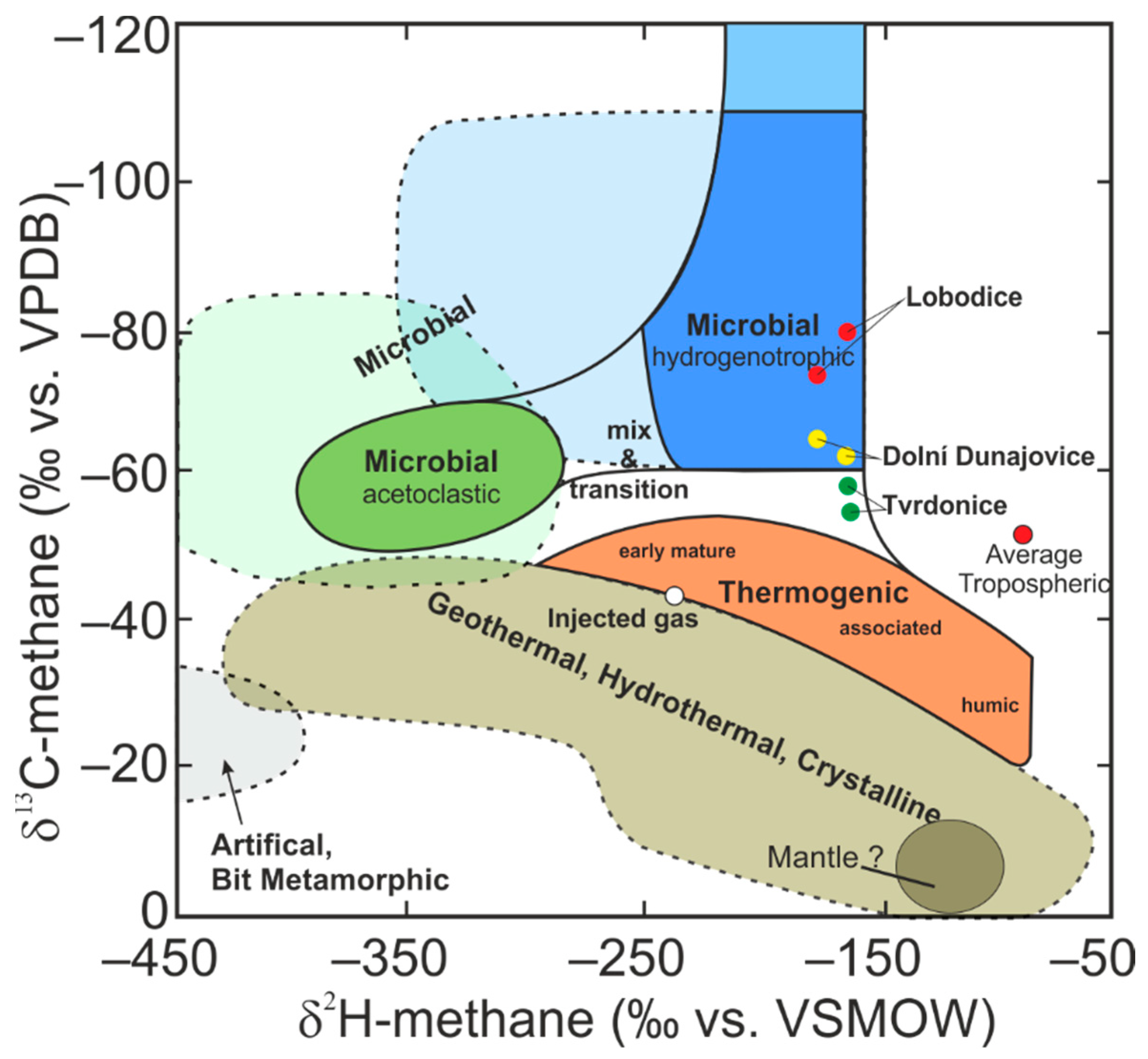
3.2. Isotopic Determination
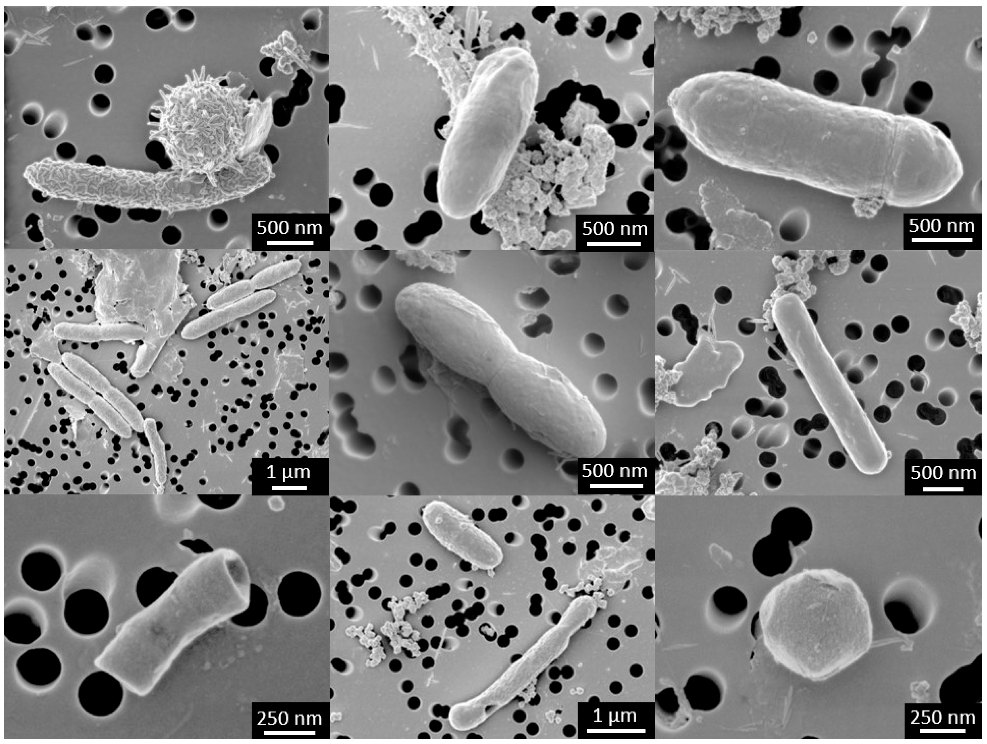
The results of the isotopic analysis of the gas and water samples indicate the origin of these samples. These results and an example of the typical isotopic composition of the injected gas are given in Table S4. The raw δD values measured in CH4 must be corrected using the δD values measured in water, because there is an equilibrium between these values, and the values in methane are affected by the values in water. The data listed in Table S4 are also shown in Figure 1. The structure in the background [42] helps to immediately assign the samples to a specific type of origin. As can be seen in Figure 1, the samples from Lobodice and Dolní Dunajovice are clearly of microbial origin. The results for the samples from Tvrdonice indicate that these degassing samples are a mixture of microbial and thermogenic gas. For comparison, the values for injected gas have also been added to this figure to illustrate the difference between the thermogenic gas typically stored in the UGS and the gases that were influenced (at least to some degree) by microbial processes.

3.3. Microscopy

The SEM-micrograph shows a wide variety of morphological types of microorganisms, predominantly the accumulation of rod-shaped cells with a length of 1–4 µm and cocci with a diameter of about 0.3 µm (Figure 2).

3.4. Quantitative Analyses of Methanogens (qPCR)

Of all sites, 14 wells were sampled, ten of which were sampled once and four of which were sampled repeatedly in different seasons, focusing on specific UGS regimes (injection–extraction gas periods) (Table S2). Of the total twenty samples from the dataset, DUN-B (December 2018), LO-H (November 2019), LO-H (October 2018), and LO-I (March 2019) were discarded due to low DNA concentration. The number of mcrA gene copies (Table S5) found in the Lobodice samples ranged from 7.20 × 101 to 2.40 × 10⁷ in 1 µL of DNA isolated from 1 L of well water. The highest number of mcrA gene copies (2.40 × 10⁷) was detected in the well LO-C (October 2018), while the lowest number (2.00 × 10²) was found in LO-F (May 2018). In Dolní Dunajovice, the mcrA gene copies ranged from 1.7 × 10⁵ to 1.03 × 10⁶, and in Tvrdonice from 1.86 × 10⁴ to 1.42 × 10⁶. Certain wells in Lobodice were repeatedly sampled at different times of the year over a two-year period. When comparing the number of mcrA gene copies, the hypothesis about the influence of the UGS regime (injection/withdrawal periods for gas) on methanogen abundance cannot be confirmed.

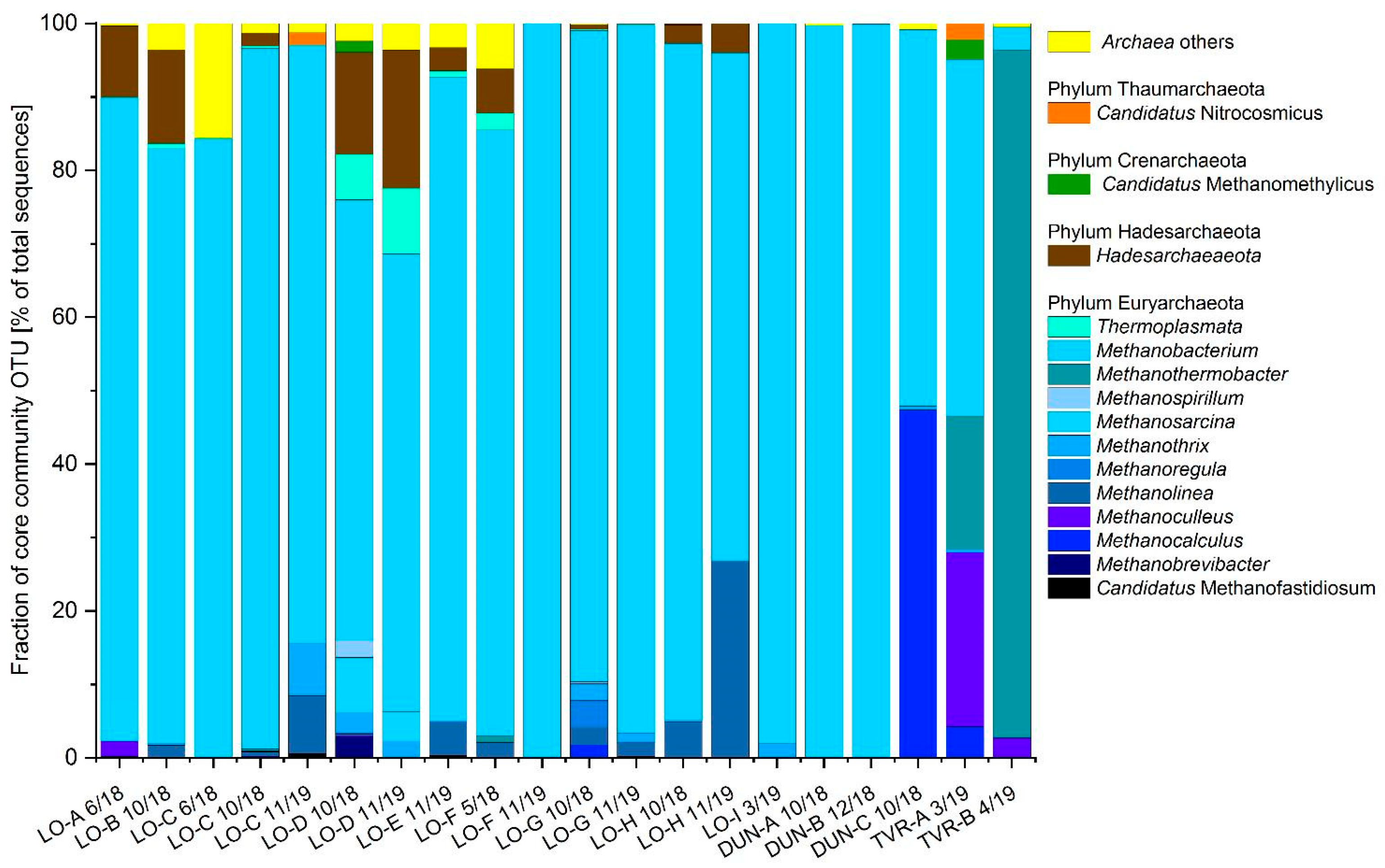
3.5. Metagenomic Analyses of UGS Archaeal Community

3.5.1. Next-Generation Sequencing (NGS) Analysis

Next-generation sequencing (NGS) analysis targeting archaeal and bacterial 16S rRNA genes was performed to elucidate microbial community structures in anoxic groundwater from deep aquifers. The 24 samples were sequenced along with 16 independent samples on Illumina MiniSeq using the MidOutput Kit (2 × 150 bp). The total run yield was 2.73 Gbp, with 88.39% of the reads passing the quality filter (>Q30), resulting in 8,689,550 reads passing the filter. The average error rate of the sequencing run was 0.98%. The DADA2 algorithm extracted 2864 unique ASVs from the 24 samples (Table S6). The sequencing depth obtained far exceeded the requirements, as shown by the rarefaction curve (Figure S3) with a minimum of 94,616 reads and a mean of 150,555 reads per sample.
For Illumina MiniSeq sequencing, the V4 region of the 16S rRNA gene was amplified [34,43]. The 16S rRNA gene is ubiquitous, occurs in several bacterial and archaeal species, and is highly conserved. Members of both domains were detected in varying unexpected proportions in all samples (archaea comprised 0.2%–75.7% of the microbial community in all sampled wells). Archaeal community composition differed slightly among the three sites, as did environmental and physicochemical conditions (Figure S4).
All samples collected were positive for the presence of methanogenic archaea. The 16S rRNA marker was used for sequence analyses and indicated the presence of an archaeal community in each well (Figure 3). The archaea were represented by five identified phyla, Euryarchaeota and Crenarchaeota, Hydrothermarchaeota, Nanoarchaeaeota and Hadesarchaeaeota, the last being a recently proposed phylum of thermophilic microorganisms found in deep mines, hot springs, marine sediments, and other subsurface environments [44]. This includes eight discovered classes, seven orders, thirteen families and fifteen genera. Sequencing of the 16S rRNA gene shows that the majority of the 140 archaeal OTUs belong to methanogens and consist of 10 genera and two recently discovered Candidatus species, whose abundance accounted for more than 5% of the total community.
The composition of the methanogenic archaea community detected in the UGS water samples shows that the major metabolic pathway in the UGS environment is hydrogenotrophic methanogenesis, with hydrogen and carbon dioxide as carbon and energy sources (Figure 4). In general, the genus Methanobacterium predominated in almost all samples, followed by the genus Methanothermobacter (present exclusively in Tvrdonice) and Methanolinea. The metabolism of these genera is exclusively hydrogenotrophic methanogenesis. The acetoclastic members of the genera Methanothrix and Methanosarcina were represented by large numbers of cells in the LO-A and LO-D wells.
Exceptionally high numbers of members of the genus Methanocalculus were found in the DUN-C well, where they make up half of the archaeal community. Methanothermobacter genera absolutely dominate in TVR-B and contribute to the archaeome in TVR-A, as do Methanoculleus members. In addition, new methanogens of interest were detected in interesting numbers in this well, namely the methylotrophs Candidatus Methanomethylicus and Candidatus Methanofastidiosum.

3.5.2. Biodiversity of Microbial Communities

Shannon and Unifrac indexes were chosen to describe the biodiversity of microbial communities. Alpha diversity refers to the diversity within a given ecosystem and is usually expressed by the number of species (or species richness). The diversity and richness of microbial communities inhabiting the different wells were determined using 16S rRNA gene analyses. Comparing the microbial diversity of each sample, Lobodice recorded the highest values with respect to all three sampling sites. Within all localities, the highest alpha diversity was found in the sample LO-D 11/19, while the opposite was found in LO-C 10/18. Relatively high values for the Shannon index were found in LO-C 11/19, LO-F 5/18, LO-F 11/19, and DUN-A 10/18 (Figure S5, Alpha biodiversity measure).
Beta diversity describes the structural complexity of the environment. It is a measure of the difference (or conversely, similarity) in species composition between communities along a given gradient of the environment, or between the community and its environment. Beta diversity is higher when a community contains uncommon species. Beta diversity shows the difference between microbial communities from different sampling sites. The obtained indices show no significant clustering among all samples. Only one distinct group formed from samples from Dolní Dunajovice and Tvrdonice, which originated from similar environments. In addition, some samples from Lobodice (LO-D 10/18, LO-E 11/19, LO-A 6/18) were clustered together with them. The most divergent samples, considering all sites, were LO-C 10/18 and LO-G 10/18 (Figures S5 and S6).

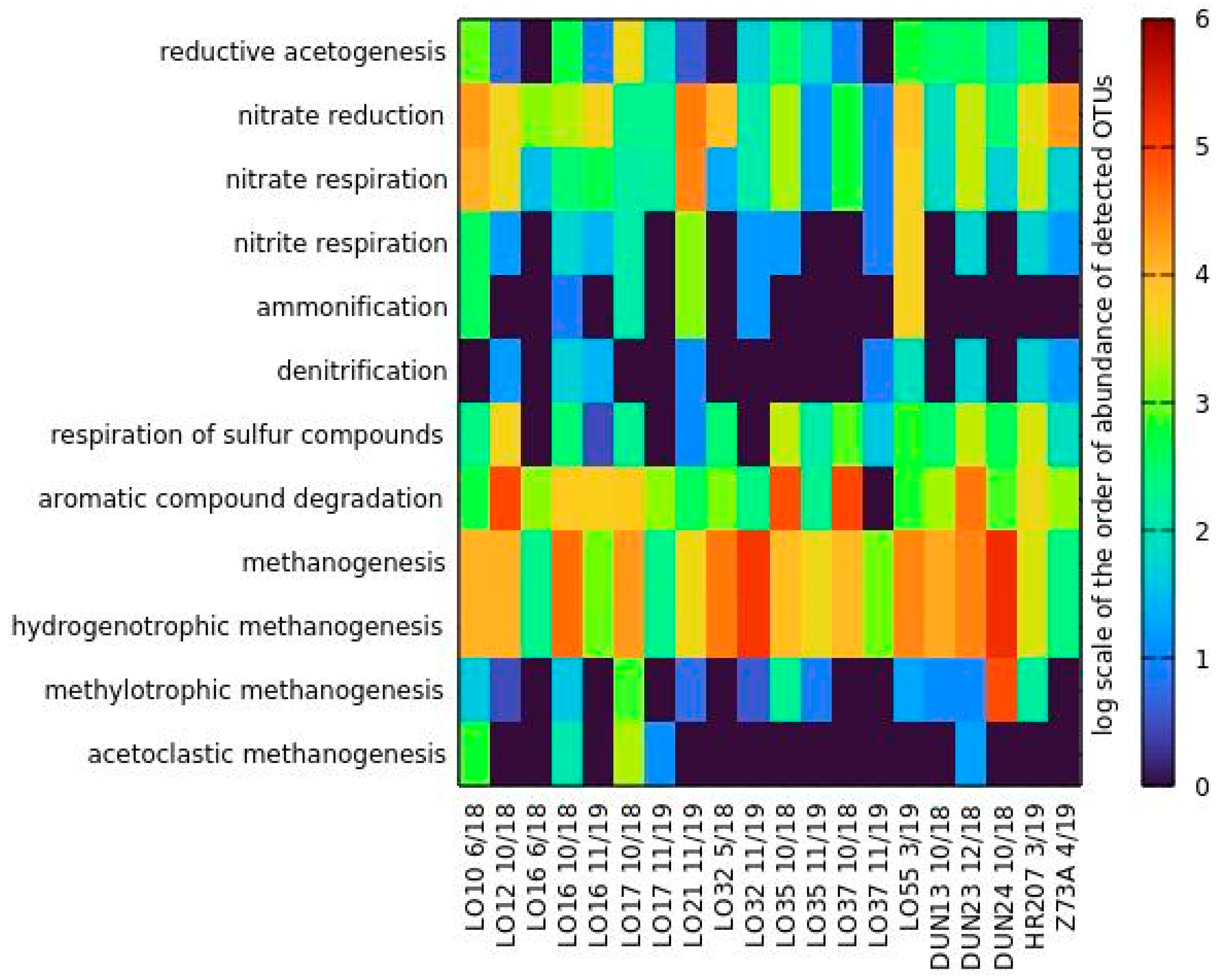
3.5.3. Metabolism Prediction—FAPROTAX

The potential metabolic functions of microorganisms were estimated using the database of functional annotations of prokaryotic organisms, FAPROTAX, which showed that the most frequent categories were related to carbon cycling under anaerobic conditions. Furthermore, metabolism involved in nitrate respiration and respiration of sulfur compounds was found to be marginal (Figure 4). During anaerobic decomposition of organic material, the hydrolysis products are decomposed into simple organic and inorganic substances (acids, alcohols, CO2, H2). Fermentation of these substances produces several reduced end products. In the next step—acetogenesis—the syntrophic acetogenic microorganisms produce hydrogen and decompose organic acids, alcohols, and some aromatic compounds. In the dataset, aromatic compound degraders, such as members of the genera Pelotomaculum or Acinetobacter, were detected in all samples, but significant levels were reached in the wells LO-B, LO-G, LO-H, and DUN-B. The presence of syntrophic acetogenic bacteria (Sporomusa, Anoxynatronum) was confirmed in the LO-D well. The dataset also reflects the composition of the methanogenic community in the well, showing a high abundance of acetoclastic methanogens. Acetogenic microorganisms are commonly found in syntrophy with methanogenic archaea, which consume the hydrogen they produce. Methanogens utilize substrates, such as carbonaceous substances (methanol, formic acid, methylamines, CO2, CO) or acetic acid. The end products of their metabolism are methane and carbon dioxide. All types of methanogenic metabolism were recorded in the collected samples, but hydrogenotrophic methanogenesis, represented mainly by the genus Methanobacterium, was predominant. A significant potential of methylotrophic methanogenesis was observed only in DUN-C (Methanocalculus, Methanospirillum). Acetotrophic methanogens, represented exclusively by the genus Methanosaeta, were detected only in the LO-D well, corresponding to high acetogenic activity. The highest methanogenic activity was then found in DUN-C and LO-F. The intensity of predicted methanogenic metabolism varied by site and sampling, showing unexpected changes in metabolic activity. In general, it appears that this very particular type of ecosystem is more dynamic than should be expected.

4. Discussion

Over the course of four years of studying biomethanation in underground water reservoirs, we have built on the results summarized in the review paper [5].
The objective of the study was to determine part of the composition of the UGS microbiome, focusing on methanogenic archaea, biological methane production, and evaluation of the biotechnological potential for biomethane production from underground hydrocarbon reservoirs and aquifers. Although methanogens have been detected in aquifers [4,18,19] or in the sediments of UGS pipelines [45], to the best of our knowledge, this is the first thorough study addressing different types of reservoirs. We hypothesize that seasonal fluctuations of gas in UGS may have an impact on the abundance of methanogens in UGS [2].
To demonstrate the dynamics of their abundance during the year, some of the wells were sampled at different times of the year, focusing on specific UGS regimes (injection/withdrawal gas). The results show that the methanogenic community is dynamic throughout the year, but no significant trend was found with respect to the UGS regime (Figure 3). The results of the sequencing analyses confirmed that each well hosts a specific methanogenic community that accounts for 70%–100% of the total archaeal community. This means that methanogen abundance in UGS is generally greater than 10%.
Hydrogenotrophic Methanobacterium sp. prevailed in most samples from the Lobodice locality, as proven by NGS and cultivation. Methanosarcina sp. were also abundant, consuming acetate or methylated compounds as a substrate for methanogenesis. The second sampling site, Dolní Dunajovice, had a different reservoir environment and thus a different microbiome composition compared to Lobodice. Members of the hydrogenotrophic order Methanomicrobiales were most abundant. The third sampling site, Tvrdonice, was strongly influenced by the higher temperature in the UGS and therefore contained thermophilic members of the Methanothermobacter genus.
Methanogenic and fermentative microorganisms are often organized in mutualistic consortia to facilitate rapid electron exchange by diffusion of hydrogen or formate [46]. In addition, electron exchange by direct electron transfer between species has been discovered. Geobacter metallireducens transfers electrons directly to Methanothrix harundinacea during methanogenic degradation of ethanol, presumably through nanowires [47]. Members of the genera Geobacter and Methanothrix were found in samples from LO-A and DUN-A.
Anaerobic syntrophy is defined as a thermodynamically interdependent extreme lifestyle in which the degradation of an organic compound occurs only when the end products (usually hydrogen, formate, and acetate) are maintained at very low concentrations. Microbial syntrophy between Bacteria and methanogenic archaea enhances the methanogenic activity and methane yield. This type of syntrophy is related to the global carbon cycle in anaerobic environments, which is based on a complex community of metabolically coupled microorganisms that are highly adapted to the environment. This was clearly demonstrated in the publication [48].
The dynamics of the conversion of H2 and CO2 to methane varied in all samples depending on the UGS environment. The key parameters were temperature and chemical composition of the UGS groundwater. Biomethane production is also influenced by the composition of the microbial community in each UGS well [49]. Our study confirms the presence and natural activity of methanogenic archaea in underground gas storages. The function of underground gas storages as natural bioreactors is confirmed by the result of our field experiment under real reservoir conditions [50]. The strategic importance of this solution is quite clear. These principles can make an important contribution to reducing the impact of transport and energy on nature and decarbonizing the economy. The use of underground reservoirs for industrial production of biological methane is one of the ways by which the Czech Republic might achieve self-sufficiency, avoiding dependence on natural gas imports.

4.1. Lobodice

The low salinity and higher pH (close to eight) of the environment provide ideal conditions for the growth of members of the genus Methanobacterium. The high prevalence of this genus was confirmed in all but one of the sampled wells. This genus is hydrogenotrophic but can also metabolize formate. Several species have already been isolated from aquifers, so these genera seem to be widespread in this environment [3,15,51,52]. Moreover, this genus has even been isolated during previous experiments at the Lobodice UGS [4]. Members of the genus Methanosarcina were found in large numbers in LO-A and LO-D. The main substrates for growth are acetate, methanol, trimethylamines, or other methyl-containing compounds. Some of the species can form methane from H2 and CO2, or they use H2 to reduce methanol to methane. Acetotrophic members of the genus Methanothrix were detected and were present in samples depending on morphological observation, especially in LO-C, LO-D, and LO-G samples. They are exclusively served by acetate as a substrate, and their affinity for this substrate is much higher than that of the genus Methanosarcina. Members of the genus Methanoculleus were found exclusively in the well LO-A. This genus usually lives in marine environments and brackish water, but is also widespread in other environments, such as bioreactors, landfills, or wastewater. Unlike other Archaea, Methanoculleus can use ethanol and some secondary alcohols as electron donors for final methane production [53]. Conditions at this site appear to support acetogenic bacteria of the genus Acetobacterium, which use H2 and CO2 to form acetate. The acetate formed does not remain in the liquid for long, being rapidly consumed by a narrow range of bacteria, or serving as a substrate for methanogenesis.

4.2. Dolní Dunajovice

The Dolní Dunajovice site is characterized by higher salinity and temperature compared to Lobodice. Due to these conditions, the composition of methanogens differs significantly from that in Lobodice. In the wells DUN-A and DUN-B, methanogens belonging to the order Methanobacteriales absolutely dominate. In the sample from the well DUN-C, two predominant archaeal taxons, Methanobacterium sp. and Methanocalculus sp., comprised around half of the organisms. Species of the genus Methanocalculus are very salt tolerant and can live at sodium chloride concentrations as high as 125 g/L [54]. Moreover, the higher temperature in the Dolní Dunajovice reservoir is ideal for them, as the optimal temperature of the species is 45 °C [55].

4.3. Tvrdonice

Tvrdonice offers unique conditions, with salinity levels somewhere between those of Lobodice and Dolní Dunajovice, at temperatures around 50 °C. The higher temperature fosters microbial communities distinct from those of mesophilic environments. The dominant methanogen, Methanothermobacter sp., was found in well TVR-B and was also detected in another deep aquifer [15]. Its predominance is likely to lead to faster reactions in methane formation. High temperatures are a key factor affecting microbial composition. The absolute prevalence of the thermophilic Methanothermobacter sp. (94%), which grows best at temperatures between 55 °C and 65 °C, was confirmed by 16S rRNA sequencing. Only two other genera were detected in well TVR-B by 16S rRNA sequencing: Methanobacterium (3.2%) and Methanoculleus (2.7%). These genera use carbon dioxide and hydrogen as substrates to produce methane for energy production.
Samples from TVR-A well water consisted of the genera Methanobacterium (approximately 50%), Methanoculeus (24%), Methanothermobacter (18%), and members of the recently proposed taxa Candidatus Methanomethylicus and Candidatus Methanofastidiosum [56]. Candidatus Methanofastidiosum is a unique methanogen that utilizes methylated thiol reduction and bridged carbon and sulfur cycles and may compete with CO2-reducing methanogens and even sulfate reducers [56,57].

5. Conclusions

Based on our results, we can conclude that the underground gas storages assessed by us showed a microbiome composition suitable for biological methane production. It can be concluded that if underground gas storages offer suitable habitat, they can be used as fermenters for the biological conversion of CO2 and H2 into biomethane anywhere in the world. From a strategic perspective, underground storages can play an important role in the energy mix, as they can be used for long-term energy storage.

Supplementary Materials

The following supporting information can be downloaded at: https://0-www-mdpi-com.brum.beds.ac.uk/article/10.3390/fermentation8060251/s1, Table S1: Overview on sampling and on physiochemical parameters measured in sampled wells, Table S2: Table of gas composition measured in sampled wells, Table S3: Primer sequences for Illumina analysis, Table S4: Results of isotopic analysis of degas water and injected gas, Table S5: Absolute quantification of mcr-A gene via qPCR (gene copies per ml), Table S6: Table of Illumina reads for DADA2 analysis, Figure S1: Piper and Durov diagrams for groundwaters sampled at UGS Lobodice. UGS Tvrdonice. and UGS Dunajovice, Figure S2: Stability pH-Eh diagrams for carbon (a) nitrogen (b) and sulfur (c) dissolved components. Diagrams were prepared for UGS Lobodice groundwater conditions, conditions of UGS Tvrdonice and UGS Dunajovice are not significantly different. (d) Speciation of carbon dissolved species in dependence on redox potential, Figure S3: Rarefaction curve, Figure S4: Physical-chemistry well water properties in different sites and composition of archaeal community, Figure S5: The Alpha biodiversity index (Shannon) of sampled wells, Figure S6: The Beta biodiversity index (unweighted UniFrac) of sampled wells.

Author Contributions

I.B., M.V., and V.O. contributed to the conception and design of the study. I.B. and A.M. wrote the first draft of the manuscript. T.V., M.V., I.U., V.O. and J.Z. wrote sections of the manuscript. I.B., A.M., M.V., T.V., N.H. collected samples. I.B., A.M., and D.N. performed molecular biology analyses. I.B., D.N., M.Č., and J.L. performed bioinformatics analyses. I.U. performed degas analyses. J.Z. performed chemical analyses of samples. J.J. and M.M. performed SEM micrographs. H.H. and L.D. revised the manuscript. All authors have read and agreed to the published version of the manuscript.

Funding

The financial support was received from innogy Gas Storage, s.r.o.

Institutional Review Board Statement

Not applicable.

Informed Consent Statement

Not applicable.

Data Availability Statement

Not applicable.

Conflicts of Interest

The authors declare no conflict of interest.

References

  1. Basso, O.; Lascourreges, J.F.; Le Borgne, F.; Le Goff, C.; Magot, M. Characterization by culture and molecular analysis of the microbial diversity of a deep subsurface gas storage aquifer. Res. Microbiol. 2009, 160, 107–116. [Google Scholar] [CrossRef] [PubMed]
  2. Ivanova, A.E.; Borzenkov, I.A.; Tarasov, A.L.; Milekhina, E.I.; Belyaev, S.S. A microbiological study of an underground gas storage in the process of gas extraction. Microbiology 2007, 76, 461–468. [Google Scholar] [CrossRef]
  3. Buzek, F.; Onderka, V.; Vančura, P.; Wolf, I. Carbon isotope study of methane production in a town gas storage reservoir. Fuel 1994, 73, 747–752. [Google Scholar] [CrossRef]
  4. Šmigáň, P.; Greksák, M.; Kozánková, J.; Buzek, F.; Onderka, V.; Wolf, I. Methanogenic bacteria as a key factor involved in changes of town gas stored in an underground reservoir. FEMS Microbiol. Lett. 1990, 73, 221–224. [Google Scholar] [CrossRef]
  5. Molíková, A.; Vítězová, M.; Vítěz, T.; Buriánková, I.; Huber, H.; Dengler, L.; Hanišáková, N.; Onderka, V.; Urbanová, I. Underground gas storage as a promising natural methane bioreactor and reservoir? J. Energy Storage 2022, 47, 103631. [Google Scholar] [CrossRef]
  6. Rittmann, S.; Seifert, A.; Herwig, C. Essential prerequisites for successful bioprocess development of biological CH4 production from CO2 and H2. Crit. Rev. Biotechnol. 2015, 35, 141–151. [Google Scholar] [CrossRef]
  7. Koytsoumpa, E.I.; Bergins, C.; Kakaras, E. The CO2 economy: Review of CO2 capture and reuse technologies. J. Supercrit. Fluids 2018, 132, 3–16. [Google Scholar] [CrossRef]
  8. Nazir, H.; Louis, C.; Jose, S.; Prakash, J.; Muthuswamy, N.; Buan, M.E.M.; Flox, C.; Chavan, S.; Shi, X.; Kauranen, P.; et al. Is the H2 economy realizable in the foreseeable future ? Part I: H2 production methods. Int. J. Hydrogen Energy 2020, 45, 13777–13788. [Google Scholar] [CrossRef]
  9. Sampath, P.; Brijesh; Reddy, K.R.; Reddy, C.V.; Shetti, N.P.; Kulkarni, R.V.; Raghu, A.V. Biohydrogen Production from Organic Waste—A Review. Chem. Eng. Technol. 2020, 43, 1240–1248. [Google Scholar] [CrossRef]
  10. Aguilera, R.F.; Aguilera, R. Revisiting the role of natural gas as a transition fuel. Miner. Econ. 2020, 33, 73–80. [Google Scholar] [CrossRef]
  11. Pedersen, K.; Arlinger, J.; Ekendahl, S.; Hallbeck, L. 16S rRNA gene diversity of attached and unattached bacteria in boreholes along the access tunnel to the Äspö hard rock laboratory, Sweden. FEMS Microbiol. Ecol. 1996, 19, 249–262. [Google Scholar] [CrossRef]
  12. Kotelnikova, S.; Pedersen, K. Evidence for methanogenic Archaea and homoacetogenic Bacteria in deep granitic rock aquifers. FEMS Microbiol. Rev. 1997, 20, 339–349. [Google Scholar] [CrossRef]
  13. Fry, N.K.; Fredrickson, J.K.; Fishbain, S.; Wagner, M.; Stahl, D.A. Population structure of microbial communities associated with two deep, anaerobic, alkaline aquifers. Appl. Environ. Microbiol. 1997, 63, 1498–1504. Available online: https://0-aem-asm-org.brum.beds.ac.uk/content/63/4/1498.short (accessed on 21 May 2019). [CrossRef] [Green Version]
  14. Shimizu, S.; Akiyama, M.; Ishijima, Y.; Hama, K.; Kunimaru, T.; Naganuma, T. Molecular characterization of microbial communities in fault-bordered aquifers in the Miocene formation of northernmost Japan. Geobiology 2006, 4, 203–213. [Google Scholar] [CrossRef] [Green Version]
  15. Kimura, H.; Nashimoto, H.; Shimizu, M.; Hattori, S.; Yamada, K.; Koba, K.; Yoshida, N.; Kato, K. Microbial methane production in deep aquifer associated with the accretionary prism in Southwest Japan. ISME J. 2010, 4, 531–541. [Google Scholar] [CrossRef]
  16. Flynn, T.M.; Sanford, R.A.; Ryu, H.; Bethke, C.M.; Levine, A.D.; Ashbolt, N.J.; Santo Domingo, J.W. Functional microbial diversity explains groundwater chemistry in a pristine aquifer. BMC Microbiol. 2013, 13, 146. [Google Scholar] [CrossRef] [Green Version]
  17. Wu, X.; Holmfeldt, K.; Hubalek, V.; Lundin, D.; Åström, M.; Bertilsson, S.; Dopson, M. Microbial metagenomes from three aquifers in the Fennoscandian shield terrestrial deep biosphere reveal metabolic partitioning among populations. ISME J. 2016, 10, 1192–1203. [Google Scholar] [CrossRef] [Green Version]
  18. Frank, Y.A.; Kadnikov, V.V.; Gavrilov, S.N.; Banks, D.; Gerasimchuk, A.L.; Podosokorskaya, O.A.; Merkel, A.Y.; Chernyh, N.A.; Mardanov, A.V.; Ravin, N.V.; et al. Stable and variable parts of microbial community in Siberian deep subsurface thermal aquifer system revealed in a long-term monitoring study. Front. Microbiol. 2016, 7, 2101. [Google Scholar] [CrossRef]
  19. Kadnikov, V.V.; Frank, Y.A.; Mardanov, A.V.; Beletsky, A.V.; Ivasenko, D.A.; Pimenov, N.V.; Karnachuk, O.V.; Ravin, N.V. Variability of the composition of the microbial community of the deep subsurface thermal aquifer in Western Siberia. Microbiology 2017, 86, 765–772. [Google Scholar] [CrossRef]
  20. Vigneron, A.; Bishop, A.; Alsop, E.B.; Hull, K.; Rhodes, I.; Hendricks, R.; Head, I.M.; Tsesmetzis, N. Microbial and isotopic evidence for methane cycling in hydrocarbon-containing groundwater from the Pennsylvania region. Front. Microbiol. 2017, 8, 593. [Google Scholar] [CrossRef] [Green Version]
  21. Evans, P.N.; Boyd, J.A.; Leu, A.O.; Woodcroft, B.J.; Parks, D.H.; Hugenholtz, P.; Tyson, G.W. An evolving view of methane metabolism in the Archaea. Nat. Rev. Microbiol. 2019, 17, 219–232. [Google Scholar] [CrossRef]
  22. Stevens, T.O.; McKinley, J.P.; Fredrickson, J.K. Bacteria Associated with Deep, Alkaline, Anaerobic Groundwaters in Southeast Washington T. Microb. Ecol. 1993, 25, 35–50. [Google Scholar] [CrossRef]
  23. Thauer, R.K.; Kaster, A.K.; Seedorf, H.; Buckel, W.; Hedderich, R. Methanogenic archaea: Ecologically relevant differences in energy conservation. Nat. Rev. Microbiol. 2008, 6, 579–591. [Google Scholar] [CrossRef]
  24. Hedderich, R.; Whitman, W.B. Physiology and biochemistry of the methane-producing Archaea. Prokaryotes 2006, 2, 1050–1079. Available online: http://www.academia.edu/download/63378241/Dworkin_The_Prokaryotes-A_Handbook_on_the_Biology_of_Bacteria_3rd_ed_Vol_220200520-24653-hqh6kx.pdf#page=1100 (accessed on 18 January 2021).
  25. Lang, K.; Schuldes, J.; Klingl, A.; Poehlein, A.; Daniel, R.; Brune, A. New mode of energy metabolism in the seventh order of methanogens as revealed by comparative genome analysis of “Candidatus Methanoplasma termitum”. Appl. Environ. Microbiol. 2015, 81, 1338–1352. [Google Scholar] [CrossRef] [Green Version]
  26. Berghuis, B.A.; Yu, F.B.; Schulz, F.; Blainey, P.C.; Woyke, T.; Quake, S.R. Hydrogenotrophic methanogenesis in archaeal phylum Verstraetearchaeota reveals the shared ancestry of all methanogens. Proc. Natl. Acad. Sci. USA 2019, 116, 5037–5044. [Google Scholar] [CrossRef] [Green Version]
  27. Hattori, S. Syntrophic acetate-oxidizing microbes in methanogenic environments. Microbes Environ. 2008, 23, 118–127. [Google Scholar] [CrossRef] [Green Version]
  28. Mosbæk, F.; Kjeldal, H.; Mulat, D.G.; Albertsen, M.; Ward, A.J.; Feilberg, A.; Nielsen, J.L. Identification of syntrophic acetate-oxidizing bacteria in anaerobic digesters by combined protein-based stable isotope probing and metagenomics. ISME J. 2016, 10, 2405–2418. [Google Scholar] [CrossRef] [Green Version]
  29. Hungate, R.E. Chapter IV A Roll Tube Method for Cultivation of Strict Anaerobes. Methods Microbiol. 1969, 3, 117–132. [Google Scholar] [CrossRef]
  30. Miller, T.L.; Wolin, M.J. A Serum Bottle Modification of the Hungate Technique for Cultivating Obligate Anaerobes. Appl. Microbiol. 1974, 27, 985–987. [Google Scholar] [CrossRef]
  31. Denman, S.E.; Tomkins, N.W.; McSweeney, C.S. Quantitation and diversity analysis of ruminal methanogenic populations in response to the antimethanogenic compound bromochloromethane. FEMS Microbiol. Ecol. 2007, 62, 313–322. [Google Scholar] [CrossRef] [PubMed] [Green Version]
  32. Steinberg, L.M.; Regan, J.M. mcrA-targeted real-time quantitative PCR method to examine methanogen communities. Appl. Environ. Microbiol. 2009, 75, 4435–4442. [Google Scholar] [CrossRef] [PubMed] [Green Version]
  33. Pichler, M.; Coskun, Ö.K.; Ortega-Arbulú, A.S.; Conci, N.; Wörheide, G.; Vargas, S.; Orsi, W.D. A 16S rRNA gene sequencing and analysis protocol for the Illumina MiniSeq platform. Microbiologyopen 2018, 7, e00611. [Google Scholar] [CrossRef] [PubMed]
  34. Thompson, L.R.; Sanders, J.G.; McDonald, D.; Amir, A.; Ladau, J.; Locey, K.J.; Prill, R.J.; Tripathi, A.; Gibbons, S.M.; Ackermann, G.; et al. A communal catalogue reveals Earth’s multiscale microbial diversity. Nature 2017, 551, 457–463. [Google Scholar] [CrossRef] [Green Version]
  35. Callahan, B.J.; Sankaran, K.; Fukuyama, J.A.; McMurdie, P.J.; Holmes, S.P. Bioconductor workflow for microbiome data analysis: From raw reads to community analyses. F1000Research 2016, 5, 1496. [Google Scholar] [CrossRef]
  36. Wang, Q.; Garrity, G.M.; Tiedje, J.M.; Cole, J.R. Naïve Bayesian classifier for rapid assignment of rRNA sequences into the new bacterial taxonomy. Appl. Environ. Microbiol. 2007, 73, 5261–5267. [Google Scholar] [CrossRef] [Green Version]
  37. Quast, C.; Pruesse, E.; Yilmaz, P.; Gerken, J.; Schweer, T.; Glo, F.O.; Yarza, P. The SILVA ribosomal RNA gene database project: Improved data processing and web-based tools. Nucleic Acids Res. 2013, 41, 590–596. [Google Scholar] [CrossRef]
  38. Schliep, K.P. Phangorn: Phylogenetic analysis in R. Bioinformatics 2011, 27, 592–593. [Google Scholar] [CrossRef] [Green Version]
  39. McMurdie, P.J.; Holmes, S. Phyloseq: An R Package for Reproducible Interactive Analysis and Graphics of Microbiome Census Data. PLoS ONE 2013, 8, e61217. [Google Scholar] [CrossRef] [Green Version]
  40. Louca, S.; Parfrey, L.W.; Doebeli, M. Decoupling function and taxonomy in the global ocean microbiome. Science 2016, 353, 1272–1277. [Google Scholar] [CrossRef]
  41. Carini, P.; Marsden, P.J.; Leff, J.W.; Morgan, E.E.; Strickland, M.S.; Fierer, N. Relic DNA is abundant in soil and obscures estimates of soil microbial diversity. Nat. Microbiol. 2016, 2, 16242. [Google Scholar] [CrossRef]
  42. Whiticar, M.J. Carbon and hydrogen isotope systematics of bacterial formation and oxidation of methane. Chem. Geol. 1999, 161, 291–314. [Google Scholar] [CrossRef]
  43. Caporaso, J.G.; Lauber, C.L.; Walters, W.A.; Berg-Lyons, D.; Huntley, J.; Fierer, N.; Owens, S.M.; Betley, J.; Fraser, L.; Bauer, M.; et al. Ultra-high-throughput microbial community analysis on the Illumina HiSeq and MiSeq platforms. ISME J. 2012, 6, 1621–1624. [Google Scholar] [CrossRef] [Green Version]
  44. Baker, B.J.; Saw, J.H.; Lind, A.E.; Lazar, C.S.; Hinrichs, K.U.; Teske, A.P.; Ettema, T.J.G. Genomic inference of the metabolism of cosmopolitan subsurface Archaea, Hadesarchaea. Nat. Microbiol. 2016, 1, 16002. [Google Scholar] [CrossRef] [Green Version]
  45. Staniszewska, A.; Kunicka-Styczyńska, A.; Otlewska, A.; Gawor, J.; Gromadka, R.; Żuchniewicz, K.; Ziemiński, K. High-throughput sequencing approach in analysis of microbial communities colonizing natural gas pipelines. Microbiologyopen 2019, 8, e00806. [Google Scholar] [CrossRef]
  46. Sieber, J.R.; McInerney, M.J.; Gunsalus, R.P. Genomic Insights into Syntrophy: The Paradigm for Anaerobic Metabolic Cooperation. Annu. Rev. Microbiol. 2012, 66, 429–452. [Google Scholar] [CrossRef]
  47. Reguera, G.; McCarthy, K.D.; Mehta, T.; Nicoll, J.S.; Tuominen, M.T.; Lovley, D.R. Extracellular electron transfer via microbial nanowires. Nature 2005, 435, 1098–1101. [Google Scholar] [CrossRef]
  48. Amend, J.P.; Shock, E.L. Energetics of overall metabolic reactions of thermophilic and hyperthermophilic Archaea and Bacteria. FEMS Microbiol. Rev. 2001, 25, 175–243. [Google Scholar] [CrossRef]
  49. Yang, G.C.; Zhou, L.; Mbadinga, S.M.; Liu, J.F.; Yang, S.Z.; Gu, J.D.; Mu, B.Z. Formate-dependent microbial conversion of CO2 and the dominant pathways of methanogenesis in production water of high-temperature oil reservoirs amended with bicarbonate. Front. Microbiol. 2016, 7, 365. [Google Scholar] [CrossRef] [Green Version]
  50. Vítězová, M.; Urbanová, I.; Molíková, A.; Buriánková, I.; Hanišáková, N.; Onderka, V.; Vítěz, T.; Javůrek, J.; Machálková, M. In situ field experiment shows the potential of methanogenic archaea for biomethane production from underground gas storage. Unpublished work.
  51. Godsy, E.M. Isolation of Methanobacterium bryantii from a deep aquifer by using a novel broth-antibiotic disk method. Appl. Environ. Microbiol. 1980, 39, 1074–1075. [Google Scholar] [CrossRef] [Green Version]
  52. Kotelnikova, S.; Macario, A.J.L.; Pedersen, K. Methanobacterium subterraneum sp. nov., a new alkaliphilic, eurythermic and halotolerant methanogen isolated from deep granitic groundwater. Int. J. Syst. Bacteriol. 1998, 48, 357–367. [Google Scholar] [CrossRef] [Green Version]
  53. Cheng, L.; Qiu, T.L.; Li, X.; Wang, W.D.; Deng, Y.; Yin, X.B.; Zhang, H. Isolation and characterization of Methanoculleus receptaculi sp. nov. from Shengli oil field, China. FEMS Microbiol. Lett. 2008, 285, 65–71. [Google Scholar] [CrossRef] [Green Version]
  54. Ollivier, B.; Fardeau, M.; Cayol, J.; Magot, M.; Patel, B.K.C.; Prensiep, G.; Garcia, J. Methanocalculus halotolerans. Int. J. Syst. Evol. Microbiol. 1998, 48, 821–828. [Google Scholar]
  55. Shlimon, A.G.; Friedrich, M.W.; Niemann, H.; Ramsing, N.B.; Finster, K. Methanobacterium aarhusense sp. nov., a novel methanogen isolated from a marine sediment (Aarhus Bay, Denmark). Int. J. Syst. Evol. Microbiol. 2004, 54, 759–763. [Google Scholar] [CrossRef] [Green Version]
  56. Vanwonterghem, I.; Evans, P.N.; Parks, D.H.; Jensen, P.D.; Woodcroft, B.J.; Hugenholtz, P.; Tyson, G.W. Methylotrophic methanogenesis discovered in the archaeal phylum Verstraetearchaeota. Nat. Microbiol. 2016, 1, 16170. [Google Scholar] [CrossRef] [Green Version]
  57. Nobu, M.K.; Narihiro, T.; Kuroda, K.; Mei, R.; Liu, W.T. Chasing the elusive Euryarchaeota class WSA2: Genomes reveal a uniquely fastidious methyl-reducing methanogen. ISME J. 2016, 10, 2478–2487. [Google Scholar] [CrossRef]
Figure 1. C–D plot [42] with measured data.
Fermentation 08 00251 g001
Figure 2. Examples of microbial morphotypes: rods and cocci found in well TVR-B captured by SEM. Inorganic nanoparticles which form conglomerates can be seen.
Fermentation 08 00251 g002
Figure 3. Archaeal community—16S rRNA gene sequence focused on methanogens detected in water samples from UGS’s (5% cut).
Fermentation 08 00251 g003
Figure 4. Selected metabolic groups predicted using the database of functional annotations of prokaryotic organisms, FAPROTAX.
Fermentation 08 00251 g004
Table 1. Selected investigations performed in similar rock environment.
Author, ReferenceYearCountryFocus of Study
Šmigán et al. [4]1989Czech Republicmethanogenic archaea
Buzek et al. [4]1994Czech Republicmicrobial methane production
Pedersen et al. [11]1996Swedenmicrobial diversity
Kotelnikova et al. [12]1997Swedenmethanogenic archaea, homoacetogenic bacteria
Fry et al. [13]1997USAmicrobial diversity
Shimizu et al. [14]2006Japanmicrobial diversity
Ivanova et al. [2]2007Russiamicrobiological
Basso et al. [1]2009Francemicrobial diversity
Kimura et al. [15]2010Japanmicrobial methane study
Flynn et al. [16]2013USAfunctional microbial diversity
Wu et al. [17]2016Swedenmicrobial diversity, metabolism
Frank et al. [18]2016Russiavariability in microbial composition
Kadnikov et al. [19]2017Russiamicrobial diversity
Vigneron et al. [20]2017USAmicrobial methane study
Publisher’s Note: MDPI stays neutral with regard to jurisdictional claims in published maps and institutional affiliations.

