
Magnetic Properties Study of Iron Oxide Nanoparticles-Loaded Poly(ε-caprolactone) Nanofibres

 , , and
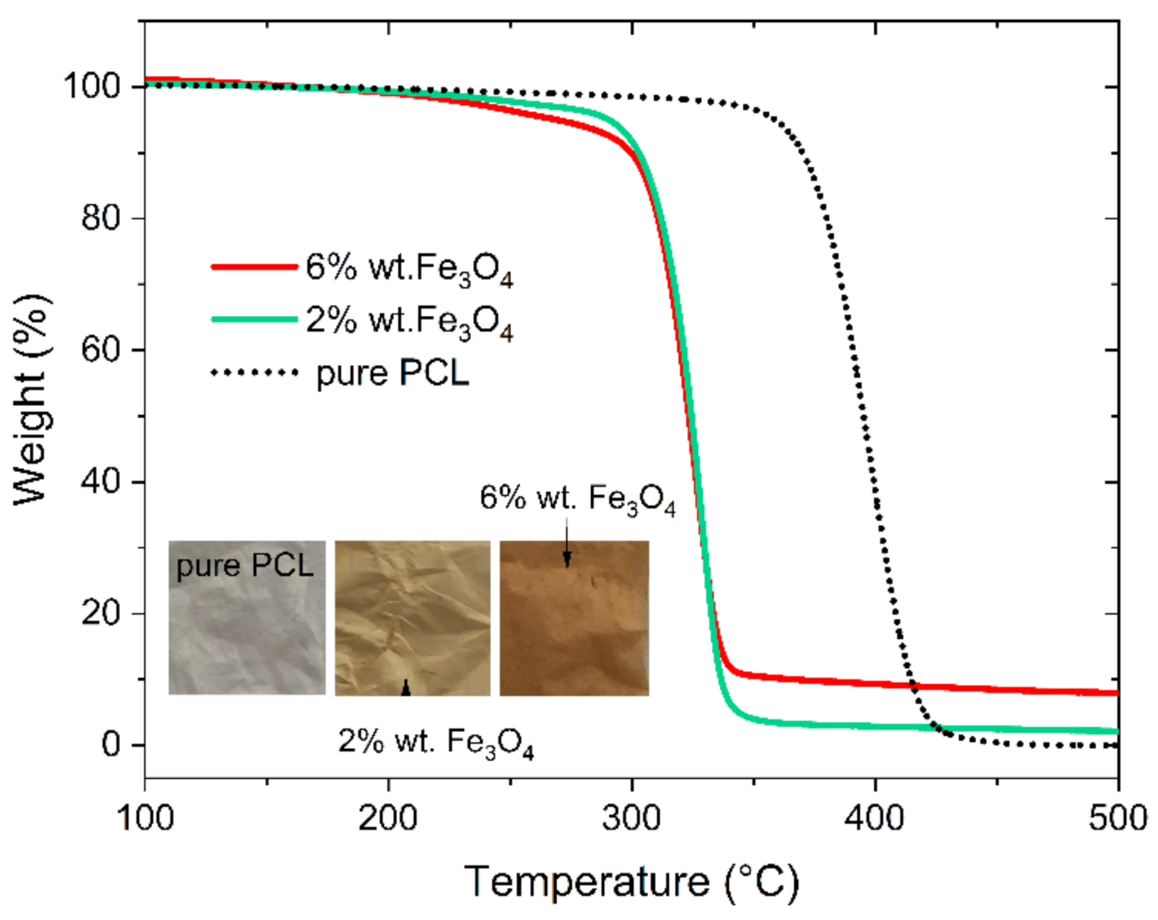
Abstract
:1. Introduction
2. Results and Discussion
2.1. Thermal Analysis
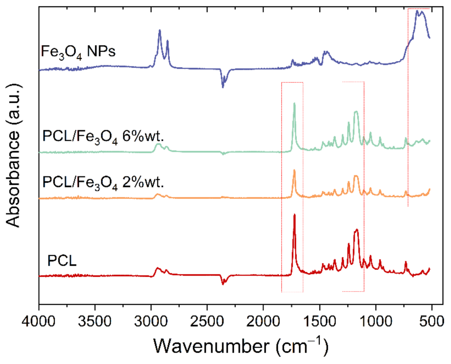
2.2. IRspectroscopy
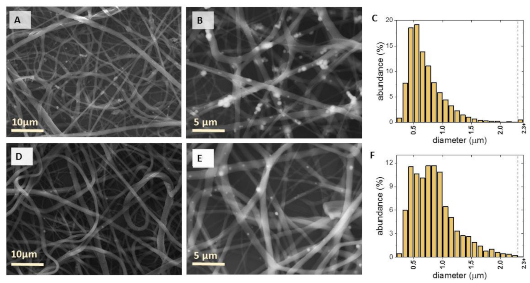
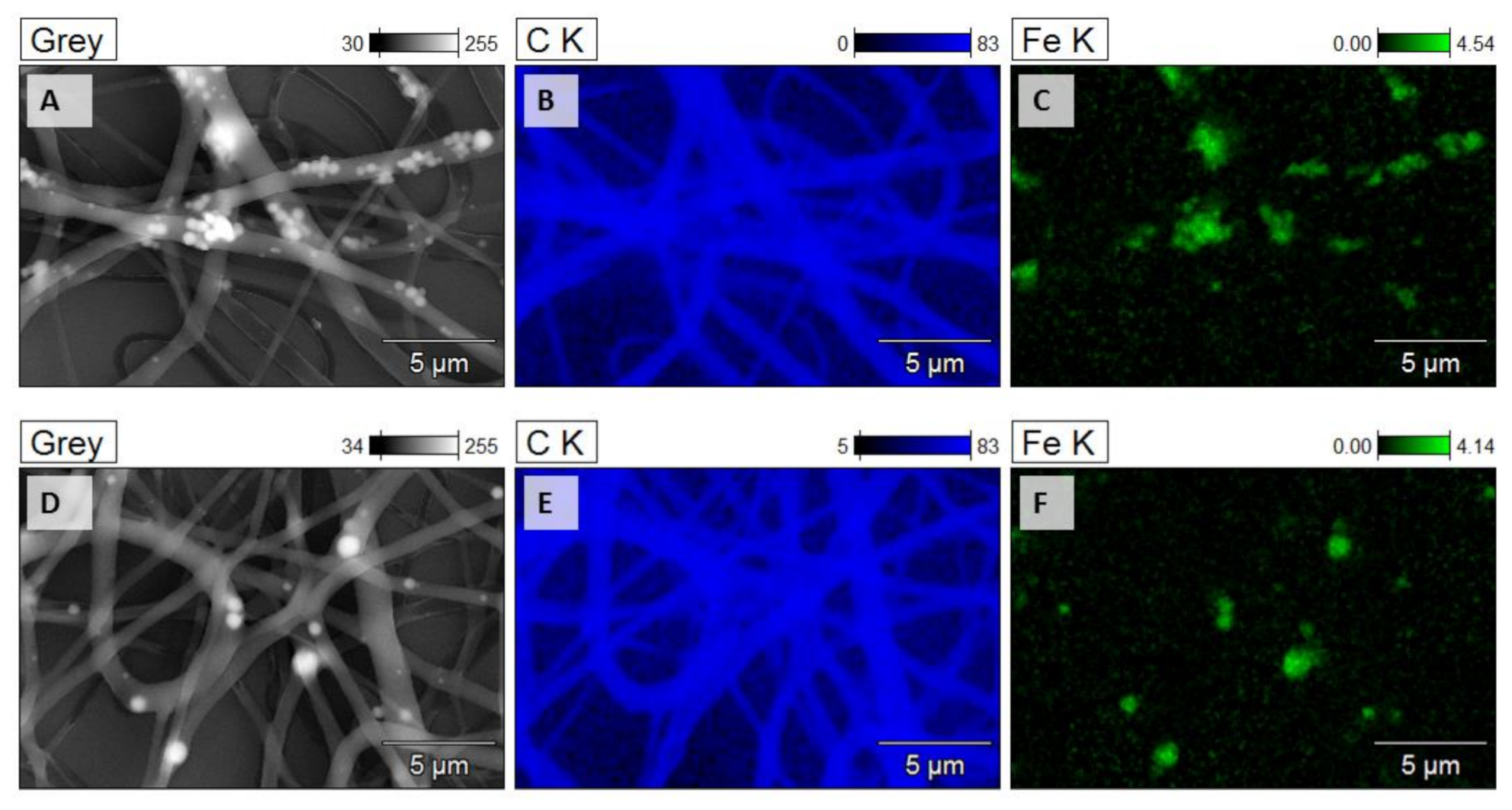
2.3. Morphology of Electrospun PCL/Fe3O4 Mats
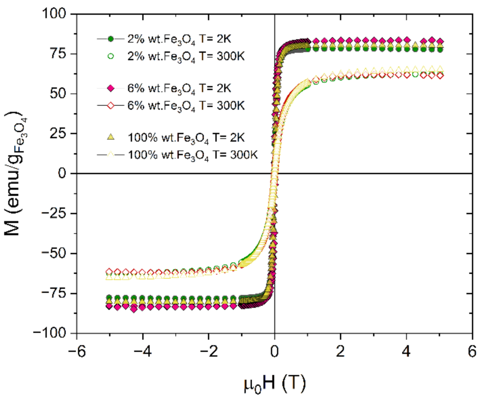
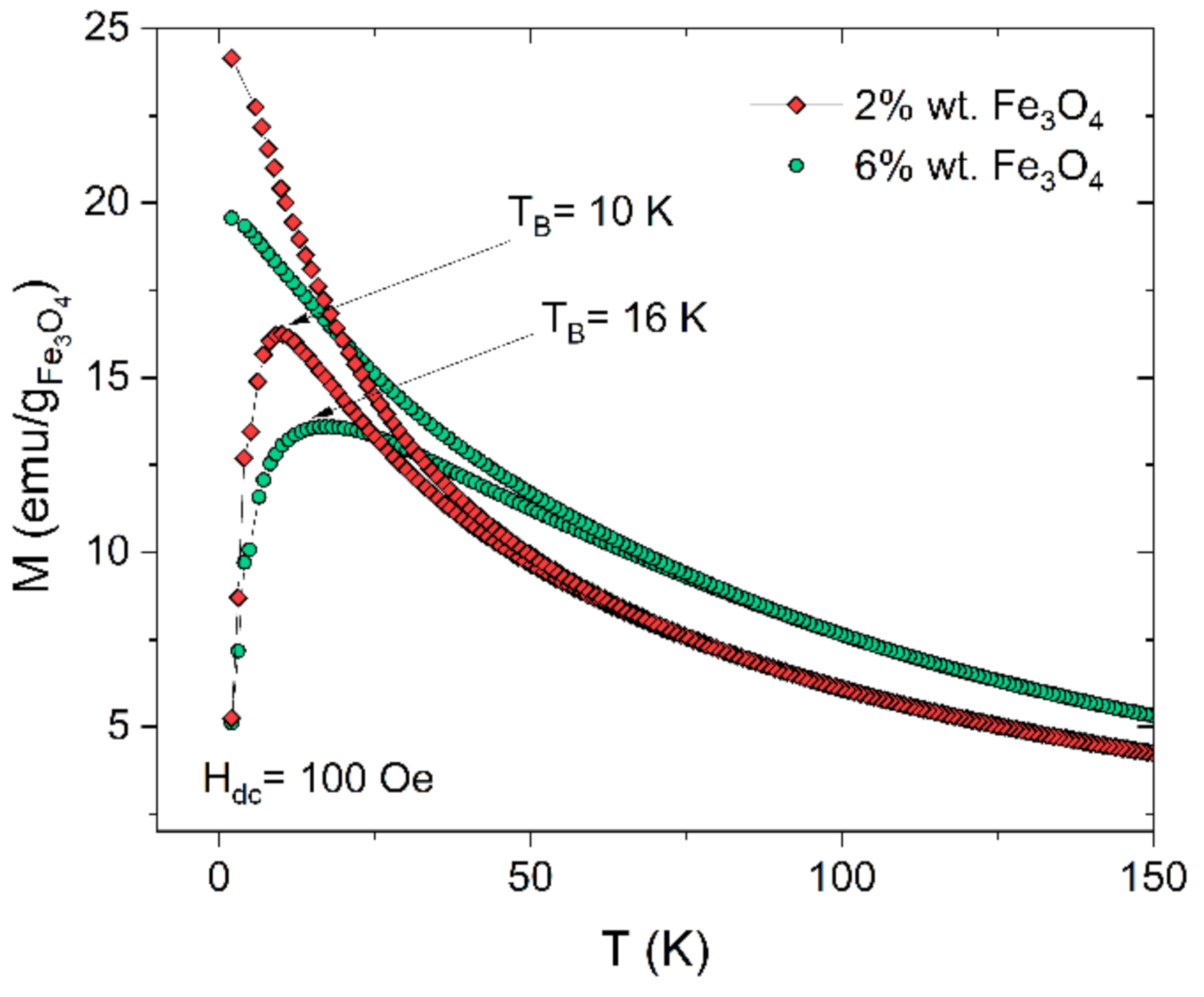
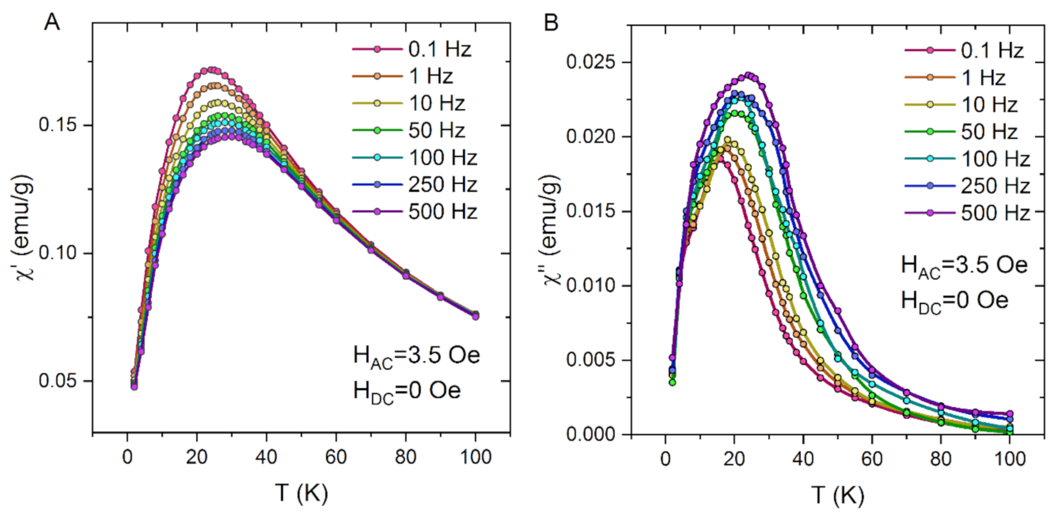
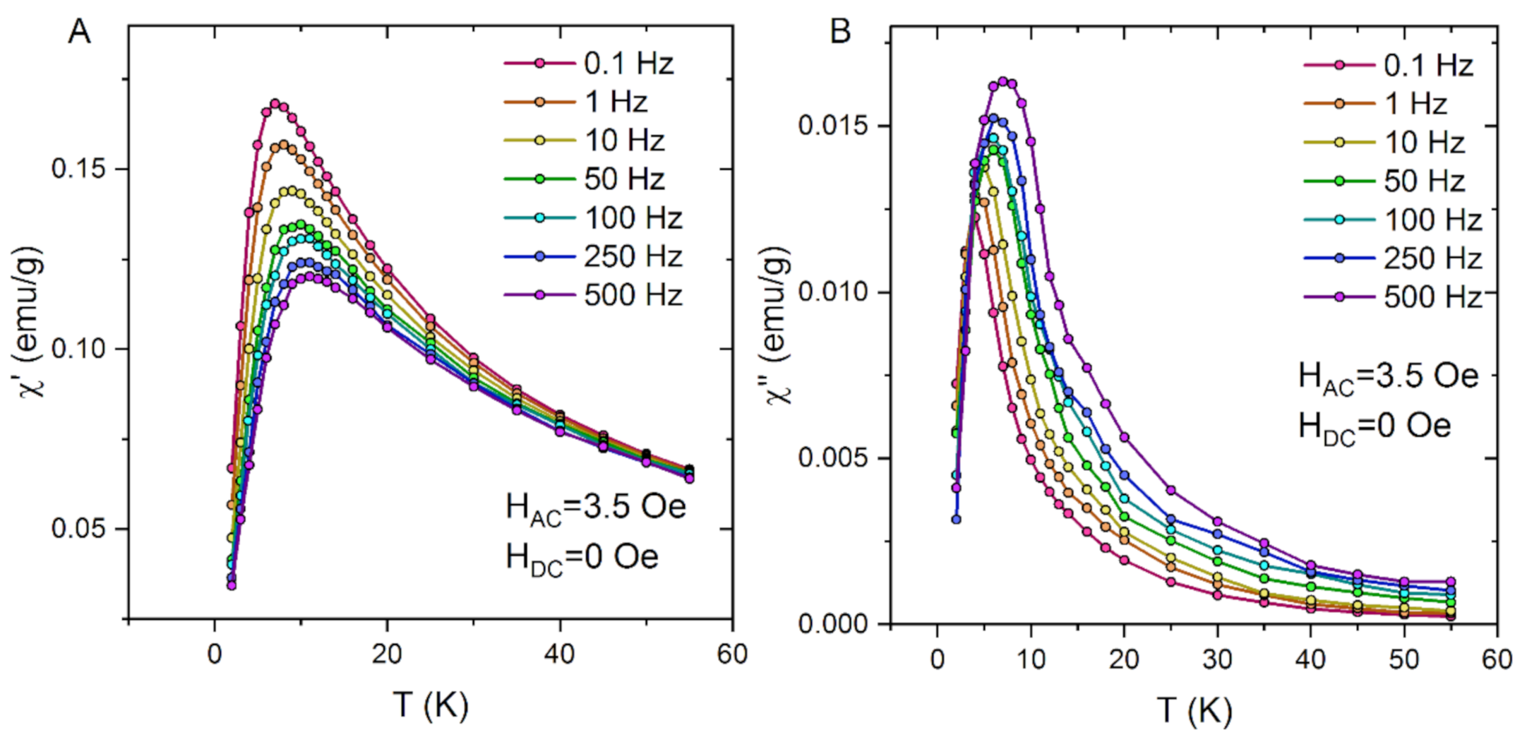
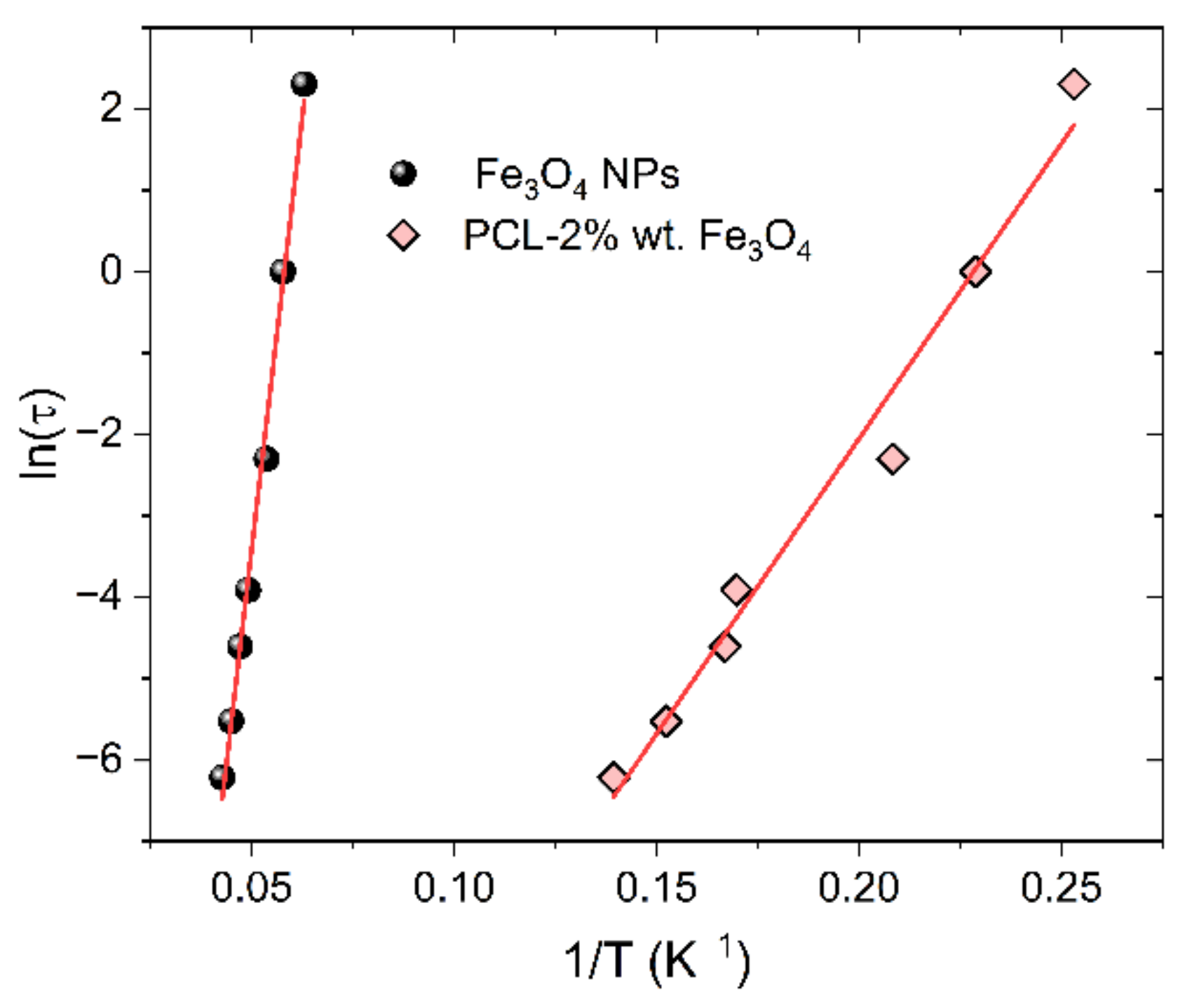
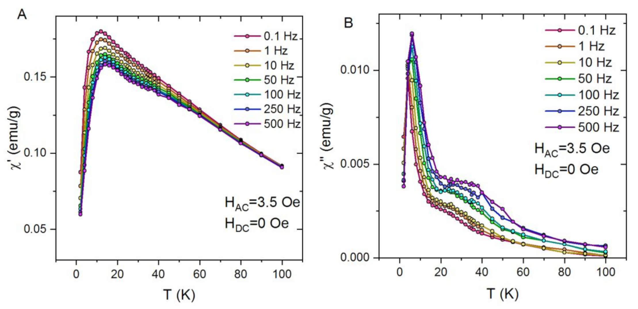
2.4. Magnetic Properties
3. Materials and Methods
3.1. Materials
3.2. Preparation of PCL and PCL-Fe3O4 Composite Fibres
3.3. Characterization Techniques
4. Conclusions
Author Contributions
Funding
Institutional Review Board Statement
Informed Consent Statement
Data Availability Statement
Conflicts of Interest
References
- Shifrina, Z.B.; Matveeva, V.G.; Bronstein, L.M. Role of Polymer Structures in Catalysis by Transition Metal and Metal Oxide Nanoparticle Composites. Chem. Rev. 2020, 120, 1350–1396. [Google Scholar] [CrossRef] [PubMed]
- Mertz, D.; Harlepp, S.; Goetz, J.; Bégin, D.; Schlatter, G.; Bégin-Colin, S.; Hébraud, A. Nanocomposite Polymer Scaffolds Responding under External Stimuli for Drug Delivery and Tissue Engineering Applications. Adv. Ther. 2020, 3, 1900143. [Google Scholar] [CrossRef]
- Bonilla, A.M.; Gonzalez, P.H. Hybrid Polymeric-Magnetic Nanoparticles in Cancer Treatments. Curr. Pharm. Des. 2018, 23. [Google Scholar] [CrossRef]
- Makarewicz, M.; Podsiadły, M.; Bałanda, M. Magnetic Investigation of Carbon Coated Co-, Ni- and Fe-Nanoparticles. Acta Phys. Pol. A 2009, 115, 568–571. [Google Scholar] [CrossRef]
- Sunny, V.; Sakthi Kumar, D.; Yoshida, Y.; Makarewicz, M.; Tabiś, W.; Anantharaman, M.R. Synthesis and properties of highly stable nickel/carbon core/shell nanostructures. Carbon NY 2010, 48, 1643–1651. [Google Scholar] [CrossRef]
- Mieloch, A.A.; Kręcisz, M.; Rybka, J.D.; Strugała, A.; Krupiński, M.; Urbanowicz, A.; Kozak, M.; Skalski, B.; Figlerowicz, M.; Giersig, M. The influence of ligand charge and length on the assembly of Brome mosaic virus derived virus-like particles with magnetic core. AIP Adv. 2018, 8, 035005. [Google Scholar] [CrossRef] [Green Version]
- Pérez, N.; Guardia, P.; Roca, A.G.; Morales, M.P.; Serna, C.J.; Iglesias, O.; Bartolomé, F.; García, L.M.; Batlle, X.; Labarta, A. Surface anisotropy broadening of the energy barrier distribution in magnetic nanoparticles. Nanotechnology 2008, 19, 475704. [Google Scholar] [CrossRef] [PubMed] [Green Version]
- Rudakov, G.A.; Tsiberkin, K.B.; Ponomarev, R.S.; Henner, V.K.; Ziolkowska, D.A.; Jasinski, J.B.; Sumanasekera, G. Magnetic properties of transition metal nanoparticles enclosed in carbon nanocages. J. Magn. Magn. Mater. 2019, 472, 34–39. [Google Scholar] [CrossRef]
- Deubel, F.; Steenackers, M.; Garrido, J.A.; Stutzmann, M.; Jordan, R. Semiconductor/Polymer Nanocomposites of Acrylates and Nanocrystalline Silicon by Laser-Induced Thermal Polymerization. Macromol. Mater. Eng. 2013, 298, 1160–1165. [Google Scholar] [CrossRef]
- John, A.; Benny, L.; Cherian, A.R.; Narahari, S.Y.; Varghese, A.; Hegde, G. Electrochemical sensors using conducting polymer/noble metal nanoparticle nanocomposites for the detection of various analytes: A review. J. Nanostructure Chem. 2021, 11, 1–31. [Google Scholar] [CrossRef]
- Chen, J.; Liu, B.; Gao, X.; Xu, D. A review of the interfacial characteristics of polymer nanocomposites containing carbon nanotubes. RSC Adv. 2018, 8, 28048–28085. [Google Scholar] [CrossRef] [Green Version]
- Tripathy, J. Polymer Nanocomposites for Biomedical and Biotechnology Applications. In Properties and Applications of Polymer Nanocomposites; Springer: Berlin/Heidelberg, Germany, 2017; pp. 57–76. [Google Scholar]
- Liu, Y.; Kumar, S. Polymer/Carbon Nanotube Nano Composite Fibers—A Review. ACS Appl. Mater. Interfaces 2014, 6, 6069–6087. [Google Scholar] [CrossRef]
- Urbina, M.C.; Zinoveva, S.; Miller, T.; Sabliov, C.M.; Monroe, W.T.; Kumar, C.S.S.R. Investigation of Magnetic Nanoparticle−Polymer Composites for Multiple-controlled Drug Delivery. J. Phys. Chem. C 2008, 112, 11102–11108. [Google Scholar] [CrossRef]
- Yar, Y.; Khodadust, R.; Akkoc, Y.; Utkur, M.; Saritas, E.U.; Gozuacik, D.; Yagci Acar, H. Development of tailored SPION-PNIPAM nanoparticles by ATRP for dually responsive doxorubicin delivery and MR imaging. J. Mater. Chem. B 2018, 6, 289–300. [Google Scholar] [CrossRef] [Green Version]
- Palanisamy, S.; Wang, Y.-M. Superparamagnetic iron oxide nanoparticulate system: Synthesis, targeting, drug delivery and therapy in cancer. Dalt. Trans. 2019, 48, 9490–9515. [Google Scholar] [CrossRef]
- Jiang, P.-C.; Chang, C.-H.-T.; Hsieh, C.-Y.; Su, W.-B.; Tsay, J.-S. A practical method for fabricating superparamagnetic films and the mechanism involved. Nanoscale 2020, 12, 14096–14105. [Google Scholar] [CrossRef]
- Vural, M.; Crowgey, B.; Kempel, L.C.; Kofinas, P. Nanostructured flexible magneto-dielectrics for radio frequency applications. J. Mater. Chem. C 2014, 2, 756–763. [Google Scholar] [CrossRef]
- Wang, C.; Wang, J.; Zeng, L.; Qiao, Z.; Liu, X.; Liu, H.; Zhang, J.; Ding, J. Fabrication of Electrospun Polymer Nanofibers with Diverse Morphologies. Molecules 2019, 24, 834. [Google Scholar] [CrossRef] [Green Version]
- Zhong, Y.; Leung, V.; Yuqin Wan, L.; Dutz, S.; Ko, F.K.; Häfeli, U.O. Electrospun magnetic nanofibre mats—A new bondable biomaterial using remotely activated magnetic heating. J. Magn. Magn. Mater. 2015, 380, 330–334. [Google Scholar] [CrossRef]
- Kim, I.; Viswanathan, K.; Kasi, G.; Sadeghi, K.; Thanakkasaranee, S.; Seo, J. Poly(Lactic Acid)/ZnO Bionanocomposite Films with Positively Charged ZnO as Potential Antimicrobial Food Packaging Materials. Polymers (Basel) 2019, 11, 1427. [Google Scholar] [CrossRef] [Green Version]
- Elzein, T.; Nasser-Eddine, M.; Delaite, C.; Bistac, S.; Dumas, P. FTIR study of polycaprolactone chain organization at interfaces. J. Colloid Interface Sci. 2004, 273, 381–387. [Google Scholar] [CrossRef] [PubMed]
- Mydosh, J.A. Spin Glasses; CRC Press: Boca Raton, FL, USA, 2014; ISBN 9781482295191. [Google Scholar]


Publisher’s Note: MDPI stays neutral with regard to jurisdictional claims in published maps and institutional affiliations. |
© 2021 by the authors. Licensee MDPI, Basel, Switzerland. This article is an open access article distributed under the terms and conditions of the Creative Commons Attribution (CC BY) license (https://creativecommons.org/licenses/by/4.0/).
Share and Cite
Sas, W.; Jasiurkowska-Delaporte, M.; Czaja, P.; Zieliński, P.M.; Fitta, M. Magnetic Properties Study of Iron Oxide Nanoparticles-Loaded Poly(ε-caprolactone) Nanofibres. Magnetochemistry 2021, 7, 61. https://0-doi-org.brum.beds.ac.uk/10.3390/magnetochemistry7050061
Sas W, Jasiurkowska-Delaporte M, Czaja P, Zieliński PM, Fitta M. Magnetic Properties Study of Iron Oxide Nanoparticles-Loaded Poly(ε-caprolactone) Nanofibres. Magnetochemistry. 2021; 7(5):61. https://0-doi-org.brum.beds.ac.uk/10.3390/magnetochemistry7050061
Chicago/Turabian StyleSas, Wojciech, Małgorzata Jasiurkowska-Delaporte, Paweł Czaja, Piotr Maciej Zieliński, and Magdalena Fitta. 2021. "Magnetic Properties Study of Iron Oxide Nanoparticles-Loaded Poly(ε-caprolactone) Nanofibres" Magnetochemistry 7, no. 5: 61. https://0-doi-org.brum.beds.ac.uk/10.3390/magnetochemistry7050061