Mesoporous Bioactive Glasses Cytocompatibility Assessment: A Review of In Vitro Studies


 ,
,  ,
,
Abstract
:1. Introduction
- -
- The formation of a biomimetic hydroxyapatite layer after glass immersion after interaction with biological fluids;
- -
- The osteogenic ability of some dissolution products and leachable compounds and ions.
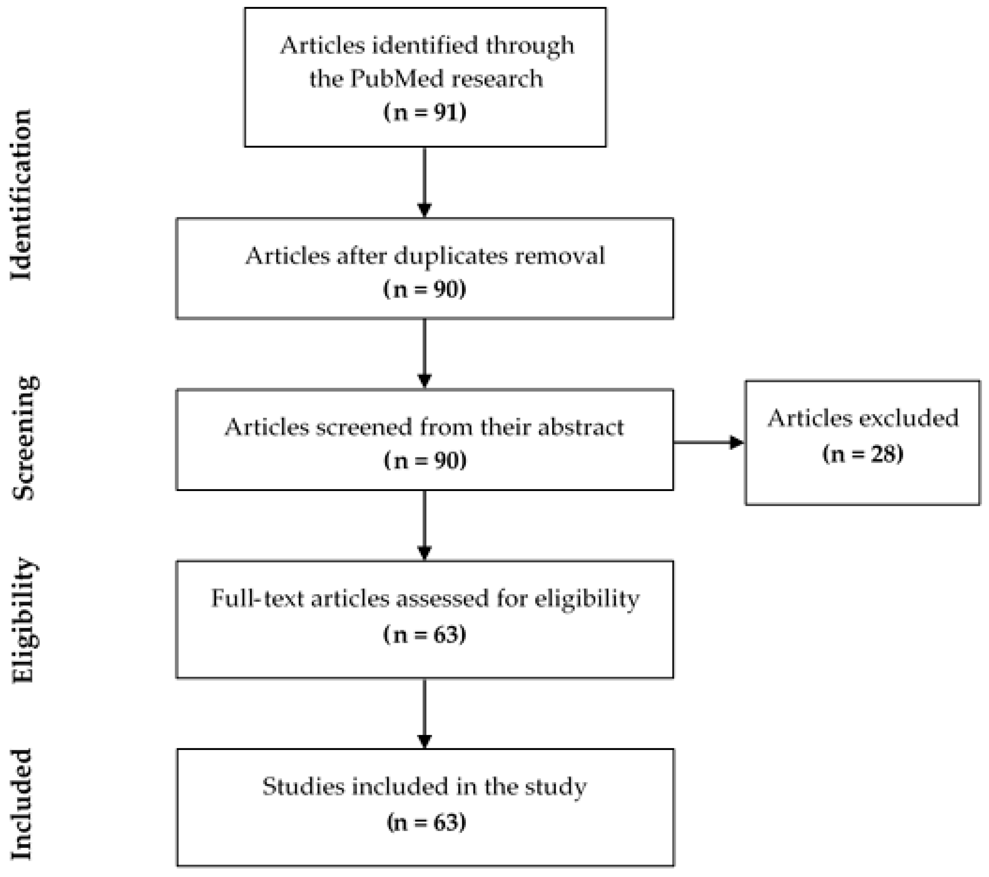
2. Methods
2.1. Research Question
2.2. Search Strategy
2.3. Keywords Selection
2.4. Inclusion and Exclusion Criteria
- (1)
- Research article;
- (2)
- Use of mesoporous bioactive glasses with and without scaffolds or composite;
- (3)
- Detailed investigation of biocompatibility in vitro.
- (1)
- Review articles;
- (2)
- Articles that used non-mesoporous bioactive glasses;
- (3)
- Articles that did not explain their biocompatibility methods;
- (4)
- Articles that did not carry out biocompatibility tests in vitro.
2.5. Paper Selection and Data Extraction
- -
- Medical application of the MBG;
- -
- MBG composition and synthesis pathway;
- -
- Cells used for biocompatibility tests;
- -
- Biocompatibility tests performed and their results.
3. Results
3.1. Control Group Used to Compare the Biocompatibility of Mesoporous Bioactive Glasses (MBGs)
3.2. Study Characteristics
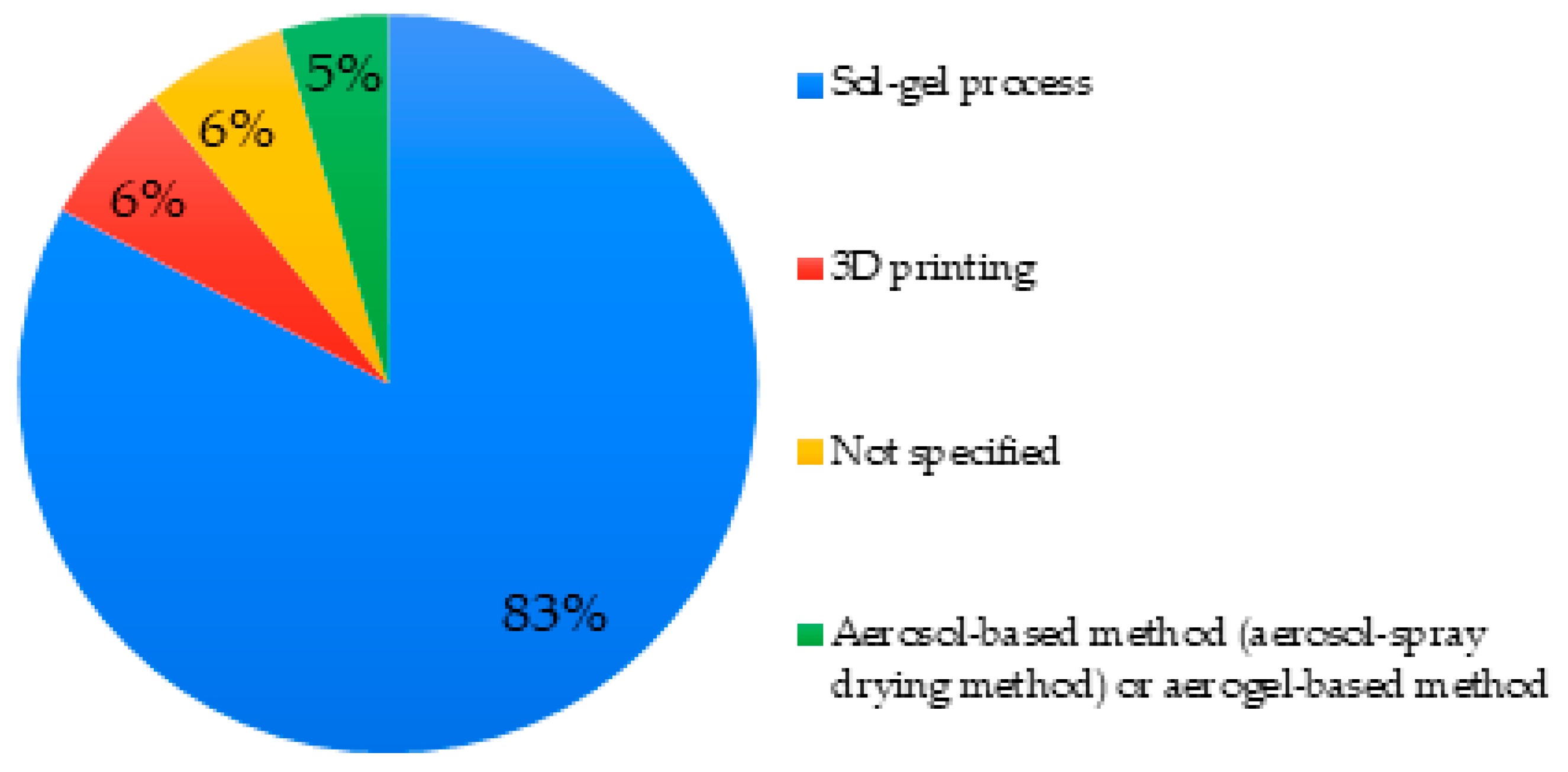
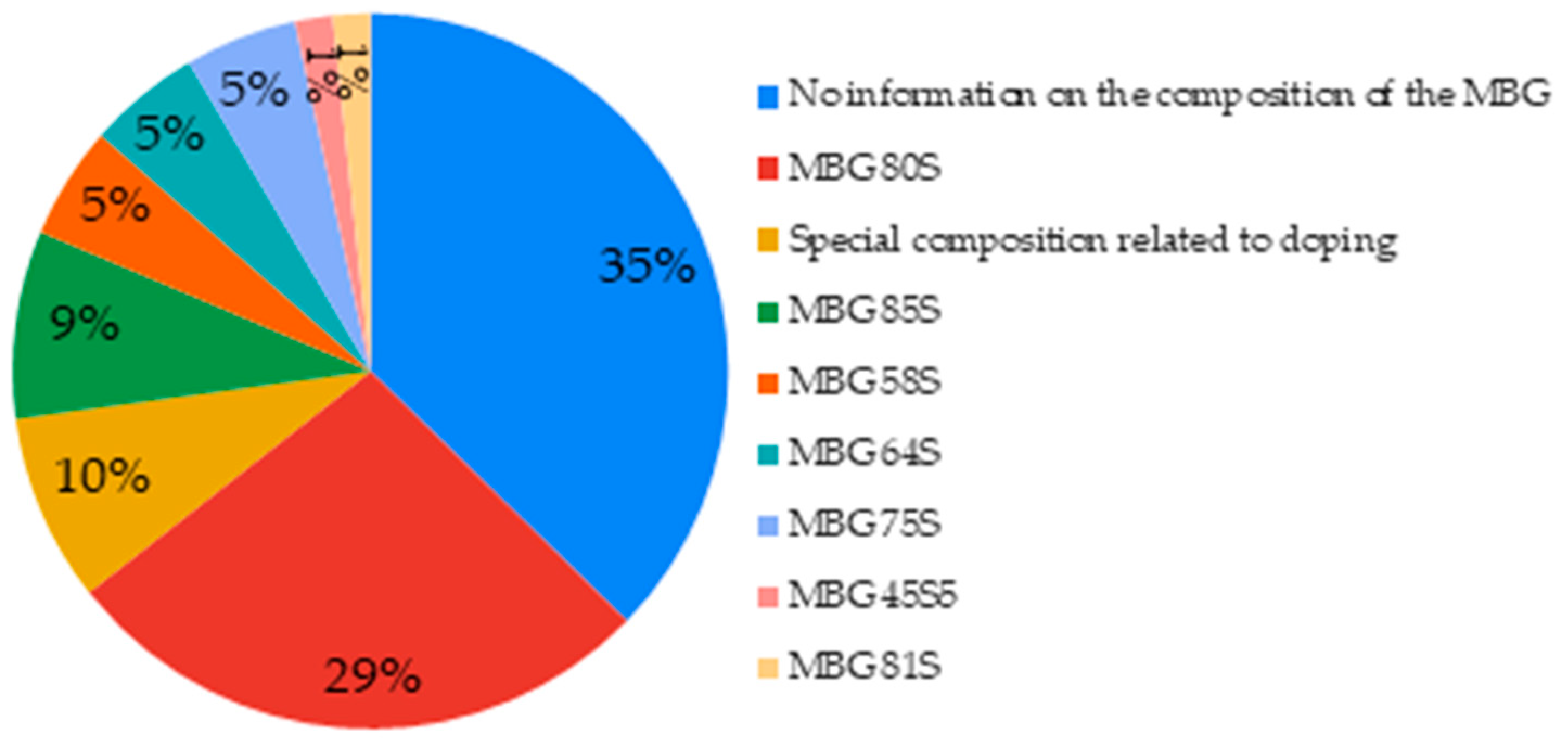
3.2.1. MBG Synthesis, Characteristics, and Application Areas
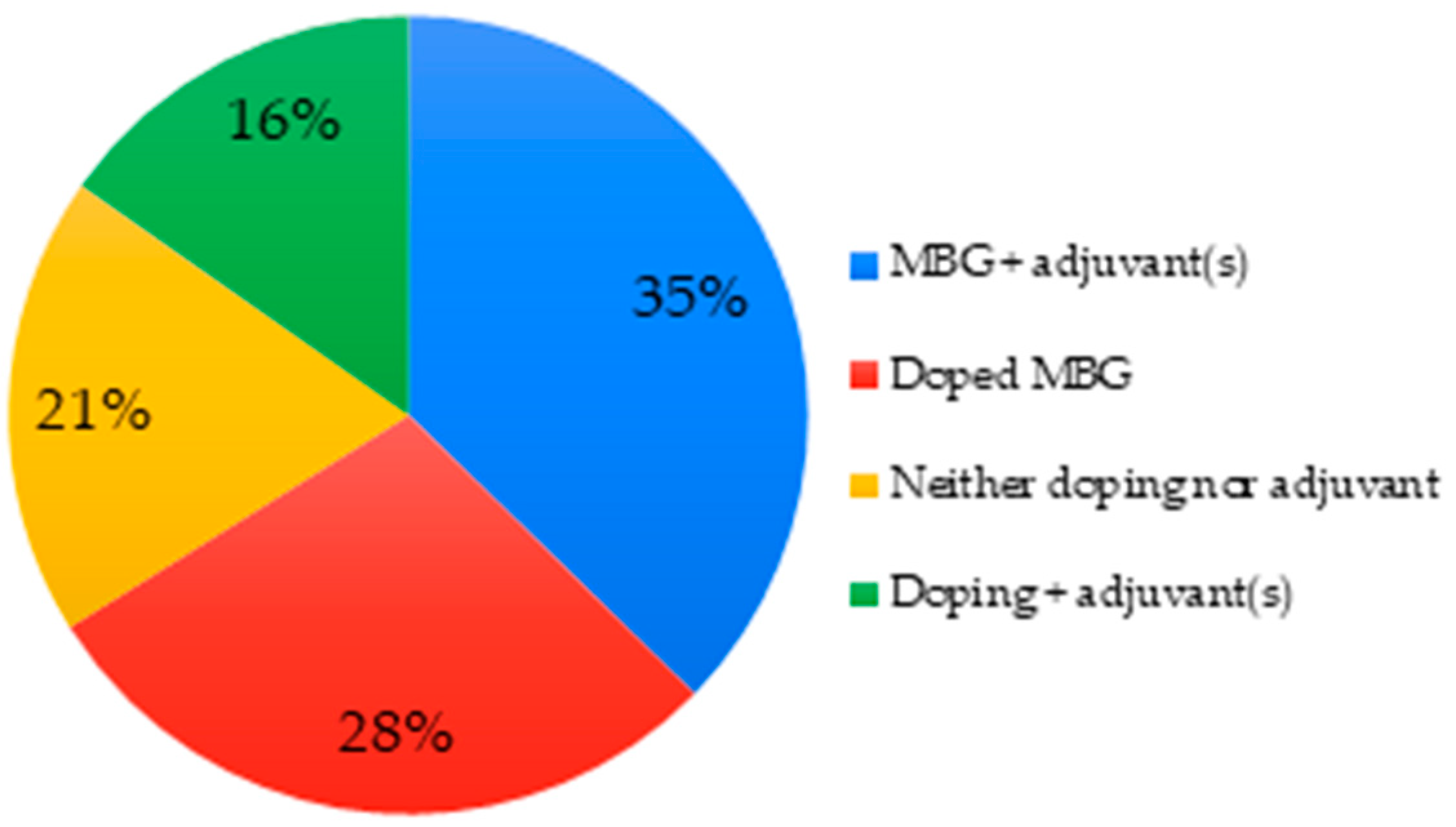
3.2.2. Doping and Adjuvants of Studied MBGs
3.3. Cells Characteristics
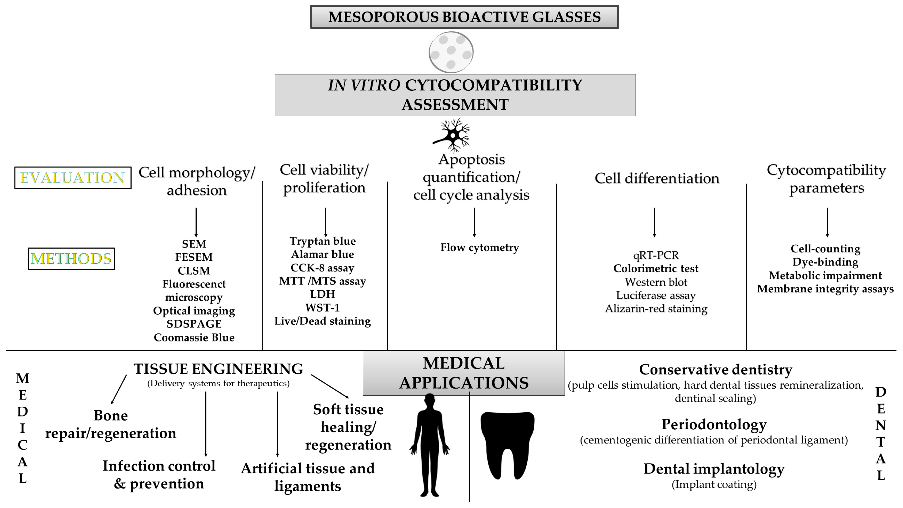
3.4. Techniques Used to Assess the MBG In Vitro Biocompatibility
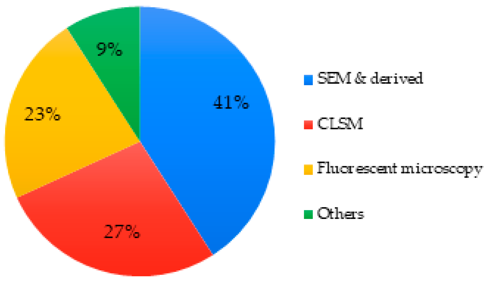
3.4.1. Cell Morphology
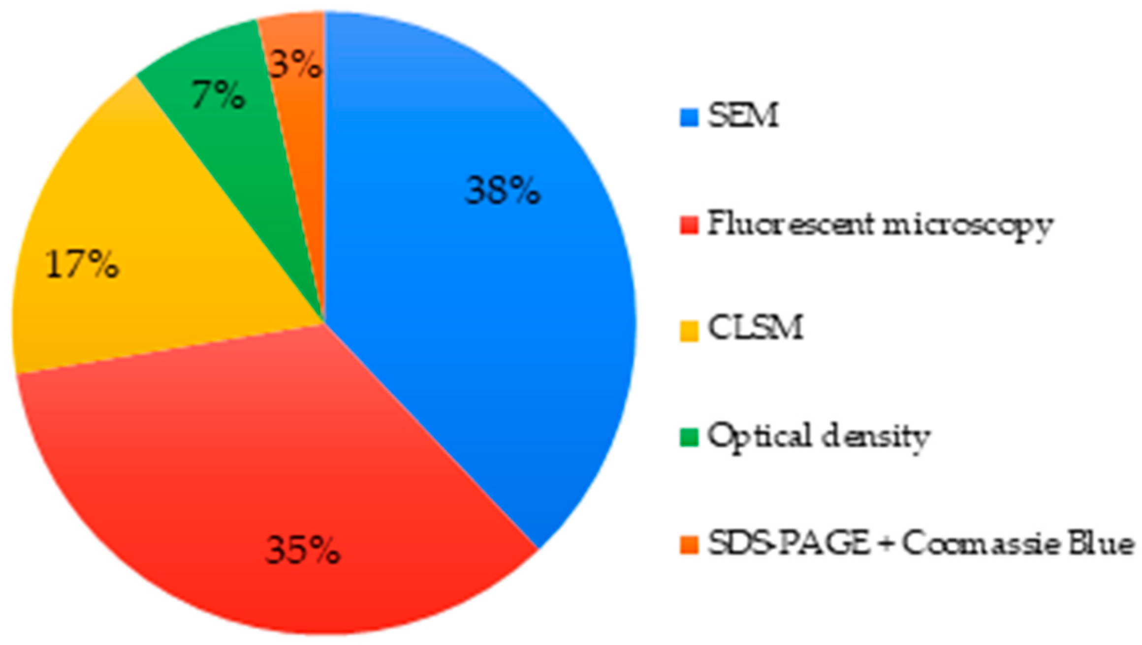
3.4.2. Cell Adhesion/Attachment
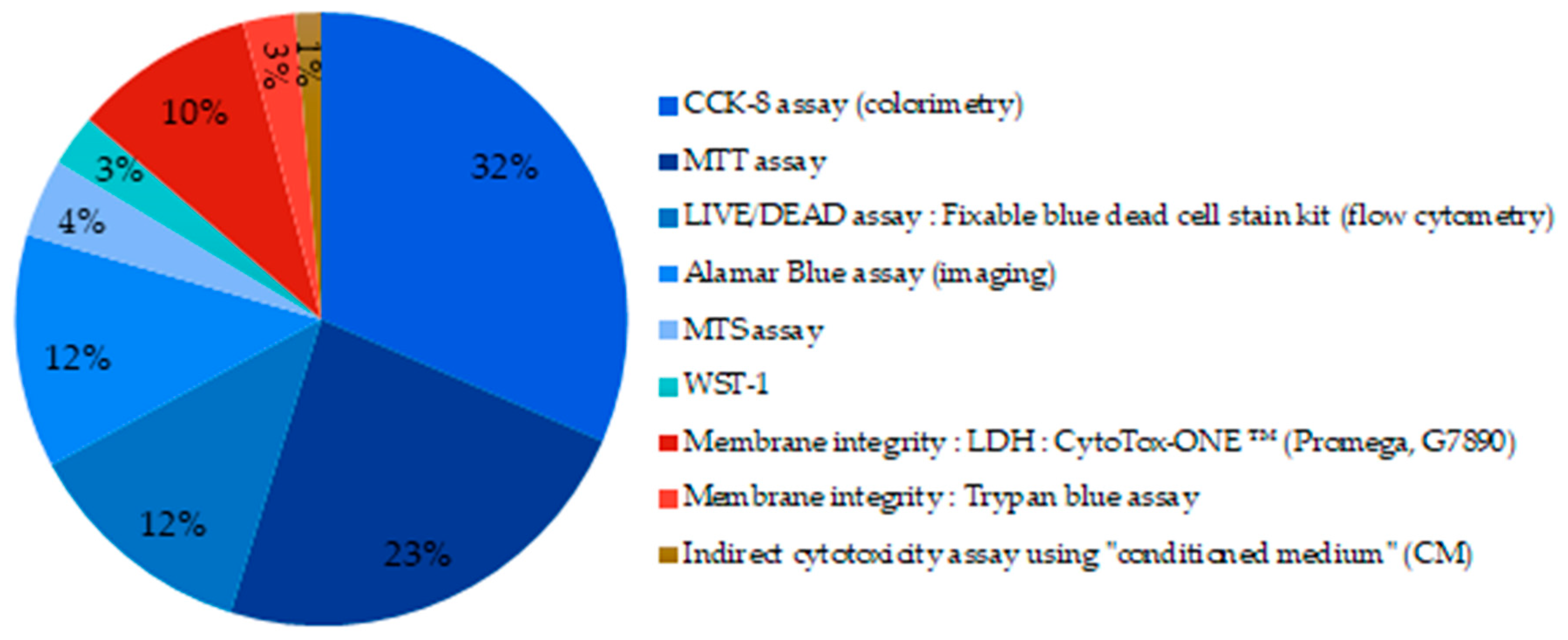
3.4.3. Cell Viability and Proliferation
3.4.4. Apoptosis Quantification, Cell-Cycle Analysis
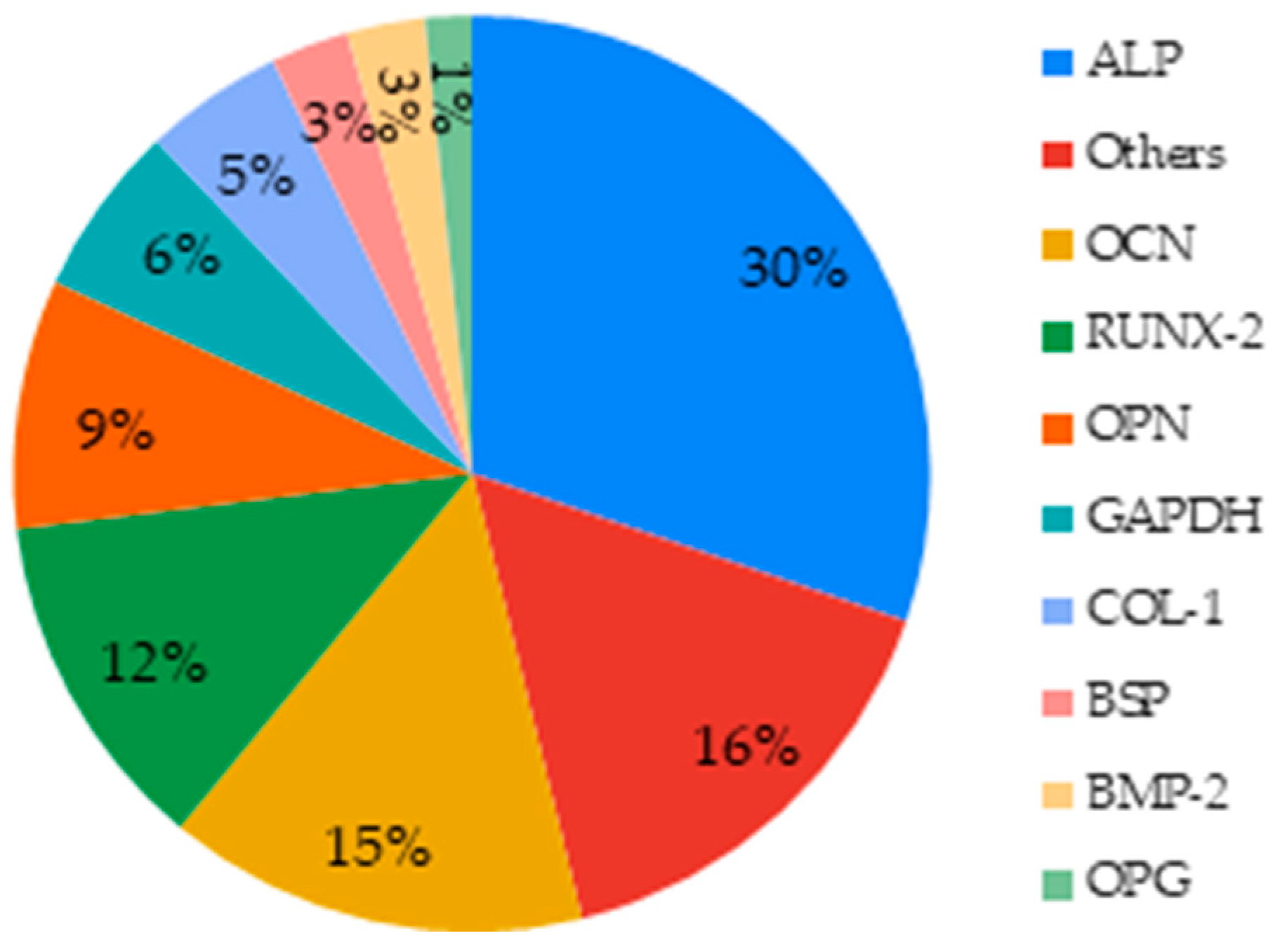
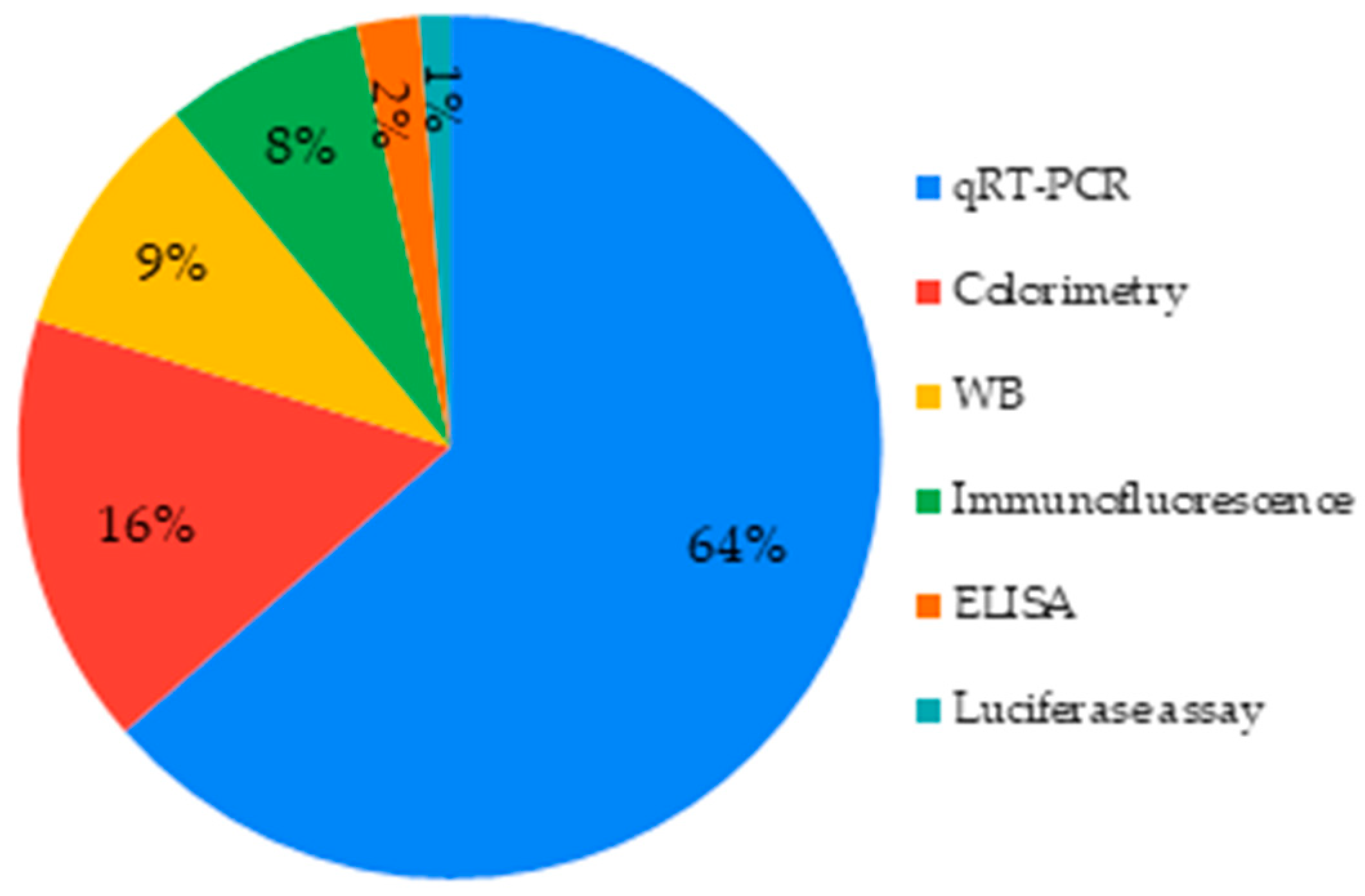
3.4.5. Cell Differentiation
3.5. Cell cultivation Setting (Direct and Indirect Contact)
4. Discussion
5. Conclusions
Author Contributions
Funding
Institutional Review Board Statement
Informed Consent Statement
Data Availability Statement
Acknowledgments
Conflicts of Interest
References
- Hench, L.L. The Story of Bioglass®. J. Mater. Sci. Mater. Med. 2006, 17, 967–978. [Google Scholar] [CrossRef] [PubMed]
- Donaruma, L.G. Definitions in Biomaterials. J. Polym. Sci. B Polym. Lett. Ed. 1988, 26, 414. [Google Scholar] [CrossRef]
- Jones, J.R. Review of Bioactive Glass: From Hench to Hybrids. Acta Biomater. 2013, 9, 4457–4486. [Google Scholar] [CrossRef] [PubMed]
- Zamet, J.S.; Darbar, U.R.; Griffiths, G.S.; Bulman, J.S.; Bragger, U.; Burgin, W.; Newman, H.N. Particulate BioglassR as a Grafting Material in the Treatment of Periodontal Intrabony Defects. J. Clin. Periodontol. 1997, 24, 410–418. [Google Scholar] [CrossRef]
- Braem, A.; Mattheys, T.; Neirinck, B.; Čeh, M.; Novak, S.; Schrooten, J.; Van der Biest, O.; Vleugels, J. Bioactive Glass–Ceramic Coated Titanium Implants Prepared by Electrophoretic Deposition. Mater. Sci. Eng. C 2012, 32, 2267–2273. [Google Scholar] [CrossRef]
- Van Oirschot, B.A.J.A.; Alghamdi, H.S.; Närhi, T.O.; Anil, S.; Al Farraj Aldosari, A.; van den Beucken, J.J.J.P.; Jansen, J.A. In Vivo Evaluation of Bioactive Glass-Based Coatings on Dental Implants in a Dog Implantation Model. Clin. Oral Impl. Res. 2014, 25, 21–28. [Google Scholar] [CrossRef]
- Pourshahrestani, S.; Kadri, N.A.; Zeimaran, E.; Towler, M.R. Well-Ordered Mesoporous Silica and Bioactive Glasses: Promise for Improved Hemostasis. Biomater. Sci. 2019, 7, 31–50. [Google Scholar] [CrossRef]
- Earl, J.S.; Leary, R.K.; Muller, K.H.; Langford, R.M.; Greenspan, D.C. Physical and Chemical Characterization of Dentin Surface Following Treatment with NovaMin Technology. J. Clin. Dent. 2011, 22, 62–67. [Google Scholar]
- Mocquot, C.; Attik, N.; Pradelle-Plasse, N.; Grosgogeat, B.; Colon, P. Bioactivity Assessment of Bioactive Glasses for Dental Applications: A Critical Review. Dent. Mater. 2020, 36, 1116–1143. [Google Scholar] [CrossRef]
- Yan, X.; Yu, C.; Zhou, X.; Tang, J.; Zhao, D. Highly Ordered Mesoporous Bioactive Glasses with Superior In Vitro Bone-Forming Bioactivities. Angew. Chem. Int. Ed. 2004, 43, 5980–5984. [Google Scholar] [CrossRef]
- Fernando, D.; Attik, N.; Cresswell, M.; Mokbel, I.; Pradelle-Plasse, N.; Jackson, P.; Grosgogeat, B.; Colon, P. Influence of Network Modifiers in an Acetate Based Sol-Gel Bioactive Glass System. Microporous Mesoporous Mater. 2018, 257, 99–109. [Google Scholar] [CrossRef]
- Pérez-Pariente, J.; Balas, F.; Román, J.; Salinas, A.J.; Vallet-Regí, M. Influence of Composition and Surface Characteristics on the in Vitro Bioactivity of SiO(2)-CaO-P(2)O(5)-MgO Sol-Gel Glasses. J. Biomed. Mater. Res. 1999, 47, 170–175. [Google Scholar] [CrossRef]
- Chen, W.-C.; Kung, J.-C.; Chen, C.-H.; Hsiao, Y.-C.; Shih, C.-J.; Chien, C.-S. Effects of Bioactive Glass with and without Mesoporous Structures on Desensitization in Dentinal Tubule Occlusion. Appl. Surf. Sci. 2013, 283, 833–842. [Google Scholar] [CrossRef]
- Soulié, J.; Hardy-Dessources, A.; Nedelec, J.-M.; Jallot, E. 3D Organized Macroporous Bioactive Glasses: A Study of Pore Size Effect on Physicochemical Reactivity by Micro-PIXE-RBS. J. Phys. Chem. C 2013, 117, 6702–6711. [Google Scholar] [CrossRef]
- Kresge, C.T.; Leonowicz, M.E.; Roth, W.J.; Vartuli, J.C.; Beck, J.S. Ordered Mesoporous Molecular Sieves Synthesized by a Liquid-Crystal Template Mechanism. Nature 1992, 359, 710–712. [Google Scholar] [CrossRef]
- Holland, B.T.; Blanford, C.F.; Do, T.; Stein, A. Synthesis of Highly Ordered, Three-Dimensional, Macroporous Structures of Amorphous or Crystalline Inorganic Oxides, Phosphates, and Hybrid Composites. Chem. Mater. 1999, 11, 795–805. [Google Scholar] [CrossRef]
- Wang, X.; Liu, Q.; Chen, W.; Liu, L. FGF Adsorbed Mesoporous Bioactive Glass with Larger Pores in Enhancing Bone Tissue Engineering. J. Mater. Sci. Mater. Med. 2019, 30, 48. [Google Scholar] [CrossRef]
- López-Noriega, A.; Arcos, D.; Izquierdo-Barba, I.; Sakamoto, Y.; Terasaki, O.; Vallet-Regí, M. Ordered Mesoporous Bioactive Glasses for Bone Tissue Regeneration. Chem. Mater. 2006, 18, 3137–3144. [Google Scholar] [CrossRef]
- Wu, L.; Wei, Z.; He, S.; Bi, Y.; Cao, Y.; Wang, W. Mesoporous Bioactive Glass Scaffold Delivers Salvianolic Acid B to Promote Bone Regeneration in a Rat Cranial Defect Model. Curr. Drug Deliv. 2020, 17. [Google Scholar] [CrossRef]
- Wang, X.; Cheng, F.; Liu, J.; Smått, J.-H.; Gepperth, D.; Lastusaari, M.; Xu, C.; Hupa, L. Biocomposites of Copper-Containing Mesoporous Bioactive Glass and Nanofibrillated Cellulose: Biocompatibility and Angiogenic Promotion in Chronic Wound Healing Application. Acta Biomater. 2016, 46, 286–298. [Google Scholar] [CrossRef]
- Mocquot, C.; Colon, P.; Fernando, D.; Jackson, P.; Pradelle-Plasse, N.; Grosgogeat, B.; Attik, N. The Infuence of Experimental Bioactive Glasses on Pulp Cells Behavior in Vitro. Dent. Mater. 2020, 36, 1322–1331. [Google Scholar] [CrossRef] [PubMed]
- Mao, C.; Chen, X.; Hu, Q.; Miao, G.; Lin, C. Acute Toxicity and in Vivo Biodistribution of Monodispersed Mesoporous Bioactive Glass Spheres in Intravenously Exposed Mice. Mater. Sci. Eng. C 2016, 58, 682–691. [Google Scholar] [CrossRef] [PubMed]
- Anand, A.; Lalzawmliana, V.; Kumar, V.; Das, P.; Devi, K.B.; Maji, A.K.; Kundu, B.; Roy, M.; Nandi, S.K. Preparation and in Vivo Biocompatibility Studies of Different Mesoporous Bioactive Glasses. J. Mech. Behav. Biomed. Mater. 2019, 89, 89–98. [Google Scholar] [CrossRef] [PubMed]
- Ghamor-Amegavi, E.P.; Yang, X.; Qiu, J.; Xie, L.; Pan, Z.; Wang, J.; Zhang, X.; Ke, X.; Zhao, T.; Zhang, L.; et al. Composition Control in Biphasic Silicate Microspheres on Stimulating New Bone Regeneration and Repair of Osteoporotic Femoral Bone Defect. J. Biomed. Mater. Res. 2020, 108, 377–390. [Google Scholar] [CrossRef] [PubMed]
- Lalzawmliana, V.; Anand, A.; Kumar, V.; Das, P.; Devi, K.B.; Mukherjee, J.; Maji, A.K.; Kundu, B.; Roy, M.; Nandi, S.K. Potential of Growth Factor Incorporated Mesoporous Bioactive Glass for in Vivo Bone Regeneration. J. Mech. Behav. Biomed. Mater. 2019, 91, 182–192. [Google Scholar] [CrossRef]
- Li, Y.; Bastakoti, B.P.; Yamauchi, Y. Smart Soft-Templating Synthesis of Hollow Mesoporous Bioactive Glass Spheres. Chem. Eur. J. 2015, 21, 8038–8042. [Google Scholar] [CrossRef]
- Garg, S.; Thakur, S.; Gupta, A.; Kaur, G.; Pandey, O.P. Antibacterial and Anticancerous Drug Loading Kinetics for (10-x)CuO-XZnO-20CaO-60SiO2-10P2O5 (2 ≤ x ≤ 8) Mesoporous Bioactive Glasses. J. Mater. Sci. Mater. Med. 2017, 28, 11. [Google Scholar] [CrossRef]
- Jiang, S.; Zhang, Y.; Shu, Y.; Wu, Z.; Cao, W.; Huang, W. Amino-Functionalized Mesoporous Bioactive Glass for Drug Delivery. Biomed. Mater. 2017, 12, 025017. [Google Scholar] [CrossRef]
- Shoaib, M.; Saeed, A.; Rahman, M.S.U.; Naseer, M.M. Mesoporous Nano-Bioglass Designed for the Release of Imatinib and in Vitro Inhibitory Effects on Cancer Cells. Mater. Sci. Eng. C 2017, 77, 725–730. [Google Scholar] [CrossRef]
- Baino, F.; Fiume, E.; Miola, M.; Leone, F.; Onida, B.; Laviano, F.; Gerbaldo, R.; Verné, E. Fe-Doped Sol-Gel Glasses and Glass-Ceramics for Magnetic Hyperthermia. Materials 2018, 11, 173. [Google Scholar] [CrossRef] [Green Version]
- Baino, F.; Potestio, I.; Vitale-Brovarone, C. Production and Physicochemical Characterization of Cu-Doped Silicate Bioceramic Scaffolds. Materials 2018, 11, 1524. [Google Scholar] [CrossRef] [PubMed] [Green Version]
- Fernando, D.; Colon, P.; Cresswell, M.; Journet, C.; Pradelle-Plasse, N.; Jackson, P.; Grosgogeat, B.; Attik, N. The Influence of Precursor Addition Order on the Porosity of Sol–Gel Bioactive Glasses. Dent. Mater. 2018, 34, 1323–1330. [Google Scholar] [CrossRef] [PubMed]
- Kargozar, S.; Baino, F.; Hamzehlou, S.; Hill, R.G.; Mozafari, M. Bioactive Glasses Entering the Mainstream. Drug Discov. Today 2018, 23, 1700–1704. [Google Scholar] [CrossRef] [PubMed]
- Nawaz, Q.; Rehman, M.A.U.; Burkovski, A.; Schmidt, J.; Beltrán, A.M.; Shahid, A.; Alber, N.K.; Peukert, W.; Boccaccini, A.R. Synthesis and Characterization of Manganese Containing Mesoporous Bioactive Glass Nanoparticles for Biomedical Applications. J. Mater. Sci. Mater. Med. 2018, 29, 64. [Google Scholar] [CrossRef]
- Liu, X.; Chen, H.-H.; Lin, Y.-C.; Nabilla, S.C.; Liu, W.-C.; Wang, W.-C.; Shih, S.-J.; Li, Y.; Lin, C.-P.; Zhao, G.; et al. Composite Polyelectrolyte Multilayer and Mesoporous Bioactive Glass Nanoparticle Coating on 316L Stainless Steel for Controlled Antibiotic Release and Biocompatibility. J. Biomed. Nanotechnol. 2018, 14, 725–735. [Google Scholar] [CrossRef]
- Mubina, M.S.K.; Shailajha, S.; Sankaranarayanan, R.; Saranya, L. In Vitro Bioactivity, Mechanical Behavior and Antibacterial Properties of Mesoporous SiO2-CaO-Na2O-P2O5 Nano Bioactive Glass Ceramics. J. Mech. Behav. Biomed. Mater. 2019, 100, 103379. [Google Scholar] [CrossRef]
- Shadjou, N.; Hasanzadeh, M. Silica-Based Mesoporous Nanobiomaterials as Promoter of Bone Regeneration Process: Bone Regeneration Process Using Silica-Based Mesoporous Nanobiomaterials. J. Biomed. Mater. Res. 2015, 103, 3703–3716. [Google Scholar] [CrossRef]
- Baino, F.; Fiorilli, S.; Vitale-Brovarone, C. Bioactive Glass-Based Materials with Hierarchical Porosity for Medical Applications: Review of Recent Advances. Acta Biomater. 2016, 42, 18–32. [Google Scholar] [CrossRef]
- Galarraga-Vinueza, M.E.; Mesquita-Guimarães, J.; Magini, R.S.; Souza, J.C.M.; Fredel, M.C.; Boccaccini, A.R. Anti-Biofilm Properties of Bioactive Glasses Embedding Organic Active Compounds: Bioactive glasses embedding organic active compounds. J. Biomed. Mater. Res. 2017, 105, 672–679. [Google Scholar] [CrossRef]
- Vichery, C.; Nedelec, J.-M. Bioactive Glass Nanoparticles: From Synthesis to Materials Design for Biomedical Applications. Materials 2016, 9, 288. [Google Scholar] [CrossRef] [Green Version]
- Fiume, E.; Barberi, J.; Verné, E.; Baino, F. Bioactive Glasses: From Parent 45S5 Composition to Scaffold-Assisted Tissue-Healing Therapies. J. Funct. Biomater. 2018, 9, 24. [Google Scholar] [CrossRef] [PubMed] [Green Version]
- Kargozar, S.; Montazerian, M.; Hamzehlou, S.; Kim, H.-W.; Baino, F. Mesoporous Bioactive Glasses: Promising Platforms for Antibacterial Strategies. Acta Biomater. 2018, 81, 1–19. [Google Scholar] [CrossRef] [PubMed]
- Kaya, S.; Cresswell, M.; Boccaccini, A.R. Mesoporous Silica-Based Bioactive Glasses for Antibiotic-Free Antibacterial Applications. Mater. Sci. Eng. C 2018, 83, 99–107. [Google Scholar] [CrossRef]
- Wu, J.; Yang, L.; Li, Y.; Guo, L. Research progress on mesoporous bioactive glass. Sheng Wu Yi Xue Gong Cheng Xue Za Zhi 2018, 4, 647–650. [Google Scholar] [CrossRef]
- Lalzawmliana, V.; Anand, A.; Roy, M.; Kundu, B.; Nandi, S.K. Mesoporous Bioactive Glasses for Bone Healing and Biomolecules Delivery. Mater. Sci. Eng. C 2020, 106, 110180. [Google Scholar] [CrossRef]
- Gisbert-Garzarán, M.; Manzano, M.; Vallet-Regí, M. Mesoporous Silica Nanoparticles for the Treatment of Complex Bone Diseases: Bone Cancer, Bone Infection and Osteoporosis. Pharmaceutics 2020, 12, 83. [Google Scholar] [CrossRef] [Green Version]
- Sistanipour, E.; Meshkini, A.; Oveisi, H. Catechin-Conjugated Mesoporous Hydroxyapatite Nanoparticle: A Novel Nano-Antioxidant with Enhanced Osteogenic Property. Colloids Surf. B Biointerf. 2018, 169, 329–339. [Google Scholar] [CrossRef]
- Wu, Y.; Zhang, W.; Zhang, J.; Mao, Z.-X.; Ding, L.; Li, H.; Ma, R.; Tang, J.-H. Methionine Functionalized Biocompatible Block Copolymers for Targeted Plasmid DNA Delivery. J. Vis. Exp. 2019, 58527. [Google Scholar] [CrossRef]
- Lin, D.; Yang, K.; Tang, W.; Liu, Y.; Yuan, Y.; Liu, C. A Poly(Glycerol Sebacate)-Coated Mesoporous Bioactive Glass Scaffold with Adjustable Mechanical Strength, Degradation Rate, Controlled-Release and Cell Behavior for Bone Tissue Engineering. Colloids Surf. B Biointerf. 2015, 131, 1–11. [Google Scholar] [CrossRef]
- Min, Z.; Shichang, Z.; Chen, X.; Yufang, Z.; Changqing, Z. 3D-Printed Dimethyloxallyl Glycine Delivery Scaffolds to Improve Angiogenesis and Osteogenesis. Biomater. Sci. 2015, 3, 1236–1244. [Google Scholar] [CrossRef]
- Gómez-Cerezo, N.; Sánchez-Salcedo, S.; Izquierdo-Barba, I.; Arcos, D.; Vallet-Regí, M. In Vitro Colonization of Stratified Bioactive Scaffolds by Pre-Osteoblast Cells. Acta Biomater. 2016, 44, 73–84. [Google Scholar] [CrossRef] [PubMed]
- Han, X.; Lin, H.; Guo, G.; Qu, F.; Chen, X.; Li, X. One-Step Method for the Preparation of Poly(Methyl Methacrylate) Modified Titanium-Bioactive Glass Three-Dimensional Scaffolds for Bone Tissue Engineering. IET Nanobiotechnol. 2016, 10, 45–53. [Google Scholar] [CrossRef] [PubMed]
- Hesaraki, S. Photocurable Bioactive Bone Cement Based on Hydroxyethyl Methacrylate-Poly(Acrylic/Maleic) Acid Resin and Mesoporous Sol Gel-Derived Bioactive Glass. Mater. Sci. Eng. C 2016, 63, 535–545. [Google Scholar] [CrossRef] [PubMed]
- Kim, T.-H.; Singh, R.K.; Kang, M.S.; Kim, J.-H.; Kim, H.-W. Inhibition of Osteoclastogenesis through SiRNA Delivery with Tunable Mesoporous Bioactive Nanocarriers. Acta Biomater. 2016, 29, 352–364. [Google Scholar] [CrossRef] [PubMed]
- Pourshahrestani, S.; Zeimaran, E.; Adib Kadri, N.; Gargiulo, N.; Samuel, S.; Naveen, S.V.; Kamarul, T.; Towler, M.R. Gallium-Containing Mesoporous Bioactive Glass with Potent Hemostatic Activity and Antibacterial Efficacy. J. Mater. Chem. B 2016, 4, 71–86. [Google Scholar] [CrossRef]
- Singh, R.K.; Patel, K.D.; Mahapatra, C.; Kang, M.S.; Kim, H.-W. C-Dot Generated Bioactive Organosilica Nanospheres in Theranostics: Multicolor Luminescent and Photothermal Properties Combined with Drug Delivery Capacity. ACS Appl. Mater. Interfaces 2016, 8, 24433–24444. [Google Scholar] [CrossRef] [PubMed]
- Tang, W.; Lin, D.; Yu, Y.; Niu, H.; Guo, H.; Yuan, Y.; Liu, C. Bioinspired Trimodal Macro/Micro/Nano-Porous Scaffolds Loading RhBMP-2 for Complete Regeneration of Critical Size Bone Defect. Acta Biomater. 2016, 32, 309–323. [Google Scholar] [CrossRef]
- Vishnu Priya, M.; Sivshanmugam, A.; Boccaccini, A.R.; Goudouri, O.M.; Sun, W.; Hwang, N.; Deepthi, S.; Nair, S.V.; Jayakumar, R. Injectable Osteogenic and Angiogenic Nanocomposite Hydrogels for Irregular Bone Defects. Biomed. Mater. 2016, 11, 035017. [Google Scholar] [CrossRef]
- Wu, T.; Cheng, N.; Xu, C.; Sun, W.; Yu, C.; Shi, B. The Effect of Mesoporous Bioglass on Osteogenesis and Adipogenesis of Osteoporotic BMSCs. J. Biomed. Mater. Res. A 2016, 104, 3004–3014. [Google Scholar] [CrossRef] [Green Version]
- Zhang, X.; Zeng, D.; Li, N.; Jiang, X.; Liu, C.; Li, Y. Large-Pore Mesoporous Ca–Si-Based Bioceramics with High in Vitro Bioactivity and Protein Adsorption Capability for Bone Tissue Regeneration. J. Mater. Chem. B 2016, 4, 3916–3924. [Google Scholar] [CrossRef]
- Zhang, X.; Zeng, D.; Li, N.; Wen, J.; Jiang, X.; Liu, C.; Li, Y. Functionalized Mesoporous Bioactive Glass Scaffolds for Enhanced Bone Tissue Regeneration. Sci. Rep. 2016, 6, 19361. [Google Scholar] [CrossRef] [PubMed] [Green Version]
- Ge, F.; Yu, M.; Yu, C.; Lin, J.; Weng, W.; Cheng, K.; Wang, H. Improved RhBMP-2 Function on MBG Incorporated TiO 2 Nanorod Films. Colloids Surf. B Biointerfaces 2017, 150, 153–158. [Google Scholar] [CrossRef] [PubMed]
- Kaur, G.; Sriranganathan, N.; Waldrop, S.G.; Sharma, P.; Chudasama, B.N. Effect of Copper on the Up-Regulation/down-Regulation of Genes, Cytotoxicity and Ion Dissolution for Mesoporous Bioactive Glasses. Biomed. Mater. 2017, 12, 045020. [Google Scholar] [CrossRef] [PubMed]
- Li, X.; Zhao, L.; Liang, Q.; Ye, J.; Komatsu, N.; Zhang, Q.; Gao, W.; Xu, M.; Chen, X. Cationic Polyarginine Conjugated Mesoporous Bioactive Glass Nanoparticles with Polyglycerol Coating for Efficient DNA Delivery. J. Biomed. Nanotechnol. 2017, 13, 280–289. [Google Scholar] [CrossRef] [PubMed]
- Luo, H.; Li, W.; Ao, H.; Li, G.; Tu, J.; Xiong, G.; Zhu, Y.; Wan, Y. Preparation, Structural Characterization, and in Vitro Cell Studies of Three-Dimensional SiO2–CaO Binary Glass Scaffolds Built Ofultra-Small Nanofibers. Mater. Sci. Eng. C 2017, 76, 94–101. [Google Scholar] [CrossRef] [PubMed]
- Luo, H.; Zhang, Y.; Li, G.; Tu, J.; Yang, Z.; Xiong, G.; Wang, Z.; Huang, Y.; Wan, Y. Sacrificial Template Method for the Synthesis of Three-Dimensional Nanofibrous 58S Bioglass Scaffold and Its in Vitro Bioactivity and Cell Responses. J. Biomater. Appl. 2017, 32, 265–275. [Google Scholar] [CrossRef] [PubMed]
- Pourshahrestani, S.; Zeimaran, E.; Kadri, N.A.; Gargiulo, N.; Jindal, H.M.; Naveen, S.V.; Sekaran, S.D.; Kamarul, T.; Towler, M.R. Potency and Cytotoxicity of a Novel Gallium-Containing Mesoporous Bioactive Glass/Chitosan Composite Scaffold as Hemostatic Agents. ACS Appl. Mater. Interfaces 2017, 9, 31381–31392. [Google Scholar] [CrossRef]
- Qi, X.; Pei, P.; Zhu, M.; Du, X.; Xin, C.; Zhao, S.; Li, X.; Zhu, Y. Three Dimensional Printing of Calcium Sulfate and Mesoporous Bioactive Glass Scaffolds for Improving Bone Regeneration in Vitro and in Vivo. Sci. Rep. 2017, 7, 42556. [Google Scholar] [CrossRef] [Green Version]
- Sánchez-Salcedo, S.; García, A.; Vallet-Regí, M. Prevention of Bacterial Adhesion to Zwitterionic Biocompatible Mesoporous Glasses. Acta Biomater. 2017, 57, 472–486. [Google Scholar] [CrossRef]
- Schumacher, M.; Reither, L.; Thomas, J.; Kampschulte, M.; Gbureck, U.; Lode, A.; Gelinsky, M. Calcium Phosphate Bone Cement/Mesoporous Bioactive Glass Composites for Controlled Growth Factor Delivery. Biomater. Sci. 2017, 5, 578–588. [Google Scholar] [CrossRef]
- Shoaib, M.; Saeed, A.; Akhtar, J.; Rahman, M.S.U.; Ullah, A.; Jurkschat, K.; Naseer, M.M. Potassium-Doped Mesoporous Bioactive Glass: Synthesis, Characterization and Evaluation of Biomedical Properties. Mater. Sci. Eng. C 2017, 75, 836–844. [Google Scholar] [CrossRef] [PubMed]
- Wang, X.; Chen, W.; Liu, Q.; Gao, K.; Wang, G.; Gao, L.; Liu, L. Function and Mechanism of Mesoporous Bioactive Glass Adsorbed Epidermal Growth Factor for Accelerating Bone Tissue Regeneration. Biomed. Mater. 2017, 12, 025020. [Google Scholar] [CrossRef] [PubMed]
- Xin, T.; Gu, Y.; Cheng, R.; Tang, J.; Sun, Z.; Cui, W.; Chen, L. Inorganic Strengthened Hydrogel Membrane as Regenerative Periosteum. ACS Appl. Mater. Interfaces 2017, 9, 41168–41180. [Google Scholar] [CrossRef] [PubMed]
- Xue, Y.; Guo, Y.; Yu, M.; Wang, M.; Ma, P.X.; Lei, B. Monodispersed Bioactive Glass Nanoclusters with Ultralarge Pores and Intrinsic Exceptionally High MiRNA Loading for Efficiently Enhancing Bone Regeneration. Adv. Healthc. Mater. 2017, 6, 1700630. [Google Scholar] [CrossRef] [PubMed]
- Yu, M.; Xue, Y.; Ma, P.X.; Mao, C.; Lei, B. Intrinsic Ultrahigh Drug/MiRNA Loading Capacity of Biodegradable Bioactive Glass Nanoparticles toward Highly Efficient Pharmaceutical Delivery. ACS Appl. Mater. Interfaces 2017, 9, 8460–8470. [Google Scholar] [CrossRef]
- Cai, L.; Lin, D.; Chai, Y.; Yuan, Y.; Liu, C. MBG Scaffolds Containing Chitosan Microspheres for Binary Delivery of IL-8 and BMP-2 for Bone Regeneration. J. Mater. Chem. B 2018, 6, 4453–4465. [Google Scholar] [CrossRef] [PubMed]
- Covarrubias, C.; Cádiz, M.; Maureira, M.; Celhay, I.; Cuadra, F.; von Marttens, A. Bionanocomposite Scaffolds Based on Chitosan–Gelatin and Nanodimensional Bioactive Glass Particles: In Vitro Properties and in Vivo Bone Regeneration. J. Biomater. Appl. 2018, 32, 1155–1163. [Google Scholar] [CrossRef]
- Fiorilli, S.; Molino, G.; Pontremoli, C.; Iviglia, G.; Torre, E.; Cassinelli, C.; Morra, M.; Vitale-Brovarone, C. The Incorporation of Strontium to Improve Bone-Regeneration Ability of Mesoporous Bioactive Glasses. Materials 2018, 11, 678. [Google Scholar] [CrossRef] [Green Version]
- Gómez-Cerezo, N.; Casarrubios, L.; Morales, I.; Feito, M.J.; Vallet-Regí, M.; Arcos, D.; Portolés, M.T. Effects of a Mesoporous Bioactive Glass on Osteoblasts, Osteoclasts and Macrophages. J. Colloid Interface Sci. 2018, 528, 309–320. [Google Scholar] [CrossRef]
- Hsu, F.-Y.; Hsu, H.-W.; Chang, Y.-H.; Yu, J.-L.; Rau, L.-R.; Tsai, S.-W. Macroporous Microbeads Containing Apatite-Modified Mesoporous Bioactive Glass Nanofibres for Bone Tissue Engineering Applications. Mater. Sci. Eng. C 2018, 89, 346–354. [Google Scholar] [CrossRef]
- Jia, X.; Miron, R.J.; Yin, C.; Xu, H.; Luo, T.; Wang, J.; Jia, R.; Wu, M.; Zhang, Y.; Li, Y. HnRNPL Inhibits the Osteogenic Differentiation of PDLCs Stimulated by SrCl 2 through Repressing Setd2. J. Cell. Mol. Med. 2019, 23, 2667–2677. [Google Scholar] [CrossRef] [PubMed] [Green Version]
- Kumar, A.; Aditya, A.; Murugavel, S. Effect of Surfactant Concentration on Textural Characteristics and Biomineralization Behavior of Mesoporous Bioactive Glasses. Mater. Sci. Eng. C 2019, 96, 20–29. [Google Scholar] [CrossRef] [PubMed]
- Mandakhbayar, N.; El-Fiqi, A.; Lee, J.-H.; Kim, H.-W. Evaluation of Strontium-Doped Nanobioactive Glass Cement for Dentin–Pulp Complex Regeneration Therapy. ACS Biomater. Sci. Eng. 2019, 5, 6117–6126. [Google Scholar] [CrossRef]
- Pourshahrestani, S.; Kadri, N.A.; Zeimaran, E.; Gargiulo, N.; Samuel, S.; Naveen, S.V.; Hasikin, K.; Kamarul, T.; Towler, M.R. Comparative Efficacy of Hemorrhage Control of a Novel Mesoporous Bioactive Glass versus Two Commercial Hemostats. Biomed. Mater. 2018, 13, 025020. [Google Scholar] [CrossRef] [PubMed]
- Qi, X.; Wang, H.; Zhang, Y.; Pang, L.; Xiao, W.; Jia, W.; Zhao, S.; Wang, D.; Huang, W.; Wang, Q. Mesoporous Bioactive Glass-Coated 3D Printed Borosilicate Bioactive Glass Scaffolds for Improving Repair of Bone Defects. Int. J. Biol. Sci. 2018, 14, 471–484. [Google Scholar] [CrossRef] [Green Version]
- Shoaib, M.; Ur Rahman, M.S.; Saeed, A.; Naseer, M.M. Mesoporous Bioactive Glass-Polyurethane Nanocomposites as Reservoirs for Sustained Drug Delivery. Colloids Surf. B Biointerfaces 2018, 172, 806–811. [Google Scholar] [CrossRef] [PubMed]
- Zeng, D.; Zhang, X.; Wang, X.; Huang, Q.; Wen, J.; Miao, X.; Peng, L.; Li, Y.; Jiang, X. The Osteoimmunomodulatory Properties of MBG Scaffold Coated with Amino Functional Groups. Artif. Cells Nanomed. Biotechnol. 2018, 46, 1425–1435. [Google Scholar] [CrossRef] [Green Version]
- Du, X.; Wei, D.; Huang, L.; Zhu, M.; Zhang, Y.; Zhu, Y. 3D Printing of Mesoporous Bioactive Glass/Silk Fibroin Composite Scaffolds for Bone Tissue Engineering. Mater. Sci. Eng. C 2019, 103, 109731. [Google Scholar] [CrossRef]
- Fu, S.; Du, X.; Zhu, M.; Tian, Z.; Wei, D.; Zhu, Y. 3D Printing of Layered Mesoporous Bioactive Glass/Sodium Alginate-Sodium Alginate Scaffolds with Controllable Dual-Drug Release Behaviors. Biomed. Mater. 2019, 14, 065011. [Google Scholar] [CrossRef]
- Gómez-Cerezo, N.; Casarrubios, L.; Saiz-Pardo, M.; Ortega, L.; de Pablo, D.; Díaz-Güemes, I.; Fernández-Tomé, B.; Enciso, S.; Sánchez-Margallo, F.M.; Portolés, M.T.; et al. Mesoporous Bioactive Glass/ɛ-Polycaprolactone Scaffolds Promote Bone Regeneration in Osteoporotic Sheep. Acta Biomater. 2019, 90, 393–402. [Google Scholar] [CrossRef]
- Li, S.; Song, C.; Yang, S.; Yu, W.; Zhang, W.; Zhang, G.; Xi, Z.; Lu, E. Supercritical CO2 Foamed Composite Scaffolds Incorporating Bioactive Lipids Promote Vascularized Bone Regeneration via Hif-1α Upregulation and Enhanced Type H Vessel Formation. Acta Biomater. 2019, 94, 253–267. [Google Scholar] [CrossRef] [PubMed]
- Liu, T.; Chen, Y.; Lai, D.; Zhang, L.; Pan, X.; Chen, J.; Weng, H. Biomimetic Fabrication of New Bioceramics-Introduced Fibrous Scaffolds: From Physicochemical Characteristics to in Vitro Biological Properties. Mater. Sci. Eng. C 2019, 94, 547–557. [Google Scholar] [CrossRef] [PubMed]
- Montalbano, G.; Borciani, G.; Pontremoli, C.; Ciapetti, G.; Mattioli-Belmonte, M.; Fiorilli, S.; Vitale-Brovarone, C. Development and Biocompatibility of Collagen-Based Composites Enriched with Nanoparticles of Strontium Containing Mesoporous Glass. Materials 2019, 12, 3719. [Google Scholar] [CrossRef] [PubMed] [Green Version]
- Pourshahrestani, S.; Zeimaran, E.; Kadri, N.A.; Gargiulo, N.; Jindal, H.M.; Hasikin, K.; Naveen, S.V.; Sekaran, S.D.; Kamarul, T. Elastomeric Biocomposite of Silver-Containing Mesoporous Bioactive Glass and Poly(1,8-Octanediol Citrate): Physiochemistry and in Vitro Antibacterial Capacity in Tissue Engineering Applications. Mater. Sci. Eng. C 2019, 98, 1022–1033. [Google Scholar] [CrossRef]
- Terzopoulou, Z.; Baciu, D.; Gounari, E.; Steriotis, T.; Charalambopoulou, G.; Tzetzis, D.; Bikiaris, D. Composite Membranes of Poly(ε-Caprolactone) with Bisphosphonate-Loaded Bioactive Glasses for Potential Bone Tissue Engineering Applications. Molecules 2019, 24, 3067. [Google Scholar] [CrossRef] [Green Version]
- Varini, E.; Sánchez-Salcedo, S.; Malavasi, G.; Lusvardi, G.; Vallet-Regí, M.; Salinas, A.J. Cerium (III) and (IV) Containing Mesoporous Glasses/Alginate Beads for Bone Regeneration: Bioactivity, Biocompatibility and Reactive Oxygen Species Activity. Mater. Sci. Eng. C 2019, 105, 109971. [Google Scholar] [CrossRef]
- Wang, W.; Liu, Y.; Yang, C.; Qi, X.; Li, S.; Liu, C.; Li, X. Mesoporous Bioactive Glass Combined with Graphene Oxide Scaffolds for Bone Repair. Int. J. Biol. Sci. 2019, 15, 2156–2169. [Google Scholar] [CrossRef] [Green Version]
- Wu, J.; Miao, G.; Zheng, Z.; Li, Z.; Ren, W.; Wu, C.; Li, Y.; Huang, Z.; Yang, L.; Guo, L. 3D Printing Mesoporous Bioactive Glass/Sodium Alginate/Gelatin Sustained Release Scaffolds for Bone Repair. J. Biomater. Appl. 2019, 33, 755–765. [Google Scholar] [CrossRef]
- Zhang, C.; Yuan, Y.; Fang, L.; Xuan, Y. Promotion of Osteogenesis by Bioactive Glass–Ceramic Coating: Possible Involvement of the Hedgehog Signaling Pathway. J. Orthop. Sci. 2019, 24, 731–736. [Google Scholar] [CrossRef]
- Zheng, K.; Kang, J.; Rutkowski, B.; Gawȩda, M.; Zhang, J.; Wang, Y.; Founier, N.; Sitarz, M.; Taccardi, N.; Boccaccini, A.R. Toward Highly Dispersed Mesoporous Bioactive Glass Nanoparticles With High Cu Concentration Using Cu/Ascorbic Acid Complex as Precursor. Front. Chem. 2019, 7, 497. [Google Scholar] [CrossRef] [Green Version]
- Berkmann, J.C.; Herrera Martin, A.X.; Pontremoli, C.; Zheng, K.; Bucher, C.H.; Ellinghaus, A.; Boccaccini, A.R.; Fiorilli, S.; Vitale Brovarone, C.; Duda, G.N.; et al. In Vivo Validation of Spray-Dried Mesoporous Bioactive Glass Microspheres Acting as Prolonged Local Release Systems for BMP-2 to Support Bone Regeneration. Pharmaceutics 2020, 12, 823. [Google Scholar] [CrossRef] [PubMed]
- Chitra, S.; Bargavi, P.; Balakumar, S. Effect of Microwave and Probe Sonication Processes on Sol–Gel-derived Bioactive Glass and Its Structural and Biocompatible Investigations. J. Biomed. Mater. Res. 2020, 108, 143–155. [Google Scholar] [CrossRef] [PubMed]
- Montalbano, G.; Borciani, G.; Cerqueni, G.; Licini, C.; Banche-Niclot, F.; Janner, D.; Sola, S.; Fiorilli, S.; Mattioli-Belmonte, M.; Ciapetti, G.; et al. Collagen Hybrid Formulations for the 3D Printing of Nanostructured Bone Scaffolds: An Optimized Genipin-Crosslinking Strategy. Nanomaterials 2020, 10, 1681. [Google Scholar] [CrossRef] [PubMed]
- Montes-Casado, M.; Sanvicente, A.; Casarrubios, L.; Feito, M.J.; Rojo, J.M.; Vallet-Regí, M.; Arcos, D.; Portolés, P.; Portolés, M.T. An Immunological Approach to the Biocompatibility of Mesoporous SiO2-CaO Nanospheres. Int. J. Mol. Sci. 2020, 21, 8291. [Google Scholar] [CrossRef]
- Pontremoli, C.; Izquierdo-Barba, I.; Montalbano, G.; Vallet-Regí, M.; Vitale-Brovarone, C.; Fiorilli, S. Strontium-Releasing Mesoporous Bioactive Glasses with Anti-Adhesive Zwitterionic Surface as Advanced Biomaterials for Bone Tissue Regeneration. J. Colloid Interface Sci. 2020, 563, 92–103. [Google Scholar] [CrossRef]
- Wang, X.; Chen, W.; Liu, Q.; Liu, L. Genistein Adsorbed Mesoporous Bioactive Glass with Enhanced Osteogenesis Properties. Biotechnol. Lett. 2020, 42, 321–328. [Google Scholar] [CrossRef]
- Zhou, J.; Xiong, Z.; Liu, M.; Yang, L.; Yao, S.; Chen, K.; Yu, K.; Qu, Y.; Sun, T.; Guo, X. Creation of Bony Microenvironment with Extracellular Matrix Doped-Bioactive Ceramics to Enhance Osteoblast Behavior and Delivery of Aspartic Acid-Modified BMP-2 Peptides. Int. J. Nanomed. 2020, 15, 8465–8478. [Google Scholar] [CrossRef]
- Zhou, L.; Fan, L.; Zhang, F.-M.; Jiang, Y.; Cai, M.; Dai, C.; Luo, Y.-A.; Tu, L.-J.; Zhou, Z.-N.; Li, X.-J.; et al. Hybrid Gelatin/Oxidized Chondroitin Sulfate Hydrogels Incorporating Bioactive Glass Nanoparticles with Enhanced Mechanical Properties, Mineralization, and Osteogenic Differentiation. Bioact. Mater. 2021, 6, 890–904. [Google Scholar] [CrossRef]
- Zemke, F.; Schölch, V.; Bekheet, M.F.; Schmidt, F. Surfactant-Assisted Sol–Gel Synthesis of Mesoporous Bioactive Glass Microspheres. Nanomater. Energy 2019, 8, 126–134. [Google Scholar] [CrossRef]
- Poliakoff, M. Green Chemistry: Science and Politics of Change. Science 2002, 297, 807–810. [Google Scholar] [CrossRef] [Green Version]
- Dang, T.H.; Bui, T.H.; Guseva, E.V.; Ta, A.T.; Nguyen, A.T.; Hoang, T.T.H.; Bui, X.V. Characterization of Bioactive Glass Synthesized by Sol-Gel Process in Hot Water. Crystals 2020, 10, 529. [Google Scholar] [CrossRef]
- Arcos, D.; López-Noriega, A.; Ruiz-Hernández, E.; Terasaki, O.; Vallet-Regí, M. Ordered Mesoporous Microspheres for Bone Grafting and Drug Delivery. Chem. Mater. 2009, 21, 1000–1009. [Google Scholar] [CrossRef]
- Wu, C.; Chang, J. Mesoporous Bioactive Glasses: Structure Characteristics, Drug/Growth Factor Delivery and Bone Regeneration Application. Interface Focus 2012, 2, 292–306. [Google Scholar] [CrossRef] [PubMed] [Green Version]
- Fernando, D.; Attik, N.; Pradelle-Plasse, N.; Jackson, P.; Grosgogeat, B.; Colon, P. Bioactive Glass for Dentin Remineralization: A Systematic Review. Mater. Sci. Eng. C 2017, 76, 1369–1377. [Google Scholar] [CrossRef] [PubMed]
- Deshmukh, K.; Kovářík, T.; Křenek, T.; Docheva, D.; Stich, T.; Pola, J. Recent Advances and Future Perspectives of Sol–Gel Derived Porous Bioactive Glasses: A Review. RSC Adv. 2020, 10, 33782–33835. [Google Scholar] [CrossRef]
- Jung, J.-H.; Park, S.-B.; Yoo, K.-H.; Yoon, S.-Y.; Bae, M.-K.; Lee, D.J.; Ko, C.-C.; Kwon, Y.H.; Kim, Y.-I. Effect of Different Sizes of Bioactive Glass-Coated Mesoporous Silica Nanoparticles on Dentinal Tubule Occlusion and Mineralization. Clin. Oral Invest. 2019, 23, 2129–2141. [Google Scholar] [CrossRef] [PubMed]
- Han, P.; Wu, C.; Chang, J.; Xiao, Y. The Cementogenic Differentiation of Periodontal Ligament Cells via the Activation of Wnt/β-Catenin Signalling Pathway by Li+ Ions Released from Bioactive Scaffolds. Biomaterials 2012, 33, 6370–6379. [Google Scholar] [CrossRef]
- Zhou, Y.; Han, S.; Xiao, L.; Han, P.; Wang, S.; He, J.; Chang, J.; Wu, C.; Xiao, Y. Accelerated Host Angiogenesis and Immune Responses by Ion Release from Mesoporous Bioactive Glass. J. Mater. Chem. B 2018, 6, 3274–3284. [Google Scholar] [CrossRef]
- Kermani, F.; Mollazadeh Beidokhti, S.; Baino, F.; Gholamzadeh-Virany, Z.; Mozafari, M.; Kargozar, S. Strontium- and Cobalt-Doped Multicomponent Mesoporous Bioactive Glasses (MBGs) for Potential Use in Bone Tissue Engineering Applications. Materials 2020, 13, 1348. [Google Scholar] [CrossRef] [Green Version]
- Miyoshi, H.; Adachi, T.; Ju, J.; Lee, S.M.; Cho, D.J.; Ko, J.S.; Uchida, G.; Yamagata, Y. Characteristics of Motility-Based Filtering of Adherent Cells on Microgrooved Surfaces. Biomaterials 2012, 33, 395–401. [Google Scholar] [CrossRef]
- Cobb, L. Cell Proliferation Assays and Cell Viability Assays. Mater. Methods 2020. [Google Scholar] [CrossRef]
- Pintor, A.V.B.; Queiroz, L.D.; Barcelos, R.; Primo, L.S.G.; Maia, L.C.; Alves, G.G. MTT versus Other Cell Viability Assays to Evaluate the Biocompatibility of Root Canal Filling Materials: A Systematic Review. Int. Endod. J. 2020, 53, 1348–1373. [Google Scholar] [CrossRef] [PubMed]
- ISO 10993. Biological Evaluation of Dental Devices; International Organization for Standardization: Geneva, Switzerland, 2018. [Google Scholar]
- Treccani, L.; Yvonne Klein, T.; Meder, F.; Pardun, K.; Rezwan, K. Functionalized Ceramics for Biomedical, Biotechnological and Environmental Applications. Acta Biomater. 2013, 9, 7115–7150. [Google Scholar] [CrossRef] [PubMed]
- Kaur, G.; Pickrell, G.; Sriranganathan, N.; Kumar, V.; Homa, D. Review and the State of the Art: Sol-Gel and Melt Quenched Bioactive Glasses for Tissue Engineering: Review and State of the Art Of Bioactive Glasses. J. Biomed. Mater. Res. 2016, 104, 1248–1275. [Google Scholar] [CrossRef]
- Baino, F.; Fiume, E. 3D Printing of Hierarchical Scaffolds Based on Mesoporous Bioactive Glasses (MBGs)—Fundamentals and Applications. Materials 2020, 13, 1688. [Google Scholar] [CrossRef] [Green Version]
- Gentleman, E.; Fredholm, Y.C.; Jell, G.; Lotfibakhshaiesh, N.; O’Donnell, M.D.; Hill, R.G.; Stevens, M.M. The Effects of Strontium-Substituted Bioactive Glasses on Osteoblasts and Osteoclasts in Vitro. Biomaterials 2010, 31, 3949–3956. [Google Scholar] [CrossRef] [Green Version]
- Abbasi, Z.; Bahrololoom, M.; Shariat, M.; Bagheri, R. Bioactive Glasses in Dentistry: A Review. J. Dent. Biomater. 2015, 2, 1–9. [Google Scholar]
- Saffarpour, M.; Mohammadi, M.; Tahriri, M.; Zakerzadeh, A. Efficacy of Modified Bioactive Glass for Dentin Remineralization and Obstruction of Dentinal Tubules. J. Dent. (Tehran) 2017, 14, 212–222. [Google Scholar]
- Chitra, S.; Bargavi, P.; Balasubramaniam, M.; Chandran, R.R.; Balakumar, S. Impact of Copper on In-Vitro Biomineralization, Drug Release Efficacy and Antimicrobial Properties of Bioactive Glasses. Mater. Sci. Eng. C 2020, 109, 110598. [Google Scholar] [CrossRef]
- Wu, C.; Xia, L.; Han, P.; Mao, L.; Wang, J.; Zhai, D.; Fang, B.; Chang, J.; Xiao, Y. Europium-Containing Mesoporous Bioactive Glass Scaffolds for Stimulating in Vitro and in Vivo Osteogenesis. ACS Appl. Mater. Interfaces 2016, 8, 11342–11354. [Google Scholar] [CrossRef]
- Izquierdo-Barba, I.; Vallet-Regí, M. Mesoporous Bioactive Glasses: Relevance of Their Porous Structure Compared to That of Classical Bioglasses. Biomed. Glasses 2015, 1. [Google Scholar] [CrossRef]
- Baino, F.; Fiorilli, S.; Vitale-Brovarone, C. Composite Biomaterials Based on Sol-Gel Mesoporous Silicate Glasses: A Review. Bioengineering 2017, 4, 15. [Google Scholar] [CrossRef] [PubMed] [Green Version]
- Anselme, K.; Davidson, P.; Popa, A.M.; Giazzon, M.; Liley, M.; Ploux, L. The Interaction of Cells and Bacteria with Surfaces Structured at the Nanometre Scale. Acta Biomater. 2010, 6, 3824–3846. [Google Scholar] [CrossRef] [PubMed]
- Alamoush, R.A.; Kushnerev, E.; Yates, J.M.; Satterthwaite, J.D.; Silikas, N. Response of Two Gingival Cell Lines to CAD/CAM Composite Blocks. Dent. Mater. 2020, 36, 1214–1225. [Google Scholar] [CrossRef] [PubMed]
- Johnson, H.J.; Northup, S.J.; Seagraves, P.A.; Garvin, P.J.; Wallin, R.F. Biocompatibility Test Procedures for Materials Evaluationin Vitro. I. Comparative Test System Sensitivity. J. Biomed. Mater. Res. 1983, 17, 571–586. [Google Scholar] [CrossRef]
- Wilkesmann, S.; Fellenberg, J.; Nawaz, Q.; Reible, B.; Moghaddam, A.; Boccaccini, A.R.; Westhauser, F. Primary Osteoblasts, Osteoblast Precursor Cells or Osteoblast-like Cell Lines: Which Human Cell Types Are (Most) Suitable for Characterizing 45S5-bioactive Glass? J. Biomed. Mater. Res. 2020, 108, 663–674. [Google Scholar] [CrossRef]
- Bragdon, B.; Burns, R.; Baker, A.H.; Belkina, A.C.; Morgan, E.F.; Denis, G.V.; Gerstenfeld, L.C.; Schlezinger, J.J. Intrinsic Sex-Linked Variations in Osteogenic and Adipogenic Differentiation Potential of Bone Marrow Multipotent Stromal Cells: Age- and Sex-Linked Variation in MSC Differentiation. J. Cell. Physiol. 2015, 230, 296–307. [Google Scholar] [CrossRef] [Green Version]
- Widholz, B.; Tsitlakidis, S.; Reible, B.; Moghaddam, A.; Westhauser, F. Pooling of Patient-Derived Mesenchymal Stromal Cells Reduces Inter-Individual Confounder-Associated Variation without Negative Impact on Cell Viability, Proliferation and Osteogenic Differentiation. Cells 2019, 8, 633. [Google Scholar] [CrossRef] [Green Version]
- Adan, A.; Kiraz, Y.; Baran, Y. Cell Proliferation and Cytotoxicity Assays. Curr. Pharmaceut. Biotechnol. 2016, 17, 1213–1221. [Google Scholar] [CrossRef]
- Bopp, S.K.; Lettieri, T. Comparison of Four Different Colorimetric and Fluorometric Cytotoxicity Assays in a Zebrafish Liver Cell Line. BMC Pharmacol 2008, 8, 8. [Google Scholar] [CrossRef]
- Cobb, L. Cell Based Assays: The Cell Cycle, Cell Proliferation and Cell Death. Mater. Methods 2013, 3. [Google Scholar] [CrossRef]
- Gong, W.; Huang, Z.; Dong, Y.; Gan, Y.; Li, S.; Gao, X.; Chen, X. Ionic Extraction of a Novel Nano-Sized Bioactive Glass Enhances Differentiation and Mineralization of Human Dental Pulp Cells. J. Endod. 2014, 40, 83–88. [Google Scholar] [CrossRef] [PubMed]
- Johnson, H.J.; Northup, S.J.; Seagraves, P.A.; Atallah, M.; Garvin, P.J.; Lin, L.; Darby, T.D. Biocompatibility Test Procedures for Materials Evaluationin Vitro. II. Objective Methods of Toxicity Assessment. J. Biomed. Mater. Res. 1985, 19, 489–508. [Google Scholar] [CrossRef] [PubMed]
- Keong, L.C.; Halim, A.S. In Vitro Models in Biocompatibility Assessment for Biomedical-Grade Chitosan Derivatives in Wound Management. Int. J. Mol. Sci. 2009, 10, 1300–1313. [Google Scholar] [CrossRef] [Green Version]
- ISO 7405. Evaluation of Biocompatibility of Medical Devices Used in Dentistry n.d. 2018. Available online: https://www.iso.org/standard/71503.html (accessed on 12 February 2020).


| Keyword Selection | Number of Articles in PubMed |
|---|---|
| ((mesoporous) AND (bioactive) AND (glass)) OR (MBG) AND (biocompatibility) | 91 |
| ((mesoporous) AND (bioactive) AND (glass)) OR (MBG) AND (biocompatibility) AND ((dental) OR (tooth) OR (teeth)) | 8 |
| Articles | Grounds for Exclusion |
|---|---|
| Mao—2016 [22] | Only in vivo biocompatibility tests |
| Anand—2019 [23] | |
| Ghamor-Amegavi—2020 [24] | |
| Lalzawmliana—2019 [25] | |
| Li—2015 [26] | No biocompatibility data |
| Garg—2017 [27] | |
| Jiang—2017 [28] | |
| Shoaib—2017 [29] | |
| Baino—2018 [30] | |
| Baino—2018 [31] | |
| Fernando—2018 [32] | |
| Kargozar—2018 [33] | |
| Nawaz—2018 [34] | |
| Liu—2018 [35] | |
| Pourshahrestani—2019 [7] | |
| Mubina—2019 [36] | |
| Shadjou—2015 [37] | Review Articles |
| Baino—2016 [38] | |
| Galarraga-Vinueza—2017 [39] | |
| Vichery—2016 [40] | |
| Fiume—2018 [41] | |
| Kargozar—2018 [42] | |
| Kaya—2018 [43] | |
| Wu—2018 [44] | |
| Lalzawmliana—2020 [45] | |
| Gisbert-Garzarán—2020 [46] | |
| Sistanipour—2018 [47] | Articles not using mesoporous bioactive glasses [47,48] |
| Wu—2019 [48] |
| References | Aims | MBG Composition | MBG Compared with Conventional BG? | MBG Elabos Method | Type of Cells Used | Culture Setting (Direct or Indirect Contact) | Techniques Used to Assess Biocompatibility | Main Results |
|---|---|---|---|---|---|---|---|---|
| Lin—2015 [49] | Bone regeneration | MBG (no information on the composition of the MBG) + polyglycerol sebacate (adjuvant) | No Blank control | Sol-gel | rBMSCs: rat bone marrow mesenchymal stem cells | Direct | Cell adhesion: Optical density Cell morphology: CLSM Cell viability: MTT assay Cell differentiation: ALP activity assay and qRT-PCR analysis (RUNX-2, OCN, OPN) | Same viability and morphology. Adhesion and proliferation increased with PGS concentration. At high concentrations, biocompatibility decreased due to acidification. |
| Min—2015 [50] | Bone regeneration | MPHS: MBG 80S15C (80SiO2: 15CaO: 5P2O5) + PHBHHx (adjuvant) | No Negative control (without treatment) | 3D printing | hBMSCs: human bone marrow mesenchymal stem cells | Direct | Cell adhesion: SEM Cell viability: CCK-8 assay Cell differentiation: ALP activity assay and qRT-PCR analysis (OCN, OPN, bFGF, SDF-1) | Cell morphology: well-extended MPHS-1.0 and MPHS-1.5 with a higher DMOG enhanced cell % rate Higher ALP activity, expression levels of OCN and OPN than MPHS scaffolds. |
| Gómez-Cerezo—2016 [51] | Bone regeneration | MBG 58S (58 SiO2: 37 CaO: 5 P2O5) + polycapro-lactone (adjuvant) | No Polycaprolactone alone | Sol-gel | MC3T3-E1: murine osteoblastic cells | Direct | Cell morphology: SEM & CLSM Cell viability: Alamar Blue assay and membrane integrity: LDH: CytoTox-ONE ™ (Promega, G7890) Cell differentiation: ALP activity assay | MBG stimulated cell proliferation, colonization, and differentiation. Cell migration affected by architectural features and enhanced by the chemical release produced during the MBG dissolution. |
| Han—2016 [52] | Bone regeneration | MBG (no information on the composition of the MBG) + PMMA (adjuvant), titanium-doped | No Negative control: no titanium | Sol-gel | U2OS: human osteosarcoma cell line | Direct | Cell viability: MTT assay | Slight cytotoxicity for all titanium concentrations. No effect on the cell proliferation. |
| Hesaraki—2016 [53] | Bone regeneration | MBG 64S (64SiO2: 31CaO: 5P2O5) + resin poly-methacrylate (adjuvant) | No Resin alone | Sol-gel | Calvarium-derived newborn rat osteoblasts | Direct | Cell morphology: SEM Cell viability: MTT assay | Bioactive glass/resin composite biocompatible and osteoconductive. |
| Kim—2016 [54] | Bone regeneration | MBG 85S (85SiO2: 15CaO) functionalized with amino groups | No Blank control | Sol-gel | RAW264.7: murine macrophages | Direct | Cell morphology: CLSM Cell viability: CCK-8 assay Cell differentiation: qRT-PCR analysis (c-fos, cathepsin-K, TRAP, NFATc1) | Cell viability dose and time dependent. Morphological characteristics reflected results of cell viability. |
| Pourshahrestani—2016 [55] | Hemostasis | Ga-MBGs: MBG 80S15C (80SiO2: 15CaO: 5P2O5) doped with gallium Si/Ca/P/Ga: 80/15/5/0; 79/15/5/1; 78/15/5/2; 77/15/5/3 | No Control: MBG without gallium | Sol-gel | HDFs: human dermal fibroblast cells | Indirect | Cell viability: MTT assay | All glasses were non-cytotoxic. Cell viability enhanced in the presence of 1% Ga-MBGs. |
| Singh—2016 [56] | Bone regeneration and drug delivery | MBG (no information on the composition of the MBG) doped C-dot | No Blank control | Sol-gel | HeLa cell line, MC3T3-E1 murine osteoblastic cells, rBMSCs: rat bone marrow mesenchymal stem cells | Direct | Cell viability: MTT assay and CCK-8 assay | High bioactivity in vitro and cell viability of the developed nanospheres, equivalent to the bioactive glass nanoparticles. |
| Tang—2016 [57] | Bone regeneration | TMS/rhBMP-2, TMS (trimodal MBG scaffold), MBG with different pore sizes (macro/micro/nano-porous) (no information on the composition of the MBG) | No Blank control | Sol-gel | rBMSCs: rat bone marrow mesenchymal stem cells and HUVECs: human umbilical vein endothelial cells | Direct | Cell morphology: CLSM & SEM Cell adhesion: CLSM Cell viability: LIVE/DEAD assay Cell differentiation: ALP activity assay and qRT-PCR analysis (RUNX-2, OCN, OPN, BSP, GAPDH) | Excellent cytocompatibility with all trimodal and bimodal scaffolds and desirable environment for cells attachment and colonization. |
| Vishnu Priya—2016 [58] | Bone regeneration | Hydrogel containing magnesium-doped bioglass (60SiO2: 30CaO:10MgO) | No Control: hydrogel without MBG | Sol-gel | HUVECs: human umbilical vein endothelial cells and ADSCs: rabbit adipose-derived stem cells | Direct | Cell proliferation: Alamar Blue Cell adhesion: Fluorescence microscopy Cell differentiation: ALP activity, immunofluorescence (ALP, OCN) | Hydrogels containing MBG showed early initiation of differentiation and higher expression of ALP and osteocalcin confirming the osteoinductive property of MBG. |
| Wang—2016 [20] | Bone regeneration | MBG 80S15C (80SiO2: 15CaO: 5P2O5) doped with copper Si/Ca/P/Cu: 78/15/5/2; 75/15/5/5 | No Control: TCPS | Sol-gel | MC3T3: mouse fibroblast cells | Direct and indirect | Cell adhesion: SEM Cell viability: MTT assay and CLSM Cell differentiation: qRT-PCR analysis (VEGF, bFGF and PDGF) | Cytotoxicity of copper dose dependent. Copper: proangiogenic (promotes differentiation) |
| Wu—2016 [59] | Bone regeneration and osteoporosis | MBG 80S15C (80 SiO2:15CaO) | No Blank control | Sol-gel | rBMSCs: bone marrow mesenchymal stem cells derived from either sham control or ovariectomized (OVX) rats | Indirect | Cell proliferation: CCK-8 assay Cell morphology and cytoskeletal structure: fluorescence microscopy Cell differentiation: ALP staining, Alizarin Red S, Oil Red-O staining, qRT-PCR (RUNX2, PPARγ, GAPDH), western blot (WB) (Runx2, PPARγ, β-actin) | Lower concentration of MBG dissolution can promote osteogenesis but inhibit adipogenesis of the sham and OVX BMSCs. |
| Zhang—2016 [60] | Bone regeneration | Large-pore MBG (no information on the composition of the MBG) | No Blank control | Not indicated | ADSCs: rat adipose-derived stem cells | Direct | Cell morphology: SEM Cell viability: MTS assay Cell differentiation: qRT-PCR analysis (ALP, OCN, OPG, PPAR gamma) | Proliferation related to ions released. Large pore mesoporous glass promotes the expression of osteogenic-related genes but also inhibit the expression of adipogenic genes. |
| Zhang—2016 [61] | Bone regeneration | MBG 80S15C (80SiO2: 15CaO: 5P2O5) functionalized with amino groups | No Blank control | Sol-gel | Rabbit BMSCs: bone marrow mesenchymal stem cells | Direct | Cell adhesion: SEM Cell viability: MTT assay Cell differentiation: ALP activity assay & qRT-PCR analysis (ALP, BSP, OCN, RUNX-2) | Amino-MBGS: the most potent proliferative effect and the most effective osteoblastic differentiation potential. |
| Ge—2017 [62] | Bone regeneration | MBG on nanoTitanium film, doped with growth factor. Special composition related to doping: SiO2/CaO/P2O5/TiO2: 80/5/5/10 | No Control without the drug | Sol-gel | rBMSCs: rat bone marrow mesenchymal stem cells | Direct | Cell differentiation: ALP activity assay and qRT-PCR analysis (COL-1, OCN) | Highest ALP activity and strong Col-I and OCN expressions on 200-MBG film cells: possibly, due to the surface of the glass that accelerates the signal transduction. |
| Kaur—2017 [63] | Bone regeneration | MBG 64S (64 SiO2: 31 CaO: 5 P2O5) doped with copper (2.5 to 10%) | No Blank control | Sol-gel | J774A.1: murine macrophage cell line | Direct | Cell viability: MTT assay and Membrane integrity: Trypan Blue assay | High concentrations of copper (from 1,95 µg/mL): toxic. With the decrease in concentration, all the MBGs increased live cell and decreased dead cell rates. |
| Li—2017 [64] | Gene delivery | MBG (no information on the composition of the MBG) + polyglycerol + Arg8 (to functionalize polymer), loaded with DNA | No Blank control | Sol-gel | Human HeLa cervical cancer cell line | Direct | Cell viability: LIVE/DEAD assay and CCK-8 assay | Good cell biocompatibility. Most cells in the complex-treated groups grew well in contact with the MBG/DOX-treated group. |
| Luo—2017 [65] | Bone regeneration | Nanofibrous MBG (no information on the composition of the MBG) | No Blank control | Sol-gel | Mouse osteoblasts | Direct | Cell morphology: SEM & Fluorescence microscopy Cell viability: LIVE/DEAD assay and CCK-8 assay Cell differentiation: ALP activity assay | 60S40C scaffolds: favorable support for cell growth, proliferation, and differentiation. |
| Luo—2017 [66] | Bone regeneration | NanoMBG 58S (58 SiO2: 37 CaO: 5 P2O5) | No Blank control | Aerogel-based method | Primary mouse osteoblast cells | Direct | Cell morphology: Fluorescence microscopy Cell adhesion: Fluorescence microscopy Cell viability: CCK-8 assay Cell differentiation: ALP activity assay | 58S scaffold: more cells growth and cell differentiation than scaffold without 58S. |
| Pourshahrestani—2017 [67] | Hemostasis | MBG 80S15C (80SiO2: 15CaO: 5P2O5) doped with gallium (Ga) + chitosan (CHT = adjuvant) Si/Ca/P/Ga: 79/15/5/1 Ga-MBG/CHT (wt.%): 10/90; 30/70; 50/50 | No Negative control: chitosan alone | Sol-gel | HDFs: human dermal fibroblast cells | Direct | Cell viability: Alamar Blue assay and LIVE/DEAD assay Cell morphology: CLSM | Good biocompatibility. No cytotoxicity. |
| Qi—2017 [68] | Bone regeneration | MBG 80S (80SiO2: 16CaO: 4P2O5) + calcium sulfate hydrate (adjuvant) | No Control: calcium sulfate hydrate alone | 3D printing | hBMSCs: human bone marrow mesenchymal stem cells | Direct | Cell adhesion: SEM Cell viability: CCK-8 assay Cell differentiation: ALP activity assay and qRT-PCR analysis (ALP, OCN, OPN, RUNX-2) | Cell viability, proliferation, and differentiation increased with increase MBG concentrations. |
| Sánchez-Salcedo—2017 [69] | Bone regeneration | MBG 75S (75SiO2: 20CaO: 5P2O5) & MBG 85S (85SiO2: 15CaO) both functionalized with amino groups or lysine | No Blank control | Sol-gel | MC3T3-E1: mouse osteoblastic cells | Direct | Cell morphology: Inverted optical microscopy Cell adhesion: Fluorescence microscopy Cell viability: MTS assay and membrane integrity: LDH: CytoTox-ONE ™ (Promega, G7890) Cell differentiation: ALP activity assay | In vitro cytocompatibility of MBGs was preserved functionalization. |
| Schumacher—2017 [70] | Bone regeneration and drug delivery | MBG 80S (80SiO2: 16CaO: 4P2O5) + CaP bone cement (calcium phosphate cement) (adjuvant) | No Control: calcium phosphate cement alone | Sol-gel | Saos2: human osteosarcoma cell line | Direct | Cell viability: WST-1 | Higher cell adhesion and metabolic activity for composites compared to pure calcium cement. |
| Shoaib—2017 [71] | Bone regeneration | MBG (49SiO2: 20CaO: 20Na2O: 7K2O: 4P2O5 mol %) doped with potassium | Yes Control: Bioglass 45S5 | Sol-gel | NHFB: normal human fibroblast cell line | Direct | Cell viability: CCK-8 assay Cell-cycle analysis: Flow cytometer Cell differentiation: ELISA (anti-OCN) and ALP activity assay | Cell viability: no differences. Cell-cycle analysis: MBG did not have any role in cell cycle dose-dependency. |
| Wang—2017 [72] | Bone regeneration | MBG (no information on the composition of the MBG) functionalized with amino groups | No Control: conventional MBG | Sol-gel | MC3T3-E1: mouse osteoblast cell line | Direct | Cell adhesion: CLSM Cell viability: CCK-8 assay Cell differentiation: ALP activity assay, Luciferase assay (RUNX-2) and qRT-PCR analysis (GAPDH, OCN, OPN) | Better cell adhesion with amino-MBGs than with conventional MBGs. Both MBGs with or without adjuvant promote osteoblastic differentiation. |
| Xin—2017 [73] | Bone regeneration | Nano MBG80S (80SiO2: 16CaO: 4P2O5) (MBGN) + methacrylicanhydride and gelatin (GelMA-G) (adjuvants) | No Blank control | Not indicated | MC3T3-E1: mouse osteoblast cell line | Direct and indirect | Cell adhesion: SEM Cell viability: CCK-8 assay Cell differentiation: ALP activity assay | GelMA-G-MBGN membrane enhanced osteogenesis differentiation. |
| Xue—2017 [74] | Bone regeneration | NanoMBG (NanoBGs) (No information on the composition of the MBG) loaded with miRNA (adjuvant) | No Blank control | Sol-gel | hBMSCs: human bone marrow mesenchymal stem cells | Direct | Cell morphology: Inverted fluorescent microscopy Cell adhesion: Inverted fluorescent microscopy Cell viability: Alamar Blue assay and LIVE/DEAD assay Cell differentiation: qRT-PCR analysis (ALP, OPN, RUNX2) | Good cell biocompatibility. NanoBGs could efficiently deliver miRNA to enhance osteogenic differentiation. |
| Yu—2017 [75] | Gene delivery | MBG 80S (80SiO2: 16CaO: 4P2O5) and silica nanoparticles | No Control: commercial transfection reagent PEI 25K and LIPO300 | Sol-gel | hBMSCs: human bone marrow mesenchymal stem cells | Direct | Cell viability: LIVE/DEAD assay Cell differentiation: ALP activity assay | NanoBGs revealed significantly lower cytotoxicity than the commercial transfection reagents. |
| Cai—2018 [76] | Bone regeneration | MBG (no information on the composition of the MBG) + trapped BMP2 (adjuvant) + microspheres of chitosan (adjuvant) (containing IL8) | No Blank control | Sol-gel | rBMSCs: rat bone marrow mesenchymal stem cells | Direct | Cell viability: CCK-8 assay Cell differentiation: ALP activity assay and qRT-PCR analysis (RUNX-2, COL-1, OPN, OCN, β-actin) | Below 1 mg/mL, microspheres exhibited little cytotoxicity to rBMSCs. |
| Covarrubias—2018 [77] | Bone regeneration | NanoMBG (nBGs) (No information on the composition of the MBG) + chitosan/gelatin (CHT/Gel 1:1) (adjuvant) | Yes nMBG/CHT/Gel, and nBG/CHT/Gel bionano-composite scaffolds | Sol-gel | DPSCs: dental pulp stem cells | Direct | Cell viability: MTS assay Cell differentiation: ALP activity assay | Cell viability and proliferation decreased with the concentration of nBGs (due to high calcium release); 5% nBGs allowed the highest cell proliferation rate. |
| Fiorilli—2018 [78] | Bone regeneration | MBG 85S (85SiO2: 15CaO) doped with strontium | No Control: polystyrene | Sol-gel and aerosol-spray drying method | J774a.1, fibroblast cell line L929, Saos-2 | Direct and indirect | Cell viability: MTT assay Cell differentiation: qRT PCR analysis (ALP, COL-1, GAPDH, OPG, RANKL, SPARC) | Good biocompatibility: reduction of the inflammatory response and stimulation of the pro-osteogenic genes’ expression. |
| Gómez-Cerezo—2018 [79] | Bone regeneration | MBG 75S (75SiO2: 20CaO: 5P2O5) | No Control: without material Then, different doses of MBG-75S (0.5 mg/mL, 1 mg/mL, 2 mg/mL) | Sol-gel | Human Saos-2, osteoclast-like cells, murine RAW 264.7 murine macrophages | Direct | Cell morphology: CLSM Cell viability: Membrane integrity: LDH: CytoTox-ONE ™ (Promega, G7890) Apoptosis quantification and cell-cycle analysis: Flow cytometry | Cytotoxicity is dose dependent. No inhibition of osteoclastogenesis. Decrease of resorption activity. |
| Hsu—2018 [80] | Bone regeneration | Apatite-modified MBG (no information on the composition of the MBG): MBGNFs (MBG nanofibers) with PMMA and sodium alginate (adjuvants) | No | Sol-gel | MG-63: human osteoblast-like | Direct | Cell morphology: Fluorescence microscopy Cell differentiation: Immunofluorescence (BSP and OCN) | Better cell adhesion but lower viability with macroporous microbeads containing MBG nanofibers than with glass beads. |
| Jia—2019 [81] | Osteoporosis | Sr-MBG (Porous strontium- incorporated mesopore- bioglass) MBG doped with 5% Sr | No Control: MBG 80S 80Si02:15CaO:5 P2O5 | Sol-gel | hPDLc: human periodontal ligament stem cells | Indirect | Cell differentiation: Remineralization (Alizarin Red staining and quantification), WB (hnRNPL, Setd2, H3K36me3, P-AKT, AKT, P-CREB) and RT qPCR analysis (hnRNPL, Setd2, ALPL, RUNX-2, GAPDH) | Sr-MBG scaffolds had visibly more new bone formation and vascular distribution in the healing area than MBG scaffolds. More frequent RUNX-2-positive cells were detected in the presence of Sr while the percentage of hnRNPL-positive cells was less in Sr-MBG group. |
| Kumar—2019 [82] | Bone regeneration | MBG 64S (64SiO2: 31CaO: 5P2O5) + surfactant | No No control at all | Sol-gel | Human Saos-2: Sarcoma osteogenic cells | Direct | Cell viability: Membrane integrity: LDH: CytoTox-ONE ™ (Promega, G7890) | Cell proliferation was significantly affected by the textural characteristics, which is related to dissolution rate of Ca, P, and Si. |
| Mandakhbayar–2019 [83] | Bone regeneration | Sr-doped (85SiO2:10CaO:5SrO) and Sr-free (85SiO2:15CaO) nanobioactive glasses | No Blank control | Sol-gel | DPSCs: dental pulp stem cells | Indirect | Cell cytotoxicity: WST-1, CCK-8 assay and CLSM Cell differentiation: ALP activity and Alizarin Red staining and quantification” | Sr-NBC: high bioactivity, excellent biodegradability, fast therapeutic ion release, and high drug loading capability, which potentiates its application in dentin−pulp complex regeneration therapy. |
| Pourshahrestani—2018 [84] | Hemostasis | Gallium-doped MBG (no information on the composition of the MBG) | No Control: other hemostatic reagents | Sol-gel | HDFs: human dermal fibroblast cells | Direct | Cell viability: MTT assay and LIVE/DEAD assay | No toxic effect. Significant increase of viability. |
| Qi—2018 [85] | Bone regeneration | 3D printed borosilicate BG (6Na2O, 8K2O, 2MgO, 6SrO, 22CaO, 36B2O3, 18SiO2, 2P2O5; mol.%) coated with MBG (no information on the composition of the MBG) | Yes BG coated with MBG compared with BG without coating | 3D printing | hBMSCs: human bone marrow mesenchymal stem cells | Direct | Cell adhesion: SEM Cell viability: CCK-8 assay Cell differentiation: ALP activity assay and qRT-PCR analysis (RUNX-2, OCN, COL1) | Well-spread cell morphology. Proliferation rates of BG-MBG > BG scaffolds (P < 0.05). ALP activity: BG-MBG > BG (P < 0.05). Upregulation of osteogenic-related genes in cells grown on BG-MBG (P < 0.05). |
| Shoaib—2018 [86] | Drug delivery | MBG doped with potassium (special composition related to doping: 49SiO2: 20CaO: 20Na2O: 7K2O: 4P2O5 mol %) with variable percentages was used as filler in arginine and starch-based PU matrices (adjuvant) | No Different percentages of MBG in the nanocomposite | Sol-gel | NHFB: normal human fibroblast cell line | Direct | Cell viability: MTT assay | Enhancement of cell adhesion. No significant difference on cell viability. |
| Zeng—2018 [87] | Bone regeneration | MBG 80S (80SiO2: 16CaO: 4P2O5) functionalized with amino groups (N-MBG) | No Blank control | Not indicated | Rabbit BMSCs: bone marrow mesenchymal stem cells | Direct and indirect | Cell viability: LIVE/DEAD assay and MTT assay Cell differentiation: ALP activity assay and qRT-PCR analysis (CaSR, RUNX-2, GAPDH, OPG, RANKL, IL-10, Arg-1) | Better cell viability and differentiation with N-MBG. Decreased gene expression after NPS2143 (CaSR signaling pathway inhibitor) treatment. |
| Du—2019 [88] | Bone regeneration | MBG (no information on the composition of the MBG) + silk fibroin (adjuvant) (MBG/SF) | No Control: MBG/PCL scaffold | Sol-gel | hBMSCs: human bone marrow mesenchymal stem cells | Direct | Cell adhesion: SEM and CLSM Cell viability: CCK-8 assay Cell differentiation: ALP activity assay and qRT-PCR analysis (BMP-2, OCN, OPN, BSP, COL-1) | Better cell adhesion and proliferation on MBG/SF composite scaffold. |
| Fu—2019 [89] | Drug delivery | MBG (no information on the composition of the MBG) + sodium alginate (SA) (adjuvant) | No SA alone | 3D printing | hBMSCs: human bone marrow mesenchymal stem cells | Direct | Cell adhesion: CLSM Cell viability: CCK-8 assay Cell differentiation: ALP activity assay and qRT-PCR analysis (OCN, COL-1, BMP-2, BSP) | More live cells, better cell proliferation and differentiation were observed on MBG/SA and MBG/SA–SA scaffolds compared to SA scaffolds. |
| Gómez-Cerezo—2019 [90] | Osteoporosis, bone regeneration and drug delivery | MBG58S (58 SiO2: 37 CaO: 5 P2O5) + e-polycapro-lactone (adjuvant) | No Blank control | Sol-gel | Human Saos-2 osteoblasts, RAW-264.7 murine macrophages | Direct and indirect | Cell morphology: CLSM Apoptosis quantification: Flow cytometer Cell-cycle analysis: Flow cytometer | Zoledronic acid released from MBG produced osteoblast apoptosis and a delay of osteoblast proliferation in a time-dependent way; caused an inflammation and fibrosis. |
| Li—2019 [91] | Bone regeneration and drug delivery | Scaffold PLGA- MBG (SiO2-CaO-P2O5, Si/Ca/P=80:15:5) and FTY/MBG-PLGA FTY (adjuvant) | No Control: PLGA without MBG | Sol-gel | rBMSCs: rat bone marrow mesenchymal stem cells and HUVECs: human umbilical vein endothelial cells | Indirect | Cell adhesion: Immunofluorescence Cell proliferation: CCK-8 assay Cell-cycle analysis: WB (Erk1/2, p-Erk1/2) Cell differentiation: ALP activity assay, Alizarin Red S, crystal violet, immunofluorescence (OCN), qRT-PCR analysis (β-actin, ALP, OCN, BMP-2, Osterix, Hif-1α, VEGF-A, CXCR4), WB (Hif-1α and β-actin) | The scaffolds exhibited sustained release of the bioactive lipids as well as the calcium and silicon ions, which were demonstrated to broadly enhance biological activities including the adhesion, proliferation, and osteogenic differentiation of rBMSCs as well as the proliferative and in vitro angiogenic ability of HUVECs. |
| Liu—2019 [92] | Bone regeneration | MBG (no information on the composition of the MBG)-hydroxyapatite + silk fibroin (adjuvant) | No Silk fibroin alone | Sol-gel | hMSCs: human mesenchymal stem cells | Direct | Cell morphology: FESEM (field emission scanning electron microscopy) Cell viability: MTT assay Cell differentiation: ALP activity assay and qRT-PCR analysis (COL-1, OCN, RUNX-2, GAPDH) | Positive osteogenic differentiation effect and upregulated osteoblastic gene expression of samples containing a high concentration of hydroxyapatite. |
| Montalbano—2019 [93] | Bone regeneration | NanoMBG_Sr4% (dopant) + Type I collagen (adjuvant), with 4-StarPEG crosslinked special composition of MBG related to doping (Sr/Ca/Si = 4/11/85 %mol) | No Control: TCPS and non-crosslinked biomaterial samples | Sol-gel | MG-63 human osteoblast-like | Direct and indirect | Cell adhesion: SEM Cell viability: Alamar Blue assay and indirect cytotoxicity assay using “conditioned medium” | Good cell adhesion, morphology, and viability. |
| Pourshahrestani—2019 [94] | Bone regeneration | AgMBG/POC: silver-doped MBG 80S15C (80SiO2: 15CaO: 5P2O5) + poly (1,8 octanediol citrate) (adjuvant) Si/Ca/P/Ag: 79/15/5/1: 1%AgMBG AgMBG/POC (wt.%): 5/95; 10/90; 20/80 | No Control: adjuvant alone | Sol-gel | HDFs: human dermal fibroblast cells | Direct | Cell viability: Alamar Blue assay | Efficient antibacterial properties while preserving a favorable biocompatibility. |
| Terzopoulou—2019 [95] | Bone regeneration | MBG 80S (80SiO2: 16CaO: 4P2O5) doped with calcium or strontium + polycapro-lactone (PCL) (adjuvant) | No Blank control | Sol-gel | WJ-MSCs: human Wharton’s jelly-derived mesenchymal stem cells | Indirect | Cell adhesion: Fluorescence microscopy Cell viability: MTT assay | No cytotoxicity observed after 24 h. Osteoinductive additives in PCL matrices facilitate the differentiation. |
| Varini—2019 [96] | Bone regeneration | Cerium-doped (0 to 5%) MBG 80S (80SiO2: 16CaO: 4P2O5) + sodium alginate (adjuvant) | No Blank control | Sol-gel | MC3T3-E1: mouse osteoblastic cells | Direct and indirect | Cell viability: Alamar Blue assay and membrane integrity: LDH: CytoTox-ONE ™ (Promega, G7890) Cell differentiation: ALP activity assay | Better proliferation. No release of cytotoxic agent. Differentiation decreased with the amount of cerium. |
| Wang—2019 [17] | Bone regeneration | MBG and MBG-L (larger pores) (no information on the composition of the MBG), FGF (fibroblast growth factor) adsorbed (adjuvant) | No Control: simple MBG | Sol-gel | MC3T3-E1: mouse osteoblast cell line | Direct | Cell adhesion: laser microscope Cell viability: CCK-8 assay Cell differentiation: ALP activity assay, Luciferase assay (RUNX-2) and qRT-PCR analysis (RUNX-2, OCN, OPN, GAPDH) | Better cell adhesion, proliferation, differentiation with large-pore MBG. Adding FGF enhanced the cell adhesion and differentiation even more. |
| Wang—2019 [97] | Bone regeneration | MBG80S15C (80SiO2: 15CaO: 5P2O5) + GO (graphene oxide) (adjuvant) | No Control: MBG alone | Sol-gel | rBMSCs: rat bone marrow mesenchymal stem cells | Direct and indirect | Cell morphology: SEM Cell adhesion: SEM Cell viability: CCK-8 assay Cell differentiation: ALP activity assay, RT-qPCR analysis (RUNX-2, ALP, OCN, COL-1, VEGF, HIF-1α) and immunofluorescence (OCN) | Better cell proliferation and differentiation with the MBG containing an adjuvant. |
| Wu—2019 [98] | Bone regeneration in osteoporosis | MBG 80S (80SiO2: 16CaO: 4P2O5) + sodium alginate (SA) + gelatin (G); soaked with calcitonin gene-related peptide (CGRP) or Naringin (NG) (adjuvants) | No Control: MBG and MBG/SA/G | Sol-gel | MG-63: human osteoblast-like | Direct and indirect | Cell adhesion: Inverted fluorescent microscopy and SEM Cell viability: CCK-8 assay Cell differentiation: qRT-PCR analysis (RUNX-2, ALP, OPN, OCN) | No difference in adhesion. Drug adjuvants enhanced proliferation and differentiation. |
| Zhang—2019 [99] | Bone regeneration | MBG (no information on the composition of the MBG) | No Blank control | Not indicated | rBMSCs: rat bone marrow mesenchymal stem cells | Direct | Cell morphology: SEM and TEM Cell proliferation: Immunofluorescence (Ki67) Cell differentiation: WB (ALP, RUNX-2, OCN, BMP-2, β-actin, Gli1, Smo), qRT-PCR analysis (ALP, RUNX-2, OCN, BMP-2, Gli1, Smo) and RNA | Bioactive glass–ceramic coating promoted proliferation and differentiation, and up regulated the expression of osteogenesis-related genes. |
| Zheng—2019 [100] | Bone regeneration, drug delivery | Cu-MBGNs: MBG 85S (85SiO2: 15CaO) doped with copper (0 to 10%) | No Blank control | Sol-gel | Mesenchymal stromal ST2 cells derived from mouse bone marrow of BC8 mice | Indirect | Cell viability: CCK-8 assay | Cytotoxicity of Cu-MBGNs was related to the concentration of Cu ions as well as the dosage of particles applied. |
| Berkmann—2020 [101] | Bone regeneration | MBG 85S(85SiO2: 15CaO) | No Blank control | Aerosol spray-drying method | hMSCs: human mesenchymal stem cells | Indirect | Cell viability: PrestoBlue, LDH Cell counting: DAPI test. Cell differentiation: Alizarin-Red staining | Ionic dissolution products amplify the osteogenic differentiation of hMSCs. |
| Chitra—2020 [102] | Bone regeneration | MBG 45S5 (45SiO2: 24.5CaO: 24.5Na2O: 6P2O5): crystalline phase: Na2Ca2Si3O9 and Na2Ca3Si6O16 | Yes | Sol-gel | MG-63: human osteoblast-like and PBMC: human peripheral blood mononuclear cell | Direct | Cell viability: MTT assay | The probe sonication enriches the biocompatibility. |
| Mocquot—2020 [21] | Bone regeneration | MBG 75S(75SiO2: 15CaO: 10P2O5) | No Blank control | Sol-gel | hDPCs: human dental pulp cells | Indirect | Cell morphology: CLSM Cell viability: Alamar Blue assay Cytotoxicity: crystal violet test Cell differentiation: ALP activity assay and immunofluorescence (OCN and DMP-1) | The MBG showed no cytotoxicity, good cytoskeletal architecture, cell spreading and adhesion. |
| Montalbano—2020 [103] | Osteoporosis | Nano MBG (85% SiO2: 11% CaO + 4% Sr) + collagen (adjuvant) | No Blank control | Sol-gel | MG-63: human osteoblast-like + SaOS-2: human osteosarcoma cell line | Direct | Cell viability: Alamar Blue assay Cell adhesion and morphology: SEM | The developed hybrid system largely proved its biocompatibility in presence of MG-63 and Saos-2 cells. |
| Montes-Casado—2020 [104] | Immunity | NanoMBG (81.44% SiO2- 18.6% CaO) | No Control: cells without NanoMBGs | Sol-gel | Murine BMDCs: Bone marrow-derived dendritic cells + SR.D10 Th2 CD4+ lymphocytes + DC2.4 dendritic cells | Direct | Cell differentiation: Flow cytometry assays (FACS + antigenic markers) Cell inflammation: cytokine expression: immunofluorescence (FACS + cytokine markers) Cell proliferation: CellTraceTM Violet dye (FACS) Apoptosis: Annexin V (FACS) Cytotoxicity: CLSM | NanoMBGs were both non-toxic and non-inflammagenic for murine lymphoid cells and myeloid DCs despite their efficient intake by the cells. |
| Pontremoli—2020 [105] | Bon regeneration | Zwitterionic or no zwitterionic Nano (_SG) and micro (_SD) particles of MBG_Sr2% (85SiO2:13CaO:2Sr) | No Control: cells without MBGs | Sol-gel | MC3T3-E1: mouse osteoblast cell line | Direct | Cell proliferation: MMT-test Cytotoxicity: LDH Cell differentiation: Alizarin Red staining and quantification Cell adhesion: SPS-PAGE and Coomassie blue for visualization | After zwitterionization the in vitro bioactivity was maintained, no cytotoxicity about Sr-MBG particles. Zwitterionic Sr-MBGs showed a significant reduction of adhesion. |
| Wang—2020 [106] | Bone regeneration | Large-pore MBG (no information on the composition of the MBG) + genistein (adjuvant) | No Control: MBG with normal pore size | Sol-gel | MC3T3-E1: mouse osteoblast cell line | Direct | Cell adhesion: Fluorescence microscopy Cell viability: CCK-8 assay Cell differentiation: ALP activity assay and qRT-PCR analysis (OPN, GAPDH) | Genistein is a molecule good for cell attachment. MBG-L/G is the better substrate for osteoblast differentiation. |
| Zhou—2020 [107] | Bone regeneration | MBG/SIS-P28: MBG (no information on the composition of the MBG) doped with SIS (porcine small intestinal submucosa) + BMP2-related peptide P28 MBG/SIS-H-P28 (heparinized MBG/SIS) | No Control: MBG/SIS | Sol-gel | MC3T3-E1: mouse osteoblast cell line | Direct | Cell proliferation: MTT-test Cell viability: CLSM Cell differentiation: ALP activity, Alizarin Red staining) and qRT-PCR analysis (GAPDH, RUNX-2, OCN, OPN, ALP) | MBG/SIS-H-P28 scaffolds exhibit a much stronger ability to stimulate bone regeneration. |
| Zhou—2021 [108] | Bone regeneration | MBGNs (no information on the composition of the mesoporous bioactive glass nanoparticles) | No Control: gelatin (Gel)/oxidized chondroitin sulfate (OCS) hydrogel without MBGN + different MBGN concentrations (0%, 5%, 10% and 15%) | Sol-gel | rBMSCs: rat bone marrow mesenchymal stem cells | Direct | Cell adhesion and spreading: CLSM Cell proliferation: CCK-8 assay Cell viability: LIVE/DEAD assay Cell differentiation: Immunostaining (RUNX-2), ALP activity assay, qRT-PCR analysis (OCN, RUNX-2, OPN, COL-1), WB (OCN, OPN), Immunofluorescence (OPN) and Alizarin Red staining | Presence of MBGNs enhanced proliferation of BMSCs and osteogenic differentiation of BMSCs grown on Gel/OCS/MBGN hydrogel surfaces. |
| Topic of the Article | Studied Markers |
|---|---|
| Bone regeneration | Osteocalcin (OCN), runt-related transcription factor 2 (RUNX-2), osteopontin (OPN), bone sialoprotein (BSP), osteoprotegerin (OPG) |
| Angiogenesis | Vascular endothelial growth factor (VEGF), hypoxia-inducible factor 1-alpha (HIF1A), basic fibroblast growth factor (bFGF), stromal-derived factor 1 (SDF-1) |
Publisher’s Note: MDPI stays neutral with regard to jurisdictional claims in published maps and institutional affiliations. |
© 2021 by the authors. Licensee MDPI, Basel, Switzerland. This article is an open access article distributed under the terms and conditions of the Creative Commons Attribution (CC BY) license (http://creativecommons.org/licenses/by/4.0/).
Share and Cite
Salètes, M.; Vartin, M.; Mocquot, C.; Chevalier, C.; Grosgogeat, B.; Colon, P.; Attik, N. Mesoporous Bioactive Glasses Cytocompatibility Assessment: A Review of In Vitro Studies. Biomimetics 2021, 6, 9. https://0-doi-org.brum.beds.ac.uk/10.3390/biomimetics6010009
Salètes M, Vartin M, Mocquot C, Chevalier C, Grosgogeat B, Colon P, Attik N. Mesoporous Bioactive Glasses Cytocompatibility Assessment: A Review of In Vitro Studies. Biomimetics. 2021; 6(1):9. https://0-doi-org.brum.beds.ac.uk/10.3390/biomimetics6010009
Chicago/Turabian StyleSalètes, Margaux, Marta Vartin, Caroline Mocquot, Charlène Chevalier, Brigitte Grosgogeat, Pierre Colon, and Nina Attik. 2021. "Mesoporous Bioactive Glasses Cytocompatibility Assessment: A Review of In Vitro Studies" Biomimetics 6, no. 1: 9. https://0-doi-org.brum.beds.ac.uk/10.3390/biomimetics6010009