Article

Novel Modification of a Confirmatory SMA Sequencing Assay that Can Be Used to Determine SMN2 Copy Number

1
New England Newborn Screening Program, University of Massachusetts Medical School, Worcester, MA 01605, USA
2
Division of Genetics, Department of Pediatrics, University of Massachusetts Medical School, Worcester, MA 01605, USA
*
Author to whom correspondence should be addressed.
Int. J. Neonatal Screen. 2021, 7(3), 47; https://0-doi-org.brum.beds.ac.uk/10.3390/ijns7030047
Submission received: 30 April 2021 / Revised: 11 July 2021 / Accepted: 16 July 2021 / Published: 21 July 2021
(This article belongs to the Special Issue Newborn Screening for Spinal Muscular Atrophy)

Abstract

:
Promising treatments for spinal muscular atrophy (SMA), the leading genetic cause of infant mortality, prompted calls for inclusion in newborn screening (NBS). In January 2018, the New England Newborn Screening Program (NENSP) began statewide screening for SMA using a tiered algorithm looking for the absence of SMN1 Exon 7. When results from the first and second tier needed reconciliation, we developed and validated a third tier DNA sequencing assay to ensure the presence or absence of SMN1 Exon 7. All nine infants referred to specialty centers through NBS showed single base substitution of c.840C>T, and were confirmed to have SMA. Further, a minor sequencing protocol modification allowed the estimation of SMN2 copy number in SMA affected patients; we developed and validated a copy-number assay yielding 100% match with seven previously characterized specimens of SMA patients. All nine SMA-affected infants found through NBS were also assayed for SMN2 copy number. Results were comparable but not 100% matched with those that were reported by independent diagnostic laboratories. In conclusion, a sequencing protocol confirms NBS findings from real-time qPCR, and its modified application allows NBS programs that have sequencing capabilities to provide SMN2 copy numbers without the need for additional instrumentation.

1. Introduction

Spinal muscular atrophy (SMA) is an autosomal recessive disorder characterized by degeneration of the motor neurons of the spinal cord, which causes proximal paralysis with muscular atrophy. SMA is associated with the absence of a functional gene called Survival of Motor Neuron 1, SMN1 (telomeric), and affects approximately 1 in 10,000 births [1,2]. The clinical phenotype of the condition has variable severity and age of onset. Patients with SMA are typically categorized into clinical Types 0–IV, where SMA Type 0 is the most severe and Type 1 is the most common among liveborn infants [3]. All subtypes of SMA are caused by DNA variants in the SMN1 gene, located on chromosome 5q13.2. The majority of the patients (95%) have a homozygous deletion of Exon 7 of SMN1, and the other 5% show other DNA variants in SMN1, either independently or as a compound heterozygote comprising an Exon 7 deletion on one chromosome and another pathogenic variant in the SMN1 copy on the other chromosome [2,3]. SMN2 is a centromeric paralogue that differs from SMN1 in the Exon 7 region by only five base pairs. The SMN2 variant, c.840C>T, lies in the coding sequence of Exon 7 and causes skipping of Exon 7 in most SMN2 transcripts. However, full-length SMN protein can be produced from SMN2, albeit at very low levels (~10–20%). Thus, SMN2 can compensate for the SMN1 deletion to some degree, and at least one SMN2 gene is retained in all living SMA patients [3,4,5]. The number of SMN2 copies varies among individuals within the general population, and higher copy numbers are inversely associated with disease in people who do not have a functional copy of SMN1; more SMN2 copies ensures that the absolute amount of SMN protein that is produced is higher [6,7,8]. However, genotype–phenotype correlation is not absolute, thus SMN2 function might not be equivalent in all subjects, indicating that other modifying factors may have a role [9,10].
Upon authorization by the Massachusetts Department of Public Health, the New England Newborn Screening Program of University of Massachusetts Medical School has offered statewide population-based newborn screening for SMA since January of 2018. Being one of the first states to offer screening, we found ourselves facing many unknowns as we worked with colleagues at the U.S. Centers for Disease Control and Prevention (CDC) who were developing assays that states would be able to use upon implementation of high-throughput screening. Specifically, as attractive as the “locked nucleic acid” approach seemed to be for enhancing specificity of primer and probe binding, there was no experience within the high-throughput environment of newborn screening programs at the time and we opted for a more conservative approach. Our screening algorithm uses modifications of two independent CDC-developed RealTime™ PCR-based assays [11,12,13]—and applies them in two assays (Assay A and Assay B) across two tiers [14]. The first tier comprises only Assay A, which is highly sensitive for the detection of absent SMN1 Exon 7. In the second tier, the tests performed on the eluate from Tier 1 and two new dried blood spot (DBS) punches comprise retests of Assay A and new tests using Assay B. Assay B is designed to determine whether the apparent absence of Exon 7 is due to a hybrid gene [15,16,17,18,19] that carries a variant in the region used to prime the amplification of Exon 7 in Assay A. Results from Tiers 1 and 2 indicating absence of Exon 7 are available the same day and immediately reported with a recommendation for referral for diagnostic evaluation (Figure 1). We also developed a DNA sequencing-based assay to detect the identity of the nucleotide at position c.840 for use in a third tier when Assay A and Assay B results are not fully reconciled. Our SMA sequencing assay is designed to sequence through both SMN1 and SMN2 Exon 7, and a region of Intron 6.
As we first began referring infants for SMA evaluation, it became clear that SMN2 copy number analysis was not always going to be covered by insurance and, at the time, was taking about two weeks for result turnaround. We sought a solution that did not require another piece of instrumentation. We quickly realized that we could utilize a modification of our sequencing assay to predict SMN2 copy number in specimens showing absence of SMN1.
We report our findings from the development and implementation of sequencing-based solutions for (1) support of Tier One and Tier Two results and (2) for provision of early and universally available SMN2 copy number reporting while requisitions for diagnostic copy number testing was being approved and testing performed. Our results reveal the utility of this novel tool for the newborn screening community which provides additional molecular information related to the SMA disorders to aid in the clinical management of the SMA disease in newborn setting.

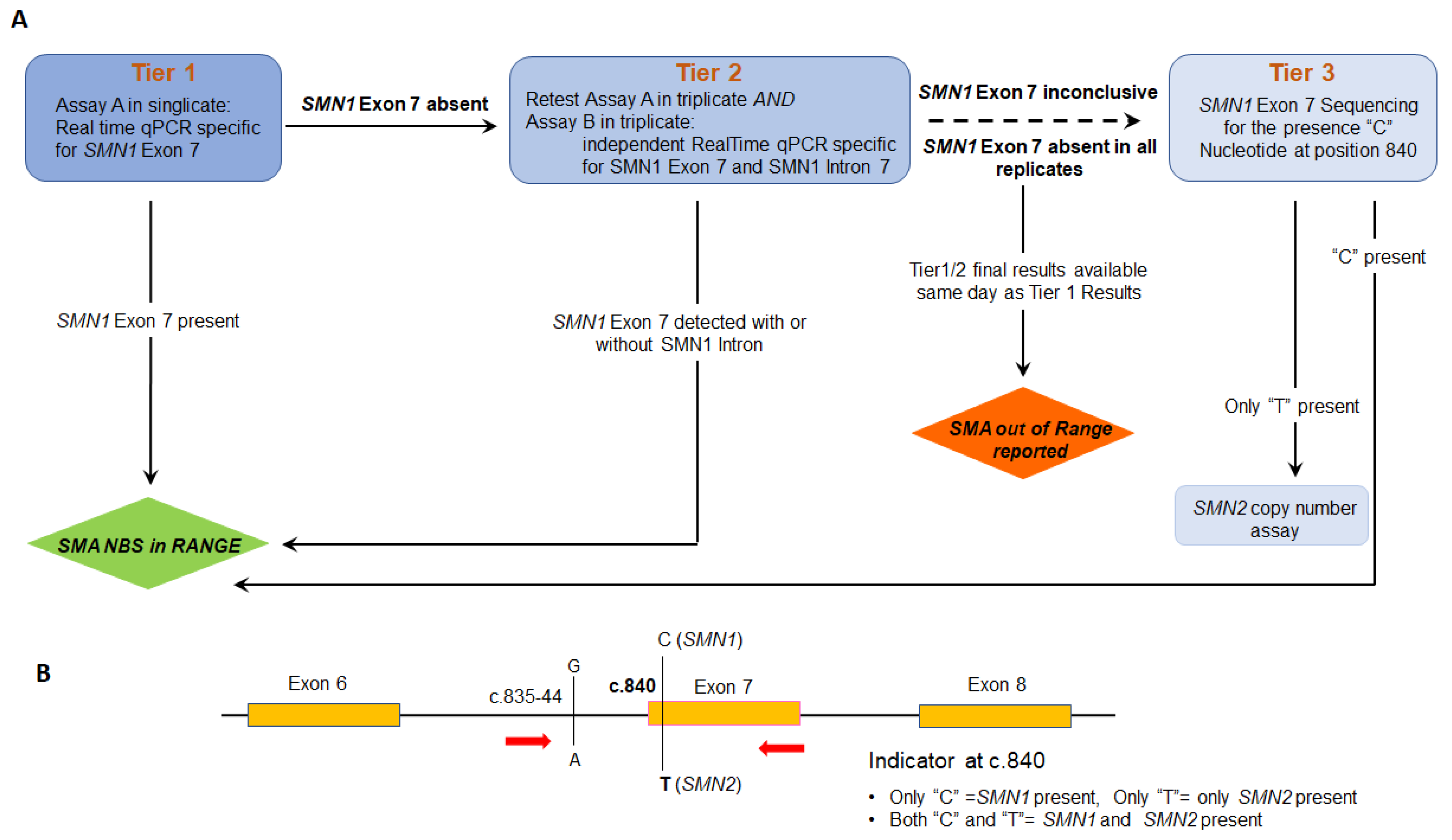
2. Materials and Methods

2.1. Materials

All the materials required to perform sequencing were purchased from Thermo Fisher Scientific (Waltham, MA, USA) including the Big Dye Direct cycle sequencing kit (Cat. 4458688), POP7 polymer (Cat. 4393714), BigDye X Terminator kit (Cat. 4376487). Primers for Exon 7 amplicon of SMN encoded genes were purchased from the IDT technology (Coralville, IA, USA). Positive control and carrier DNA for the SMA assays were purchased from Coriell Institute (Camden, NJ, USA). DBS from SMA-affected patients and their parents for development and validation were provided by Biogen (Cambridge, MA, USA).

2.2. DNA Isolation from Dry Blood Spot

DNA was prepared in accordance with our routine protocols as previously published [20].

2.3. DNA Primers and Sequencing

Forward and reverse primers that amplify the SMN gene (no discrimination between 1 and 2) are shown in Figure 1B. PCR is performed as follows; 35 cycles at 96 °C for 3 s, 62 °C for 15 s and 68 °C for 30 s, followed by extension at 72 °C for 2 min. PCR products were sized by gel electrophoresis, followed by sequencing according to the manufacturer’s instruction. Variant Reporter™ Software was used for the genetic analysis of Exon 7, using the wild-type SMN1 gene as the reference sequence.

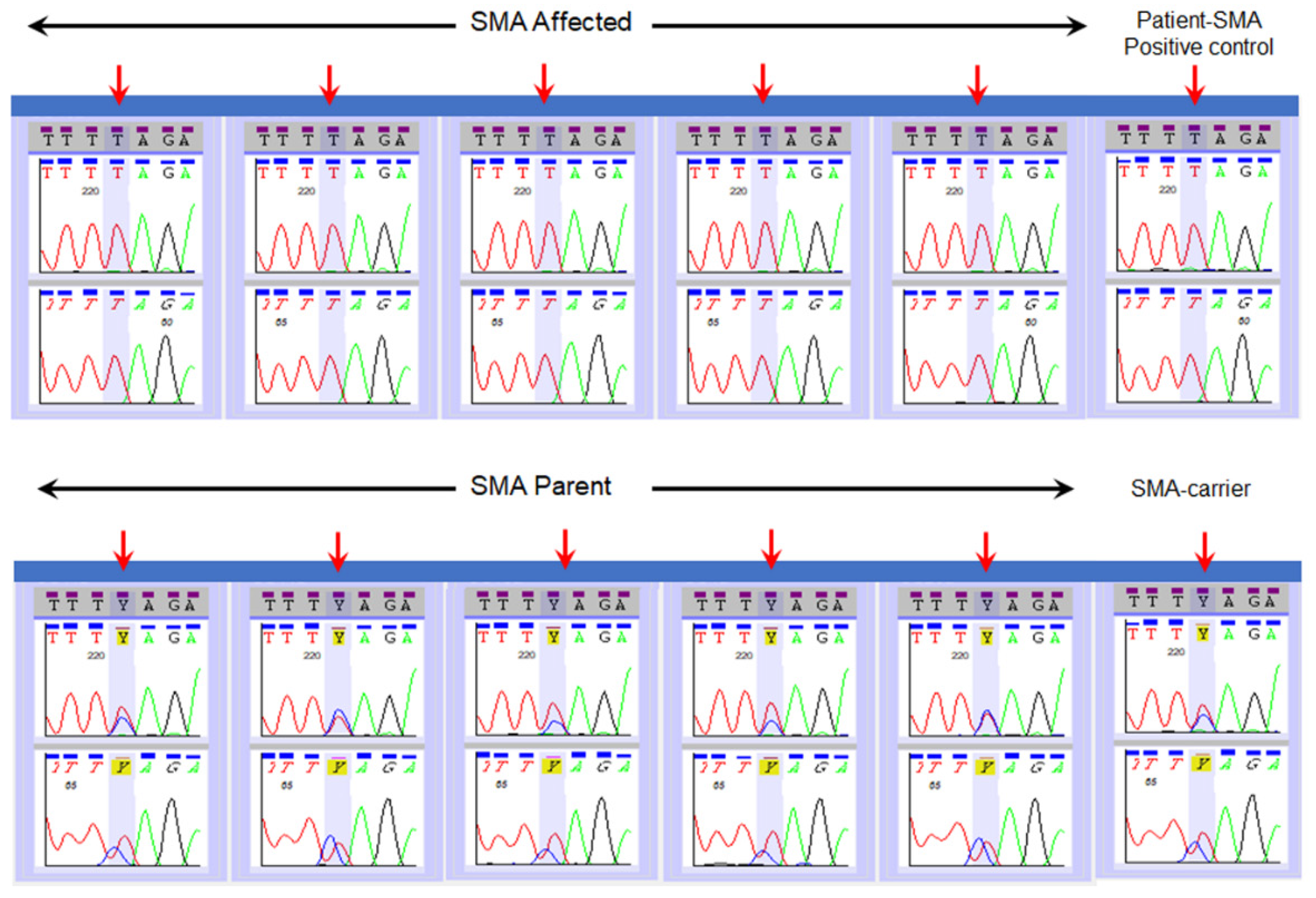
2.4. Validation of SMN1 Sequencing Assay

Thirty reference specimens with various genotypes (affected and parent) provided by Biogen were selected and coded for the validation. Upon completion, decoding was compared with the diagnostic genotype at c.840. A total of 100% concordance was required to pass.

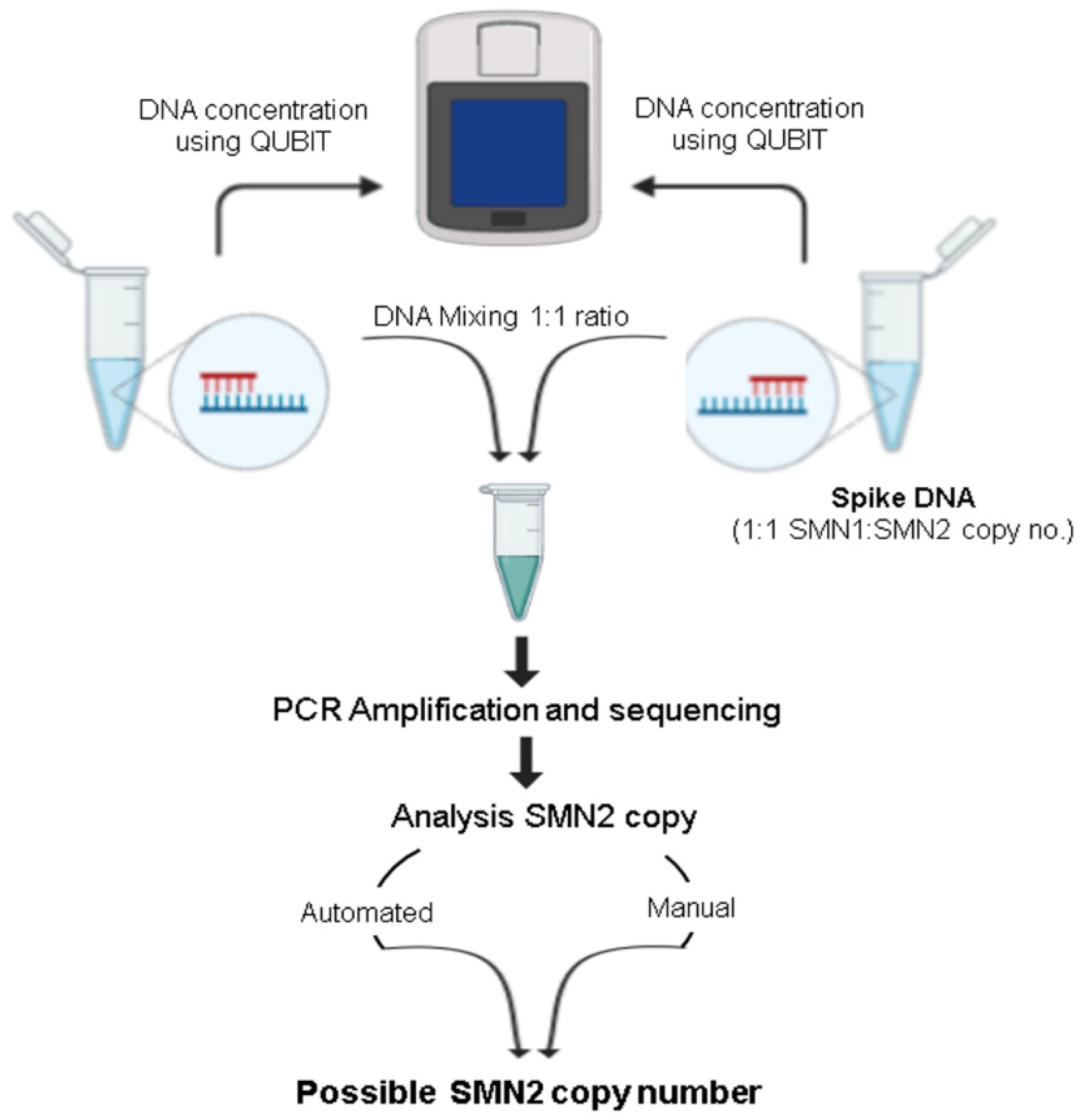
2.5. Modification of Sequencing Assay for Determination of SMN2 Copy Number
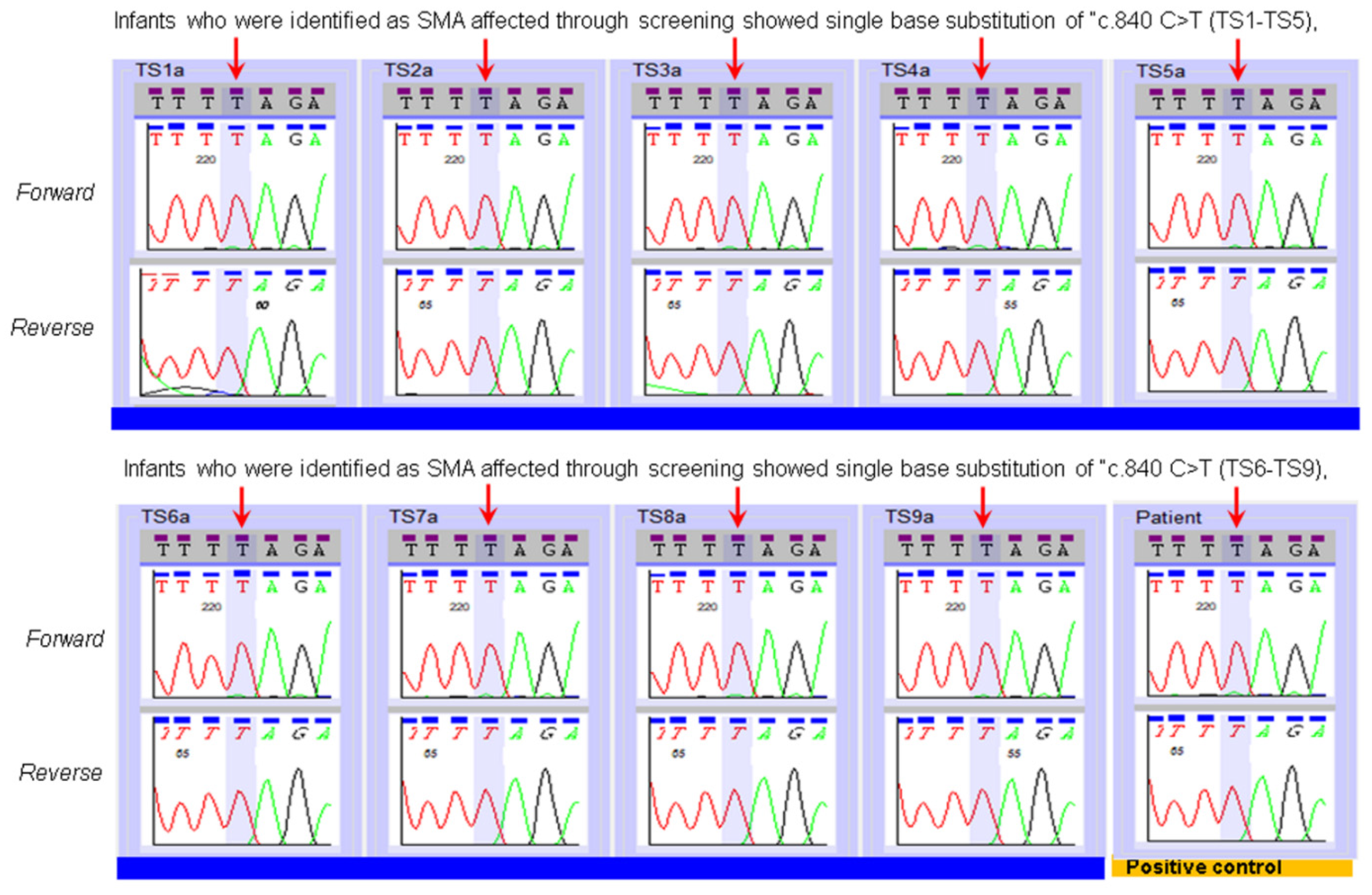
DNA from a specimen previously characterized as having one copy of SMN1 for each single copy of SMN2 (1:1) was prepared and the DNA concentration was quantified using QUBIT (Life Technology). Likewise, DNA from each of the specimens of SMA-affected patients that had been previously characterized for SMN2 copy number (reference test specimens; n = 7) was also prepared and quantified using QUBIT fluorometer. To determine the copy number of SMN2 in the test specimen, an equal amount of DNA from the test specimen was first mixed with that of the 1:1 specimen. This mixed DNA was then subjected to PCR using the same PCR conditions described above, followed by bidirectional sequencing. Reference purified DNA purchased from Coriell was used as an accuracy control. Variant Reporter™ Software generated traces used for quality assessment and genotyping. The genotyping of mixed DNA allowed the calculation of the relative ratio of SMN1:SMN2, and extrapolation from that to define the copy number of SMN2 genes in the specimen of the SMA-affected patient. Two independent methods, an automated mixed sequence reader program and a manual fluorescence intensity calculation, were used to determine the SMN2 copy number. The automated mixed sequence reader detects heterogeneity in chromatographic traces and will identify the mixed bases between the homologous sequences (SMN1/SMN2) and calculates the mixed base ratio within the sequences (http://0-dx-doi-org.brum.beds.ac.uk/10.1100/2012/365104; 15 July 2021). For the manual calculation, the traces were imported to the Variant Reporter™ Software, and the fluorescence intensity of “C” and “T” nucleotides at the c.840 position of SMN gene was recorded along with the background, and the ratio is calculated using mathematical formula A-N/B-N, where A is fluorescence intensity of nucleotide “T”, B is fluorescence intensity of nucleotide “C”, and N is background noise of the sequencing. The final copy number for SMN2 was achieved by reducing the average calculated copy number by one because one copy of SMN2 was from the mixing. For example, if the average SMN2 copy number calculated after analysis is four, the final SMN2 copy number present in the specimen is three (4-1).

3. Results

Table 1 and Figure 2 show results from the validation of Sanger Sequencing method using the 30 previously characterized specimens. All specimens from the previously characterized SMA-affected patients (n = 14) showed nucleotide “T” at c.840 position of SMN. As expected, sequencing of specimens from 16 parent specimens showed both “C” and “T” nucleotides at position c.840 (Table 1, Figure 2). Note, in two of the 16 parent specimens, mixed bases at c.840 were clearly noticeable, but the analysis software only noted the presence of a “T” nucleotide. This is due to the software’s cut off set to make a call for mixed bases. We validated a manual correction prior to clinical use. The results from these 30 specimens validated the SMA sequencing assay to differentiate individuals affected with SMA from individuals who possess one or more copies of an intact Exon 7 in the SMN1 gene. All nine infants who were diagnosed with SMA after an out-of-range NBS result were shown to have only the “T” nucleotide at c.840 of the SMN gene (Figure 3). Sequencing results followed the report of an out of range SMA NBS to pediatrician result by a few days.
During the validation of sequencing, we noted that unaffected SMA specimens reported to have different copy numbers of SMN1 and SMN2 behave differently at c.840 position of SMN1 in terms of fluorescence intensity of nucleotide “T” or “C”. We took advantage of this differential fluorescence intensity, and hypothesized that if we mixed the DNA from an individual with a known ratio of SMN1:SMN2 (1:1) with the DNA of an SMA-affected individual SMN1:SMN2 (0:?), the fluorescence intensity at c.840 or neighboring mixed bases position might provide a ratio of two nucleotides that could be used to extrapolate the SMN2 copy number in the specimen of the affected individual (Figure 4).
For validation of our copy number assay, nine well-characterized dried blood spot specimens provided by Biogen and for which we had confirmed genotypes with copy numbers were selected (Supplementary Figure S1). Seven were reference specimens from SMA-affected patients (reference test specimens) and two were parent specimens with a 1:1 ratio of SMN1 and SMN2 (1:1), which were to be used for mixing with the test specimens. DNAs from each of the reference test specimens were mixed with a 1:1 specimen, sequenced, and fluorescent-intensity analysis was performed using two independent methods as described in materials and methods. The observed copy numbers were rounded to the nearest integer and matched exactly with the copy numbers that had been reported for each of the reference patient specimens (Figure 5). Similar results were observed when using another source of 1:1 DNA. (Supplementary Figure S2). Table 2 shows our SMN2 copy number findings for the 9 SMA-affected infants who were identified by newborn screening compared to results obtained from diagnostic testing using digital droplet PCR. Results from six of the infants matched, including one who we reported as “at least three copies” and diagnostic testing reported four copies. For the other three infants, our results showed three copies of SMN2 while the results of diagnostic testing showed only two copies.

4. Discussion

Our sequencing assay has provided invaluable reassurance when the results from our Assays A and B are not fully concordant with no confirmed explanation (e.g., poor extraction, poor amplification, SMN1 hybrid). We modified our utilization of an SMA sequencing assay for the determination of SMN2 copy number to provide an early estimate or parallel finding of the copy number that would be obtained by diagnostic testing. We recognize that our copy number assay has some limitations: because we are relying on the DNA from the 1:1 specimen to contribute an SMN1 copy to determine a relative ratio, its one copy of SMN2 is also added to the affected SMA specimen and our assay cannot discriminate among copy numbers that are more than four. In addition, our assay is only semi quantitative in nature, yielding a copy number that is always approximate to the absolute ratio (e.g., three copies derived from ratios of 3.25 or 2.75). In light of these limitations, our copy number assay should be an assay of second choice and its use in NBS should be followed by another copy number assay on an independent specimen.
Specialists are increasingly relying on copy number analyses for prognosis and management of infants who are being identified with SMA by NBS before classical signs and symptoms present [21]. Early on, four or more copies of SMN2 disqualified a patient from treatment so discrimination among copy numbers higher than four was not immediately critical to the management of the infant and our approximations seemed sufficient for short-term decision making [22]. That said, the variety of assays being used for copy number assessment is wide and we note recent publications documenting discrepancies among laboratories using a single method (MLPA) and different lots of a kit [23,24,25,26]. Our own experience shows that three of nine SMA-affected infants had NBS DBS specimens with SMN2 copy number results that are discordant from those of diagnostic testing. In still other cases of SMA, discordant genotype–phenotype correlations have been attributed to intragenic SMN2 variants that influence the amount of full-length SMN protein [27]. Thankfully, we have entered a new era in which SMA-affected infants are identified early and offered new treatments. However, this means that classical characterization of an infant’s innate SMA type by clinical presentation is no longer effective. Without accurate measures and documentation of an infant’s baseline genotype prior to treatment (e.g., how many SMN2 paralogs are there? How many and which intragenic SMN2 variants are present?), we will never know the effectiveness of the treatments given and some infants could be undertreated or overtreated. It is incumbent upon screening and diagnostic laboratories to ensure accurate measurements.
In our validation, we compared our results to those well characterized specimens and observed 100% concordance. From the time we began providing copy number results on patients we referred to specialists, we observed some minor discordances between our assay results and those from some external commercial laboratories, which is not surprising. There is a need for well-designed studies of available copy-number assays, which will facilitate the availability of proficiency testing programs for those laboratories seeking to offer copy number testing. Despite its limitations, this novel assay provides reasonable results that can be useful to NBS programs and specialists until confirmatory copy number results are available. We found that recommendations from our results were consistent with those from diagnostic laboratory testing in seven of the nine SMA infants who qualified for treatment by the 2018 guidelines and with nine of the nine who qualified by the 2020 guidelines.

5. Conclusions

Since our statewide implementation in January 2018, our use of a sequencing assay and its modification for SMN2 copy-number analysis has facilitated the early referral and universal access to supplemental information used by specialists in the diagnosis and treatment of infants with SMA.

Supplementary Materials

The following are available online at https://0-www-mdpi-com.brum.beds.ac.uk/article/10.3390/ijns7030047/s1, Figure S1: Genotype confirmation on the specimens subjected for SMN2 copy validation. Figure S2: Determination of SMN2 copy number assay validation using SMA affected specimens using another spike DNA (parent 2).

Author Contributions

Conceptualization, A.M.C. (Anne Marie Comeau) and B.K.; methodology, A.M.C. (Anne Marie Comeau) and B.K.; software, A.M.C. (Anne Marie Comeau), B.K., S.B. and J.K.; validation, A.M.C. (Anne Marie Comeau), B.K., S.B. and J.K.; formal analysis, A.M.C. (Anne Marie Comeau), B.K., S.B. and J.K.; investigation, A.M.C. (Anne Marie Comeau), B.K., S.B., J.K. and A.M.C. (Anne M. Counihan), J.E.H. and R.B.E.; resources, A.M.C. (Anne Marie Comeau), B.K. and R.B.E.; data curation, A.M.C. (Anne Marie Comeau), B.K., S.B., J.K., J.E.H. and A.M.C. (Anne M. Counihan); writing—original draft preparation, B.K.; writing—review and editing, A.M.C. (Anne Marie Comeau), B.K., J.E.H., A.M.C. (Anne M. Counihan), J.K., S.B. and R.B.E.; visualization, A.M.C. (Anne Marie Comeau), B.K., S.B., J.E.H. and A.M.C. (Anne M. Counihan); supervision, A.M.C. (Anne Marie Comeau) and B.K.; project administration, A.M.C. (Anne Marie Comeau) and B.K.; funding acquisition, A.M.C. (Anne Marie Comeau) and R.B.E. All authors have read and agreed to the published version of the manuscript.

Funding

This research was partially funded by the Centers for Disease Control and Prevention, Grant Number 1 NU88EH001323-01-00.

Institutional Review Board Statement

The study was conducted according to the guidelines of the Declaration of Helsinki, and approved by the Institutional Review Boards of the Massachusetts Department of Public Health (318695-13 and date of approval 9/29/2017) and the University of Massachusetts Medical School (3712 and date of approval 8/16/2017).

Informed Consent Statement

Informed consent was obtained by parent proxy from all subjects involved in the study.

Data Availability Statement

Data beyond what is presented in the article are not publicly available as they are derived from private health information.

Acknowledgments

Jennifer Navas has since moved to a private company after contributing to this work. We want to thank Lan Ji for her initial work on the screening assay and Ana Navas for her technical input during the validation of the assays. We want to thank Biogen (Cambridge, MA, USA) for providing the well-characterized reference specimens for this work. Finally, we would like to acknowledge the Massachusetts Department of Public Health, which thoughtfully evaluated SMA NBS through its Newborn Screening Advisory Committee, and which authorized the statewide offering of SMA NBS that was operationalized by the New England Newborn Screening Program of the University of Massachusetts Medical School.

Conflicts of Interest

The authors declare no conflict of interest.

References

  1. Kolb, S.J.; Kissel, J.T. Spinal Muscular Atrophy. Neurol Clin. 2015, 33, 831–846. [Google Scholar] [CrossRef] [PubMed] [Green Version]
  2. Ogino, S.; Wilson, R.B. Genetic testing and risk assessment for spinal muscular atrophy (SMA). Hum. Genet. 2002, 111, 477–500. [Google Scholar] [CrossRef] [PubMed]
  3. Wirth, B. An update of the mutation spectrum of the survival motor neuron gene (SMN1) in autosomal recessive spinal muscular atrophy (SMA). Hum. Mutat. 2000, 15, 228–237. [Google Scholar] [CrossRef]
  4. Nurputra, D.K.; Lai, P.S.; Harahap, N.I.; Morikawa, S.; Yamamoto, T.; Nishimura, N.; Kubo, Y.; Takeuchi, A.; Saito, T. Spinal muscular atrophy: From gene discovery to clinical trials. Ann. Hum. Genet. 2013, 77, 435–463. [Google Scholar] [CrossRef] [PubMed]
  5. Ogino, S.; Wilson, R.B.; Gold, B. New insight on the evolution of the SMN1 and SMN2 region: Simulation and meta-analysis for allele and haplotype frequency calculations. Eur J. Hum. Genet. 2004, 12, 1015–1023. [Google Scholar] [CrossRef]
  6. Mailman, M.D.; Heinz, J.W.; Papp, A.C.; Snyder, P.J.; Sedra, M.S.; Wirth, B.; Burghes, A.H.M.; Prior, T.W. Molecular analysis of spinal muscular atrophy and modification of the phenotype by SMN2. Genet. Med. 2002, 4, 20–26. [Google Scholar] [CrossRef] [Green Version]
  7. Prior, T.W.; Nagan, N.; Sugarman, E.A.; Batish, S.D.; Braastad, C. Technical standards and guidelines for spinal muscular atrophy testing. Genet. Med. 2011, 13, 686–694. [Google Scholar] [CrossRef] [Green Version]
  8. Prior, T.W.; Krainer, A.R.; Hua, Y.; Swoboda, K.J.; Snyder, P.C.; Bridgeman, S.J.; Kissel, J.T. A positive modifier of spinal muscular atrophy in the SMN2 gene. Am. J. Hum. Genet. 2009, 85, 408–413. [Google Scholar] [CrossRef] [Green Version]
  9. Calucho, M.; Bernal, S.; Alías, L.; March, F.; Venceslá, A.; Rodríguez-Álvarez, F.J.; Aller, E.; Fernández, R.M.; Borrego, S.; Millán, J.M.; et al. Correlation between SMA type and SMN2 copy number revisited: An analysis of 625 unrelated Spanish patients and a compilation of 2834 reported cases. Neuromuscular Disord. 2018, 28, 208–215. [Google Scholar] [CrossRef]
  10. Feldkötter, M.; Schwarzer, V.; Wirth, R.; Wienker, T.F.; Wirth, B. Quantitative analyses of SMN1 and SMN2 based on real-time lightcycler PCR: Fast and highly reliable carrier testing and prediction of severity of spinal muscular atrophy. Am. J. Hum. Genet. 2002, 70, 358–368. [Google Scholar] [CrossRef] [Green Version]
  11. Taylor, J.L.; Lee, F.K.; Yazdanpanah, G.K.; Staropoli, J.F.; Liu, M.; Carulli, J.P.; Sun, C.; Dobrowolski, S.F.; Hannon, W.H.; Vogt, R.F. Newborn blood spot screening test using multiplexed real-time PCR to simultaneously screen for spinal muscular atrophy and severe combined immunodeficiency. Clin. Chem 2015, 61, 412–419. [Google Scholar] [CrossRef] [Green Version]
  12. Lee, F.K. Newborn Screening for Spinal Muscular Atrophy (SMA) in the US. Presented at Spinal Muscular Atrophy: Overview 324 of Available Screening Methods, Webinar. 28 June 2018. Available online: https://www.newsteps.org/sites/default/files/re-325sources/download/aphl_sma_webinar_slides_webinarslides_june2018_kh.pdf (accessed on 27 April 2021).
  13. Mercer, K. Newborn screening for spinal muscular atrophy. In Proceedings of the Newborn Screening and Genetic Testing Sym-321 posium, New Orleans, LA, USA, 10–13 September 2017; Available online: https://www.aphl.org/conferences/proceedings/Doc-322uments/Mercer.pdf (accessed on 27 April 2021).
  14. Hale, J.E.; Darras, B.T.; Swoboda, K.J.; Estrella, E.; Chen, J.Y.H.; Abbott, M.A.; Hay, B.N.; Kumar, B.; Counihan, A.M.; Gerstel-Thompson, J.; et al. Massachusetts’ findings from statewide newborn screening for spinal muscular atrophy. Int J. Neonatal Screen. 2021, 23, 26. [Google Scholar] [CrossRef]
  15. Chen, T.H.; Tzeng, C.C.; Wang, C.C.; Wu, S.M.; Chang, J.G.; Yang, S.N.; Hung, C.H.; Jong, Y.J. Identification of bidirectional gene conversion between SMN1 and SMN2 by simultaneous analysis of SMN dosage and hybrid genes in a Chinese population. J. Neurol Sci. 2011, 308, 83–87. [Google Scholar] [CrossRef]
  16. Niba, E.T.E.; Nishio, H.; Wijaya, Y.O.S.; Lai, P.S.; Tozawa, T.; Chiyonobu, T.; Yamadera, M.; Okamoto, K.; Awano, H.; Takeshima, Y.; et al. Clinical phenotypes of spinal muscular atrophy patients with hybrid SMN gene. Brain Dev. 2021, 43, 294–302. [Google Scholar] [CrossRef]
  17. Velasco, E.; Valero, C.; Valero, A.; Moreno, F.; Hernández-Chico, C. Molecular analysis of the SMN and NAIP genes in Spanish spinal muscular atrophy (SMA) families and correlation between number of copies of c BCD541and SMA phenotype. Hum. Mol. Genet. 1996, 5, 257–263. [Google Scholar] [CrossRef] [PubMed] [Green Version]
  18. Chien, Y.H.; Chiang, S.C.; Weng, W.C.; Lee, N.C.; Lin, C.J.; Hsieh, W.S.; Lee, W.T.; Jong, Y.J.; Ko, T.M.; Hwu, W.L. Presymptomatic Diagnosis of Spinal Muscular Atrophy Through Newborn Screening. J. Pediatr. 2017, 190, 124–129. [Google Scholar] [CrossRef]
  19. Huang, C.H.; Chang, Y.Y.; Chen, C.H.; Kuo, Y.S.; Hwu, W.L.; Gerdes, T.; Ko, T.M. Copy number analysis of survival motor neuron genes by multiplex ligation-dependent probe amplification. Genet. Med. 2007, 9, 241–248. [Google Scholar] [CrossRef] [PubMed] [Green Version]
  20. Gerstel-Thompson, J.L.; Wilkey, J.F.; Baptiste, J.C.; Navas, J.S.; Pai, S.; Pass, K.A.; Eaton, R.B.; Comeau, A.M. High-throughput multiplexed T-cell-receptor excision circle quantitative PCR assay with internal controls for detection of severe combined immunodeficiency in population-based newborn screening. Clin. Chem. 2010, 56, 1466–1474. [Google Scholar] [CrossRef] [PubMed] [Green Version]
  21. Glascock, J.; Sampson, J.; Connolly, A.M.; Darras, B.T.; Day, J.W.; Finkel, R.; Jarecki, J. Revised recommendations for the treatment of infants diagnosed with spinal muscular atrophy via newborn screening who have 4 copies of SMN2. J. Neuromuscul. Dis. 2020, 7, 97–100. [Google Scholar] [CrossRef] [Green Version]
  22. Glascock, J.; Sampson, J.; Haidet-Phillips, A.; Connolly, A.; Darras, B.; Day, J.; Jarecki, J. Treatment algorithm for infants diagnosed with spinal muscular atrophy through newborn screening. J. Neuromuscul. Dis. 2018, 5, 145–158. [Google Scholar] [CrossRef] [Green Version]
  23. Schorling, D.C.; Becker, J.; Pechmann, A.; Langer, T.; Wirth, B.; Kirschner, J. Discrepancy in redetermination of SMN2 copy numbers in children with SMA. Neurology 2019, 93, 267–269. [Google Scholar] [CrossRef]
  24. Dangouloff, T.; Boemer, F.; Dideberg, V.; Caberg, J.H.; Servais, L. Reader response: Discrepancy in redetermination of SMN2 copy numbers in children with SMA. Neurology 2020, 95, 144–145. [Google Scholar] [CrossRef] [PubMed]
  25. Kirschner, J.; Becker, J.; Schorling, D.; Pechmann, A.; Wirth, B. Author response: Discrepancy in redetermination of SMN2 copy numbers in children with SMA. Neurology 2020, 95, 145. [Google Scholar] [CrossRef] [PubMed]
  26. Aravind Ganesh, A.; Steven Galetta, S. Editors’ note: Discrepancy in redetermination of SMN2 copy numbers in children with SMA. Neurology 2020, 95, 144. [Google Scholar]
  27. Blasco-Pérez, L.; Paramonov, I.; Leno, J.; Bernal, S.; Alias, L.; Fuentes-Prior, P.; Cuscó, I.; Tizzano, E.F. Beyond copy number: A new, rapid, and versatile method for sequencing the entire SMN2 gene in SMA patients. Hum. Mutat. 2021, 42, 787–795. [Google Scholar] [CrossRef] [PubMed]
Figure 1. (A) Flow chart of current SMA screening algorithm at New England Newborn Screening program. (B) Primer locus targeting SMN1 gene during SMA sequencing. Primers were tagged with M13, and indicated in bold. SMA seq-F- 5′-TGTAAAACGACGGCCAGT-AACCTTAACTGCAGCCTAATAATTG-3′ and SMA seq-R- 5′-CAGGAAACAGCTATGACC-GCTGGCAGACTTACTCCTTAAT-3′ were used for sequencing.
Ijns 07 00047 g001
Figure 2. Typical examples of SMA sequencing assay on reference samples from SMA affected patients and their parents.
Ijns 07 00047 g002
Figure 3. Typical example of SMA sequencing of specimens from nine SMA-affected infants who we referred to specialty centers through screening; all showed single base substitution of c.840C>T.
Ijns 07 00047 g003
Figure 4. Flow diagram for determination of SMN2 copy number of SMA affected specimen. DNAs were measured using QUBIT, and equal amount of spike DNA and test DNA were mixed, followed by bidirectional sequencing.
Ijns 07 00047 g004
Figure 5. Validation of use of modified sequencing for “determination of SMN2 copy number” using eight previously characterized SMA affected specimens. DNA from SMA affected specimens, and a Coriell DNA (positive control) were mixed in equal concentration with spike DNA, and bidirectional sequencing was performed. The SMN1 gene sequence was used as the reference sequence, and data were analyzed to determine the SMN2 copy number in the test specimen as described in the methods section.
Ijns 07 00047 g005
Table 1. NENSP genotype determinations of reference specimens used for validation of SMA sequencing.
Type of Reference SpecimensNumber of SpecimensNENSP Detection of Nucleotide at c.840 of SMN1
Affectedn = 14“T”
Parentsn = 16“C” and “T”
Carrier “C” and “T”
Positive control (known SMA) “T”
Table 2. Comparison of SMN2 copy numbers in newly diagnosed infants generated by our modified sequencing assay and by diagnostic laboratories.
SMN2 Copy Number Assay Applied to the Specimens of Infants Identified by NBS as SMA Affected.
MA Patient
Specimens
SMA StatusSMN2 Copy Number Prediction by NENSP from DBSSMN2 Copy Number Prediction by Diagnostic Lab Test of an Independent Specimen
TS1Affected32
TS2AffectedMore than 3 copies4
TS3Affected22
TS4Affected32
TS5Affected22
TS6Affected22
TS7Affected44
TS8Affected32
TS9Affected22
Publisher’s Note: MDPI stays neutral with regard to jurisdictional claims in published maps and institutional affiliations.

