Concentration and Quantification of SARS-CoV-2 RNA in Wastewater Using Polyethylene Glycol-Based Concentration and qRT-PCR

 , , ,
, , ,
Abstract
:1. Introduction
2. Experimental Design
2.1. Materials
- PEG8000 (Sigma Aldrich, St. Louis, MO, USA, Cat. No. P5413).
- NaCl (Sigma Aldrich, St. Louis, MO, USA, Cat. No. S7653).
- Deionised water or reverse osmosis water with purity ≥18 MΩ resistance.
- Sterile phosphate buffered saline (PBS), pH 7.4 (Sigma Aldrich, St. Louis, MO, USA, Cat. No. P4417).
- 1 M NaOH and 0.5 M HCl for pH adjustment.
- pH 6.0–10.0 pH strips (VWR, Radnor, PA, USA, Cat No. 85414.601P) or equivalent if pH meter is not available.
- NucliSens lysis buffer (BioMerieux, Marcy-l’Étoile, France, Cat No. 280,134 or 200292).
- NucliSens extraction reagent kit (BioMerieux, Marcy-l’Étoile, France, Cat. No. 200293), containing:
- ○
- Magnetic beads mix (Cat No. 280133)
- ○
- Wash Buffer 1 (Cat No. 280130)
- ○
- Wash Buffer 2 (Cat No. 280131)
- ○
- Wash Buffer 3 (Cat No. 280132)
- ○
- Elution Buffer (Cat No. 280132)
- ■
- Elution buffer and Wash Buffer 3 are the same reagents which are aliquoted in separate tubes in the extraction reagent kit.
- Optional control viruses:
- ○
- Whole process control: non-human enveloped virus stock, e.g., PRRSV (American Type Culture Collection (ATCC), Manassas, VA, USA, Cat. No. VR-2385).
- ○
- RNA extraction control: non-human RNA virus stock, e.g., MNV (ATCC, Cat. No. VR-1937).
- RNA UltraSense™ One-Step Quantitative RT-PCR System (Life Technologies, Carlsbad, CA, USA, Cat. No. 11732927), containing:
- ○
- 5× reaction mix
- ○
- 20× enzyme mix
- ○
- 20× bovine serum albumin (BSA)
- ○
- 50 nM MgSO4
- ○
- ROX reference dye
- qPCR primers and probes as detailed in Table 1.
- DNA standards incorporating the target sequences or equivalent:
- ○
- SARS-CoV-2 N1: 2019-nCoV_N_positive control (Integrated DNA Technologies (IDT), Coralville, IA, USA, Cat. No. 10006625)
- ○
- SARS-CoV-2 E: 2019-nCoV_E control (IDT, Coralville, IA, USA, Cat. No. N/A)
- ○
- MNV/PRRSV: custom Ultramer DNA oligo (IDT, Coralville, IA, USA)
2.2. Equipment
- Temperature-controlled benchtop centrifuge with fixed-angle rotor and sealed buckets with 50–200 mL tube inserts and capable of speeds up to 10,000× g (Eppendorf, Hamburg, Germany).
- Temperature-controlled benchtop centrifuge with swinging-bucket rotor and sealed buckets with 50–200 mL tube inserts and capable of speeds up to 3000× g (Eppendorf, Hamburg, Germany).
- pH meter, pocket size (Ichiro Corporation, Kotoku, Tokyo, Japan, Cat. No. S2K992) or equivalent.
- Microbiological Safety Cabinet (CL2/BSL2 compliant).
- Vortex mixer.
- Thermoshaker operating at 60 °C and 1400 rpm or equivalent.
- Magnetic rack for 1.5 mL tubes.
- Optional: NucliSens MiniMAG instrument or other (semi-)automated liquid handling system (BioMerieux, Marcy-l’Étoile, France).
- Optional: aspirator or equivalent apparatus for removing supernatant.
- Quant Studio Flex 6 real-time PCR machine (Applied Biosystems Inc., Waltham, MA, USA) or equivalent (real-time PCR machine with minimum four channels).
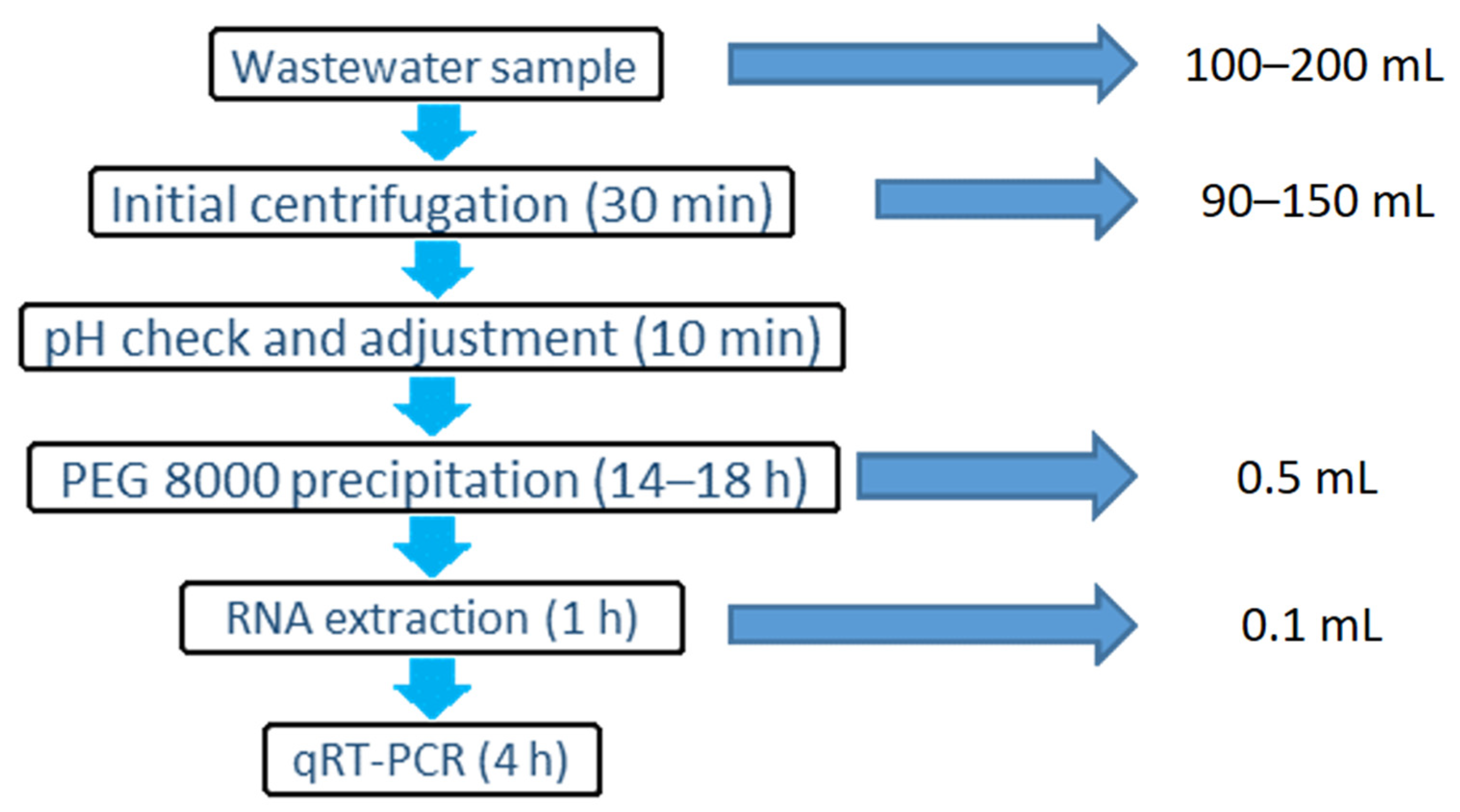
3. Procedure
3.1. Samples and Controls
- Samples: 100/200 mL untreated wastewater. Samples should be delivered to laboratory chilled within 24 h of sampling. Polypropylene bottles are recommended for sample collection and shipping. Sample volume can be reduced or increase based on the turbidity of the sample.
- Process negative control: 100/200 mL sterile water.
- Process positive control: 90/150 mL sterile water spiked with approx. 105 gc PRRSV or equivalent.
- Extraction negative control: 0.5 mL PBS.
- Extraction positive control: 0.5 mL PBS spiked with approx. 105 gc MNV or equivalent.
- qRT-PCR non-template control (NTC): 10 µL molecular-grade water.
3.2. Sample Concentration. Time for Completion: 20–28 h, Bench Time: 1.5 h
- Aliquot 100/200 mL per wastewater sample into individual 50 mL polypropylene centrifuge tubes. Use one negative control in each batch of samples (100–200 mL water) and treat as a sample.
- Centrifuge samples at 3000× g at 4 °C for 30 min. Alternatively, a 10-min centrifugation at 10,000× g at 4 °C can be used. Adjust break speed to 0–5 to avoid resuspension of the pellet.
- Remove and combine 90/150 mL of the supernatant in a new sterile 250 mL polypropylene bottle. Do not disturb the pellet. The volume of liquid may be changed, depending on the amount of pellet. Discard pellet.
- Optional step: spike supernatant with approx. 105 gc equivalent PRRSV. Spike 90/150 mL sterile water as well as a positive control.
- Adjust the pH of the 90/150 mL supernatant to 7–7.5 (usually up to 1–3 drops of 1 M NaOH) to enhance protein binding.
- Add 1:3 ratio of 40% PEG 8000, 8% NaCl solution (as described in Section 5) to each sample (pH checked) to reach a final concentration of 10% PEG 8000, 2% NaCl. Invert several times to mix.
- Incubate samples at 4 °C for 14–18 h.
- Make a mark on the tube on the side where the pellet will form as it is often difficult to see. Alternatively, place the tubes in the centrifuge with label facing up. Keep the tube in same orientation across all centrifuge steps to ensure the pellet builds up in the same location.
- Centrifuge at 10,000× g for 30 min at 4 °C. Adjust break speed to 0–5 to avoid the resuspension of the pellet.
- Discard supernatant by decanting and pipetting.
- Thoroughly resuspend the pellet in 0.5 mL PBS and proceed with nucleic acid extraction or store the concentrate at 4 °C for up to three days, −20° for seven days or −80 °C for long term. Alternatively, 2 mL Nuclisens lysis buffer may be added directly to the pellet then either extract immediately or store at −80 °C. However, mixing the pellet with the buffer may result in excessive foaming.
3.3. Viral RNA Extraction. Time for Completion: 1.5 h, Bench Time: 1.5 h
- Optional step: add approx. 105 gc equivalent murine norovirus control to each sample. Each day of extractions, include one negative (MNV- 0.5 mL water) and one positive control (MNV+ 0.5 mL water spiked with MNV).
- Add 2 mL of NucliSens lysis buffer to a 15 mL centrifuge tube. Add 500 μL of sample and mix by vortexing (skip this step if lysis buffer was added directly to the pellet in step 3.2.10).
- Incubate for 10 min at room temperature.
- Add 50 μL of well-mixed magnetic bead solution from the extraction kit to the tube and mix by vortexing briefly.
- Incubate for 10 min at room temperature.
- Centrifuge for 2 min at 1500× g then carefully discard supernatant by pipetting or aspiration.
- Add 385 μL wash buffer 1 and resuspend the pellet by pipetting/vortexing.
- Transfer suspension to a 1.5 mL centrifuge tube, vortex for 30 s at low speed and separate the beads using a magnetic rack.
- Alternatively, when the MiniMag system is used, cut the tube caps off or usescrew-cap tubes and perform a wash using preset ‘C1 continuous’ or ‘0.5 step’ for 30 s using the MiniMAG extraction systems or by vortexing.
- After washing, allow silica beads to settle using a magnetic rack or by raising the magnet of the MiniMAG extraction system. Discard supernatant by pipetting or aspiration.
- Separate tubes from the magnet, then add 385 μL Wash Buffer 1. Resuspend the pellet, wash for 30 s (Step 8 or 9), allow silica beads to settle using the magnet then discard supernatant.
- Separate tubes from the magnet, then add 485 μL Wash Buffer 2. Resuspend pellet, wash for 30 s (Step 8 or 9), allow silica beads to settle using magnet then discard supernatant. Repeat.
- Separate tubes from magnet, then add 500 μL Wash Buffer 3. Wash for 15 s (step 8), allow silica beads to settle using magnet then discard supernatant.Note: samples should not be left in Wash Buffer 3 for longer than strictly necessary.
- Add 100 μL elution buffer. Cap tubes and transfer to thermoshaker or equivalent.
- Incubate for 5 min at 60 °C with shaking at 1400 rpm.
- Place tubes in magnetic rack and allow silica beads to settle, then transfer eluate to a clean, labelled 0.5 mL tube. Continue to qRT-PCR or store the extracts at −80 °C.
3.4. qRT-PCR Quantification of Viral Genome Copies. Time for Completion: 4.5 h, Bench Time: 1–1.5 h
- Prepare and aliquot primer and probe mixes to avoid repeated freezing/thawing.
- Prepare qRT-PCR master mix as described in Table 2. Each sample should be run in duplicate. Duplicates of standard dilution series (at least five points in the concentration range of 106–100 gc/µL) and of a no template control (NTC) consisting of molecular biology grade water or Tris-EDTA (TE) buffer should be included in each run.
- Use run conditions described in Table 3 for the one-step qRT-PCR reaction.
3.5. Data Analysis
- Process negative control, extraction negative control and NTC should be negative for all gene targets.
- qRT-PCR standard curve should be in line with the recommendations described in The MIQE Guidelines [14].
- Ct values > 40 should be considered negative. We rcommend using 45 cycles of amplification to assess the amplification curve for samples with Ct values of 38–40.
- Control virus recovery should exceed 1%, as suggested by previous standard method for viral detection in shellfish (ISO 15216-2:2019). When viral recovery is lower than 1%, the qRT-PCR should be repeated with 2 µL samples added to the reaction mix to assess potential inhibition.
4. Expected Results
4.1. Method Efficiency
4.2. Applications and Recommendations
5. Reagents Setup
- 40% PEG 8000, 8% NaCl solution
- PEG 8000: 400 g;
- NaCl: 80 g;
- Deionised water: 1000 mL;
- Primer/probe mixes
- Forward primer, 100 µM: 100 µL;
- Reverse primer, 100 µM: 200 µL;
- Probe, 100 µM: 50 µL;
- Molecular-grade water: 650 µL;
Supplementary Materials
Author Contributions
Funding
Institutional Review Board Statement
Informed Consent Statement
Acknowledgments
Conflicts of Interest
References
- Sims, N.; Kasprzyk-Hordern, B. Future Perspectives of Wastewater-Based Epidemiology: Monitoring Infectious Disease Spread and Resistance to the Community Level. Environ. Int. 2020, 139, 105689. [Google Scholar] [CrossRef] [PubMed]
- Miura, T.; Lhomme, S.; Le Saux, J.C.; Le Mehaute, P.; Guillois, Y.; Couturier, E.; Izopet, J.; Abranavel, F.; Le Guyader, F.S. Detection of Hepatitis E Virus in Sewage After an Outbreak on a French Island. Food Environ. Virol. 2016, 8, 194–199. [Google Scholar] [CrossRef] [PubMed] [Green Version]
- Farkas, K.; Cooper, D.M.; McDonald, J.E.; Malham, S.K.; de Rougemont, A.; Jones, D.L.; de Rougemont, A.; Jones, D.L.; de Rougemont, A.; Jones, D.L. Seasonal and spatial dynamics of enteric viruses in wastewater and in riverine and estuarine receiving waters. Sci. Total Environ. 2018, 634, 1174–1183. [Google Scholar] [CrossRef] [PubMed]
- Bisseux, M.; Colombet, J.; Mirand, A.; Roque-Afonso, A.M.; Abravanel, F.; Izopet, J.; Archimbaud, C.; Peigue-Lafeuille, H.; Debroas, D.; Bailly, J.L.; et al. Monitoring human enteric viruses in wastewater and relevance to infections encountered in the clinical setting: A one-year experiment in central France, 2014 to 2015. Eurosurveillance 2018, 23, 1–11. [Google Scholar] [CrossRef] [PubMed] [Green Version]
- Ahmed, W.; Angel, N.; Edson, J.; Bibby, K.; Brien, J.W.O.; Choi, P.M.; Kitajima, M.; Simpson, L.; Li, J.; Tscharke, B.; et al. First confirmed detection of SARS-CoV-2 in untreated wastewater in Australia: A proof of concept for the wastewater surveillance of COVID-19 in the community. Sci. Total Environ. 2020, 138764. [Google Scholar] [CrossRef] [PubMed]
- Medema, G.; Heijnen, L.; Elsinga, G.; Italiaander, R.; Brouwer, A. Presence of SARS-Coronavirus-2 RNA in sewage and correlation with reported COVID-19 prevalence in the early stage of the epidemic in the Netherlands. Environ. Sci. Technol. Lett. 2020, 7, 511–516. [Google Scholar] [CrossRef]
- Ahmed, W.; Bivins, A.; Bertsch, P.M.; Bibby, K.; Choi, P.M.; Farkas, K.; Gyawali, P.; Hamilton, K.A.; Haramoto, E.; Kitajima, M.; et al. Surveillance of SARS-CoV-2 RNA in wastewater: Methods optimisation and quality control are crucial for generating reliable public health information. Curr. Opin. Environ. Sci. Heal. 2020. [Google Scholar] [CrossRef] [PubMed]
- Farkas, K.; Hillary, L.S.; Malham, S.K.; McDonald, J.E.; Jones, D.L. Wastewater and public health: The potential of wastewater surveillance for monitoring COVID-19. Curr. Opin. Environ. Sci. Heal. 2020. [Google Scholar] [CrossRef] [PubMed]
- Centers for Disease Control and Prevention. 2019-Novel Coronavirus (2019-nCoV) Real-Time rRT-PCR Panel Primers and Probes; Centers for Disease Control and Prevention: Atlanta, GA, USA, 2019.
- Corman, V.; Bleicker, T.; Brunink, S.; Drosten, C. Diagnostic Detection of Wuhan Coronavirus 2019 by Real-Time RT-PCR; World Health Organization: Geneva, Switzerland, 17 January 2020. [Google Scholar]
- Kitajima, M.; Oka, T.; Takagi, H.; Tohya, Y.; Katayama, H.; Takeda, N.; Katayama, K. Development and application of a broadly reactive real-time reverse transcription-PCR assay for detection of murine noroviruses. J. Virol. Methods 2010, 169, 269–273. [Google Scholar] [CrossRef] [PubMed]
- Chen, N.; Ye, M.; Xiao, Y.; Li, S.; Huang, Y.; Li, X.; Tian, K.; Zhu, J. Development of universal and quadruplex real-time RT-PCR assays for simultaneous detection and differentiation of porcine reproductive and respiratory syndrome viruses. Transbound. Emerg. Dis. 2019, 66, 2271–2278. [Google Scholar] [CrossRef] [PubMed]
- WHO. Laboratory Biosafety Manual; World Health Organization: Geneva, Switzerland, 2004; ISBN 9941059586. [Google Scholar]
- Bustin, S.; Benes, V.; Garson, J.A.; Hellemans, J.; Huggett, J.; Kubista, M.; Mueller, R.; Nolan, T.; Pfaffl, M.; Shipley, G.L.; et al. The MIQE Guidelines: Minimum Information for Publication of Quantitative Real-Time PCR Experiments. Clin. Chem. 2009, 55, 611–622. [Google Scholar] [CrossRef] [PubMed] [Green Version]
- Bofill-Mas, S.; Rusiñol, M. Recent trends on methods for the concentration of viruses from water samples. Curr. Opin. Environ. Sci. Heal. 2020, 16, 7–13. [Google Scholar] [CrossRef]
- Wang, X.W.; Li, J.S.; Guo, T.K.; Zhen, B.; Kong, Q.X.; Yi, B.; Li, Z.; Song, N.; Jin, M.; Xiao, W.J.; et al. Concentration and detection of SARS coronavirus in sewage from Xiao Tang Shan Hospital and the 309th Hospital. J. Virol. Methods 2005, 128, 156–161. [Google Scholar] [CrossRef]
- Gundy, P.M.; Gerba, C.P.; Pepper, I.L. Survival of Coronaviruses in water and wastewater. Food Environ. Virol. 2009, 1, 10–14. [Google Scholar] [CrossRef] [Green Version]
- Wu, F.; Zhang, J.; Xiao, A.; Gu, X.; Lee, W.L.; Armas, F.; Kauffman, K.; Hanage, W.; Matus, M.; Ghaeli, N.; et al. SARS-CoV-2 Titers in Wastewater Are Higher than Expected from Clinically Confirmed Cases. mSystems 2020, 5, 1–9. [Google Scholar] [CrossRef] [PubMed]
- Ahmed, W.; Bertsch, P.M.; Bivins, A.; Bibby, K.; Farkas, K.; Gathercole, A.; Haramoto, E.; Gyawali, P.; Korajkic, A.; McMinn, B.R.; et al. Comparison of virus concentration methods for the RT-qPCR-based recovery of murine hepatitis virus, a surrogate for SARS-CoV-2 from untreated wastewater. Sci. Total Environ. 2020, 739, 139960. [Google Scholar] [CrossRef] [PubMed]
- Ye, Y.; Ellenberg, R.M.; Graham, K.E.; Wigginton, K.R. Survivability, Partitioning, and Recovery of Enveloped Viruses in Untreated Municipal Wastewater. Environ. Sci. Technol. 2016, 50, 5077–5085. [Google Scholar] [CrossRef] [PubMed]
- Farkas, K.; Hassard, F.; McDonald, J.E.; Malham, S.K.; Jones, D.L. Evaluation of molecular methods for the detection and quantification of pathogen-derived nucleic acids in sediment. Front. Microbiol. 2017, 8, 53. [Google Scholar] [CrossRef] [PubMed] [Green Version]


| Target | Primer/Probe | Sequence (5′-3′) | Reference |
|---|---|---|---|
| SARS-CoV-2 N1 gene region | Forward primer | GACCCCAAAATCAGCGAAAT | [9] |
| Reverse primer | TCTGGTTACTGCCAGTTGAATCTG | ||
| Probe 1 | [FAM]ACCCCGCATTACGTTTGGTGGACC[MGB] | ||
| SARS-CoV-2 E gene region | Forward primer | ACAGGTACGTTAATAGTTAATAGCGT | [10] |
| Reverse primer | ATATTGCAGCAGTACGCACACA | ||
| Probe 1 | [VIC]ACACTAGCCATCCTTACTGCGCTTCG[QSY] | ||
| MNV ORF1-2 | Forward primer | CCGCAGGAACGCTCAGCAG | [11] |
| Reverse primer | GGYTGAATGGGGACGGCCTG | ||
| Probe 1 | [ABY]ATGAGTGATGGCGCA[QSY] | ||
| PRRSV ORF7 | Forward primer | CAGGACTTCGGAGCCTCGT | [12] |
| Reverse primer | AGCAACTGGCACAGTTGATTGA | ||
| Probe 1 | [ABY] ACGAGCTGTTAAACGAGGA[QSY] |
| Reagent | Concentration | μL/Reaction Mix |
|---|---|---|
| Water, molecular grade | - | 8.35 |
| Reaction mix | 5× | 5 |
| Enzyme mix | 20× | 1.25 |
| Bovine serum albumin (BSA) | 20× | 1.25 |
| MgSO4 | 50 mM | 0.4 |
| Primer/probe mix, N1 | 5–20 μM | 1.25 |
| Primer/probe mix, E | 5–20 μM | 1.25 |
| Primer/probe mix, MNV or PRRSV | 5–20 μM | 1.25 |
| Sample/NTC/standard | - | 5 |
| Sum | 25 |
| Temperature | Time | Number of Cycles |
|---|---|---|
| 55 °C | 60 min | 1 |
| 95 °C | 5 min | 1 |
| 95 °C | 15 s | 45 |
| 60 °C | 1 min | |
| 65 °C | 1 min * |
| Target | LOD gc/Reaction | LOD gc/µL RNA Extract | LOQ gc/Reaction | LOQ gc/µL RNA Extract |
|---|---|---|---|---|
| SARS-CoV-2 N1 | 9 | 1.7 | 59 | 11.8 |
| SARS-CoV-2 E | 19 | 3.8 | 126 | 25.1 |
| MNV | 16 | 3.1 | 164 | 32.1 |
| Wastewater Sample | PRRSV Recovery % | MNV Recovery % | N1 Gene Sewage | E Gene Sewage |
|---|---|---|---|---|
| D1 | 55.94 | 37.31 | detected | detected |
| D2 | 31.52 | 32.05 | detected | detected |
| L1 | 3.34 | 11.45 | detected | not detected |
| L2 | 3.86 | 17.15 | not detected | detected |
| L3 | 3.97 | 9.70 | detected | detected |
| M1 | 9.81 | 16.43 | not detected | not detected |
| M2 | 5.58 | 16.56 | not detected | not detected |
| M3 | 3.41 | 13.21 | not detected | not detected |
Publisher’s Note: MDPI stays neutral with regard to jurisdictional claims in published maps and institutional affiliations. |
© 2021 by the authors. Licensee MDPI, Basel, Switzerland. This article is an open access article distributed under the terms and conditions of the Creative Commons Attribution (CC BY) license (http://creativecommons.org/licenses/by/4.0/).
Share and Cite
Farkas, K.; Hillary, L.S.; Thorpe, J.; Walker, D.I.; Lowther, J.A.; McDonald, J.E.; Malham, S.K.; Jones, D.L. Concentration and Quantification of SARS-CoV-2 RNA in Wastewater Using Polyethylene Glycol-Based Concentration and qRT-PCR. Methods Protoc. 2021, 4, 17. https://0-doi-org.brum.beds.ac.uk/10.3390/mps4010017
Farkas K, Hillary LS, Thorpe J, Walker DI, Lowther JA, McDonald JE, Malham SK, Jones DL. Concentration and Quantification of SARS-CoV-2 RNA in Wastewater Using Polyethylene Glycol-Based Concentration and qRT-PCR. Methods and Protocols. 2021; 4(1):17. https://0-doi-org.brum.beds.ac.uk/10.3390/mps4010017
Chicago/Turabian StyleFarkas, Kata, Luke S. Hillary, Jamie Thorpe, David I. Walker, James A. Lowther, James E. McDonald, Shelagh K. Malham, and Davey L. Jones. 2021. "Concentration and Quantification of SARS-CoV-2 RNA in Wastewater Using Polyethylene Glycol-Based Concentration and qRT-PCR" Methods and Protocols 4, no. 1: 17. https://0-doi-org.brum.beds.ac.uk/10.3390/mps4010017