Article

Effects of Different Temperatures on the Antibacterial, Immune and Growth Performance of Crucian Carp Epidermal Mucus

1
School of Agricultural Engineering, Jiangsu University, Zhenjiang 212013, China
2
Key Laboratory of Modern Agricultural Equipment and Technology, Ministry of Education, School of Agricultural Engineering, Jiangsu University, Zhenjiang 212013, China
*
Author to whom correspondence should be addressed.
Submission received: 8 October 2021 / Revised: 10 November 2021 / Accepted: 18 November 2021 / Published: 22 November 2021

Abstract

:
Fish is one of the important sources of energy and protein, and proper water temperature is key to successful fish breeding. The authors of this study evaluated crucian carp growth, mucus antibacterial properties, and immune indicators at 17, 21, 24, 27, and 31 °C. The results indicated that in the range of 17–31 °C, the resistance of epidermal mucus to Vibrio harveyi decreased with temperature rising. At 24 and 27 °C, the activities of lysozyme and catalase significantly increased; alkaline phosphatase activity, superoxide dismutase activity, and total protein concentration first increased and then decreased with rising temperature; the highest values were observed at 24 °C, with increases of 56.55%, 26.64%, and 44.52%, respectively, compared to those under the 17 °C treatment. When the treatment reached 27 °C, the temperature had an effect on the growth and antibacterial properties of crucian carp, and the activities of alkaline phosphatase and superoxide dismutase were significantly reduced. At temperatures of 17–24 °C, the survival rate of crucian carp could reach more than 93%, and at the temperature of 24 °C, the specific growth rate reached the highest value of 43.29%. Therefore, the most favorable temperature for the long-term breeding of crucian carp was found to be 24 °C. This study provides a favorable experimental basis for the establishment of intelligent aquaculture systems and the setting of water environment parameters.

Graphical Abstract

1. Introduction

Crucian carp is considered threatened in many European countries, mainly due to external pressures such as invasive carp and goldfish. Therefore, there is an urgent need to find aquaculture methods that meet the biological characteristics of this species [1]. Appropriate water temperature and food conditions are the key factors for successful crucian carp breeding [2], which means that for maximum growth, crucian carp should be raised at the optimal growth temperature and concentrated on the best feed in an effective way [3].
Epidermal mucus is of great significance to the life and survival of fish [4]. It is the first line of defense for fish against environmental threats [5]. It is also an important innate immune system. It has many physiological and mechanical functions. It protects the host from the surrounding environment by preventing many pathogens from entering the body [6]. Epidermal mucus contains lysozyme, immunoglobulin, alkaline phosphatase, lectin, and different types of proteases including trypsin, cathepsin, and antimicrobial peptides [7]. These ingredients help to maintain the health of fish. After being secreted, they remain on the cell surface to form a matrix that can capture and contain various antibacterial molecules [8]. This matrix has antibacterial and antiproliferative effects [9], and threonine, which promotes fish growth and improves immunity, is also present in the epidermal mucus [7]. However, the environment can affect the number of cells in the skin epidermis. When the immune system is attacked, the life and health of fish can also be affected [10,11].
Temperature is a key factor affecting the composition of fish epidermal mucus [12], and it not only manifests itself in behavior and physiology but also affects fish to varying degrees in terms of disease [5,13,14], such as damage to fish health, the hinderance of growth [15,16], and ultimately death [17]. In addition, temperature affects the hormone secretion and immune response of fish [18,19]. Temperature exceeding the tolerance range of fish causes excessive pain and bacterial invasion [20]. However, fish are unlikely to obtain relief beyond adaptation to temperature and pressure through physiological and behavioral conditions [21,22]. Therefore, temperature stress is an important environmental factor that affects the growth of fish. In addition, the antibacterial and immune response of fish epidermal mucus are important biomarkers under temperature stress, pollutants, and toxic drugs [23,24]. Superoxide dismutase (SOD), catalase (CAT), lysozyme (LZM), and alkaline phosphatase (ALP) are important heat stress indicators in fish [25], so temperature stress affects the activities of a variety of enzymes in fish epidermal mucus, thereby disturbing the growth status of fish.
In aquaculture systems, temperature stress affects fish growth by interfering with fish appetite by changing the proliferation rate of aquatic bacteria to affect the antibacterial properties of fish epidermal mucus and by changing the activity of enzymes in crucian epidermal mucus to affect its immunity. Therefore, the authors of this study first conducted cytotoxicity tests on crucian carp epidermal mucus extracted after different temperature treatments, then conducted antibacterial tests on marine pathogens, and then measured four enzyme activities and total protein concentration; together with the growth indicators of crucian carp, these measurements could allow the authors to determine the optimum temperature for the aquaculture system. Therefore, the purpose of the research was to determine the optimal temperature for the growth of crucian carp from the perspective of fish health and welfare. In addition, these results provide a theoretical basis for the subsequent setting of environmental parameters of aquaculture systems.

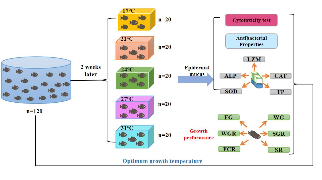
2. Materials and Methods

We promise that this study was conducted in accordance with the relevant regulations of the Animal Management Committee of Jiangsu University, with no violations of ethical and moral regulations, and there was no cruelty to animals. They were all raised normally. In the experiment, only the epidermal mucus of the crucian carp was extracted for testing. When the experiment was over, all the used crucian carp were released into the lake.

2.1. Experimental Materials

The experiment was carried out at the Key Laboratory of Modern Agricultural Equipment and Technology (119°45′ E; 32°20′ N) at Jiangsu University, Zhenjiang City, Jiangsu Province, from 10 June 2021 to 30 July 2021, for a total of 50 days. The average temperature of the laboratory air was 24.5 °C, and the relative humidity was 32.6%. We took 120 three-month-old male crucian carp (9.12 ± 0.5 g) from the Linghu fish base in Huzhou, Zhejiang. Before starting the experiment, these crucian carp were adapted to the laboratory conditions for two weeks in a 400 L water tank (water temperature: 17 ± 0.5 °C). After acclimatization, robust fish of similar size were picked out and were randomly put into 5 water tanks with a volume of 100 L each (initial water temperature 17 ± 0.5 °C), with 20 fish in each tank. The five water tanks were each increased by 3 °C every 48 h to reach the experimental water temperature, which was controlled at 17, 21, 24, 27, and 31 °C, respectively. During the experiment, commercial pellets containing 35% protein (Table 1 for nutrients) were used to feed the fish twice a day, the dissolved oxygen of the water was maintained at 6.8 ± 0.1 mg L−1, the pH was maintained at 7.6 ± 0.1, the ammonia nitrogen content was maintained at 0–0.12 mg/L, the nitrite content was maintained at 0–0.12 mg/L, and the light conditions were 12 h of darkness and 12 h of light. The experimental fish were fed twice a day at a rate of 5% of the fish’s body weight. Fish feces and uneaten fish feed were cleaned up, and 30% of water was exchanged every day.

2.2. Extraction of Epidermal Mucus

Three healthy crucian carp were randomly selected in each tank and gathered together (n = 3), using a sterile spatula to scrape the front and back skin on the back surface to prepare a mucus sample [7]. The collected mucus was thoroughly mixed with an equal amount of sterile physiological saline and centrifuged at 3000× g for 10 min at 4 °C to remove insoluble particles. The supernatant was filtered and sterilized with a 0.22 μm filter membrane and stored at −80 °C [12].

2.3. Cytotoxicity Tests

The human pancreatic cancer cell line PATU-8988T was obtained from Shanghai YuBo Biotech Co., Ltd. (Shanghai, China) and cultured in H-DMEM with 10% FBS. The pancreatic cancer cells, 5000 8988T cells/well, were placed into 96-well plates; six duplicate wells were set for each condition, and then the supernatant was carefully discarded when the cells were settled overnight, after which fresh cell culture solutions with pre-added skin mucus at different concentrations were added. The cell plates were slightly shaken and moved to an incubator with a temperature of 37 °C for 24 h of treatment. For the measurements, the supernatants were removed by leaving adherent cells minimally disturbed, and fresh culture solutions containing a 10% Cell Counting Kit-8 (CCK8, Kumamoto, Japan) reagent were added for 2 h of incubation at 37 °C. The absorbance of each well was detected with a microplate reader.

2.4. Antibacterial Activity

The ability of bacterial isolates to grow in fish epidermal mucus was examined in a 96-well plate to study the use of marine pathogens (V. harveyi) to determine the bactericidal activity in mucus samples. V. harveyi was grown on agar plates at 25 ± 2 °C. Then, we picked fresh single colonies and diluted them with a 5 mL nutrient broth, and then we incubated them in an orbital incubator at 28 °C and 150–200 rpm for 12 h. Then, the mucus bactericidal activity was measured [26]. A portion of the supernatant mucus was taken and autoclaved at 121 °C for 15 min. Bacterial suspension and mucus samples were serially 2-fold diluted with PBS. Then, 100 µL of bacterial diluent and an equal volume of autoclaved epidermal mucus were placed in a 96-well plate. The positive well contained 100 μL of bacterial suspension and 100 μL of nutrient broth. Negative wells contained 100 μL of autoclaved epidermal mucus and 100 μL of a nutrient solution. The experiment was carried out in triplicate. The plates were incubated at 28 ± 2 °C. Every 1 h, the optical density (OD) at 600 nm was measured with a microplate reader for a total of 12 h. The bacterial survival rate was calculated by the ratio of the optical density of the experimental group and the positive control group, and the result is expressed as a percentage (100%).

2.5. Determination of Enzyme Activity

A kit was used to determine the protein concentration (TP) and the lysozyme (ZLM), superoxide dismutase (SOD), catalase (CAT), and alkaline phosphatase (ALP) activity with a microplate reader according to the manufacturer’s instructions. The BCA microplate method was used to determine the protein concentration at 562 nm [27]. The lysozyme activity was determined with a turbidimetric method at 530 nm [28]. The superoxide dismutase activity was determined with a hydroxylamine method at 550 nm [29]. The catalase activity was determined with an ammonium molybdate method at 405 nm [30]. The alkaline phosphatase activity was measured with colorimetry at 520 nm [31]. The kits were acquired from Nanjing Jiancheng Institute of Bioengineering.

2.6. Growth Performance

Each three-month-old male crucian carp was starved for 24 h before sampling and weighing. At the end of the feeding experiment, the following parameters were determined:
WG = FW − IW
where WG (g) represents weight gain, FW (g) represents the final weight of crucian carp, and IW (g) represents the initial weight of the crucian carp.
WGR = 100% × WG/IW
where WGR (%) represents the relative growth rate.
SGR = 100% × (lnFW − lnIW)/T
where SGR (%) represents the specific growth rate, ln represents the natural logarithm, and T represents the number of feeding days.
FCR = 100% × (FI/WG)
where FCR (%) represents the feed conversion rate, and FI(g) represents feed intake.
SR = 100% × (N2/N1)
where SR (%) represents the survival rate, N2 represents the final number of crucian carp, and N1 represents the initial number of crucian carp.

2.7. Statistical Analyses

All data are shown as the mean plus standard deviation (±SD). All statistical analyses were performed with SPSS software. The statistical differences between groups were analyzed by using ANOVA. The least significant difference (LSD) [32] test was used to determine significance at a significance level of p < 0.05.

3. Results

3.1. Cytotoxicity

As shown in Figure 1, crucian carp epidermal mucous cells under five temperature treatments were tested for toxicity to human pancreatic cancer cell line 8988t. The results showed that as the concentration of epidermal mucus increased, the survival rate of cell lines showed a downward trend within a certain range. When the fish epidermal mucus was 1000 μg mL−1, the survival rate of the cell line reached more than 65%. The epidermal mucus under five temperature treatments had no toxic effect on the cell line [33].

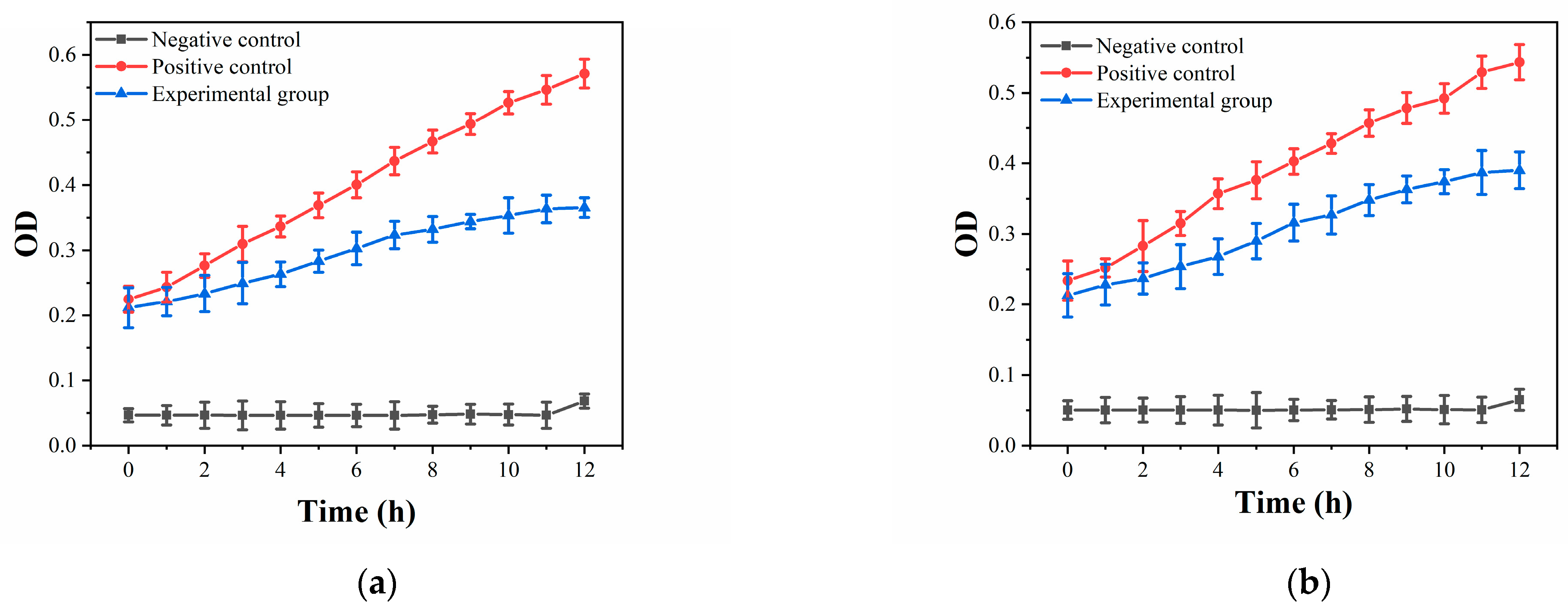
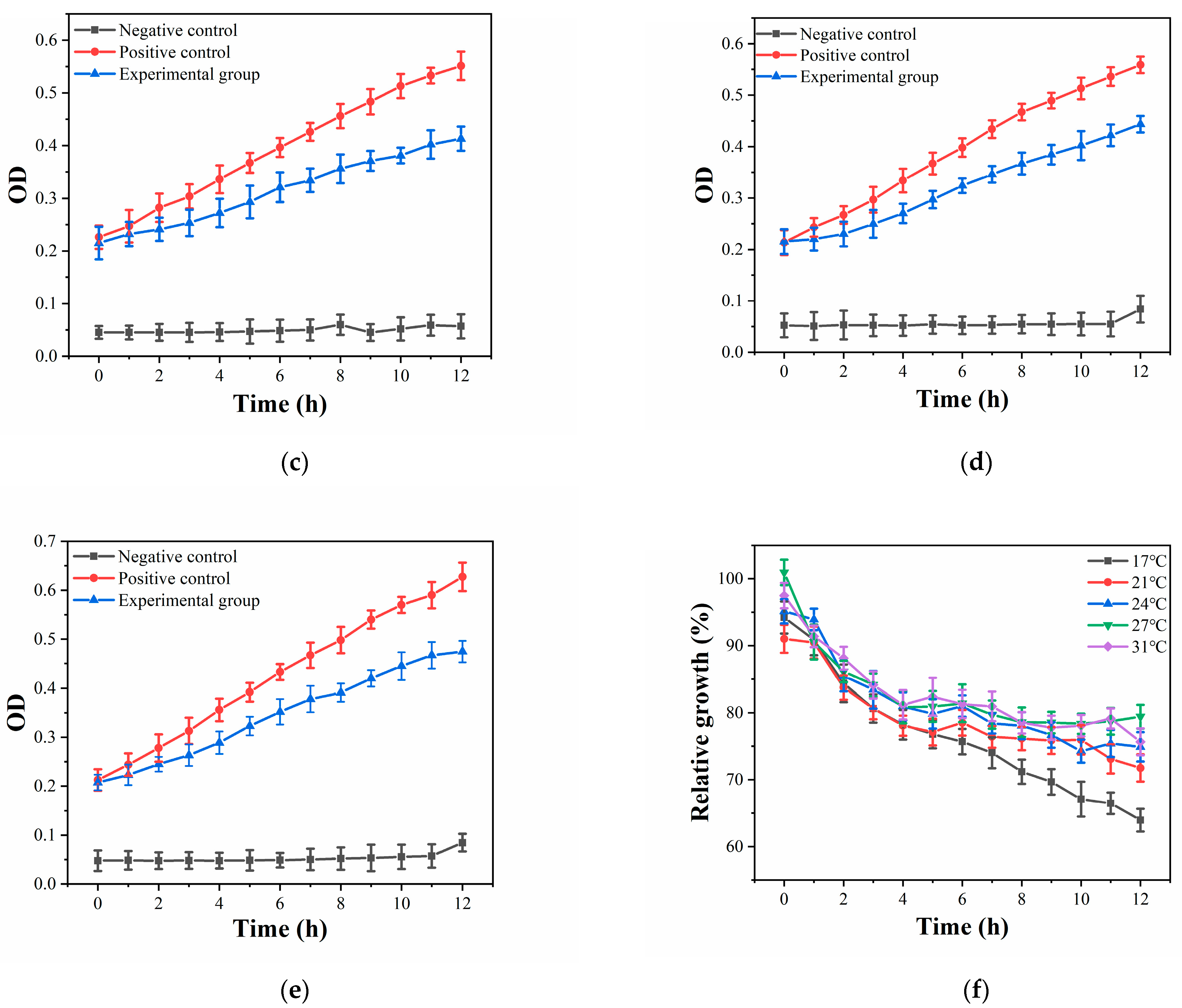
3.2. Antibacterial Properties

The growth of bacteria was analyzed in crucian carp epidermal mucus under five temperature treatments, and the V. harveyi selected in this study is a common pathogen of marine organisms. The results in Figure 2 show that during the culture period, the growth of the number of V. harveyi in the nutrient broth of the positive control group was observed to gradually increase over time. As shown in Figure 2a–e, the number of V. harveyi in the experimental group gradually increased under different epidermal mucus conditions, but the increase in the same period was less than that of the positive control group. The crucian carp epidermal mucus under the five temperature treatments all had similar results. In addition, comparing the five experimental groups in Figure 2f shows that as the temperature increased, the antibacterial ability of the crucian carp epidermal mucus gradually weakened, though the values were all significantly higher than those of the negative control group. This indicates that within a certain temperature range, the epidermal mucus of different treatments had antibacterial activity, and as the temperature increased, the survival rate of V. harveyi gradually increased (p < 0.05). On the contrary, when the temperature decreased, the metabolic activity of V. harveyi was reduced, and the survival rate of V. harveyi was also reduced (p < 0.05).

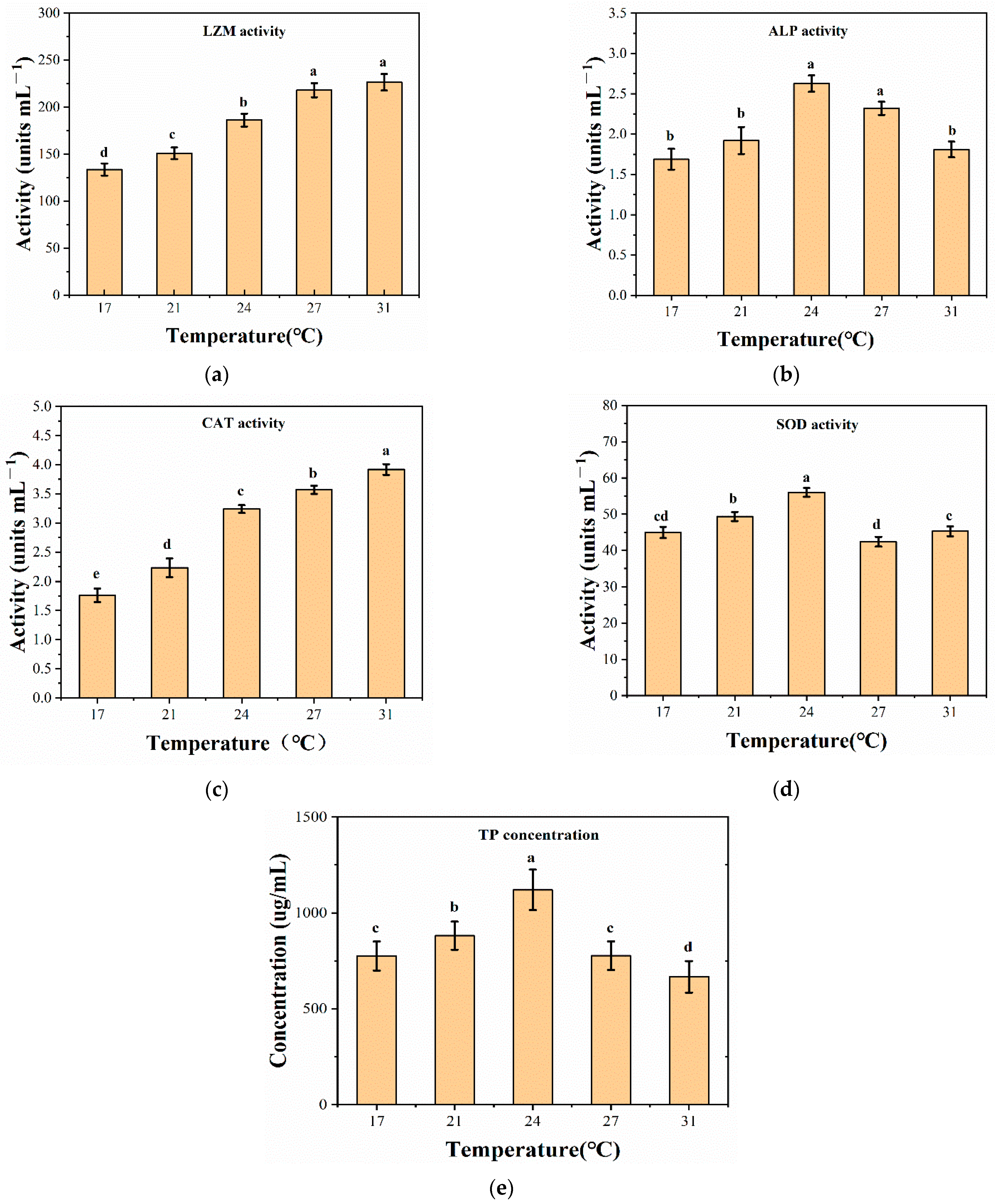
3.3. Enzyme Activity

The TP concentration and four enzyme activities in crucian carp epidermal mucus under five different treatments were tested. The activity of LZM gradually increased with the increase of temperature. Compared to the control group (17 °C), LZM activity increased by 12.78%, 39.85%, 63.91%, and 69.92% under the other four temperature treatments (Figure 3a). the ALP activity first increased and then decreased with the increase of temperature. Compared to the control group (17 °C), the ALP activity under the other four temperature treatments increased by 14.29%, 56.55%, 38.09%, and 7.74% (Figure 3b). Compared to CAT (17 °C), the other four temperature treatments increased by 14.6%, 53.36%, 67.92%, and 84.43% (Figure 3c). SOD activity first increased and then decreased with increasing temperature. Compared to the control group (17 °C), the SOD activity under the other four temperature treatments increased by 9.79%, 24.64%, −5.3%, and 0.73% (Figure 3d). The SOD level increased from 17 to 24 °C, then decreased at 27 °C, returned to the control level at 31 °C, and stabilized at 27 and 31 °C. Compared to the control group (17 °C), the total protein concentration under the other four temperature treatments increased by 13.68%, 44.52%, 0.22%, and −14.06% (Figure 3e). The total protein concentration first increased and then decreased with the increase of temperature, and it reached its highest point at 24 °C.

3.4. Growth Performance

The growth performance indexes (initial weight, final weight, weight gain, relative growth rate, specific growth rate, feed conversion rate, and survival rate) of crucian carp after 3 and 6 weeks of rearing under different temperature treatments are shown in Table 2. The growth performance of crucian carp reared at 24 °C was significantly improved. Crucian carp reared at 24 °C had the best weight gain, relative growth rate, feed conversion rate, and specific growth rate (p < 0.05). The growth status of crucian carp reared at 31 °C was the worst (p > 0.05). The survival rates of crucian carp reared at 17, 21, and 24 °C were higher than those of other groups (p < 0.05): 95.00%, 93.35%, and 93.35%, respectively.

4. Discussion

Temperature is a key factor affecting fish physiology and growth [2]. An improper temperature environment will affect the activity of enzymes in epidermal mucus [34,35]. Different types of enzymes in fish epidermal mucus can exert inhibitory or lytic activity against pathogens, which helps the host defend against bacterial infections [36,37]. Therefore, temperature can affect the disease resistance, immunity, and growth parameters of fish [38,39].
As the mucus concentration increases, the state of a cell will be affected more or less because it is still different from a cell culture medium to a certain extent. Generally speaking, we believe that when the concentration of a stimulus reaches a certain level, the survival rate of the cell is about 70% and the toxicity of the stimulus to the cell is basically negligible [33].
A large number of studies have shown that the enzymes contained in the epidermal mucus may exhibit antibacterial activity due to one or more joint actions [12,40]. We found that as the temperature rose within a certain temperature range, the enzyme and antibacterial activity of crucian carp epidermal mucus increased. However, in the range of 17–31 °C, the higher the temperature was, the more and more bacteria proliferated. When the temperature was 17 °C, the proliferation was the least. This may be because the enzyme activity increased with the increase of temperature, though the proliferation rate of bacteria is also affected by temperature. The optimum growth temperature of V. harveyi is 25–30 °C [41]; under these conditions, the proliferation rate of V. harveyi may exceed the antibacterial effect of the enzyme. At 17 °C, the enzyme activity was found to be the weakest, but the proliferation rate of V. harveyi was found to be reduced by low temperatures, so temperature exhibits strong antibacterial properties.
The main indicator of fish health maintenance is the activity of enzymes in the epidermal mucus [40]. The activity of enzymes is most affected by temperature, so temperature has an effect on the antibacterial ability of fish. LZM has been found to have the highest activity among the four enzymes secreted by the epidermis of crucian carp and is known to play a major non-specific defense role [42]. Within a certain range, higher temperatures will enhance the activity of LZM [43], so as temperature rises, the LZM activity of crucian carp under different treatments gradually increases. Here, the enzyme activity significantly changed at 24 and 27 °C, increasing by 39.85% and 63.91%, respectively, compared to that at 17 °C. Because LZM is a natural anti-infective substance, it has a bactericidal effect. Lysozymes display hydrolytic activity to specifically cleave the β-1, 4-glycosidic bonds between the N-acetylglucosamine and N-acetylmuramic acid of peptidoglycan. Therefore, LZM can destroy the peptidoglycan in the bacterial cell wall and cause its cell lysis, thereby achieving the purpose of sterilization [44]. Its strong antibacterial properties ensure the healthy growth of crucian carp.
ALP protects against bacteria by changing the surface structure of pathogens [40]. It plays an important role in the absorption of calcium in water, the formation of calcium phosphate, and the secretion and formation of chitin. ALP activity is also affected by temperature; we found that with the increase of temperature, the ALP activity of crucian carp under different treatments first increased and then decreased. The change was relatively significant at 24 and 27 °C, with increases of 56.55% and 38.09%, respectively, compared to that at 17 °C, and ALP is known to have the highest activity at 24 °C and promoted the metabolism, bone formation, and fat synthesis of crucian carp [45]. At the same time, it also enhances the destructive resistance of crucian carp skin [46].
Hydrogen peroxide is a cytotoxic substance produced by the redox reaction catalyzed by oxidase. CAT can protect cells from the damage of hydrogen peroxide. Therefore, CAT activity reflects the oxidation and health of fish epidermal mucus [40]. Temperature has a greater influence on the CAT activity of epidermal mucus [47]. We found that with the increase of temperature, the enzyme activity under the 24, 27, and 31 °C treatments significantly increased by 53.36%, 67.92%, and 84.43%, respectively. Therefore, these conditions are helpful for crucian carp to avoid poisoning by hydrogen peroxide.
The superoxide anion free radicals produced by crucian carp under normal conditions are necessary for life-sustaining activities, but if their content is too high, the crucian carp will be affected. SOD can scavenge and maintain this balance of oxygen free radicals and prevent crucian carp from being damaged [40]. In Figure 3d, it can be seen that SOD activity in this study was the highest at 24 °C. This shows that when the water temperature reached about 24 °C, the water environment exerted pressure on the crucian carp. Through the crucian carp’s own regulation system, SOD can enhance the antioxidant capacity of the crucian carp epidermal mucus by adjusting the ratio of superoxide anions [43], thus avoiding injury in high temperature environments.
We observed that the concentration of TP first increased and then decreased with the increase of temperature. The TP concentration reached its highest point at 24 °C, which was an increase of 44.52% compared to that at 17 °C. At an optimum temperature, the thickness and number of epidermal mucus cells, skin secretions, and epidermal cells of crucian carp are increased [48]. Therefore, the antibacterial peptides, lectins, and other substances in the mucus are increased, and the mucus increases resistance to pathogens and enhances its immune function [49]. In addition, changes in the composition of the mucus proteins and pathogen invasion of the body’s surface may also reasons for changes in protein concentration [50].
Water temperature can interfere with the growth of crucian carp by affecting appetite and thereby weight gain. The weight gain, relative growth rate, and survival rate of crucian carp at 27–31 °C were found to be significantly lower than those of the other three groups. The feed conversion rate was found to be much lower than that at 24 °C and slightly lower than that at 17 and 21 °C, so increasing the water temperature from 27 to 31 °C does not improve the growth of fish [50]. At 17 °C, we found that the crucian carp had the highest survival rate, so this temperature is the most suitable survival temperature rather than growth temperature. The survival rates at 21 and 24 °C were the same, so it can be concluded that the optimum growth temperature of crucian carp is between 21 and 25 °C [50]. In addition, at 21, 24, and 27 °C, the crucian carp had higher weight gains and specific growth rates.

5. Conclusions

The results of this study showed that with an increase of temperature, the antibacterial ability of crucian carp mucus gradually decreased. The activity of LZM and CAT was positively correlated with temperature, and their activity was relatively high at 24 °C. Additionally, the activity of AKP and SOD, as well as the concentration of TP, reached their maxima at 24 °C, which is beneficial to the growth of crucian carp. At the same time, the growth indicators of crucian carp fed at 24 °C were better than other conditions, except for the survival rate. However, the survival rate of crucian carp fed at 17–21 °C could reach more than 93%. In summary, this study shows that the optimum temperature for the safe growth of crucian carp is about 24 °C. The research results provide a good basis for the construction of aquaculture environmental control systems.

Author Contributions

Conceptualization, B.W. and H.M.; methodology, B.W., G.M., Y.W. and Y.L.; software, B.W., G.M. and Y.W.; validation, B.W. and X.D.; data curation, B.W. and G.M.; writing—original draft preparation, B.W., Y.L. and X.D.; writing—review and editing, B.W., Y.W., Q.S. and G.M.; project administration, H.M.; funding acquisition, H.M. All authors have read and agreed to the published version of the manuscript.

Funding

This research was funded by the National Natural Science Foundation of China (32071905), the National Natural Science Foundation of China (61771224) and Postgraduate Research & Practice Innovation Program of Jiangsu Province KYCX19_1619.

Institutional Review Board Statement

This study only extracted a small amount of epidermal mucus of crucian carp, which did not violate ethical regulations and did not involve animal abuse. After the test, all experimental fish were released. Therefore, ethical review and approval were waived.

Acknowledgments

The authors would like to thank the Key Laboratory of Agricultural Engineering in Jiangsu University for supporting the experimental conditions of the research.

Conflicts of Interest

The authors declare no conflict of interest.

References

  1. Kapusta, A.; Partyka, K.; Szczepkowska, B.; Jarmołowicz, S.; Hopko, M.; Piotrowska, I.; Kowalska, A.; Zakęś, Z. Impact of diet and culture conditions on the body shape of crucian carp (Carassius carassius L.). J. Appl. Anim. Res. 2013, 41, 462–469. [Google Scholar] [CrossRef] [Green Version]
  2. Sikorska, J.; Kondera, E.; Kamiński, R.; Ługowska, K.; Witeska, M.; Wolnicki, J. Effect of four rearing water temperatures on some performance parameters of larval and juvenile crucian carp, Carassius carassius, under controlled conditions. Aquac. Res. 2018, 49, 3874–3880. [Google Scholar] [CrossRef]
  3. Wolnicki, J.; Kamiński, R.; Sikorska, J. Combined effects of water temperature and daily food availability period on the growth and survival of tench (Tinca tinca) larvae. Aquac. Res. 2017, 48, 3809–3816. [Google Scholar] [CrossRef]
  4. Alshammari, E.; Patel, M.; Sachidanandan, M.; Kumar, P.; Adnan, M. Potential Evaluation and Health Fostering Intrinsic Traits of Novel Probiotic Strain Enterococcus durans F3 Isolated from the Gut of Fresh Water Fish Catla catla. Food Sci. Anim. Resour. 2019, 39, 844–861. [Google Scholar] [CrossRef] [Green Version]
  5. Cabillon, N.A.R.; Lazado, C.C. Mucosal Barrier Functions of Fish under Changing Environmental Conditions. Fishes 2019, 4, 2. [Google Scholar] [CrossRef] [Green Version]
  6. Fæste, C.K.; Tartor, H.; Moen, A.; Kristoffersen, A.B.; Dhanasiri, A.K.S.; Anonsen, J.H.; Furmanek, T.; Grove, S. Proteomic profiling of salmon skin mucus for the comparison of sampling methods. J. Chromatogr. B Anal. Technol. Biomed. Life Sci. 2020, 1138, 121965. [Google Scholar] [CrossRef]
  7. Abolfathi, M.; Akbarzadeh, A.; Hajimoradloo, A.; Joshaghani, H.R. Seasonal changes of hydrolytic enzyme activities in the skin mucus of rainbow trout, Oncorhynchus mykiss at different body sizes. Dev. Comp. Immunol. 2020, 103, 103499. [Google Scholar] [CrossRef]
  8. Mori, M.; Ito, T.; Washio, R.; Shibasaki, Y.; Namba, A.; Yabu, T.; Iwazaki, D.; Wada, N.; Anzai, H.; Shiba, H.; et al. Enhancement of immune proteins expression in skin mucus of Japanese flounder Paralicthys olivaceus upon feeding a diet supplemented with high concentration of ascorbic acid. Fish Shellfish Immunol. 2021, 114, 20–27. [Google Scholar] [CrossRef]
  9. Patel, M.; Ashraf, M.S.; Siddiqui, A.J.; Ashraf, S.A.; Sachidanandan, M.; Snoussi, M.; Adnan, M.; Hadi, S. Profiling and Role of Bioactive Molecules from Puntius sophore (Freshwater/Brackish Fish) Skin Mucus with Its Potent Antibacterial, Antiadhesion, and Antibiofilm Activities. Biomolecules 2020, 10, 920. [Google Scholar] [CrossRef] [PubMed]
  10. Ordóñez-Grande, B.; Guerreiro, P.M.; Sanahuja, I.; Fernández-Alacid, L.; Ibarz, A. Environmental Salinity Modifies Mucus Exudation and Energy Use in European Sea Bass Juveniles. Animals 2021, 11, 1580. [Google Scholar] [CrossRef]
  11. Sridhar, A.; Manikandan, D.B.; Palaniyappan, S.; Sekar, R.K.; Ramasamy, T. Correlation Between Three Freshwater Fish Skin Mucus Antiproliferative Effect and Its Elemental Composition Role in Bacterial Growth. Turk. J. Fish. Aquat. Sci. 2021, 21, 233–244. [Google Scholar] [CrossRef]
  12. Ceballos-Francisco, D.; Cuesta, A.; Esteban, M. Ángeles Effect of Light–Dark Cycle on Skin Mucosal Immune Activities of Gilthead Seabream (Sparus aurata) and European Sea Bass (Dicentrarchus labrax). Fishes 2020, 5, 10. [Google Scholar] [CrossRef] [Green Version]
  13. Almroth, B.C.; de Souza, K.B.; Jönsson, E.; Sturve, J. Oxidative stress and biomarker responses in the Atlantic halibut after long term exposure to elevated CO2 and a range of temperatures. Comp. Biochem. Physiol. Part B Biochem. Mol. Biol. 2019, 238, 110321. [Google Scholar] [CrossRef] [PubMed]
  14. Watson, S.A.; Allan, B.J.M.; McQueen, D.E.; Nicol, S.; Parsons, D.M.; Pether, S.M.J.; Pope, S.; Setiawan, A.N.; Smith, N.; Wilson, C.; et al. Ocean warming has a greater effect than acidification on the early life history development and swimming performance of a large circumglobal pelagic fish. Glob. Chang. Biol. 2018, 24, 4368–4385. [Google Scholar] [CrossRef] [PubMed]
  15. Islam, M.J.; Kunzmann, A.; Bögner, M.; Meyer, A.; Thiele, R.; Slater, M.J. Metabolic and molecular stress responses of European seabass, Dicentrarchus labrax at low and high temperature extremes. Ecol. Indic. 2020, 112, 106118. [Google Scholar] [CrossRef]
  16. Yilmaz, H.A.; Turkmen, S.; Kumlu, M.; Eroldogan, O.T.; Perker, N. Alteration of Growth and Temperature Tolerance of European Sea Bass (Dicentrarchus labrax Linnaeus 1758) in Different Temperature and Salinity Combinations. Turk. J. Fish. Aquat. Sci. 2020, 20, 331–340. [Google Scholar] [CrossRef]
  17. Nadermann, N.; Seward, R.K.; Volkoff, H. Effects of potential climate change -induced environmental modifications on food intake and the expression of appetite regulators in goldfish. Comp. Biochem. Physiol. Part A Mol. Integr. Physiol. 2019, 235, 138–147. [Google Scholar] [CrossRef]
  18. Masroor, W.; Farcy, E.; Blondeau-Bidet, E.; Venn, A.; Tambutté, E.; Lorin-Nebel, C. Effect of salinity and temperature on the expression of genes involved in branchial ion transport processes in European sea bass. J. Therm. Biol. 2019, 85, 102422. [Google Scholar] [CrossRef]
  19. Masroor, W.; Farcy, E.; Gros, R.; Lorin-Nebel, C. Effect of combined stress (salinity and temperature) in European sea bass Dicentrarchus labrax osmoregulatory processes. Comp. Biochem. Physiol. Part A Mol. Integr. Physiol. 2018, 215, 45–54. [Google Scholar] [CrossRef]
  20. Gamperl, A.K.; Ajiboye, O.O.; Zanuzzo, F.S.; Sandrelli, R.M.; Peroni, E.D.F.C.; Beemelmanns, A. The impacts of increasing temperature and moderate hypoxia on the production characteristics, cardiac morphology and haematology of Atlantic Salmon (Salmo salar). Aquaculture 2020, 519, 734874. [Google Scholar] [CrossRef]
  21. Antonopoulou, E.; Chatzigiannidou, I.; Feidantsis, K.; Kounna, C.; Chatzifotis, S. Effect of water temperature on cellular stress responses in meagre (Argyrosomus regius). Fish Physiol. Biochem. 2020, 46, 1075–1091. [Google Scholar] [CrossRef] [PubMed]
  22. Campos, D.F.; Braz-Mota, S.; Val, A.L.; Almeida-Val, V.M.F. Predicting thermal sensitivity of three Amazon fishes exposed to climate change scenarios. Ecol. Indic. 2019, 101, 533–540. [Google Scholar] [CrossRef]
  23. Islam, M.A.; Uddin, M.H.; Uddin, M.J.; Shahjahan, M. Temperature changes influenced the growth performance and physiological functions of Thai pangas Pangasianodon hypophthalmus. Aquacult. Rep. 2019, 13, 100179. [Google Scholar] [CrossRef]
  24. Islam, M.J.; Kunzmann, A.; Thiele, R.; Slater, M.J. Effects of extreme ambient temperature in European seabass, Dicentrarchus labrax acclimated at different salinities: Growth performance, metabolic and molecular stress responses. Sci. Total. Environ. 2020, 735, 139371. [Google Scholar] [CrossRef]
  25. Islam, M.J.; Slater, M.J.; Bögner, M.; Zeytin, S.; Kunzmann, A. Extreme ambient temperature effects in European seabass, Dicentrarchus labrax: Growth performance and hemato-biochemical parameters. Aquaculture 2020, 522, 735093. [Google Scholar] [CrossRef]
  26. Van Doan, H.; Lumsangkul, C.; Hoseinifar, S.H.; Tongsiri, S.; Chitmanat, C.; Musthafa, M.S.; El-Haroun, E.; Ringo, E. Modulation of growth, innate immunity, and disease resistance of Nile tilapia (Oreochromis niloticus) culture under biofloc system by supplementing pineapple peel powder and Lactobacillus plantarum. Fish Shellfish Immunol. 2021, 115, 212–220. [Google Scholar] [CrossRef]
  27. Rogatsky, E. Pandora box of BCA assay. Investigation of the accuracy and linearity of the microplate bicinchoninic protein assay: Analytical challenges and method modifications to minimize systematic errors. Anal. Biochem. 2021, 631, 114321. [Google Scholar] [CrossRef]
  28. Liao, Y.H.; Brown, M.B.; Martin, G.P. Turbidimetric and HPLC assays for the determination of formulated lysozyme activity. J. Pharm. Pharmacol. 2001, 53, 549–554. [Google Scholar] [CrossRef]
  29. Li, N.; Huang, H.-Q.; Zhang, G.-S.; Cui, W. Effect of 5-AZn-2′-deoxycytidine on proliferation of human lung adenocarcinoma cell line A549 in vitro. Asian Pac. J. Trop. Med. 2013, 6, 982–985. [Google Scholar] [CrossRef] [Green Version]
  30. Wu, T.; Shen, X. Response of Wumeng Semi-Fine Wool Sheep to Copper-Contaminated Environment. Pol. J. Environ. Stud. 2020, 29, 2917–2924. [Google Scholar] [CrossRef]
  31. Yang, Q.; Wang, X.; Peng, H.; Arabi, M.; Li, J.; Xiong, H.; Choo, J.; Chen, L. Ratiometric fluorescence and colorimetry dual-mode assay based on manganese dioxide nanosheets for visual detection of alkaline phosphatase activity. Sensors Actuators B Chem. 2020, 302, 127176. [Google Scholar] [CrossRef]
  32. Wang, Y.; Ma, G.; Du, X.; Liu, Y.; Wang, B.; Xu, G.; Mao, H. Effects of Nutrient Solution Irrigation Quantity and Downy Mildew Infection on Growth and Physiological Traits of Greenhouse Cucumber. Agronomy 2020, 10, 1921. [Google Scholar] [CrossRef]
  33. Cai, H.Z.; Chen, Y.X.; Xu, L.S.; Zou, Y.P.; Zhou, X.L.; Liang, G.X.; Wang, D.Q.; Tao, Z.M. Differently PEGylated Polymer Nanoparticles for Pancreatic Cancer Delivery: Using a Novel Near-Infrared Emissive and Biodegradable Polymer as the Fluorescence Tracer. Front. Bioeng. Biotechnol. 2021, 9, 699610. [Google Scholar] [CrossRef]
  34. Dominguez, M.; Takemura, A.; Tsuchiya, M.; Nakamura, S. Impact of different environmental factors on the circulating immunoglobulin levels in the Nile tilapia, Oreochromis niloticus. Aquaculture 2004, 241, 491–500. [Google Scholar] [CrossRef]
  35. Firmino, J.P.; Fernández-Alacid, L.; Vallejos-Vidal, E.; Salomón, R.; Sanahuja, I.; Tort, L.; Ibarz, A.; Reyes-López, F.E.; Gisbert, E. Carvacrol, Thymol, and Garlic Essential Oil Promote Skin Innate Immunity in Gilthead Seabream (Sparus aurata) Through the Multifactorial Modulation of the Secretory Pathway and Enhancement of Mucus Protective Capacity. Front. Immunol. 2021, 12, 633621. [Google Scholar] [CrossRef]
  36. Guardiola, F.A.; Cuesta, A.; Abellán, E.; Meseguer, J.; Esteban, M.A. Comparative analysis of the humoral immunity of skin mucus from several marine teleost fish. Fish Shellfish Immunol. 2014, 40, 24–31. [Google Scholar] [CrossRef]
  37. Palaksha, K.; Shin, G.-W.; Kim, Y.-R.; Jung, T.S. Evaluation of non-specific immune components from the skin mucus of olive flounder (Paralichthys olivaceus). Fish Shellfish Immunol. 2008, 24, 479–488. [Google Scholar] [CrossRef]
  38. Valero, Y.; García-Alcázar, A.; Esteban, M.Á.; Cuesta, A.; Chaves-Pozo, E. Seasonal variations of the humoral immune parameters of European sea bass (Dicentrarchus labrax L.). Fish Shellfish Immunol. 2014, 39, 185–187. [Google Scholar] [CrossRef] [PubMed]
  39. Leal, M.J.; Clark, B.E.; Van Eenennaam, J.P.; Schreier, A.D.; Todgham, A.E. The effects of warm temperature acclimation on constitutive stress, immunity, and metabolism in white sturgeon (Acipenser transmontanus) of different ploidies. Comp. Biochem. Physiol. Part A Mol. Integr. Physiol. 2018, 224, 23–34. [Google Scholar] [CrossRef] [PubMed] [Green Version]
  40. Cheng, C.-H.; Guo, Z.-X.; Ye, C.-X.; Wang, A.-L. Effect of dietary astaxanthin on the growth performance, non-specific immunity, and antioxidant capacity of pufferfish (Takifugu obscurus) under high temperature stress. Fish Physiol. Biochem. 2018, 44, 209–218. [Google Scholar] [CrossRef]
  41. Mougin, J.; Roquigny, R.; Flahaut, C.; Bonnin-Jusserand, M.; Grard, T.; Le Bris, C. Abundance and spatial patterns over time of Vibrionaceae and Vibrio harveyi in water and biofilm from a seabass aquaculture facility. Aquaculture 2021, 542, 736862. [Google Scholar] [CrossRef]
  42. Ndong, D.; Chen, Y.Y.; Lin, Y.H.; Vaseeharan, B.; Chen, J.C. The immune response of tilapia Oreochromis mossambicus and its susceptibility to Streptococcus iniae under stress in low and high temperatures. Fish Shellfish Immunol. 2007, 22, 686–694. [Google Scholar] [CrossRef]
  43. Huang, Z.-H.; Ma, A.-J.; Wang, X.-A. The immune response of turbot, Scophthalmus maximus (L.), skin to high water temperature. J. Fish Dis. 2011, 34, 619–627. [Google Scholar] [CrossRef]
  44. Souza, D.C.D.M.; Dos Santos, M.C.; Chagas, E.C. Immune response of teleost fish to helminth parasite infection. Rev. Bras. Parasitol. Vet. 2019, 28, 533–547. [Google Scholar] [CrossRef] [PubMed]
  45. Davies, S.J.; El-Haroun, E.R.; Hassaan, M.S.; Bowyer, P.H. A Solid-State Fermentation (SSF) supplement improved performance, digestive function and gut ultrastructure of rainbow trout (Oncorhynchus mykiss) fed plant protein diets containing yellow lupin meal. Aquaculture 2021, 545, 737177. [Google Scholar] [CrossRef]
  46. Kumari, U.; Verma, N.; Nigam, A.K.; Mittal, S.; Mittal, A.K. Wound-healing potential of curcumin in the carp, Labeo rohita. Aquac. Res. 2016, 48, 2411–2427. [Google Scholar] [CrossRef]
  47. Esteban, M.A.; Cuesta, A.; Rodriguez, A.; Meseguer, J. Effect of photoperiod on the fish innate immune system: A link between fish pineal gland and the immune system. J. Pineal Res. 2006, 41, 261–266. [Google Scholar] [CrossRef] [PubMed]
  48. Karlsen, C.; Ytteborg, E.; Timmerhaus, G.; Høst, V.; Handeland, S.; Jørgensen, S.M.; Krasnov, A. Atlantic salmon skin barrier functions gradually enhance after seawater transfer. Sci. Rep. 2018, 8, 1–12. [Google Scholar] [CrossRef]
  49. Espinosa-Ruíz, C.; Esteban, M.Á. Wound-Induced Changes in Antioxidant Enzyme Activities in Skin Mucus and in Gene Expression in the Skin of Gilthead Seabream (Sparus aurata L.). Fishes 2021, 6, 15. [Google Scholar] [CrossRef]
  50. Ai-Jun, M.; Zhi-Hui, H.; Xin-An, W. Changes in protein composition of epidermal mucus in turbot Scophthalmus maximus (L.) under high water temperature. Fish Physiol. Biochem. 2013, 39, 1411–1418. [Google Scholar] [CrossRef] [PubMed]
Figure 1. Results of a 24 h cytotoxicity test of crucian carp epiderma mucus on the 8988t human pancreatic cancer cell line at five different temperatures.
Fishes 06 00066 g001
Figure 2. Growth curves of V. harveyi in epidermal mucus and nutrient solution at different temperatures: (a) 17 °C, (b) 21 °C, (c) 24 °C, (d) 27 °C, and (e) 31 °C. (f) A comparison of the increase in the number of V. harveyi in crucian carp epidermal mucus under five temperature cultures. Values are represented as mean ± SD of pooled data from triplicates per treatment (n = 3). Means in the same row with different letters are significantly different (p < 0.05).
Fishes 06 00066 g002aFishes 06 00066 g002b
Figure 3. Determination of enzyme activities and total protein concentration in epidermal mucus treated at different temperatures: (a) LZM, (b) ALP, (c) CAT, (d) SOD, and (e) TP. Values are represented as mean ± SD of pooled data from triplicates per treatment (n = 3). Means in the same row with different letters are significantly different (p < 0.05).
Fishes 06 00066 g003
Table 1. Feed nutritional components.
Crude Protein
(%) ≥
Crude Fiber
(%) ≤
Crude Fat
(%) ≥
Crude Ash
(%) ≤
Total Phosphors
(%) ≥
Moisture
(%) ≤
Lysine
(%) ≥
35.012.05.015.01.012.51.6
Table 2. Growth performance of crucian carp under five temperature treatments.
17 °C21 °C24 °C27 °C31 °C
IW (g)9.19 ± 1.099.07 ± 0.819.11 ± 1.149.49 ± 0.998.93 ± 1.40
FG (g)
3 weeks11.99 ± 0.96 bc12.73 ± 0.81 ab13.33 ± 1.06 a12.16 ± 0.94 bc11.17 ± 1.33 c
6 weeks13.96 ± 0.87 b14.92 ± 1.60 b16.36 ± 1.03 a14.25 ± 0.99 b12.73 ± 1.33 c
WG (g)
3 weeks2.80 ± 0.96 bc3.66 ± 0.81 ab4.22 ± 1.06 a2.67 ± 0.94 bc2.24 ± 1.33 c
6 weeks4.77 ± 0.87 b5.85 ± 1.60 b7.25 ± 1.03 a4.76 ± 0.99 b3.80 ± 1.33 c
WGR (%)
3 weeks32.16 ± 2.02 b40.69 ± 4.75 ab47.49 ± 1.29 a29.87 ± 2.03 b25.81 ± 6.13 c
6 weeks53.27 ± 1.37 b65.34 ± 3.08 b81.28 ± 5.61 a50.70 ± 6.17 bc43.79 ± 5.22 c
SGR (%)
3 weeks1.27 ± 0.03 b1.61 ± 0.02 b1.81 ± 0.01 a1.18 ± 0.02 b1.07 ± 0.01 c
6 weeks0.97 ± 0.01 b1.19 ± 0.03 b1.39 ± 0.01 a1.01 ± 0.01 b0.84 ± 0.02 c
FCR (%)
3 weeks1.01 ± 0.02 b1.03 ± 0.02 b1.04 ± 0.01 a1.03 ± 0.01 b1.01 ± 0.01 b
6 weeks1.07 ± 0.01 b1.08 ± 0.01 b1.12 ± 0.02 a1.06 ± 0.02 b1.04 ± 0.02 b
SR (%)
3 weeks10010010010095.60
6 weeks95.00 ± 0.8293.35 ± 1.2593.35 ± 1.2586.65 ± 1.2584.21 ± 0.82
Values are represented as mean ± SD of pooled data from triplicates per treatment (n = 3). Different letters in the same column indicate that the value was found to be significant when p < 0.05 by the least significant difference (LSD) multiple comparison method.
Publisher’s Note: MDPI stays neutral with regard to jurisdictional claims in published maps and institutional affiliations.

