Fatigability and Cardiorespiratory Impairments in Parkinson’s Disease: Potential Non-Motor Barriers to Activity Performance


Abstract
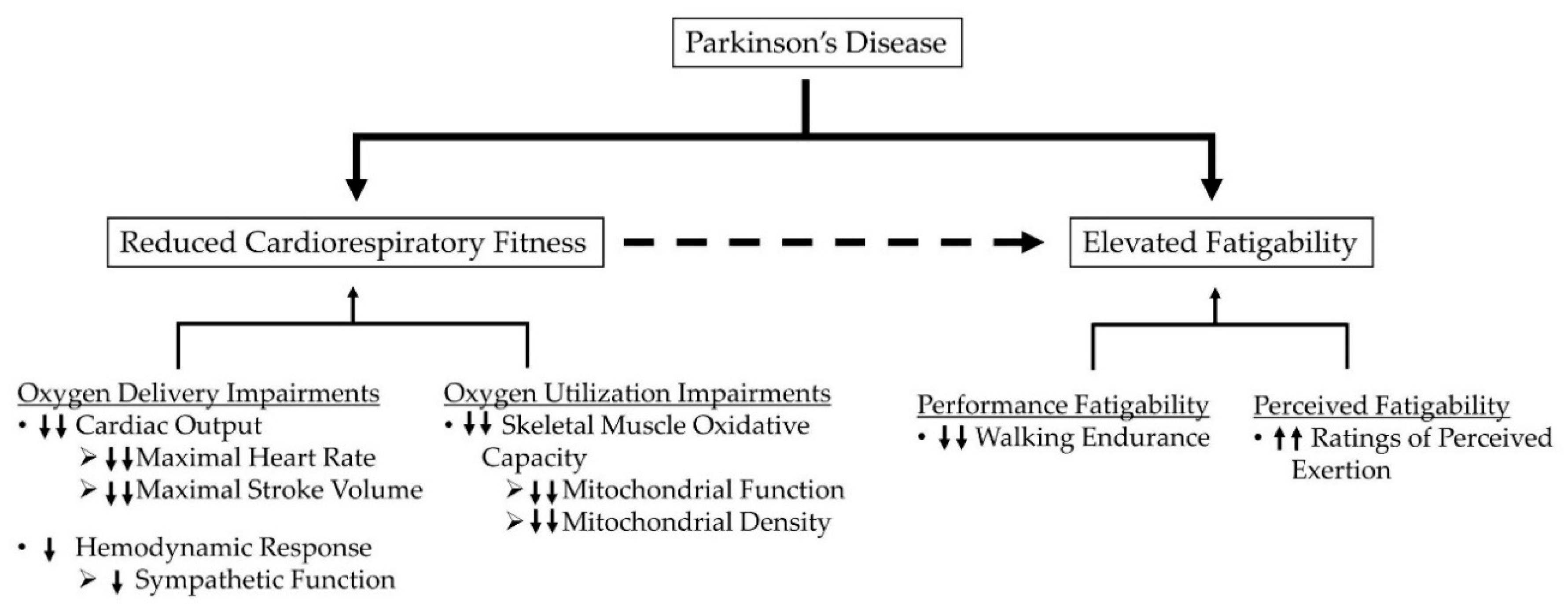
:1. Introduction
2. Fatigue versus Fatigability
2.1. Fatigue as a Symptom
2.2. Fatigability: An Activity-Induced Construct
3. Evidence of Elevated Fatigability in Parkinson’s Disease
4. Fatigability from a Bioenergetic Perspective
5. Cardiorespiratory Impairments Contributing to Fatigability in Parkinson’s Disease
5.1. Oxygen Delivery
5.2. Oxygen Utilization
6. Clinical Considerations
7. Summary
Author Contributions
Funding
Conflicts of Interest
References
- Ascherio, A.; Schwarzschild, M.A. The epidemiology of Parkinson’s disease: Risk factors and prevention. Lancet Neurol. 2016, 15, 1257–1272. [Google Scholar] [CrossRef]
- Kowal, S.L.; Dall, T.M.; Chakrabarti, R.; Storm, M.V.; Jain, A. The current and projected economic burden of Parkinson’s disease in the United States: Economic Burden of PD in the US. Mov. Disord. 2013, 28, 311–318. [Google Scholar] [CrossRef]
- Kalia, L.V.; Lang, A.E. Parkinson’s disease. Lancet 2015, 386, 896–912. [Google Scholar] [CrossRef]
- Chaudhuri, K.R.; Schapira, A.H. Non-motor symptoms of Parkinson’s disease: Dopaminergic pathophysiology and treatment. Lancet Neurol. 2009, 8, 464–474. [Google Scholar] [CrossRef]
- Friedman, J.H.; Beck, J.C.; Chou, K.L.; Clark, G.; Fagundes, C.P.; Goetz, C.G.; Herlofson, K.; Kluger, B.; Krupp, L.B.; Lang, A.E.; et al. Fatigue in Parkinson’s disease: Report from a mutidisciplinary symposium. NPJ Parkinson’s Dis. 2016, 2, 1–6. [Google Scholar] [CrossRef] [PubMed] [Green Version]
- Kluger, B.M.; Herlofson, K.; Chou, K.L.; Lou, J.-S.; Goetz, C.G.; Lang, A.E.; Weintraub, D.; Friedman, J. Parkinson’s disease-related fatigue: A case definition and recommendations for clinical research. Mov. Disord. 2016, 31, 625–631. [Google Scholar] [CrossRef]
- Nimwegen, M.; Speelman, A.D.; Hofman-van Rossum, E.J.M.; Overeem, S.; Deeg, D.J.H.; Borm, G.F.; Horst, M.H.L.; Bloem, B.R.; Munneke, M. Physical inactivity in Parkinson’s disease. J. Neurol. 2011, 258, 2214–2221. [Google Scholar] [CrossRef] [PubMed] [Green Version]
- Eldadah, B.A. Fatigue and fatigability in older adults. PM&R 2010, 2, 406–413. [Google Scholar]
- Friedman, J.H.; Brown, R.G.; Comella, C.; Garber, C.E.; Krupp, L.B.; Lou, J.-S.; Marsh, L.; Nail, L.; Shulman, L.; Taylor, C.B. Fatigue in Parkinson’s disease: A review. Mov. Disord. 2007, 22, 297–308. [Google Scholar] [CrossRef]
- Schrag, A.; Horsfall, L.; Walters, K.; Noyce, A.; Petersen, I. Prediagnostic presentations of Parkinson’s disease in primary care: A case-control study. Lancet Neurol. 2015, 14, 57–64. [Google Scholar] [CrossRef] [Green Version]
- Alves, G.; Wentzel-Larsen, T.; Larsen, J.P. Is fatigue an independent and persistent symptom in patients with Parkinson disease? Neurology 2004, 63, 1908–1911. [Google Scholar] [CrossRef] [PubMed]
- Dogan, V.B.; Koksal, A.; Dirican, A.; Baybas, S.; Dirican, A.; Dogan, G.B. Independent effect of fatigue on health-related quality of life in patients with idiopathic Parkinson’s disease. Neurol. Sci. 2015, 36, 2221–2226. [Google Scholar] [CrossRef] [PubMed]
- Gallagher, D.A.; Lees, A.J.; Schrag, A. What are the most important nonmotor symptoms in patients with Parkinson’s disease and are we missing them? Mov. Disord. 2010, 25, 2493–2500. [Google Scholar] [CrossRef]
- Gofton, T.E.; Kumar, H.; Roberts-South, A.; Speechley, M.; Jog, M.S. Validity, reliability, and insights from applying the McGill Quality of Life Questionnaire to People Living with Parkinson’s Disease (MQoL-PD). J. Palliat. Care 2015, 31, 213–220. [Google Scholar] [CrossRef]
- Prakash, K.M.; Nadkarni, N.V.; Lye, W.-K.; Yong, M.-H.; Tan, E.-K. The impact of non-motor symptoms on the quality of life of Parkinson’s disease patients: A longitudinal study. Eur. J. Neurol. 2016, 23, 854–860. [Google Scholar] [CrossRef]
- Kluger, B.M. Chapter twenty-four–fatigue in Parkinson’s disease. In International Review of Neurobiology; Nonmotor Parkinson’s: The Hidden Face; Chaudhuri, K.R., Titova, N., Eds.; Academic Press: Cambridge, MA, USA, 2017; Volume 133, pp. 743–768. [Google Scholar]
- Kotagal, V.; Szpara, A.; Albin, R.L.; Bohnen, N.I. Fatigue in parkinson’s disease associates with lower ambulatory diastolic blood pressure. J. Parkinsons. Dis. 2019, 9, 575–581. [Google Scholar] [CrossRef] [Green Version]
- Pont-Sunyer, C.; Hotter, A.; Gaig, C.; Seppi, K.; Compta, Y.; Katzenschlager, R.; Mas, N.; Hofeneder, D.; Brücke, T.; Bayés, A.; et al. The onset of nonmotor symptoms in Parkinson’s disease (the ONSET PD study). Mov. Disord. 2015, 30, 229–237. [Google Scholar]
- Loprinzi, P.D.; Herod, S.M.; Cardinal, B.J.; Noakes, T.D. Physical activity and the brain: A review of this dynamic, bi-directional relationship. Brain Res. 2013, 1539, 95–104. [Google Scholar] [CrossRef]
- Ahlskog, J.E. Aerobic exercise: Evidence for a direct brain effect to slow parkinson disease progression. Mayo Clin. Proc. 2018, 93, 360–372. [Google Scholar] [CrossRef] [Green Version]
- Ahlskog, J.E. Does vigorous exercise have a neuroprotective effect in Parkinson disease? Neurology 2011, 77, 288–294. [Google Scholar] [CrossRef] [PubMed] [Green Version]
- Petzinger, G.M.; Fisher, B.E.; McEwen, S.; Beeler, J.A.; Walsh, J.P.; Jakowec, M.W. Exercise-enhanced neuroplasticity targeting motor and cognitive circuitry in Parkinson’s disease. Lancet Neurol. 2013, 12, 716–726. [Google Scholar] [CrossRef] [Green Version]
- Garber, C.E.; Friedman, J.H. Effects of fatigue on physical activity and function in patients with Parkinson’s disease. Neurology 2003, 60, 1119–1124. [Google Scholar] [CrossRef] [PubMed]
- Lou, J.-S.; Kearns, G.; Oken, B.; Sexton, G.; Nutt, J. Exacerbated physical fatigue and mental fatigue in Parkinson’s disease. Mov. Disord. 2001, 16, 190–196. [Google Scholar] [CrossRef] [PubMed]
- Enoka, R.M.; Duchateau, J. Translating fatigue to human performance. Med. Sci. Sports Exerc. 2016, 48, 2228–2238. [Google Scholar] [CrossRef] [Green Version]
- Whitson, H.E.; Duan-Porter, W.; Schmader, K.E.; Morey, M.C.; Cohen, H.J.; Colón-Emeric, C.S. Physical resilience in older adults: Systematic review and development of an emerging construct. J. Gerontol. Series A Biol. Sci. Med Sci. 2016, 71, 489–495. [Google Scholar] [CrossRef] [Green Version]
- Schrack, J.A.; Simonsick, E.M.; Glynn, N.W. Fatigability: A Prognostic indicator of phenotypic aging. J. Gerontol. A Biol. Sci. Med. Sci. 2020, 75, e63–e66. [Google Scholar] [CrossRef]
- Kluger, B.M.; Krupp, L.B.; Enoka, R.M. Fatigue and fatigability in neurologic illnesses. Neurology 2013, 80, 409–416. [Google Scholar] [CrossRef]
- Protas, E.J.; Stanley, R.K.; Jankovic, J.; MacNeill, B. Cardiovascular and metabolic responses to upper- and lower-extremity exercise in men with idiopathic Parkinson’s disease. Phys. Ther. 1996, 76, 34–40. [Google Scholar] [CrossRef] [Green Version]
- Stanley, R.K.; Protas, E.J.; Jankovic, J. Exercise performance in those having Parkinson’s disease and healthy normals. Med. Sci. Sports Exerc. 1999, 31, 761–766. [Google Scholar] [CrossRef]
- Reuter, I.; Engelhardt, M.; Freiwaldt, J.; Baas, H. Exercise test in Parkinson’s disease. Clin. Auton. Res. 1999, 9, 129–134. [Google Scholar] [CrossRef]
- Canning, C.G.; Ada, L.; Johnson, J.J.; McWhirter, S. Walking capacity in mild to moderate Parkinson’s disease. Arch. Phys. Med. Rehabil. 2006, 87, 371–375. [Google Scholar] [CrossRef] [PubMed]
- Werner, W.G.; DiFrancisco-Donoghue, J.; Lamberg, E.M. Cardiovascular response to treadmill testing in parkinson disease. J. Neurol. Phys. Ther. 2006, 30, 68. [Google Scholar] [CrossRef] [PubMed]
- Christiansen, C.L.; Schenkman, M.L.; McFann, K.; Wolfe, P.; Kohrt, W.M. Walking economy in people with Parkinson’s disease. Mov. Disord. 2009, 24, 1481–1487. [Google Scholar] [CrossRef] [Green Version]
- DiFrancisco-Donoghue, J.; Elokda, A.; Lamberg, E.M.; Bono, N.; Werner, W.G. Norepinephrine and cardiovascular responses to maximal exercise in Parkinson’s disease on and off medication. Mov. Disord. 2009, 24, 1773–1778. [Google Scholar] [CrossRef]
- Maggioni, M.A.; Veicsteinas, A.; Rampichini, S.; Cè, E.; Nemni, R.; Riboldazzi, G.; Merati, G. Energy cost of spontaneous walking in Parkinson’s disease patients. Neurol. Sci. 2012, 33, 779–784. [Google Scholar] [CrossRef]
- Speelman, A.D.; Groothuis, J.T.; van Nimwegen, M.; van der Scheer, E.S.; Borm, G.F.; Bloem, B.R.; Hopman, M.T.E.; Munneke, M. Cardiovascular responses during a submaximal exercise test in patients with Parkinson’s disease. J. Parkinsons. Dis. 2012, 2, 241–247. [Google Scholar] [CrossRef] [PubMed]
- Strano, S.; Fanciulli, A.; Rizzo, M.; Marinelli, P.; Palange, P.; Tiple, D.; Vincentis, G.D.; Calcagnini, G.; Censi, F.; Meco, G.; et al. Cardiovascular dysfunction in untreated Parkinson’s disease: A multi-modality assessment. J. Neurol. Sci. 2016, 370, 251–255. [Google Scholar] [CrossRef] [PubMed]
- Kanegusuku, H.; Silva-Batista, C.; Peçanha, T.; Nieuwboer, A.; Silva, N.D.; Costa, L.A.; de Mello, M.T.; Piemonte, M.E.; Ugrinowitsch, C.; Forjaz, C.L. Blunted maximal and submaximal responses to cardiopulmonary exercise tests in patients with parkinson disease. Arch. Phys. Med. Rehabil. 2016, 97, 720–725. [Google Scholar] [CrossRef]
- Roberson, K.B.; Signorile, J.F.; Singer, C.; Jacobs, K.A.; Eltoukhy, M.; Ruta, N.; Mazzei, N.; Buskard, A.N.L. Hemodynamic responses to an exercise stress test in Parkinson’s disease patients without orthostatic hypotension. Appl. Physiol. Nutr. Metab. 2019, 44, 751–758. [Google Scholar] [CrossRef]
- Nakamura, T.; Hirayama, M.; Yamashita, F.; Uchida, K.; Hama, T.; Watanabe, H.; Sobue, G. Lowered cardiac sympathetic nerve performance in response to exercise in Parkinson’s disease. Mov. Disord. 2010, 25, 1183–1189. [Google Scholar] [CrossRef]
- Hortobágyi, T.; Mizelle, C.; Beam, S.; DeVita, P. Old adults perform activities of daily living near their maximal capabilities. J. Gerontol. A Biol. Sci. Med. Sci. 2003, 58, M453–M460. [Google Scholar] [CrossRef] [PubMed] [Green Version]
- Santanasto, A.J.; Glynn, N.W.; Jubrias, S.A.; Conley, K.E.; Boudreau, R.M.; Amati, F.; Mackey, D.C.; Simonsick, E.M.; Strotmeyer, E.S.; Coen, P.M.; et al. Skeletal muscle mitochondrial function and fatigability in older adults. J. Gerontol. A Biol. Sci. Med. Sci. 2015, 70, 1379–1385. [Google Scholar] [CrossRef] [Green Version]
- Coen, P.M.; Jubrias, S.A.; Distefano, G.; Amati, F.; Mackey, D.C.; Glynn, N.W.; Manini, T.M.; Wohlgemuth, S.E.; Leeuwenburgh, C.; Cummings, S.R.; et al. Skeletal muscle mitochondrial energetics are associated with maximal aerobic capacity and walking speed in older adults. J. Gerontol. A Biol. Sci. Med. Sci. 2013, 68, 447–455. [Google Scholar] [CrossRef]
- Tanaka, H.; Seals, D.R. Endurance exercise performance in Masters athletes: Age-associated changes and underlying physiological mechanisms. J. Physiol. 2008, 586, 55–63. [Google Scholar] [CrossRef] [PubMed]
- Keyser, R.E.; Christensen, E.J.; Chin, L.M.K.; Woolstenhulme, J.G.; Drinkard, B.; Quinn, A.; Connors, G.; Weir, N.A.; Nathan, S.D.; Chan, L.E. Changes in fatigability following intense aerobic exercise training in patients with interstitial lung disease. Respir. Med. 2015, 109, 517–525. [Google Scholar] [CrossRef] [PubMed] [Green Version]
- Chin, L.M.K.; Chan, L.; Drinkard, B.; Keyser, R.E. Oxygen uptake on-kinetics before and after aerobic exercise training in individuals with traumatic brain injury. Disabil. Rehabil. 2019, 41, 2949–2957. [Google Scholar] [CrossRef] [PubMed]
- Gollie, J.M.; Herrick, J.E.; Keyser, R.E.; Chin, L.M.K.; Collins, J.P.; Shields, R.K.; Panza, G.S.; Guccione, A.A. Fatigability, oxygen uptake kinetics and muscle deoxygenation in incomplete spinal cord injury during treadmill walking. Eur. J. Appl. Physiol. 2017, 117, 1989–2000. [Google Scholar] [CrossRef]
- Lepers, R.; Stapley, P.J. Master athletes are extending the limits of human endurance. Front. Physiol. 2016, 7, 613. [Google Scholar] [CrossRef] [Green Version]
- Kelly, J.O.; Kilbreath, S.L.; Davis, G.M.; Zeman, B.; Raymond, J. Cardiorespiratory fitness and walking ability in subacute stroke patients. Arch. Phys. Med. Rehabil. 2003, 84, 1780–1785. [Google Scholar] [CrossRef]
- Keyser, R.E.; Woolstenhulme, J.G.; Chin, L.M.K.; Nathan, S.D.; Weir, N.A.; Connors, G.; Drinkard, B.; Lamberti, J.; Chan, L. Cardiorespiratory function before and after aerobic exercise training in patients with interstitial lung disease. J. Cardiopulm. Rehabil. Prev. 2015, 35, 47–55. [Google Scholar] [CrossRef]
- Bassett, D.R.; Howley, E.T. Limiting factors for maximum oxygen uptake and determinants of endurance performance. Med. Sci. Sports Exerc. 2000, 32, 70–84. [Google Scholar] [CrossRef] [PubMed]
- Poole, D.C.; Ferreira, L.F.; Behnke, B.J.; Barstow, T.J.; Jones, A.M. The final frontier: Oxygen flux into muscle at exercise onset. Exerc. Sport Sci. Rev. 2007, 35, 166–173. [Google Scholar] [CrossRef] [PubMed]
- Souron, R.; Voirin, A.; Kennouche, D.; Espeit, L.; Millet, G.Y.; Rupp, T.; Lapole, T. Task failure during sustained low-intensity contraction is not associated with a critical amount of central fatigue. Scand J. Med. Sci. Sports 2020. [Google Scholar] [CrossRef] [PubMed]
- Sundberg, C.W.; Fitts, R.H. Bioenergetic basis of skeletal muscle fatigue. Curr. Opin. Physiol. 2019, 10, 118–127. [Google Scholar] [CrossRef]
- Keyser, R.E. Peripheral Fatigue: High-Energy Phosphates and Hydrogen Ions. PM&R 2010, 2, 347–358. [Google Scholar]
- Fitts, R.H. Cellular, molecular, and metabolic basis of muscle fatigue. In Comprehensive Physiology; Terjung, R., Ed.; John Wiley & Sons, Inc.: Hoboken, NJ, USA, 2011; ISBN 978-0-470-65071-4. [Google Scholar]
- Allen, D.G.; Lamb, G.D.; Westerblad, H. Skeletal muscle fatigue: Cellular mechanisms. Physiol. Rev. 2008, 88, 287–332. [Google Scholar] [CrossRef] [Green Version]
- Gandevia, S.C. Spinal and supraspinal factors in human muscle fatigue. Physiol. Rev. 2001, 81, 1725–1789. [Google Scholar] [CrossRef]
- Taylor, J.L.; Amann, M.; Duchateau, J.; Meeusen, R.; Rice, C.L. Neural contributions to muscle fatigue: From the brain to the muscle and back again. Med. Sci. Sports Exerc. 2016, 48, 2294–2306. [Google Scholar] [CrossRef] [Green Version]
- Amann, M. Significance of group III and IV muscle afferents for the endurance exercising human. Clin. Exp. Pharmacol. Physiol. 2012, 39, 831–835. [Google Scholar] [CrossRef] [Green Version]
- Mavrommati, F.; Collett, J.; Franssen, M.; Meaney, A.; Sexton, C.; Dennis-West, A.; Betts, J.F.; Izadi, H.; Bogdanovic, M.; Tims, M.; et al. Exercise response in Parkinson’s disease: Insights from a cross-sectional comparison with sedentary controls and a per-protocol analysis of a randomised controlled trial. BMJ Open 2017, 7, e017194. [Google Scholar] [CrossRef] [Green Version]
- Driver, J.A.; Logroscino, G.; Gaziano, J.M.; Kurth, T. Incidence and remaining lifetime risk of Parkinson disease in advanced age. Neurology 2009, 72, 432–438. [Google Scholar] [CrossRef] [PubMed]
- Van Den Eeden, S.K.; Tanner, C.M.; Bernstein, A.L.; Fross, R.D.; Leimpeter, A.; Bloch, D.A.; Nelson, L.M. Incidence of Parkinson’s Disease: Variation by Age, Gender, and Race/Ethnicity. Am. J. Epidemiol. 2003, 157, 1015–1022. [Google Scholar] [CrossRef] [PubMed]
- Kaminsky, L.A.; Arena, R.; Myers, J. Reference standards for cardiorespiratory fitness measured with cardiopulmonary exercise testing: Data from the fitness registry and the importance of exercise national database. Mayo Clin. Proc. 2015, 90, 1515–1523. [Google Scholar] [CrossRef] [PubMed] [Green Version]
- Betik, A.C.; Hepple, R.T. Determinants of VO2 max decline with aging: An integrated perspective. Appl. Physiol. Nutr. Metab. 2008, 33, 130–140. [Google Scholar] [CrossRef] [Green Version]
- Katzel, L.I.; Ivey, F.M.; Sorkin, J.D.; Macko, R.F.; Smith, B.; Shulman, L.M. Impaired Economy of Gait and Decreased Six-Minute Walk Distance in Parkinson’s Disease. Available online: https://www.hindawi.com/journals/pd/2012/241754/ (accessed on 12 June 2019).
- Poole, D.C.; Musch, T.I. Mechanistic insights into how advanced age moves the site of (.)VO2 kinetics limitation upstream. J. Appl. Physiol. 2010, 108, 5–6. [Google Scholar] [CrossRef] [PubMed] [Green Version]
- Behnke, B.J.; Delp, M.D. Aging blunts the dynamics of vasodilation in isolated skeletal muscle resistance vessels. J. Appl. Physiol. 2010, 108, 14–20. [Google Scholar] [CrossRef] [Green Version]
- Behnke, B.J.; Delp, M.D.; Dougherty, P.J.; Musch, T.I.; Poole, D.C. Effects of aging on microvascular oxygen pressures in rat skeletal muscle. Respir. Physiol. Neurobiol. 2005, 146, 259–268. [Google Scholar] [CrossRef]
- Musch, T.I.; Eklund, K.E.; Hageman, K.S.; Poole, D.C. Altered regional blood flow responses to submaximal exercise in older rats. J. Appl. Physiol. 2004, 96, 81–88. [Google Scholar] [CrossRef]
- Koga, S.; Rossiter, H.B.; Heinonen, I.; Musch, T.I.; Poole, D.C. Dynamic heterogeneity of exercising muscle blood flow and O2 utilization. Med. Sci. Sports Exerc. 2014, 46, 860–876. [Google Scholar] [CrossRef]
- Conley, K.E. Mitochondria to motion: Optimizing oxidative phosphorylation to improve exercise performance. J. Exp. Biol. 2016, 219, 243–249. [Google Scholar] [CrossRef] [Green Version]
- Conley, K.E.; Amara, C.E.; Jubrias, S.A.; Marcinek, D.J. Mitochondrial function, fibre types and ageing: New insights from human muscle in vivo. Exp. Physiol. 2007, 92, 333–339. [Google Scholar] [CrossRef]
- Robertson, R.J.; Noble, B.J. Perception of physical exertion: Methods, mediators, and applications. Exerc. Sport Sci. Rev. 1997, 25, 407–452. [Google Scholar] [CrossRef]
- Brooks, G.A.; Fahey, T.D.; Baldwin, K.M. Exercise Physiology: Human Bioenergetics and Its Applications, 4th ed.; McGraw-Hill: New York, NY, USA, 2006. [Google Scholar]
- Orimo, S.; Ozawa, E.; Oka, T.; Nakade, S.; Tsuchiya, K.; Yoshimoto, M.; Wakabayashi, K.; Takahashi, H. Different histopathology accounting for a decrease in myocardial MIBG uptake in PD and MSA. Neurology 2001, 57, 1140–1141. [Google Scholar] [CrossRef]
- Orimo, S.; Oka, T.; Miura, H.; Tsuchiya, K.; Mori, F.; Wakabayashi, K.; Nagao, T.; Yokochi, M. Sympathetic cardiac denervation in Parkinson’s disease and pure autonomic failure but not in multiple system atrophy. J. Neurol. Neurosurg. Psychiatry 2002, 73, 776–777. [Google Scholar] [CrossRef]
- Goldstein, D.S.; Holmes, C.; Li, S.T.; Bruce, S.; Metman, L.V.; Cannon, R.O. Cardiac sympathetic denervation in Parkinson disease. Ann. Intern. Med. 2000, 133, 338–347. [Google Scholar] [CrossRef] [PubMed]
- Goldstein, D.S.; Holmes, C.; Cannon, R.O.; Eisenhofer, G.; Kopin, I.J. Sympathetic cardioneuropathy in dysautonomias. N. Engl. J. Med. 1997, 336, 696–702. [Google Scholar] [CrossRef]
- Goldstein, D.S.; Holmes, C.S.; Dendi, R.; Bruce, S.R.; Li, S.-T. Orthostatic hypotension from sympathetic denervation in Parkinson’s disease. Neurology 2002, 58, 1247–1255. [Google Scholar] [CrossRef] [PubMed]
- Li, S.-T.; Dendi, R.; Holmes, C.; Goldstein, D.S. Progressive loss of cardiac sympathetic innervation in Parkinson’s disease. Ann. Neurol. 2002, 52, 220–223. [Google Scholar] [CrossRef]
- Akincioglu, C.; Unlu, M.; Tunc, T. Cardiac innervation and clinical correlates in idiopathic Parkinson’s disease. Nucl. Med. Commun. 2003, 24, 267–271. [Google Scholar] [CrossRef] [PubMed]
- Taki, J.; Nakajima, K.; Hwang, E.H.; Matsunari, I.; Komai, K.; Yoshita, M.; Sakajiri, K.; Tonami, N. Peripheral sympathetic dysfunction in patients with Parkinson’s disease without autonomic failure is heart selective and disease specific. Eur. J. Nucl. Med. 2000, 27, 566–573. [Google Scholar] [CrossRef] [PubMed]
- Yoshita, M.; Hayashi, M.; Hirai, S. Iodine 123-labeled meta-iodobenzylguanidine myocardial scintigraphy in the cases of idiopathic Parkinson’s disease, multiple system atrophy, and progressive supranuclear palsy. Rinsho Shinkeigaku 1997, 37, 476–482. [Google Scholar]
- Orimo, S.; Ozawa, E.; Nakade, S.; Sugimoto, T.; Mizusawa, H. 123I-metaiodobenzylguanidine myocardial scintigraphy in Parkinson’s disease. J. Neurol. Neurosurg. Psychiatry 1999, 67, 189–194. [Google Scholar] [CrossRef] [PubMed] [Green Version]
- Takatsu, H.; Nishida, H.; Matsuo, H.; Watanabe, S.; Nagashima, K.; Wada, H.; Noda, T.; Nishigaki, K.; Fujiwara, H. Cardiac sympathetic denervation from the early stage of Parkinson’s disease: Clinical and experimental studies with radiolabeled MIBG. J. Nucl. Med. 2000, 41, 71–77. [Google Scholar] [PubMed]
- Senard, J.M.; Brefel-Courbon, C.; Rascol, O.; Montastruc, J.L. Orthostatic hypotension in patients with Parkinson’s disease: Pathophysiology and management. Drugs Aging 2001, 18, 495–505. [Google Scholar] [CrossRef] [PubMed]
- Braune, S.; Reinhardt, M.; Bathmann, J.; Krause, T.; Lehmann, M.; Lücking, C.H. Impaired cardiac uptake of meta-[123I]iodobenzylguanidine in Parkinson’s disease with autonomic failure. Acta Neurol. Scand. 1998, 97, 307–314. [Google Scholar] [CrossRef] [PubMed]
- Braune, S.; Reinhardt, M.; Schnitzer, R.; Riedel, A.; Lücking, C.H. Cardiac uptake of [123I]MIBG separates Parkinson’s disease from multiple system atrophy. Neurology 1999, 53, 1020–1025. [Google Scholar] [CrossRef] [PubMed]
- Reinhardt, M.J.; Jüngling, F.D.; Krause, T.M.; Braune, S. Scintigraphic differentiation between two forms of primary dysautonomia early after onset of autonomic dysfunction: Value of cardiac and pulmonary iodine-123 MIBG uptake. Eur. J. Nucl. Med. 2000, 27, 595–600. [Google Scholar] [CrossRef]
- Kanzaki, N.; Sato, K.; Hayabara, T. Improved cardiac iodine-123 metaiodobenzylguanidine accumulation after drug therapy in a patient with Parkinson’s disease. Clin. Nucl. Med. 1997, 22, 697–699. [Google Scholar] [CrossRef]
- Yoshita, M.; Hayashi, M.; Hirai, S. Decreased myocardial accumulation of 123I-meta-iodobenzyl guanidine in Parkinson’s disease. Nucl. Med. Commun. 1998, 19, 137–142. [Google Scholar] [CrossRef]
- Druschky, A.; Hilz, M.J.; Platsch, G.; Radespiel-Tröger, M.; Druschky, K.; Kuwert, T.; Neundörfer, B. Differentiation of Parkinson’s disease and multiple system atrophy in early disease stages by means of I-123-MIBG-SPECT. J. Neurol. Sci. 2000, 175, 3–12. [Google Scholar] [CrossRef]
- Satoh, A.; Serita, T.; Seto, M.; Tomita, I.; Satoh, H.; Iwanaga, K.; Takashima, H.; Tsujihata, M. Loss of 123I-MIBG uptake by the heart in Parkinson’s disease: Assessment of cardiac sympathetic denervation and diagnostic value. J. Nucl. Med. 1999, 40, 371–375. [Google Scholar]
- Serita, T.; Satoh, A.; Tanigawa, M.; Seto, M.; Seto, S.; Tsujihata, M.; Yano, K. Total Defect of Metaiodobenzylguanidine (MIBG) Imaging on Heart in Parkinson’s Disease (PD). J. Nuclear Cardiol. 1997, 1, S101. [Google Scholar] [CrossRef]
- Orimo, S.; Takahashi, A.; Uchihara, T.; Mori, F.; Kakita, A.; Wakabayashi, K.; Takahashi, H. Degeneration of cardiac sympathetic nerve begins in the early disease process of Parkinson’s disease. Brain Pathol. 2007, 17, 24–30. [Google Scholar] [CrossRef]
- Goldstein, D.S. Orthostatic hypotension as an early finding in Parkinson’s disease. Clin. Auton. Res. 2006, 16, 46–54. [Google Scholar] [CrossRef]
- Goldstein, D.S.; Sharabi, Y.; Karp, B.I.; Bentho, O.; Saleem, A.; Pacak, K.; Eisenhofer, G. Cardiac sympathetic denervation preceding motor signs in Parkinson disease. Clin. Auton. Res. 2007, 17, 118–121. [Google Scholar] [CrossRef] [PubMed]
- Jain, S.; Goldstein, D.S. Cardiovascular dysautonomia in Parkinson disease: From pathophysiology to pathogenesis. Neurobiol. Dis. 2012, 46, 572–580. [Google Scholar] [CrossRef] [PubMed] [Green Version]
- Bryant, M.S.; Jackson, G.R.; Hou, J.G.; Protas, E.J. Treadmill exercise tests in persons with Parkinson’s disease: Responses and disease severity. Aging Clin. Exp. Res. 2016, 28, 1009–1014. [Google Scholar] [CrossRef]
- Gai, W.P.; Geffen, L.B.; Denoroy, L.; Blessing, W.W. Loss of C1 and C3 epinephrine-synthesizing neurons in the medulla oblongata in Parkinson’s disease. Ann. Neurol. 1993, 33, 357–367. [Google Scholar] [CrossRef]
- Forno, L.S.; Alvord, E.C. Depigmentation in the nerve cells of the substantia nigra and locus ceruleus in Parkinsonism. Adv. Neurol. 1974, 5, 195–202. [Google Scholar] [PubMed]
- Kish, S.J.; Shannak, K.S.; Rajput, A.H.; Gilbert, J.J.; Hornykiewicz, O. Cerebellar norepinephrine in patients with Parkinson’s disease and control subjects. Arch. Neurol. 1984, 41, 612–614. [Google Scholar] [CrossRef] [PubMed]
- Zweig, R.M.; Cardillo, J.E.; Cohen, M.; Giere, S.; Hedreen, J.C. The locus ceruleus and dementia in Parkinson’s disease. Neurology 1993, 43, 986–991. [Google Scholar] [CrossRef] [PubMed]
- Jellinger, K.A. Pathology of Parkinson’s disease. Changes other than the nigrostriatal pathway. Mol. Chem. Neuropathol. 1991, 14, 153–197. [Google Scholar] [CrossRef]
- Wakabayashi, K.; Toyoshima, Y.; Awamori, K.; Anezaki, T.; Yoshimoto, M.; Tsuji, S.; Takahashi, H. Restricted occurrence of Lewy bodies in the dorsal vagal nucleus in a patient with late-onset parkinsonism. J. Neurol. Sci. 1999, 165, 188–191. [Google Scholar] [CrossRef]
- Halliday, G.M.; Li, Y.W.; Blumbergs, P.C.; Joh, T.H.; Cotton, R.G.; Howe, P.R.; Blessing, W.W.; Geffen, L.B. Neuropathology of immunohistochemically identified brainstem neurons in Parkinson’s disease. Ann. Neurol. 1990, 27, 373–385. [Google Scholar] [CrossRef] [PubMed]
- Goldstein, D.S. Dysautonomia in Parkinson’s disease: Neurocardiological abnormalities. Compr. Physiol. 2014, 4, 805–826. [Google Scholar] [CrossRef]
- Wakabayashi, K.; Takahashi, H. Neuropathology of autonomic nervous system in Parkinson’s disease. Eur. Neurol. 1997, 38 (Suppl. 2), 2–7. [Google Scholar] [CrossRef]
- Low, D.A.; Vichayanrat, E.; Iodice, V.; Mathias, C.J. Exercise hemodynamics in Parkinson’s disease and autonomic dysfunction. Parkinsonism Relat. Disord. 2014, 20, 549–553. [Google Scholar] [CrossRef]
- Sabino-Carvalho, J.L.; Teixeira, A.L.; Samora, M.; Daher, M.; Vianna, L.C. Blunted cardiovascular responses to exercise in Parkinson’s disease patients: Role of the muscle metaboreflex. J. Neurophysiol. 2018, 120, 1516–1524. [Google Scholar] [CrossRef] [Green Version]
- Sabino-Carvalho, J.L.; Vianna, L.C. Altered cardiorespiratory regulation during exercise in patients with Parkinson’s disease: A challenging non-motor feature. SAGE Open Med. 2020, 8, 2050312120921603. [Google Scholar] [CrossRef]
- Ludin, S.M.; Steiger, U.H.; Ludin, H.P. Autonomic disturbances and cardiovascular reflexes in idiopathic Parkinson’s disease. J. Neurol. 1987, 235, 10–15. [Google Scholar] [CrossRef]
- Krämer, H.H.; Lautenschläger, G.; de Azevedo, M.; Doppler, K.; Schänzer, A.; Best, C.; Oertel, W.H.; Reuter, I.; Sommer, C.; Birklein, F. Reduced central sympathetic activity in Parkinson’s disease. Brain Behav. 2019, 9, e01463. [Google Scholar] [CrossRef] [Green Version]
- Shindo, K. Age and duration related changes in muscle sympathetic nerve activity in Parkinson’s disease. J. Neurol. Neurosurg. Psychiatry 2003, 74, 1407–1411. [Google Scholar] [CrossRef] [PubMed]
- Sharabi, Y.; Imrich, R.; Holmes, C.; Pechnik, S.; Goldstein, D.S. Generalized and neurotransmitter-selective noradrenergic denervation in Parkinson’s disease with orthostatic hypotension. Mov. Disord. 2008, 23, 1725–1732. [Google Scholar] [CrossRef] [PubMed]
- Senard, J.M.; Rascol, O.; Durrieu, G.; Tran, M.A.; Berlan, M.; Rascol, A.; Montastruc, J.L. Effects of yohimbine on plasma catecholamine levels in orthostatic hypotension related to parkinson disease or multiple system atrophy. Clin. Neuropharmacol. 1993, 16, 70–76. [Google Scholar] [CrossRef]
- Joyner, M.J.; Casey, D.P. Regulation of increased blood flow (hyperemia) to muscles during exercise: A hierarchy of competing physiological needs. Physiol. Rev. 2015, 95, 549–601. [Google Scholar] [CrossRef] [PubMed] [Green Version]
- Thomas, G.D.; Segal, S.S. Neural control of muscle blood flow during exercise. J. Appl. Physiol. 2004, 97, 731–738. [Google Scholar] [CrossRef]
- Behnke, B.J.; Padilla, D.J.; Ferreira, L.F.; Delp, M.D.; Musch, T.I.; Poole, D.C. Effects of arterial hypotension on microvascular oxygen exchange in contracting skeletal muscle. J. Appl. Physiol. 2006, 100, 1019–1026. [Google Scholar] [CrossRef] [Green Version]
- Wright, J.R.; McCloskey, D.I.; Fitzpatrick, R.C. Effects of muscle perfusion pressure on fatigue and systemic arterial pressure in human subjects. J. Appl. Physiol. 1999, 86, 845–851. [Google Scholar] [CrossRef]
- Wright, J.R.; McCloskey, D.I.; Fitzpatrick, R.C. Effects of systemic arterial blood pressure on the contractile force of a human hand muscle. J. Appl. Physiol. 2000, 88, 1390–1396. [Google Scholar] [CrossRef] [Green Version]
- Fitzpatrick, R.; Taylor, J.L.; McCloskey, D.I. Effects of arterial perfusion pressure on force production in working human hand muscles. J. Physiol. 1996, 495, 885–891. [Google Scholar] [CrossRef] [Green Version]
- Ahlqvist, G.; Landin, S.; Wroblewski, R. Ultrastructure of skeletal muscle in patients with Parkinson’s disease and upper motor lesions. Lab. Invest. 1975, 32, 673–679. [Google Scholar] [PubMed]
- Bindoff, L.A.; Birch-Machin, M.A.; Cartlidge, N.E.; Parker, W.D.; Turnbull, D.M. Respiratory chain abnormalities in skeletal muscle from patients with Parkinson’s disease. J. Neurol. Sci. 1991, 104, 203–208. [Google Scholar] [CrossRef]
- Shoffner, J.M.; Watts, R.L.; Juncos, J.L.; Torroni, A.; Wallace, D.C. Mitochondrial oxidative phosphorylation defects in parkinson’s disease. Ann. Neurol. 1991, 30, 332–339. [Google Scholar] [CrossRef] [PubMed]
- Nakagawa-Hattori, Y.; Yoshino, H.; Kondo, T.; Mizuno, Y.; Horai, S. Is Parkinson’s disease a mitochondrial disorder? J. Neurol. Sci. 1992, 107, 29–33. [Google Scholar] [CrossRef]
- Cardellach, F.; Martí, M.J.; Fernández-Solá, J.; Marín, C.; Hoek, J.B.; Tolosa, E.; Urbano-Márquez, A. Mitochondrial respiratory chain activity in skeletal muscle from patients with Parkinson’s disease. Neurology 1993, 43, 2258–2262. [Google Scholar] [CrossRef]
- Blin, O.; Desnuelle, C.; Rascol, O.; Borg, M.; Peyro Saint Paul, H.; Azulay, J.P.; Billé, F.; Figarella, D.; Coulom, F.; Pellissier, J.F. Mitochondrial respiratory failure in skeletal muscle from patients with Parkinson’s disease and multiple system atrophy. J. Neurol. Sci. 1994, 125, 95–101. [Google Scholar] [CrossRef]
- Wiedemann, F.R.; Winkler, K.; Lins, H.; Wallesch, C.W.; Kunz, W.S. Detection of respiratory chain defects in cultivated skin fibroblasts and skeletal muscle of patients with Parkinson’s disease. Ann. N. Y. Acad. Sci. 1999, 893, 426–429. [Google Scholar] [CrossRef]
- Anderson, J.J.; Bravi, D.; Ferrari, R.; Davis, T.L.; Baronti, F.; Chase, T.N.; Dagani, F. No evidence for altered muscle mitochondrial function in Parkinson’s disease. J. Neurol. Neurosurg. Psychiatry 1993, 56, 477–480. [Google Scholar] [CrossRef] [Green Version]
- DiDonato, S.; Zeviani, M.; Giovannini, P.; Savarese, N.; Rimoldi, M.; Mariotti, C.; Girotti, F.; Caraceni, T. Respiratory chain and mitochondrial DNA in muscle and brain in Parkinson’s disease patients. Neurology 1993, 43, 2262–2268. [Google Scholar] [CrossRef]
- Reichmann, H.; Janetzky, B.; Bischof, F.; Seibel, P.; Schöls, L.; Kuhn, W.; Przuntek, H. Unaltered respiratory chain enzyme activity and mitochondrial dna in skeletal muscle from patients with idiopathic parkinson’s syndrome. ENE 1994, 34, 263–267. [Google Scholar] [CrossRef]
- Taylor, D.J.; Krige, D.; Barnes, P.R.J.; Kemp, G.J.; Carroll, M.T.; Mann, V.M.; Cooper, J.M.; Marsden, C.D.; Schapira, A.H.V. A 31P magnetic resonance spectroscopy study of mitochondrial function in skeletal muscle of patients with Parkinson’s disease. J. Neurol. Sci. 1994, 125, 77–81. [Google Scholar] [CrossRef]
- Mann, V.M.; Cooper, J.M.; Krige, D.; Daniel, S.E.; Schapira, A.H.; Marsden, C.D. Brain, skeletal muscle and platelet homogenate mitochondrial function in Parkinson’s disease. Brain 1992, 115, 333–342. [Google Scholar] [CrossRef]
- Winkler-Stuck, K.; Kirches, E.; Mawrin, C.; Dietzmann, K.; Lins, H.; Wallesch, C.-W.; Kunz, W.S.; Wiedemann, F.R. Re-evaluation of the dysfunction of mitochondrial respiratory chain in skeletal muscle of patients with Parkinson’s disease. J. Neural. Transm. 2005, 112, 499–518. [Google Scholar] [CrossRef] [PubMed]
- Ercan, A.; Kulaksiz, G.; Dalmizrak, O.; Muftuoglu, M.; Ogus, İ.; Cavdar, L.; İnan, L.; Nazmi, O. Mitochondrial Respiratory Chain Enzyme Activities, mtDNA Variants and Gene Expression Levels in Idiopathic Parkinson’s Disease. Turk. J. Biochem. 2009, 34, 97–104. [Google Scholar]
- Gdynia, H.-J.; Sperfeld, A.-D.; Unrath, A.; Ludolph, A.C.; Sabolek, M.; Storch, A.; Kassubek, J. Histopathological analysis of skeletal muscle in patients with Parkinson’s disease and “dropped head’/’bent spine” syndrome. Parkinsonism Relat. Disord. 2009, 15, 633–639. [Google Scholar] [CrossRef] [PubMed]
- Afshari, M.; Yang, A.; Bega, D. Motivators and barriers to exercise in Parkinson’s disease. J. Parkinson’s Dis. 2017, 7, 703–711. [Google Scholar] [CrossRef]
- Flach, A.; Jaegers, L.; Krieger, M.; Bixler, E.; Kelly, P.; Weiss, E.P.; Ahmad, S.O. Endurance exercise improves function in individuals with Parkinson’s disease: A meta-analysis. Neurosci. Lett. 2017, 659, 115–119. [Google Scholar] [CrossRef]
- Armstrong, M.J.; Okun, M.S. Diagnosis and treatment of Parkinson disease: A Review. JAMA 2020, 323, 548. [Google Scholar] [CrossRef]
- Aburub, A.; Ledger, S.J.; Sim, J.; Hunter, S.M. Cardiopulmonary Function and Aerobic Exercise in Parkinson’s: A systematic review of the literature. Mov. Disord. Clin. Pract. 2020, 7, 599–606. [Google Scholar] [CrossRef]


| Author(s), Year | Sample Size (n), Age (Years) | Assessment | Results: Performance Fatigability | Results: Perceived Fatigability |
|---|---|---|---|---|
| Protas et al., 1996 [29] | PD: n = 8, 61.4 (6.9) CON: n = 7, not reported | Leg cycle ergometer to volitional exhaustion (20 W increase every 2 min). Arm cycle ergometer to volitional exhaustion (10 W increase every 2 min) | Peak power output significantly lower in PD vs. CON during leg and arm cycle ergometer tests. | n/a |
| Stanley et al., 1999 [30] | PD: n = 20, not reported CON: n = 23, not reported | Leg cycle ergometer with 20 W increase every 2 min until volitional exhaustion | Peak exercise time significantly lower in PD men vs. CON men. No difference in peak exercise time in PD women vs. CON women. | n/a |
| Reuter et al., 1999 [31] | PD: n = 15, 63 (6.17) CON: n = 15, 63.8 (5.38) | Leg cycle ergometer with 4 min stages (25 W, 50 W, 75 W, 100 W, 125 W, 150 W) | % completed each stage (PD vs. CON): (75 W: 100% vs. 100%) (100 W: 80% vs. 100%) (125 W: 47% vs. 80%) (150 W: 47% vs. 60%) | n/a |
| Canning et al., 2006 [32] | PD: n = 16, 65 (6.9) CON: n = 22, 66.3 (6.6) | 6-Minute Walk Test | Significantly lower total distance walked in PD vs. CON. | Perceived leg fatigue at end of test significantly higher in PD vs. CON. Mean perceived shortness of breath at end of test higher in PD vs. CON. |
| Werner et al., 2006 [33] | PD: n = 16, not reported CON: n = 11, 66.1 (9.4) | Treadmill Modified Bruce Protocol | Mean peak work rate lower in PD vs. CON. | RPE significantly higher at peak exercise in PD vs. CON. |
| Christiansen et al., 2009 [34] | PD: n = 90, 64.4 (10.3) CON: n = 44, 64.6 (7.3) | Treadmill walking at different speeds (1 mph, 1.5 mph, 2 mph, 2.5 mph, 3 mph, 3.5 mph) | n/a | RPE significantly higher during walking in PD vs. CON (2 mph, 2.5 mph, 3 mph, 3.5 mph). |
| DiFransisco-Donoghue et al., 2009 [35] | PD: n = 14, 67.7 (6.8) CON: n = 15, 67.1 (4.4) | Treadmill Modified Bruce Protocol | Peak exercise time significantly lower in PD vs. CON. | Mean RPE higher at peak exercise in PD vs. CON. |
| Maggioni et al., 2012 [36] | PD: n = 14, 67.9 (8.1) CON: n = 14, 66.6 (5.3) | 5-Minute Walk Test at both “self-selected” and “as fast as possible” speeds | Total distance walked significantly lower in PD vs. CON at “self-selected” and “as fast as possible” speeds. | n/a |
| Speelman et al., 2012 [37] | PD: n = 546, not reported CON: n = 29, not reported | Astrand–Rhyming submaximal leg cycle ergometer exercise test | % test completion (PD vs. CON): (46% vs 86%). | n/a |
| Strano et al., 2016 [38] | PD: n = 18, 59.3 (10.5) CON: n = 18, not reported | Leg cycle ergometer using individualized ramp protocols designed to promote volitional exhaustion in 6–12 min | Peak power output significantly lower in PD vs. CON. | n/a |
| Kanegusuku et al., 2016 [39] | PD: n = 48, 66 (8) CON: n = 20, 64 (9) | Leg cycle ergometer using individualized ramp protocols designed to promote volitional exhaustion in 8–12 min | Workload at RCP significantly lower in PD vs. CON. Mean workload at AT lower in PD vs. CON. | n/a |
| Roberson et al., 2019 [40] | PD: n = 14, 68 (12) CON: n = 16, 66 (7) | Leg cycle ergometer using 3 min stages (35 W, 55 W, 75 W, 95 W, 115 W) | % completed each stage (PD vs. CON): (stage 1: 100% vs. 100%) (stage 2: 93% vs. 100%) (stage 3: 71% vs. 81%) (stage 4: 43% vs. 75%) (stage 5: 0% vs. 38%). | Mean RPE higher at peak exercise in PD vs. CON. |
Publisher’s Note: MDPI stays neutral with regard to jurisdictional claims in published maps and institutional affiliations. |
© 2020 by the authors. Licensee MDPI, Basel, Switzerland. This article is an open access article distributed under the terms and conditions of the Creative Commons Attribution (CC BY) license (http://creativecommons.org/licenses/by/4.0/).
Share and Cite
Pechstein, A.E.; Gollie, J.M.; Guccione, A.A. Fatigability and Cardiorespiratory Impairments in Parkinson’s Disease: Potential Non-Motor Barriers to Activity Performance. J. Funct. Morphol. Kinesiol. 2020, 5, 78. https://0-doi-org.brum.beds.ac.uk/10.3390/jfmk5040078
Pechstein AE, Gollie JM, Guccione AA. Fatigability and Cardiorespiratory Impairments in Parkinson’s Disease: Potential Non-Motor Barriers to Activity Performance. Journal of Functional Morphology and Kinesiology. 2020; 5(4):78. https://0-doi-org.brum.beds.ac.uk/10.3390/jfmk5040078
Chicago/Turabian StylePechstein, Andrew E., Jared M. Gollie, and Andrew A. Guccione. 2020. "Fatigability and Cardiorespiratory Impairments in Parkinson’s Disease: Potential Non-Motor Barriers to Activity Performance" Journal of Functional Morphology and Kinesiology 5, no. 4: 78. https://0-doi-org.brum.beds.ac.uk/10.3390/jfmk5040078