Recent Advances in Radioisotope Imaging Technology for Plant Science Research in Japan


1
Department of Radiation-Applied Biology Research, National Institutes for Quantum and Radiological Science and Technology (QST), Takasaki, Gunma 370-1292, Japan
2
Graduate School of Agricultural and Life Sciences, the University of Tokyo, Bunkyo-ku, Tokyo 113-8657, Japan
3
Faculty of Life and Environmental Sciences, University of Tsukuba, Tsukuba, Ibaraki 305-8577, Japan
*
Author to whom correspondence should be addressed.
Quantum Beam Sci. 2019, 3(3), 18; https://0-doi-org.brum.beds.ac.uk/10.3390/qubs3030018
Submission received: 5 July 2019
/
Revised: 2 August 2019
/
Accepted: 16 August 2019
/
Published: 25 August 2019
(This article belongs to the Special Issue Ion Beams in Biology and Medicine)
Abstract
:Soil provides most of the essential elements required for the growth of plants. These elements are absorbed by the roots and then transported to the leaves via the xylem. Photoassimilates and other nutrients are translocated from the leaves to the maturing organs via the phloem. Non-essential elements are also transported via the same route. Therefore, an accurate understanding of the movement of these elements across the plant body is of paramount importance in plant science research. Radioisotope imaging is often utilized to understand element kinetics in the plant body. Live plant imaging is one of the recent advancements in this field. In this article, we recapitulate the developments in radioisotope imaging technology for plant science research in Japanese research groups. This collation provides useful insights into the application of radioisotope imaging technology in wide domains including plant science.
1. Introduction
As the plant body absorbs and accumulates various elements present in the soil and air, accurate monitoring of element kinetics is essential to understand their physiology. Accordingly, in the field of plant science, there is a significant need to investigate the physiological functions responsible for absorbing, transferring, and accumulating the essential and harmful elements present in environments of varying temperature, humidity, light intensity, and atmospheric composition.
Autoradiography methods, despite a major limitation in terms of terminating the physiological activity of the test plant, have contributed immensely to the field of plant science [1,2]. In recent years, various imaging technologies have been developed for investigating living systems. A very popular technique is radioisotope (RI) imaging, which utilizes images of the absorption of the RIs of a target element from roots to leaves to monitor the movements and distributions within the plant body. This article presents an outline of RI imaging technology in plants with a special focus on investigating element kinetics.
RI imaging systems employed in plant science research are based either on the introduction of radiotracers or on the improvement of existing medical or preclinical RI imaging systems [3]. However, in contrast to the medical field (which focuses on the distribution of drugs and compounds in the body), the understanding of element kinetics is the main concern in plant science research. For example, in nuclear medicine, positron emission tomography (PET) and single photon emission computed tomography (SPECT) involve radiotracers designed to congregate at specific sites. This implies that the imaging apparatus must quickly identify the point where the positron-emitting nuclides (11C and 18F) aggregate. On the contrary, in vivo tracking of the movement of various elements is of interest in plant sciences, and therefore, multiple images of the corresponding radioisotopes need to be simultaneously monitored. Most of the medical or preclinical RI imaging systems acquire images using a limited variety of RIs such as single gamma-emitting radionuclides of 99mTc, positron-emitting radionuclides of 18F and 11C, etc. Furthermore, the radiation signals from RIs vary between beta rays, X-rays, and gamma rays due to their energy differences. Therefore, the investigations in plant sciences require an imaging system capable of covering a wide variety of elements. This is the central focus in the development of plant RI imaging technologies.
In recent years, there has been a dramatic evolution of fundamental technologies for radiation detectors such as scintillator materials, semiconductor detectors, and electronic circuits, leading to a high degree of freedom in their design. For example, silicon photomultipliers (SiPMs), which are the advancement of photomultiplier tubes (PMTs), are widely used [4]. Compared to PMTs, SiPMs have several advantages, such as the utilization of a small operating voltage, high durability, a smaller size, lighter weight, and higher magnetic field resistance. However, SiPMs sometimes require the development of application-specific integrated circuits (ASICs), which could be a cost-prohibitive factor. The development of ASICs has recently become more straightforward, and several novel RI imaging systems with integrated SiPMs have been developed.
Depending on the task involved in target plant physiologies, a specific plant RI imaging device with a measurement principle best suited for radiation detection should be used for studying element kinetics. In other words, for plant imaging experiments, multiple highly versatile technologies are required rather than one state-of-the-art technology for a specific application.
2. RI Imaging Technologies for Plant Science
In Japan, research groups at The University of Tokyo, University of Tsukuba, and Takasaki Advanced Radiation Research Institute of the National Institutes for Quantum and Radiological Science and Technology (QST) have developed a variety of RI imaging technologies for plant science. In this section, we present an outline of the following imaging techniques: 1. Real-time radioisotope imaging systems (RRISs), 2. Positron imaging technology, which is called plant PET or a positron-emitting tracer imaging system (PETIS), 3. Gamma camera imaging technology, and 4. Prospective RI imaging technologies.
As each of the plant imaging techniques aims to acquire the in vivo kinetics of the target element, a contactless approach is critical. Even though medical and preclinical imaging techniques such as PET and SPECT are minimally invasive in nature, their application in plant sciences requires a stringent level of “non-invasiveness.”
2.1. Beta-Rays Imaging
To visualize beta rays and soft X-rays through plant roots and leaves, real-time radioisotope imaging systems (RRISs) have been developed [5,6,7]. In general, it is difficult to visualize beta rays in thick samples because of strong interactions between them. However, the leaves and roots of a plant are so thin that the dynamic range of detection in an RRIS is 1000 times better than that in traditional systems [8]. An RRIS can be used to study the solute transport in a juvenile plant. Using this system, the kinetics of 14C [9,10], 22Na [8,11], 28Mg [11,12,13], 32P [5,11,13,14,15,16], 33P [17], 35S [11,18], 42K [11,19], 45Ca [11,13,18,20], 54Mn [11], 55Fe [18,21], 65Zn [8], 86Rb [8], 109Cd [8,14,22], and 137Cs [8,11] have been visualized.
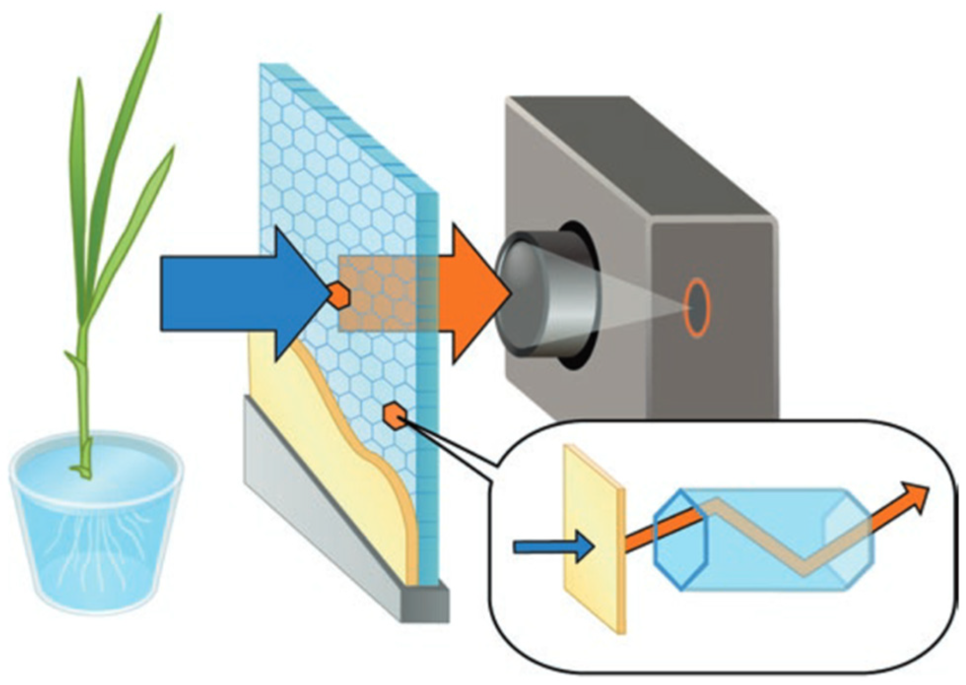
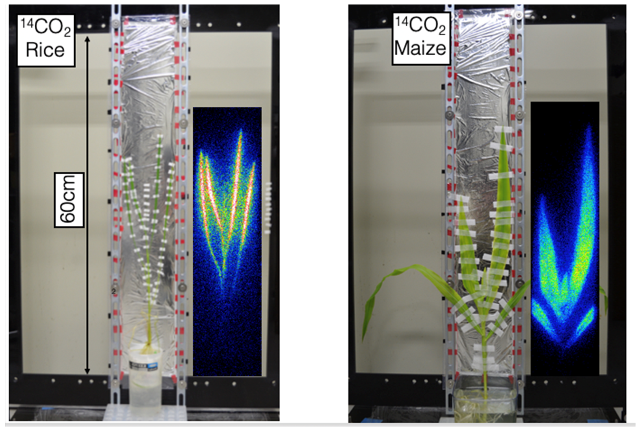
The visualization mechanism in an RRIS system (shown in Figure 1) can be summarized briefly in the following steps. (1) Radiation (beta rays and soft X-rays) is emitted from the absorbed radionuclides in leaves and roots. (2) This radiation is converted to visible light using scintillators. (3) The visible light is captured by a charge-coupled device (CCD) camera. When the fiber optic plate with a CsI scintillator (FOS) is implemented in the system, the image size is 10 cm × 20 cm with a resolution of several millimeters. Recently, we employed plastic scintillators to visualize larger plants (Figure 2). For example, we obtained an image size of 50 cm × 60 cm for 14C and 32P kinetics in plants [10,16].
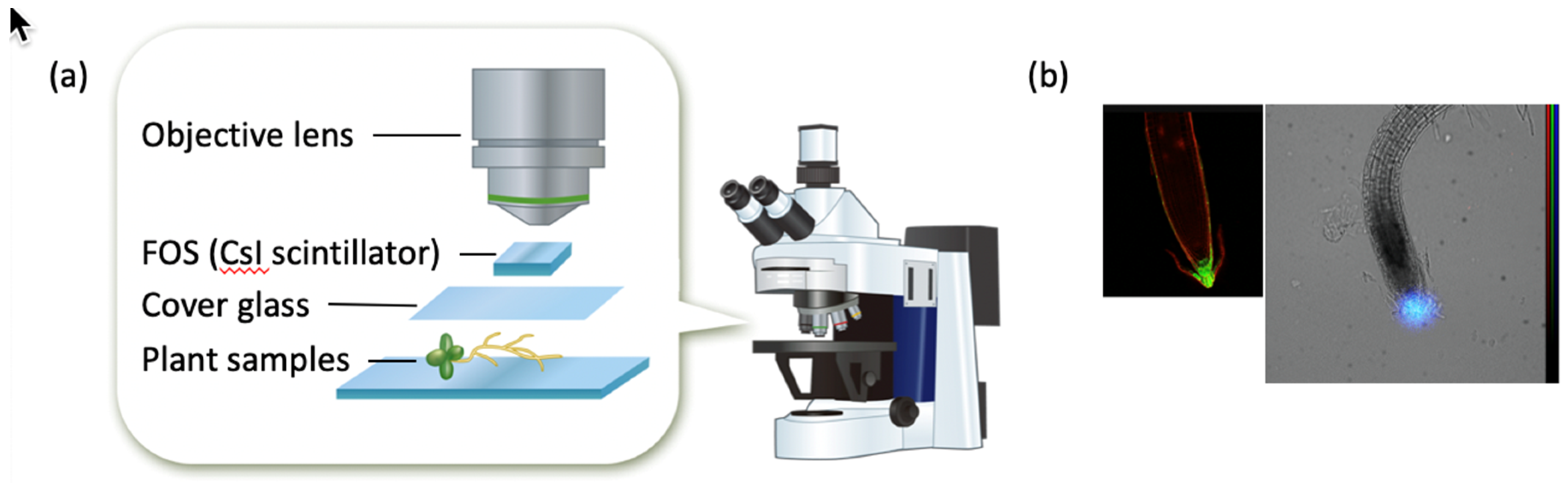
Furthermore, an RRIS was integrated with a fluorescent microscopy system [7,18,21] to observe the minute details. A resolution of 1 mm–100 μm was obtained with this set-up. The main advantage of this microscopy system is that it can acquire images of radioisotopes as well as the fluorescence from the sample (Figure 3).
2.2. Positron Imaging
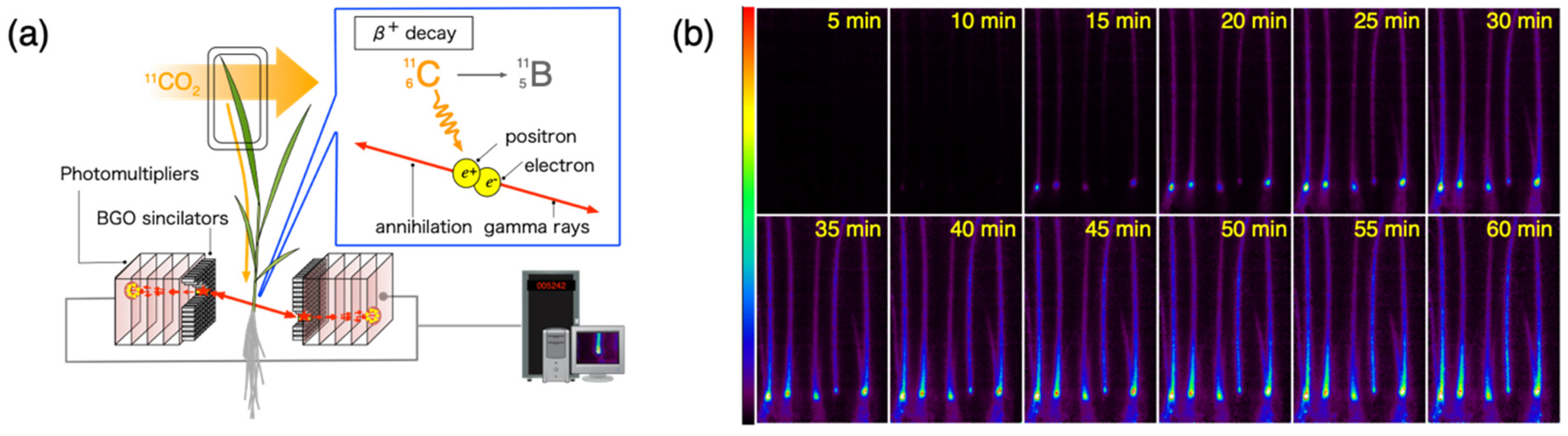
The plant PET developed at QST, which is called PETIS, utilizes the physical characteristics of positron-emitting radionuclides (which are simultaneously released by the annihilation of two 511-keV gamma rays in opposite (~180º degree) directions) to estimate the location of the chemical element of interest [24,25]. The positron imaging technology has a very high degree of sensitivity compared to other radionuclide-based imaging techniques. Therefore, imaging of several mega-becquerel of RIs distributed in the field of view is possible in a few seconds. In addition, the image of fast element kinetics, such as that of phloem flow shifting by several centimeters, can be acquired using 11C labeled carbon dioxide in 1 min. The migration of 11C-labeled sucrose produced by photosynthesis and the carbon dynamics of photosynthesis can also be traced. This can help to enhance the understanding of the photosynthesis process and the productivity of crops. Experimental systems using 11C and a PETIS are expected to produce useful results in the future (Figure 4).
The spatial resolution obtained in the positron imaging method is generally of the order of a few millimeters. This is mostly limited by the size of the scintillator that can stop high-energy gamma rays of 511 keV. In particular, the range of the positron (the distance from the release point of the positron to the annihilation point) depends on the peripheral density of the RI. When very thin herbaceous plants are the subject of the experiment, the positrons might escape outside the body. Therefore, this measurement requires extreme caution, especially while setting the plant and the experimental parameters. Unlike the molecular biology methods aimed at identifying the transport proteins of the element of interest, the migration of elements from outside to inside or the accumulation between organs or tissues is set as the imaging target in the plant RI imaging method; therefore, an effective imaging experiment can be designed even at this spatial resolution. The wide dynamic range and linear responsiveness between the image intensity and radioactivity are important for the performance of the imaging system.
In an imaging experiment with a PETIS, we utilized beta-plus RIs such as 22Na, 52Mn, 52Fe, and 107Cd, as opposed to an RI-labeled drug for PET synthesized with 11C, 13N, 15O, and 18F (having a positron emission rate of almost 100%), to elucidate the kinetics of essential elements and the elements related to environmental pollutants in plants. Cadmium is the most severe environmental pollutant in Japan. In order to control its absorption/migration by plants, we carried out investigations to image 107Cd in rice [26,27,28], oilseed rape plant [29], fern [30], and sedum [31]. 107Cd has a positron emission rate as low as 0.2%. However, as the migration of cadmium is essentially very slow (about several centimeters per hour), there is enough time to trace its kinetics in the plant body. Thus, the sensitivity of the imaging device is not of great importance as far as performance is concerned. The success of an imaging experiment to trace the slow-moving elements from the synthesis of the 107Cd tracer is primarily decided by the ability of the technology to maintain the measurement environment over several days. Examples of experimental techniques are setting a view in anticipation of the growth of the plant, devising means to keep the water surface of hydroponics containing radioactive tracer constant, and devising an efficient method to transport tracers within the plant body.
Most positron-emitting nuclides need to be manufactured using a cyclotron and purified by the researchers themselves. However, the commercially available 22Na (half-timelife, t1/2 = 2.6 years) and 65Zn (t1/2 = 244 days) emit sufficient positrons for imaging. Therefore, positron imaging can be implemented to study the dynamics of sodium or zinc in a plant body without a cyclotron. Fujimaki et al. visualized the dynamics of sodium in a reed, a salt-tolerant grass plant, using 22Na and a PETIS. Consequently, a contrasting image was obtained in which sodium did not stay and migrated to the upper leaf in the rice plant, i.e., sodium gathered at the stem base of the reed and hardly migrated to the stem and leaf. After removing 22Na from the hydroponic solution, the migration of 22Na was observed in other parts of the plant. In rice, sodium in the root continued to migrate to the upper leaves, whereas it moved towards the root tip in a reed. These results suggested that the salt tolerance mechanism of reed maintained a low concentration of sodium in the ground part by returning the sodium absorbed from the root [32]. Positron imaging of zinc has been performed using 62Zn (t1/2 = 9.2 hours) [33]. However, it was recently reported that positron imaging can be conducted with 65Zn (1.4%) as well, which has a low positron emission rate leading to long-term visualization of zinc dynamics [34]. Wongkaew et al. succeeded in directly visualizing the increased zinc transfer to the aboveground part of transgenic Arabidopsis with increased glutathione content using 65Zn and a PETIS [35].
2.3. Gamma Camera Imaging
In RI imaging experiments for plant science, all elements of interest are not necessarily beta-plus or beta-minus nuclides. In such cases, gamma camera imaging technology, where the direction of the arrival of gamma rays is estimated using a physical collimator, is a reliable method to use. As the energy of the gamma rays increases, collimation becomes difficult, leading to deterioration in the spatial resolution. Furthermore, as the collimator reduces the amount of radiation reaching the detector, the sensitivity of this technique is significantly inferior to that of positron imaging. Therefore, gamma cameras can be used in botanical research to visualize elemental dynamics in units of a few hours or days with long half-life RIs.
We have earlier mentioned our investigations aimed at the environmental pollutant cadmium. However, since the Fukushima nuclear accident, radioactive cesium has become the major pollutant of concern. Analysis of physiological functions, such as the absorption, transfer, and accumulation of radioactive cesium by food crops, and the utilization of plants with high levels of cesium for soil remediation have gained critical interest recently.
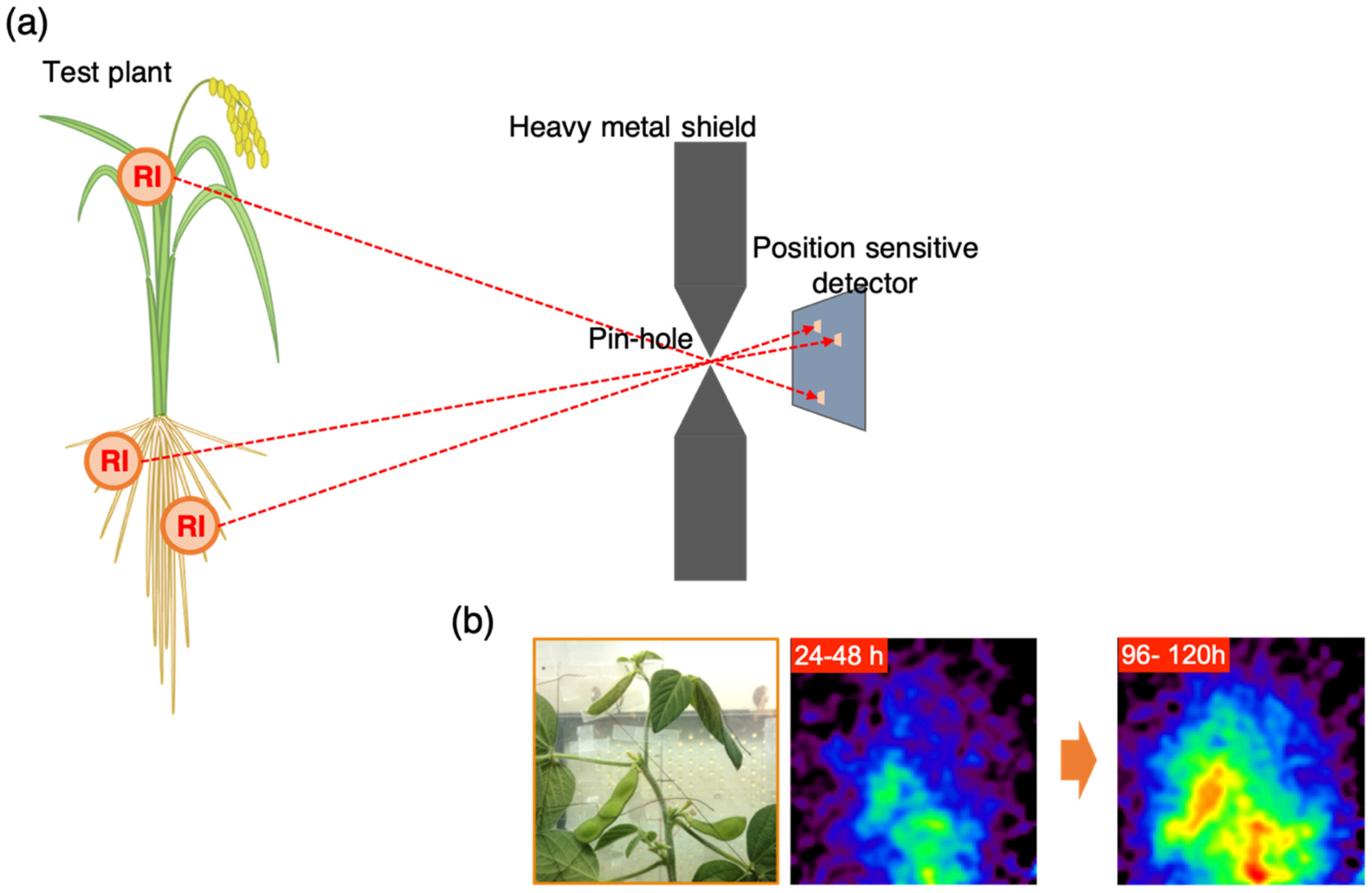
Therefore, in order to acquire images of 137Cs dynamics inside the plant body, we developed a pinhole-type gamma camera. This gamma camera uses the same mechanism as the so-called pinhole camera. Here, 622-keV gamma rays emitted from 137Cs are narrowed by a pinhole, constructing an image of radioactive cesium (Figure 5). Various types of gamma cameras that can monitor radioactive cesium in the environment have been developed; however, plant RI imaging technology requires the development of devices that are focused on the quantification of the radioactivity. In our scheme, the spatial resolution of the pinhole-type gamma camera for 137Cs was nearly several centimeters. Imaging experiments targeting soybeans revealed distribution patterns such as a slow transition rate, as opposed to the vessel flow and accumulation, at specific sites during the growth period [36].
When a target element of RI emits low-energy gamma rays and characteristic X-rays, pinhole-type gamma cameras can obtain excellent spatial resolution (in the orders of submillimeters or greater) because radiation penetration in the physical collimator is minimized. This is not realistic when the imaging target is a small animal or a human due to the huge loss in the amount of radiation signal reaching the detector from the body. However, if the target is a thin herbaceous plant, this imaging technique can be very useful. The development of such systems has already started in Japan [37,38], which could provide a detailed analysis of element kinetics.
2.4. Prospective Imaging Techniques
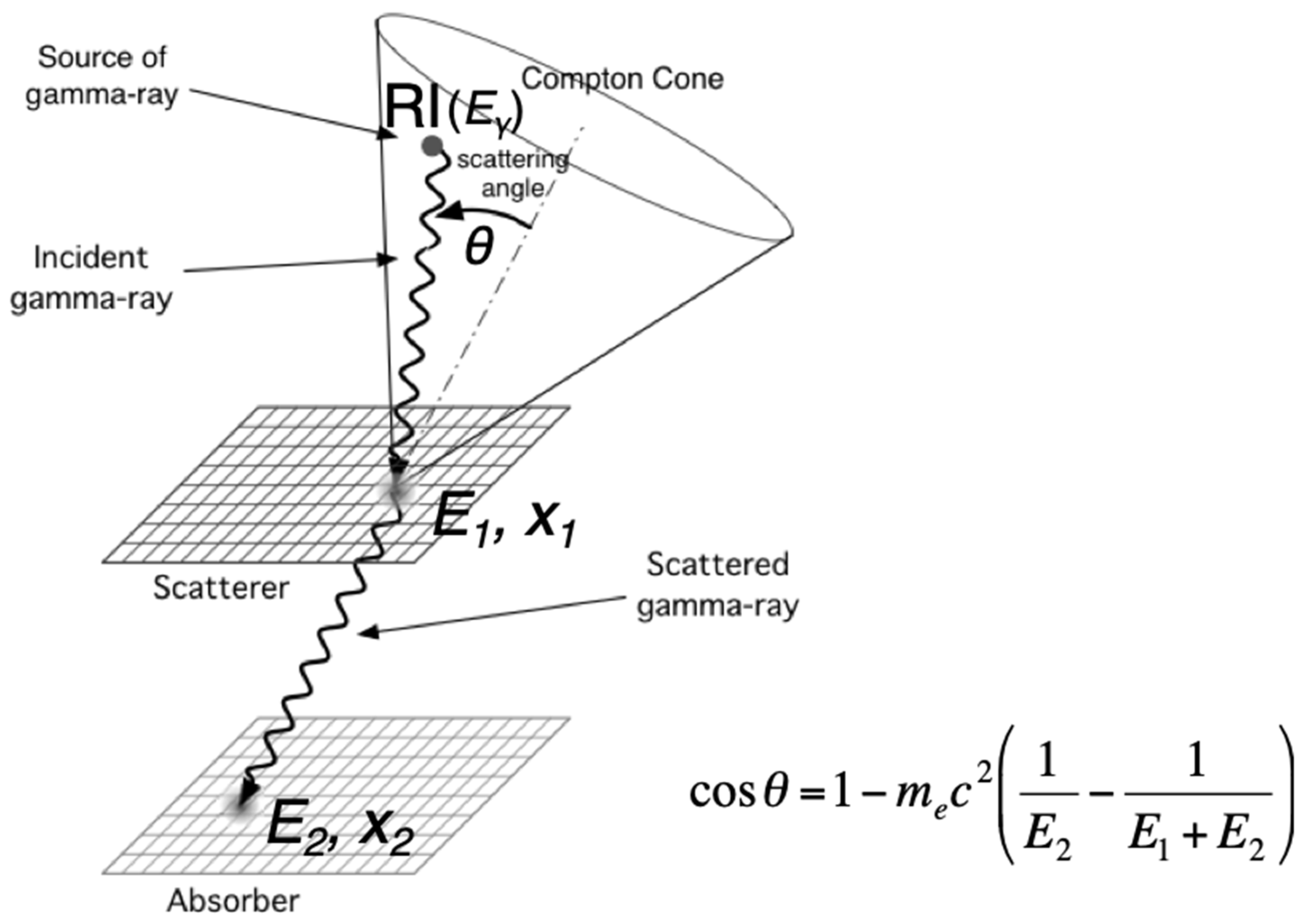
Recently, Compton cameras have garnered much attention as the next-generation non-invasive RI imaging apparatus as they overcome the limitations of the other technologies such as the limited usable nuclides in positron imaging and the low sensitivity of the gamma camera. This can allow simultaneous kinetic analysis of multiple elements in a test plant body. Several Compton cameras were developed as imaging devices for the environmental monitoring of 137Cs after the Fukushima disaster [39,40,41,42,43]. The Compton imaging method is based on the idea of precisely measuring Compton scattering in the detector and obtaining the distribution of radioactive isotopes by estimating the direction of arrival of gamma rays (Figure 6).
Therefore, the Compton camera is indispensable for the development of detector elements (with high spatial and energy resolution) and electronic circuits that can handle enormous amounts of measurement data. Compton imaging was discovered at the same time as PET. However, as the development of ASICs, electronic circuits, and image reconstruction algorithms was a major challenge, this camera became a reality in recent years only. It is believed that Compton camera imaging technology, as well as some types of conventional SPECT imaging methods, can facilitate the simultaneous imaging of multiple elements in vivo to study the interactions of elements and substances in the plant body. Moreover, because Compton interaction primarily occurs in the energy band ranging from 100 keV to 1–2 MeV, Compton cameras can detect gamma rays with a wide energy range from a hundred and several tens keV to several MeV. This allows plant researchers to select a variety of RI elements in the RI imaging experiments. The research group at the University of Tsukuba focuses on the normalization of the behavior of multiple elements within a plant. In some cases, the environmental pollutants, such as cadmium and cesium, are absorbed and transferred via mineral uptake pathways for essential elements. Therefore, the movements of pollutants normalized by those of essential elements can provide important information for comparing or understanding the behavior of pollutants under specific conditions. Recent studies indicated both similarity and specificity in the behavior of potassium and cesium in plants [44,45]. The difference in their behavior suggests the unique nature of plant transporters towards elemental affinity, even if the elements have a high chemical similarity. These approaches could be promising as RI imaging technologies, and the development of the corresponding methodology is necessary for plant science.
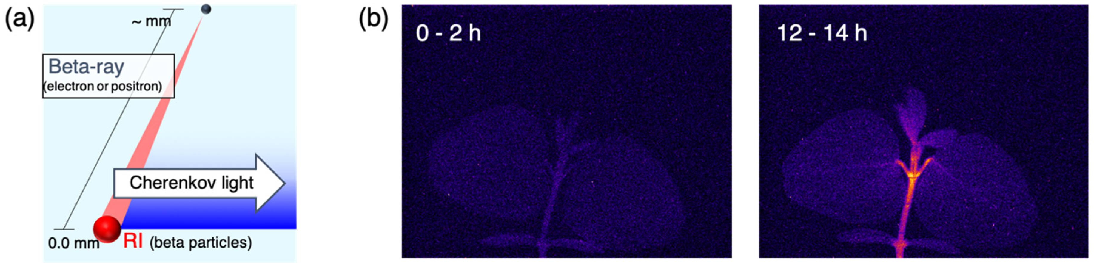
As mentioned earlier, a very high spatial resolution is unnecessary for plant RI imaging technology. The improvement of spatial resolution poses an inevitable challenge for researchers. However, for the detection of X-rays and gamma rays passing through a living body, dramatic improvements in spatial resolution cannot be expected given the range of photons in the detection element. Instead, a so-called “transformation of ideas” is necessary. In imaging devices used for detecting beta-plus or beta-minus rays, an error is generated in the quantitative value due to the tissue permeability of electrons. For example, beta-minus rays originating from deeper tissue layers are weak, because they are attenuated in tissue. On the other hand, beta-plus rays originating from shallower tissue underneath the surface are penetrating to the air and have little chance to emit annihilation gamma rays, so that errors of positron imaging increase. Therefore, an imaging technique was developed recently that focused on weak Cherenkov light (Figure 7), which is generated when beta rays pass through tissue [46,47]. Cherenkov light is emitted closer to the RIs than beta rays because of the energy threshold of the light emission. Further, the light beam is easy to concentrate and can be enlarged/reduced using optical technology. Therefore, this technique can be considered as a promising imaging technology to realize resolution of the order of several hundred microns.
3. Conclusions
RI imaging technology that targets plant science research has a wide scope of applications. This technology may be used for the advancement of applied research areas. For example, radioisotope imaging for plant science can be used for the imaging of elements, water, and gas. It is useful not only for nuclear medicine but also for material science. However, the efforts of interdisciplinary research teams are indispensable for this technological development, and we hope that more researchers will be interested in contributing to this field. We have discussed several imaging technologies in this article, which aside from RRIS and positron imaging, are at a nascent stage. We believe that the advancement of experimental techniques employed in RI imaging will further promote developmental research in the field of plant science as well as other research areas that thrive on imaging technology.
Funding
No funds related to this article.
Conflicts of Interest
The authors declare no conflict of interest.
References
- Rennie, E.A.; Turgeon, R. A comprehensive picture of phloem loading strategies. Proc. Natl. Acad. Sci. USA 2009, 106, 14162–14167. [Google Scholar] [CrossRef] [PubMed] [Green Version]
- Hubeau, M.; Mincke, J.; Vanhove, C.; Gorel, A.P.; Fayolle, A.; Epila, J.; Leroux, O.; Vandenberghe, S.; Steppe, K. 11C-Autoradiographs to Image Phloem Loading. Front. For. Glob. Chang. 2019, 2. [Google Scholar] [CrossRef]
- Hubeau, M.; Steppe, K. Plant-PET Scans: In Vivo Mapping of Xylem and Phloem Functioning. Trends Plant. Sci. 2015, 20, 676–685. [Google Scholar] [CrossRef] [PubMed]
- Schaart, D.R.; van Dam, H.T.; Seifert, S.; Vinke, R.; Dendooven, P.; Löhner, H.; Beekman, F.J. A novel, SiPM-array-based, monolithic scintillator detector for PET. Phys. Med. Biol. 2009, 54, 3501–3512. [Google Scholar] [CrossRef] [PubMed]
- Kanno, S.; Rai, H.; Ohya, T.; Hayashi, Y.; Tanoi, K.; Nakanishi, T.M. Real-time imaging of radioisotope labeled compounds in a living plant. J. Radioanal. Nucl. Chem. 2007, 272, 565–570. [Google Scholar] [CrossRef]
- Nakanishi, T.M.; Yamawaki, M.; Kannno, S.; Nihei, N.; Masuda, S.; Tanoi, K. Real-time imaging of ion uptake from root to above-ground part of the plant using conventional beta-ray emitters. J. Radioanal. Nucl. Chem. 2009, 282, 265. [Google Scholar] [CrossRef]
- Kanno, S.; Arrighi, J.-F.; Chiarenza, S.; Bayle, V.; Berthomé, R.; Péret, B.; Javot, H.; Delannoy, E.; Marin, E.; Nakanishi, T.M.; et al. A novel role for the root cap in phosphate uptake and homeostasis. eLife 2016, 5, e14577. [Google Scholar] [CrossRef] [PubMed]
- Sugita, R.; Kobayashi, N.I.; Hirose, A.; Tanoi, K.; Nakanishi, T.M. Evaluation ofin vivodetection properties of 22Na, 65Zn, 86Rb, 109Cd and 137Cs in plant tissues using real-time radioisotope imaging system. Phys. Med. Biol. 2014, 59, 837–851. [Google Scholar] [CrossRef] [PubMed]
- Sugita, R.; Kobayashi, N.I.; Hirose, A.; Ohmae, Y.; Tanoi, K.; Nakanishi, T.M. Nondestructive real-time radioisotope imaging system for visualizing 14C-labeled chemicals supplied as CO2 in plants using Arabidopsis thaliana. J. Radioanal. Nucl. Chem. 2013, 298, 1411–1416. [Google Scholar] [CrossRef]
- Sugita, R.; Sugahara, K.; Kobayashi, N.I.; Hirose, A.; Nakanishi, T.M.; Furuta, E.; Sensui, M.; Tanoi, K. Evaluation of plastic scintillators for live imaging of 14C-labeled photosynthate movement in plants. J. Radioanal. Nucl. Chem. 2018, 318, 579–584. [Google Scholar] [CrossRef]
- Sugita, R.; Kobayashi, N.I.; Hirose, A.; Saito, T.; Iwata, R.; Tanoi, K.; Nakanishi, T.M. Visualization of Uptake of Mineral Elements and the Dynamics of Photosynthates in Arabidopsis by a Newly Developed Real-Time Radioisotope Imaging System (RRIS). Plant. Cell Physiol. 2016, 57, 743–753. [Google Scholar] [CrossRef] [PubMed]
- Sugita, R.; Kobayashi, N.I.; Saito, T.; Hirose, A.; Iwata, R.; Tanoi, K.; Nakanishi, T.M. Quantitative Analysis of 28Mg in Arabidopsis using Real-time Radioisotope Imaging System(RRIS). Radioisotopes 2014, 63, 227–237. [Google Scholar] [CrossRef]
- Sugita, R.; Kobayashi, N.I.; Hirose, A.; Iwata, R.; Suzuki, H.; Tanoi, K.; Nakanishi, T.M. Visualization of how light changes affect ion movement in rice plants using a real-time radioisotope imaging system. J. Radioanal. Nucl. Chem. 2017, 312, 717–723. [Google Scholar] [CrossRef]
- Yamawaki, M.; Kanno, S.; Ishibashi, H.; Noda, A.; Hirose, A.; Tanoi, K.; Nakanishi, T.M. The development of real-time RI imaging system for plant under light environment. J. Radioanal. Nucl. Chem. 2009, 282, 275. [Google Scholar] [CrossRef]
- Nussaume, L.; Kanno, S.; Javot, H.; Marin, E.; Nakanishi, T.M.; Thibaud, M.-C. Phosphate Import in Plants: Focus on the PHT1 Transporters. Front. Plant. Sci. 2011, 2. [Google Scholar] [CrossRef] [PubMed]
- Sugahara, K.; Sugita, R.; Kobayashi, N.I.; Hirose, A.; Nakanishi, T.M.; Furuta, E.; Sensui, M.; Tanoi, K. Plastic Scintillators Enable Live Imaging of 32P-labelled Phosphorus Movement in Large Plants. Radioisotopes 2019, 68, 73–82. [Google Scholar] [CrossRef]
- Kanno, S.; Cuyas, L.; Javot, H.; Bligny, R.; Gout, E.; Dartevelle, T.; Hanchi, M.; Nakanishi, T.M.; Thibaud, M.-C.; Nussaume, L. Performance and Limitations of Phosphate Quantification: Guidelines for Plant Biologists. Plant. Cell Physiol. 2016, 57, 690–706. [Google Scholar] [CrossRef] [PubMed] [Green Version]
- Kanno, S.; Yamawaki, M.; Ishibashi, H.; Kobayashi, N.I.; Hirose, A.; Tanoi, K.; Nussaume, L.; Nakanishi, T.M. Development of real-time radioisotope imaging systems for plant nutrient uptake studies. Philos. Trans. R. Soc. B Biol. Sci. 2012, 367, 1501–1508. [Google Scholar] [CrossRef] [PubMed]
- Aramaki, T.; Sugita, R.; Hirose, A.; Kobayashi, N.I.; Tanoi, K.; Nakanishi, T.M. Application of 42K to Arabidopsis Tissues Using Real-Time Radioisotope Imaging System(RRIS). Radioisotopes 2015, 64, 169–176. [Google Scholar] [CrossRef]
- Hirose, A.; Yamawaki, M.; Kanno, S.; Igarashi, S.; Sugita, R.; Ohmae, Y.; Tanoi, K.; Nakanishi, T.M. Development of a 14C detectable real-time radioisotope imaging system for plants under intermittent light environment. J. Radioanal. Nucl. Chem. 2013, 296, 417–422. [Google Scholar] [CrossRef]
- Kobayashi, N.I.; Tanoi, K.; Kanno, S.; Nakanishi, T.M. Analysis of the Iron Movement in the Root Tip Part Using Real-time Imaging System. Radioisotopes 2012, 61, 121–128. [Google Scholar] [CrossRef] [Green Version]
- Yamawaki, M.; Hirose, A.; Kanno, S.; Ishibashi, H.; Noda, A.; Tanoi, K.; Nakanishi, T.M. Evaluation of 109Cd Detection Performance of a Real-Time RI Imaging System for Plant Research. Radioisotopes 2010, 59, 155–162. [Google Scholar] [CrossRef]
- Sugita, R.; Kobayashi, N.I.; Hirose, A.; Tanoi, K.; Nakanishi, T.M. Visualization of Ion Transport in Plants. In Agricultural Implications of the Fukushima Nuclear Accident (III): After 7 Years; Nakanishi, T.M., O’Brien, M., Tanoi, K., Eds.; Springer: Singapore, 2019; pp. 221–231. [Google Scholar] [Green Version]
- McKay, R.M.L.; Palmer, G.R.; Ma, X.P.; Layzell, D.B.; McKee, B.T.A. The use of positron emission tomography for studies of long-distance transport in plants: uptake and transport of 18F. Plant. Cell Environ. 1988, 11, 851–861. [Google Scholar] [CrossRef]
- Kume, T.; Matsuhashi, S.; Shimazu, M.; Ito, H.; Fujimura, T.; Adachi, K.; Uchida, H.; Shigeta, N.; Matsuoka, H.; Osa, A.; et al. Uptake and transport of positron-emitting tracer (18F) in plants. Appl. Radiat. Isot. 1997, 48, 1035–1043. [Google Scholar] [CrossRef]
- Fujimaki, S.; Suzui, N.; Ishioka, N.S.; Kawachi, N.; Ito, S.; Chino, M.; Nakamura, S. Tracing cadmium from culture to spikelet: Non-invasive imaging and quantitative characterization of absorption, transport and accumulation of cadmium in an intact rice plant. Plant. Physiol. 2010, 152, 1796–1806. [Google Scholar] [CrossRef]
- Ishikawa, S.; Suzui, N.; Ito-Tanabata, S.; Ishii, S.; Igura, M.; Abe, T.; Kuramata, M.; Kawachi, N.; Fujimaki, S. Real-time imaging and analysis of differences in cadmium dynamics in rice cultivars (Oryza sativa) using positron-emitting107Cd tracer. BMC Plant. Biol. 2011, 11, 172. [Google Scholar] [CrossRef] [PubMed]
- Fontanili, L.; Lancilli, C.; Suzui, N.; Dendena, B.; Yin, Y.-G.; Ferri, A.; Ishii, S.; Kawachi, N.; Lucchini, G.; Fujimaki, S.; et al. Kinetic Analysis of Zinc/Cadmium Reciprocal Competitions Suggests a Possible Zn-Insensitive Pathway for Root-to-Shoot Cadmium Translocation in Rice. Rice 2016, 9, 1–13. [Google Scholar] [CrossRef] [Green Version]
- Nakamura, S.-I.; Suzui, N.; Nagasaka, T.; Komatsu, F.; Ishioka, N.S.; Ito-Tanabata, S.; Kawachi, N.; Rai, H.; Hattori, H.; Chino, M.; et al. Application of glutathione to roots selectively inhibits cadmium transport from roots to shoots in oilseed rape. J. Exp. Bot. 2013, 64, 1073–1081. [Google Scholar] [CrossRef]
- Yoshihara, T.; Suzui, N.; Ishii, S.; Kitazaki, M.; Yamazaki, H.; Kitazaki, K.; Kawachi, N.; Yin, Y.-G.; Ito-Tanabata, S.; Hashida, S.-N.; et al. A kinetic analysis of cadmium accumulation in a Cd Hyper-accumulator Fern, Athyrium Yokoscense and tobacco plants. Plant. Cell Environ. 2014, 37, 1086–1096. [Google Scholar] [CrossRef]
- Hu, P.; Yin, Y.-G.; Ishikawa, S.; Suzui, N.; Kawachi, N.; Fujimaki, S.; Igura, M.; Yuan, C.; Huang, J.; Li, Z.; et al. Nitrate facilitates cadmium uptake, transport and accumulation in the hyperaccumulator Sedum plumbizincicola. Environ. Sci. Pollut. Res. 2013, 20, 6306–6316. [Google Scholar] [CrossRef]
- Fujimaki, S.; Maruyama, T.; Suzui, N.; Kawachi, N.; Miwa, E.; Higuchi, K. Base to Tip and Long-Distance Transport of Sodium in the Root of Common Reed [Phragmites australis (Cav.) Trin. ex Steud.] at Steady State Under Constant High-Salt Conditions. Plant. Cell Physiol. 2015, 56, 943–950. [Google Scholar] [CrossRef] [PubMed] [Green Version]
- Suzuki, M.; Takahashi, M.; Tsukamoto, T.; Watanabe, S.; Matsuhashi, S.; Yazaki, J.; Kishimoto, N.; Kikuchi, S.; Nakanishi, H.; Mori, S. Biosynthesis and secretion of mugineic acid family phytosiderophores in zinc-deficient barley. Plant. J. 2006, 48, 85–97. [Google Scholar] [CrossRef] [PubMed]
- Suzui, N.; Yin, Y.-G.; Ishii, S.; Sekimoto, H.; Kawachi, N. Visualization of zinc dynamics in intact plants using positron imaging of commercially available 65Zn. Plant. Methods 2017, 13, 40. [Google Scholar] [CrossRef] [PubMed] [Green Version]
- Wongkaew, A.; Nakamura, S.-I.; Suzui, N.; Yin, Y.-G.; Ishii, S.; Kawachi, N.; Kojima, K.; Sekimoto, H.; Yokoyama, T.; Ohkama-Ohtsu, N. Elevated glutathione synthesis in leaves contributes to zinc transport from roots to shoots in Arabidopsis. Plant. Sci. 2019, 283, 416–423. [Google Scholar] [CrossRef] [PubMed]
- Kawachi, N.; Yin, Y.-G.; Suzui, N.; Ishii, S.; Yoshihara, T.; Watabe, H.; Yamamoto, S.; Fujimaki, S. Imaging of radiocesium uptake dynamics in a plant body by using a newly developed high-resolution gamma camera. J. Environ. Radioact. 2016, 151, 461–467. [Google Scholar] [CrossRef] [PubMed] [Green Version]
- Yamamoto, S.; Kamada, K.; Yoshikawa, A. Use of YAP(Ce) in the development of high spatial resolution radiation imaging detectors. Radiat. Meas. 2018, 119, 184–191. [Google Scholar] [CrossRef]
- Ando, K.; Yamaguchi, M.; Yamamoto, S.; Toshito, T.; Kawachi, N. Development of a low-energy X-ray camera for the imaging of secondary electron bremsstrahlung x-ray emitted during proton irradiation for range estimation. Phys. Med. Biol. 2017, 62, 5006–5020. [Google Scholar] [CrossRef] [PubMed]
- Kabuki, S.; Hattori, K.; Kohara, R.; Kunieda, E.; Kubo, A.; Kubo, H.; Miuchi, K.; Nakahara, T.; Nagayoshi, T.; Nishimura, H.; et al. Development of Electron Tracking Compton Camera using micro pixel gas chamber for medical imaging. Nucl. Instrum. Methods Phys. Res. Sect. A: Accel. Spectrom. Assoc. Equip. 2007, 580, 1031–1035. [Google Scholar] [CrossRef] [Green Version]
- Takeda, S.; Aono, H.; Okuyama, S.; Ishikawa, S.; Odaka, H.; Watanabe, S.; Kokubun, M.; Takahashi, T.; Nakazawa, K.; Tajima, H.; et al. Experimental Results of the Gamma-Ray Imaging Capability With a Si/CdTe Semiconductor Compton Camera. IEEE Trans. Nucl. Sci. 2009, 56, 783–790. [Google Scholar] [CrossRef]
- Alnaaimi, M.A.; Royle, G.J.; Ghoggali, W.; Banoqitah, E.; Cullum, I.; Speller, R.D. Performance evaluation of a pixellated Ge Compton camera. Phys. Med. Biol. 2011, 56, 3473–3486. [Google Scholar] [CrossRef]
- Motomura, S.; Kanayama, Y.; Hiromura, M.; Fukuchi, T.; Ida, T.; Haba, H.; Watanabe, Y.; Enomoto, S. Improved imaging performance of a semiconductor Compton camera GREI makes for a new methodology to integrate bio-metal analysis and molecular imaging technology in living organisms. J. Anal. At. Spectrom. 2013, 28, 934–939. [Google Scholar] [CrossRef]
- Kishimoto, A.; Kataoka, J.; Taya, T.; Tagawa, L.; Mochizuki, S.; Ohsuka, S.; Nagao, Y.; Kurita, K.; Yamaguchi, M.; Kawachi, N.; et al. First demonstration of multi-color 3-D in vivo imaging using ultra-compact Compton camera. Sci. Rep. 2017, 7, 2110. [Google Scholar] [CrossRef] [PubMed]
- Nieves-Cordones, M.; Mohamed, S.; Tanoi, K.; Kobayashi, N.I.; Takagi, K.; Vernet, A.; Guiderdoni, E.; Périn, C.; Sentenac, H.; Véry, A.-A. Production of low-Cs+ rice plants by inactivation of the K+ transporter OsHAK1 with the CRISPR-Cas system. Plant. J. 2017, 92, 43–56. [Google Scholar] [CrossRef] [PubMed]
- Rai, H.; Yokoyama, S.; Satoh-Nagasawa, N.; Furukawa, J.; Nomi, T.; Ito, Y.; Fujimura, S.; Takahashi, H.; Suzuki, R.; Yousra, E.; et al. Cesium Uptake by Rice Roots Largely Depends Upon a Single Gene, HAK1, Which Encodes a Potassium Transporter. Plant. Cell Physiol. 2017, 58, 1486–1493. [Google Scholar] [CrossRef] [PubMed] [Green Version]
- Yamamoto, S.; Ogata, Y.; Kawachi, N.; Suzui, N.; Yin, Y.-G.; Fujimaki, S. Ultra-high resolution of radiocesium distribution detection based on Cherenkov light imaging. Nucl. Instrum. Methods Phys. Res. Sect. A: Accel. Spectrom. Assoc. Equip. 2015, 777, 102–109. [Google Scholar] [CrossRef]
- Kurita, K.; Suzui, N.; Yin, Y.-G.; Ishii, S.; Watabe, H.; Yamamoto, S.; Kawachi, N. Development of a Cherenkov light imaging system for studying the dynamics of radiocesium in plants. J. Nucl. Sci. Technol. 2017, 54, 662–667. [Google Scholar] [CrossRef]
Figure 1.
Schematic diagram of a real-time radioisotope imaging system (RRIS) including a fiber optic plate with a CsI scintillator (FOS). The radiation from the nuclides in a living plant is converted to visible light by the CsI scintillator. The visible light is captured by the charge-coupled device (CCD) camera. This schematic is reproduced from the study by Sugita et al. 2019 [23].

Figure 2.
Large sizes of the RRIS using plastic scintillators. The plants were set on the system after exposure to 14CO2. This picture is reproduced from the studies by Sugita et al. 2018 [10] and Sugahara et al. 2019 [16].

Figure 3.
(a) Schematic diagram of the micro-RRIS integrated with a fluorescent microscope. This figure is reproduced from the study by Sugita et al. 2016 [11]; (b) Images obtained from the system. Left: Fluorescent image of an Arabidopsis root. In the mutant plant, the phosphate transport system is active only at the root tip, which is indicated by the green color. Right: Corresponding phosphate (32P) image. These pictures are reproduced from the study by Kanno et al. 2016 [7].

Figure 4.
(a) Measurement principle of positron imaging method; (b) Representative example of a serial image of 11C-labeled photoassimilate kinetics in five rice plants.
Figure 4.
(a) Measurement principle of positron imaging method; (b) Representative example of a serial image of 11C-labeled photoassimilate kinetics in five rice plants.

Figure 5.
(a) Measurement principle of the pinhole-type gamma camera; (b) Representative example of the images obtained by this camera. This frame shows snapshots of a test plant and serial images of 137Cs dynamics after feeding the radiotracer into the hydroponic culture.
Figure 5.
(a) Measurement principle of the pinhole-type gamma camera; (b) Representative example of the images obtained by this camera. This frame shows snapshots of a test plant and serial images of 137Cs dynamics after feeding the radiotracer into the hydroponic culture.

Figure 6.
Schematic diagram for the principle of Compton imaging, which is used to detect the direction of incoming gamma photons from the radiation source. The kinematics between the scattering angle and the dropped energies in the scatterer are represented the equation, where θ, me, c, E1, and E2 indicate the scattering angle, electron mass, light velocity, imparted energy to the front detector by the scattering of the incident photon, and remaining energy of the scattered photon, respectively. The deposited energies and positions in the two consecutive events are measured as (E1, x1) in the front detector of the scatterer and (E2, x2) in the rear detector of the absorber, where x1 and x2 denote the positions. The probable location of the radiation source is estimated as a “Compton cone”. Finally, the image of the distribution of the radiation source is generated [40].

Figure 7.
(a) Measurement principle of Cherenkov optical imaging; (b) Representative example of serial images of 137Cs dynamics after feeding the radiotracer into the hydroponic culture obtained through a CCD camera. As the target of measurement is light, it is possible to use an optical lens instead of directly measuring the radiation from the radioactive isotope. Moreover, CCD cameras are inexpensive compared to radiation imaging apparatus.

© 2019 by the authors. Licensee MDPI, Basel, Switzerland. This article is an open access article distributed under the terms and conditions of the Creative Commons Attribution (CC BY) license (http://creativecommons.org/licenses/by/4.0/).
Share and Cite
MDPI and ACS Style
Suzui, N.; Tanoi, K.; Furukawa, J.; Kawachi, N. Recent Advances in Radioisotope Imaging Technology for Plant Science Research in Japan. Quantum Beam Sci. 2019, 3, 18. https://0-doi-org.brum.beds.ac.uk/10.3390/qubs3030018
AMA Style
Suzui N, Tanoi K, Furukawa J, Kawachi N. Recent Advances in Radioisotope Imaging Technology for Plant Science Research in Japan. Quantum Beam Science. 2019; 3(3):18. https://0-doi-org.brum.beds.ac.uk/10.3390/qubs3030018
Chicago/Turabian StyleSuzui, Nobuo, Keitaro Tanoi, Jun Furukawa, and Naoki Kawachi. 2019. "Recent Advances in Radioisotope Imaging Technology for Plant Science Research in Japan" Quantum Beam Science 3, no. 3: 18. https://0-doi-org.brum.beds.ac.uk/10.3390/qubs3030018