
Experimental Setup for Irradiation of Cell Cultures at L2A2
 ,
,  , , , , , , , ,
  ,
,  and
and
Abstract
:1. Introduction
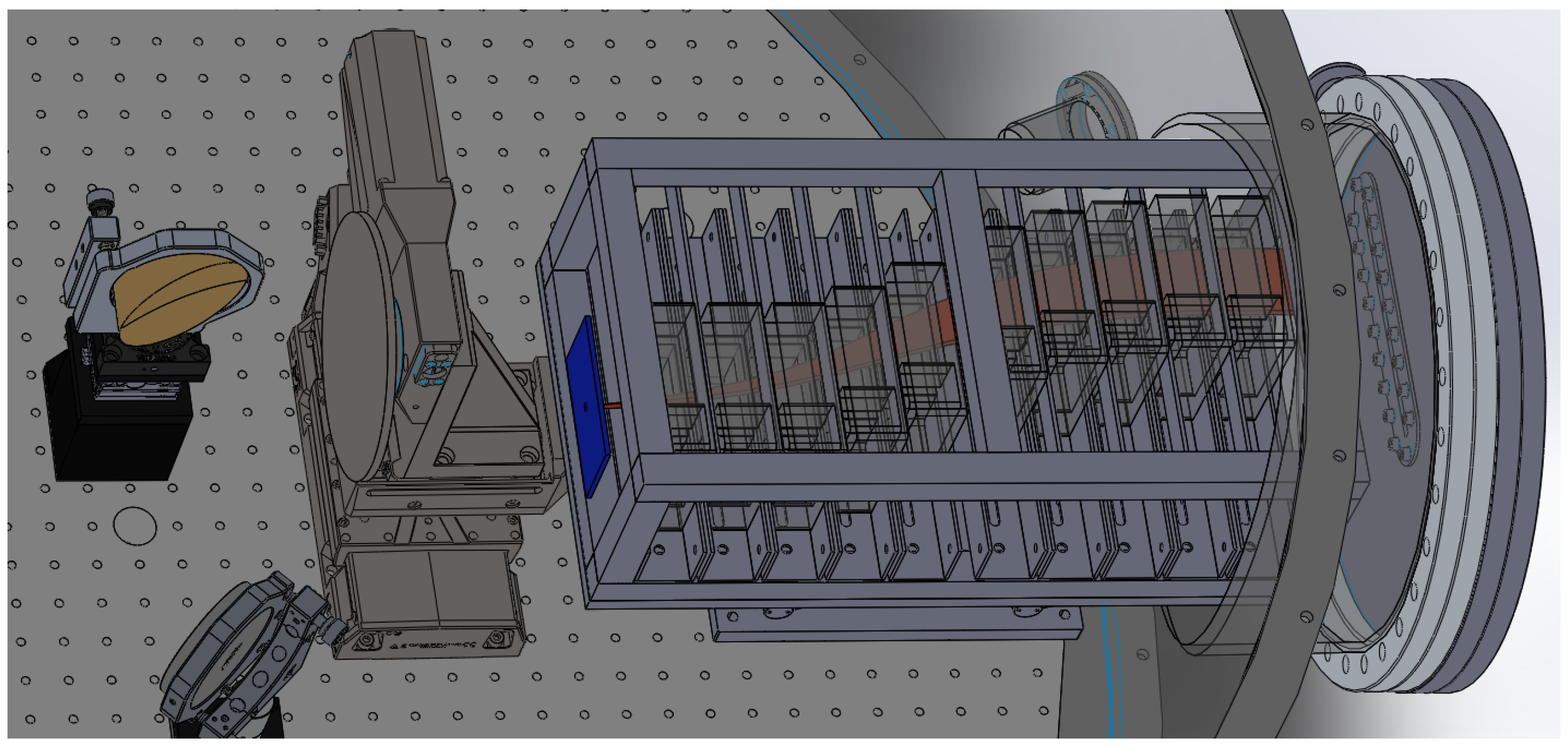
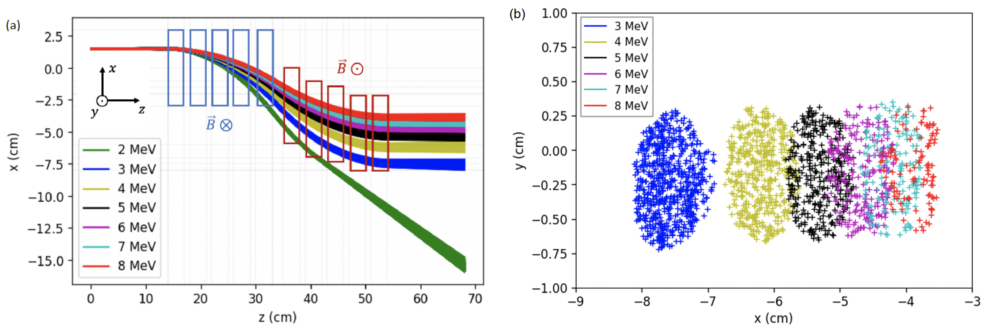
2. Energy Selector
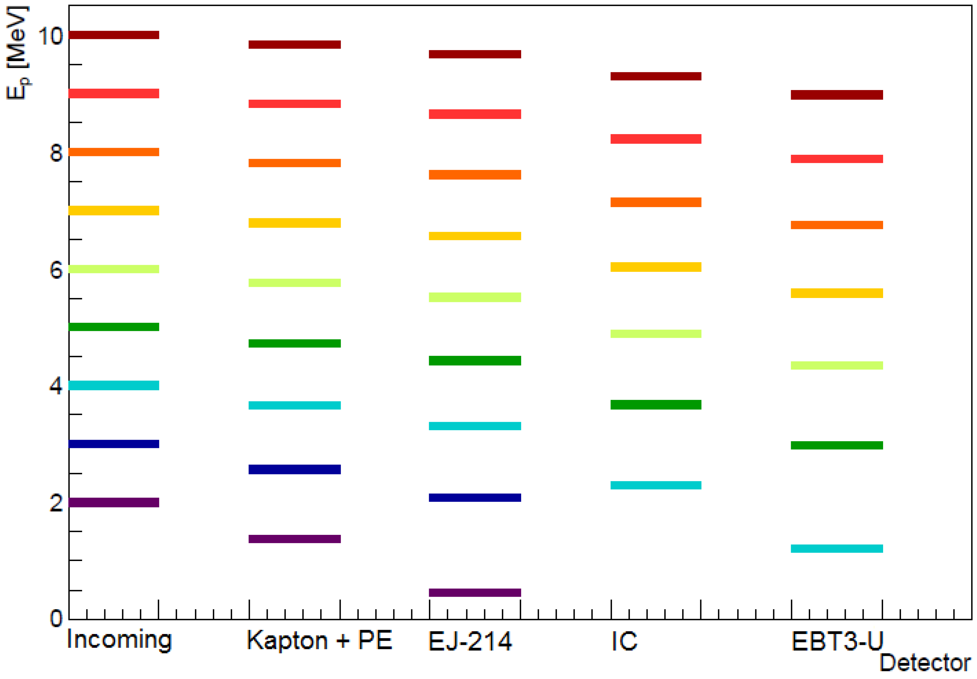
3. Particle Fluence Detectors
4. Cell Cultures
5. Conclusions and Outlook
Author Contributions
Funding
Data Availability Statement
Conflicts of Interest
References
- Friedrich, T.; Scholz, U.; Elsässer, T.; Durante, M.; Scholz, M. Systematic analysis of RBE and related quantities using a database of cell survival experiments with ion beam irradiation. J. Radiat. Res. 2012, 54, 494–514. [Google Scholar] [CrossRef] [PubMed] [Green Version]
- Friedrich, T.; Pfuhl, T.; Scholz, M. Update of the particle irradiation data ensemble (PIDE) for cell survival. J. Radiat. Res. 2021, 62, 645–655. [Google Scholar] [CrossRef] [PubMed]
- Henthorn, N.T.; Sokol, O.; Durante, M.; De Marzi, L.; Pouzoulet, F.; Miszczyk, J.; Olko, P.; Brandenburg, S.; van Goethem, M.J.; Barazzuol, L.; et al. Mapping the Future of Particle Radiobiology in Europe: The INSPIRE Project. Front. Phys. 2020, 8, 565055. [Google Scholar] [CrossRef]
- Patera, V.; Prezado, Y.; Azaiez, F.; Battistoni, G.; Bettoni, D.; Brandenburg, S.; Bugay, A.; Cuttone, G.; Dauvergne, D.; de France, G.; et al. Biomedical Research Programs at Present and Future High-Energy Particle Accelerators. Front. Phys. 2020, 8, 380. [Google Scholar] [CrossRef] [PubMed]
- Wéra, A.C.; Riquier, H.; Heuskin, A.C.; Michiels, C.; Lucas, S. In vitro irradiation station for broad beam radiobiological experiments. Nucl. Instrum. Methods Phys. Res. Sect. B Beam Interact. Mater. At. 2011, 269, 3120–3124. [Google Scholar] [CrossRef] [Green Version]
- Manti, L.; Campajola, L.; Perozziello, F.M.; Kavanagh, J.N.; Schettino, G. Development of a low-energy particle irradiation facility for the study of the biological effectiveness of the ion track end. J. Phys. Conf. Ser. 2012, 373, 012019. [Google Scholar] [CrossRef] [Green Version]
- Baratto-Roldán, A.; Jiménez-Ramos, M.d.C.; Jimeno, S.; Huertas, P.; García-López, J.; Gallardo, M.I.; Cortés-Giraldo, M.A.; Espino, J.M. Preparation of a radiobiology beam line at the 18 MeV proton cyclotron facility at CNA. Phys. Medica Eur. J. Med Phys. 2020, 74, 19–29. [Google Scholar] [CrossRef]
- Viñals, S.; Sánchez-Parcerisa, D.; Fraile, L.M.; España, S.; García, G.; García-Díaz, M.; SánchezTembleque, V.; Udías, J.M. Characterization of the proton pulsed beam at CMAM. EPJ Web Conf. 2021, 253, 04027. [Google Scholar] [CrossRef]
- Auer, S.; Hable, V.; Greubel, C.; Drexler, G.A.; Schmid, T.E.; Belka, C.; Dollinger, G.; Friedl, A.A. Survival of tumor cells after proton irradiation with ultra-high dose rates. Radiat. Oncol. 2011, 6, 139. [Google Scholar] [CrossRef] [Green Version]
- Hughes, J.R.; Parsons, J.L. FLASH Radiotherapy: Current Knowledge and Future Insights Using Proton-Beam Therapy. Int. J. Mol. Sci. 2020, 21, 6492. [Google Scholar] [CrossRef]
- Ledingham, K.; Bolton, P.; Shikazono, N.; Ma, C.M. Towards Laser Driven Hadron Cancer Radiotherapy: A Review of Progress. Appl. Sci. 2014, 4, 402–443. [Google Scholar] [CrossRef]
- Yogo, A.; Sato, K.; Nishikino, M.; Mori, M.; Teshima, T.; Numasaki, H.; Murakami, M.; Demizu, Y.; Akagi, S.; Nagayama, S.; et al. Application of laser-accelerated protons to the demonstration of DNA double-strand breaks in human cancer cells. Appl. Phys. Lett. 2009, 94, 181502. [Google Scholar] [CrossRef]
- Chaudhary, P.; Milluzzo, G.; Ahmed, H.; Odlozilik, B.; McMurray, A.; Prise, K.M.; Borghesi, M. Radiobiology Experiments With Ultra-high Dose Rate Laser-Driven Protons: Methodology and State-of-the-Art. Front. Phys. 2021, 9, 75. [Google Scholar] [CrossRef]
- Rösch, T.F.; Szabó, Z.; Haffa, D.; Bin, J.; Brunner, S.; Englbrecht, F.S.; Friedl, A.A.; Gao, Y.; Hartmann, J.; Hilz, P.; et al. A feasibility study of zebrafish embryo irradiation with laser-accelerated protons. Rev. Sci. Instrum. 2020, 91, 063303. [Google Scholar] [CrossRef] [PubMed]
- Brack, F.E.; Kroll, F.; Gaus, L.; Bernert, C.; Beyreuther, E.; Cowan, T.E.; Karsch, L.; Kraft, S.; Kunz-Schughart, L.A.; Lessmann, E.; et al. Spectral and spatial shaping of laser-driven proton beams using a pulsed high-field magnet beamline. Sci. Rep. 2020, 10, 9118. [Google Scholar] [CrossRef] [PubMed]
- Kraft, S.D.; Richter, C.; Zeil, K.; Baumann, M.; Beyreuther, E.; Bock, S.; Bussmann, M.; Cowan, T.E.; Dammene, Y.; Enghardt, W.; et al. Dose-dependent biological damage of tumour cells by laser-accelerated proton beams. New J. Phys. 2010, 12, 085003. [Google Scholar] [CrossRef]
- Bin, J.; Allinger, K.; Assmann, W.; Dollinger, G.; Drexler, G.A.; Friedl, A.A.; Habs, D.; Hilz, P.; Hoerlein, R.; Humble, N.; et al. A laser-driven nanosecond proton source for radiobiological studies. Appl. Phys. Lett. 2012, 101, 243701. [Google Scholar] [CrossRef]
- Zeil, K.; Baumann, M.; Beyreuther, E.; Burris-Mog, T.; Cowan, T.E.; Enghardt, W.; Karsch, L.; Kraft, S.D.; Laschinsky, L.; Metzkes, J.; et al. Dose-controlled irradiation of cancer cells with laser-accelerated proton pulses. Appl. Phys. B 2013, 110, 437–444. [Google Scholar] [CrossRef] [Green Version]
- Raschke, S.; Spickermann, S.; Toncian, T.; Swantusch, M.; Boeker, J.; Giesen, U.; Iliakis, G.; Willi, O.; Boege, F. Ultra-short laser-accelerated proton pulses have similar DNA-damaging effectiveness but produce less immediate nitroxidative stress than conventional proton beams. Sci. Rep. 2016, 6, 32441. [Google Scholar] [CrossRef] [Green Version]
- Pommarel, L.; Vauzour, B.; Mégnin-Chanet, F.; Bayart, E.; Delmas, O.; Goudjil, F.; Nauraye, C.; Letellier, V.; Pouzoulet, F.; Schillaci, F.; et al. Spectral and spatial shaping of a laser-produced ion beam for radiation-biology experiments. Phys. Rev. Accel. Beams 2017, 20, 032801. [Google Scholar] [CrossRef] [Green Version]
- Peñas, J.; Cortina-Gil, D.; Martín, L.; Ruiz, C.; Seimetz, M.; Benlliure, J. A Multi-Shot Wheel-Target Assembly for Laser-Plasma Proton Acceleration. High Power Laser Sci. Eng. 2021, submitted.
- Roth, M.; Schollmeier, M. Ion Acceleration-Target Normal Sheath Acceleration. CERN Yellow Rep. 2016, 1, 231. [Google Scholar]
- Fiorini, F.; Kirby, D.; Borghesi, M.; Doria, D.; Jeynes, J.C.G.; Kakolee, K.F.; Kar, S.; Litt, S.K.; Kirkby, K.J.; Merchant, M.J.; et al. Dosimetry and spectral analysis of a radiobiological experiment using laser-driven proton beams. Phys. Med. Biol. 2011, 56, 6969–6982. [Google Scholar] [CrossRef] [PubMed] [Green Version]
- Doria, D.; Kakolee, K.F.; Kar, S.; Litt, S.K.; Fiorini, F.; Ahmed, H.; Green, S.; Jeynes, J.C.G.; Kavanagh, J.; Kirby, D.; et al. Biological effectiveness on live cells of laser driven protons at dose rates exceeding 109 Gy/s. AIP Adv. 2012, 2, 011209. [Google Scholar] [CrossRef]
- Hanton, F.; Chaudhary, P.; Doria, D.; Gwynne, D.; Maiorino, C.; Scullion, C.; Ahmed, H.; Marshall, T.; Naughton, K.; Romagnani, L.; et al. DNA DSB Repair Dynamics following Irradiation with Laser-Driven Protons at Ultra-High Dose Rates. Sci. Rep. 2019, 9, 4471. [Google Scholar] [CrossRef]
- Yogo, A.; Maeda, T.; Hori, T.; Sakaki, H.; Ogura, K.; Nishiuchi, M.; Sagisaka, A.; Kiriyama, H.; Okada, H.; Kanazawa, S.; et al. Measurement of relative biological effectiveness of protons in human cancer cells using a laser-driven quasimonoenergetic proton beamline. Appl. Phys. Lett. 2011, 98, 053701. [Google Scholar] [CrossRef]
- Schillaci, F.; Pommarel, L.; Romano, F.; Cuttone, G.; Costa, M.; Giove, D.; Maggiore, M.; Russo, A.; Scuderi, V.; Malka, V.; et al. Characterization of the ELIMED Permanent Magnets Quadrupole system prototype with laser-driven proton beams. J. Instrum. 2016, 11, T07005. [Google Scholar] [CrossRef]
- Fritzler, S.; Malka, V.; Grillon, G.; Rousseau, J.P.; Burgy, F.; Lefebvre, E.; d’Humières, E.; McKenna, P.; Ledingham, K.W.D. Proton beams generated with high-intensity lasers: Applications to medical isotope production. Appl. Phys. Lett. 2003, 83, 3039–3041. [Google Scholar] [CrossRef]
- Manti, L.; Perozziello, F.; Borghesi, M.; Candiano, G.; Chaudhary, P.; Cirrone, G.; Doria, D.; Gwynne, D.; Leanza, R.; Prise, K.M.; et al. The radiobiology of laser-driven particle beams: Focus on sub-lethal responses of normal human cells. J. Instrum. 2017, 12, C03084. [Google Scholar] [CrossRef] [Green Version]
- Bayart, E.; Flacco, A.; Delmas, O.; Pommarel, L.; Levy, D.; Cavallone, M.; Megnin-Chanet, F.; Deutsch, E.; Malka, V. Fast dose fractionation using ultra-short laser accelerated proton pulses can increase cancer cell mortality, which relies on functional PARP1 protein. Sci. Rep. 2019, 9, 10132. [Google Scholar] [CrossRef] [Green Version]
- Berger, M.J.; Coursey, J.S.; Zucker, M.A.; Chang, J. Stopping-power and range tables for electrons, protons, and helium ions. In NIST Standard Reference Database 124; NIST: Gaithersburg, MD, USA, 1998. [Google Scholar] [CrossRef]
- Ziegler, J.F.; Ziegler, M.D.; Biersack, J.P. SRIM – The Stopping Range of Ions in Matter (2010). Nucl. Instrum. Meth. Phys. Res. Sect. B Beam Interact. Mater. At. 2010, 268, 1818–1823. [Google Scholar] [CrossRef] [Green Version]
- Seimetz, M.; Peñas, J.; Llerena, J.; Benlliure, J.; García López, J.; Millán-Callado, M.; Benlloch, J. PADC nuclear track detector for ion spectroscopy in laser-plasma acceleration. Phys. Medica Eur. J. Med. Phys. 2020, 76, 72–76. [Google Scholar] [CrossRef] [PubMed]
- Milluzzo, G.; Ahmed, H.; Romagnani, L.; Doria, D.; Chaudhary, P.; Maiorino, C.; McIlvenny, A.; McMurray, A.; Polin, K.; Katzir, Y.; et al. Dosimetry of laser-accelerated carbon ions for cell irradiation at ultra-high dose rate. J. Phys. Conf. Ser. 2020, 1596, 012038. [Google Scholar] [CrossRef]
- Seimetz, M.; Bellido, P.; Soriano, A.; García López, J.; Jiménez-Ramos, M.C.; Fernández, B.; Conde, P.; Crespo, E.; González, A.J.; Hernández, L.; et al. Calibration and Performance Tests of Detectors for Laser-Accelerated Protons. IEEE Trans. Nucl. Sci. 2015, 62, 3216–3224. [Google Scholar] [CrossRef]
- Richter, C.; Karsch, L.; Dammene, Y.; Kraft, S.D.; Metzkes, J.; Schramm, U.; Schürer, M.; Sobiella, M.; Weber, A.; Zeil, K.; et al. A dosimetric system for quantitative cell irradiation experiments with laser-accelerated protons. Phys. Med. Biol. 2011, 56, 1529–1543. [Google Scholar] [CrossRef]
- Di Martino, F.; Giannelli, M.; Traino, A.C.; Lazzeri, M. Ion recombination correction for very high dose-per-pulse high-energy electron beams. Med. Phys. 2005, 32, 2204–2210. [Google Scholar] [CrossRef]
- Suckert, T.; Nexhipi, S.; Dietrich, A.; Koch, R.; Kunz-Schughart, L.A.; Bahn, E.; Beyreuther, E. Models for Translational Proton Radiobiology-From Bench to Bedside and Back. Cancers 2021, 13, 4216. [Google Scholar] [CrossRef]


| (mm) | (cm) | E (MeV) | (mGy) |
|---|---|---|---|
| MeV: | |||
| 50 | 2.9–4.9 | ||
| 70 | 3.1–4.7 | ||
| 100 | 3.3–4.4 | ||
| MeV: | |||
| 50 | 3.7–6.4 | ||
| 70 | 3.8–6.1 | ||
| 100 | 4.0–5.8 | ||
| MeV: | |||
| 50 | 4.3–8.1 | ||
| 70 | 4.7–7.6 | ||
| 100 | 5.0–7.2 | ||
Publisher’s Note: MDPI stays neutral with regard to jurisdictional claims in published maps and institutional affiliations. |
© 2022 by the authors. Licensee MDPI, Basel, Switzerland. This article is an open access article distributed under the terms and conditions of the Creative Commons Attribution (CC BY) license (https://creativecommons.org/licenses/by/4.0/).
Share and Cite
Torralba, A.; Palenciano, L.; Reija, A.; Rigla, J.P.; Peñas, J.; Llerena, J.J.; Contreras-Martínez, R.; Benlliure, J.; Vega, A.; Aguado-Barrera, M.E.; et al. Experimental Setup for Irradiation of Cell Cultures at L2A2. Quantum Beam Sci. 2022, 6, 10. https://0-doi-org.brum.beds.ac.uk/10.3390/qubs6010010
Torralba A, Palenciano L, Reija A, Rigla JP, Peñas J, Llerena JJ, Contreras-Martínez R, Benlliure J, Vega A, Aguado-Barrera ME, et al. Experimental Setup for Irradiation of Cell Cultures at L2A2. Quantum Beam Science. 2022; 6(1):10. https://0-doi-org.brum.beds.ac.uk/10.3390/qubs6010010
Chicago/Turabian StyleTorralba, Alberto, Lidia Palenciano, Alicia Reija, Juan Pablo Rigla, Juan Peñas, Juan José Llerena, Ramiro Contreras-Martínez, José Benlliure, Ana Vega, Miguel Elías Aguado-Barrera, and et al. 2022. "Experimental Setup for Irradiation of Cell Cultures at L2A2" Quantum Beam Science 6, no. 1: 10. https://0-doi-org.brum.beds.ac.uk/10.3390/qubs6010010