Article

Design and Medical Effects of a Vaginal Cleaning Device Generating Plasma-Activated Water with Antimicrobial Activity on Bacterial Vaginosis

1
SJ Global, Bucheon-si, Gyeonggi-do 14559, Korea
2
5T International Patent Law Firm, Seoul 05836, Korea
3
Institute of Convergent Chemical Engineering and Technology, Sungkyunkwan University, Suwon-si, Gyeonggi-do 16419, Korea
4
Department of Media Communication, Hanyang University, Seoul 04736, Korea
5
Department of Obstetrics and Gynecology, Hanyang University College of Medicine, Seoul 04736, Korea
6
Department of Biomedical Engineering, Hanyang University, Seoul 04736, Korea
*
Author to whom correspondence should be addressed.
Submission received: 16 October 2020 / Revised: 10 November 2020 / Accepted: 16 November 2020 / Published: 17 November 2020

Abstract

:
Bacterial vaginosis is a common female disease caused by a vaginal infection due to an overgrowth of bacteria that naturally live in the vaginal tract. Bacterial vaginosis has frequently been treated with the oral or vaginal administration of antibiotics and topical disinfectants. However, hygienic application of topical treatment deep in the vagina remains difficult. Herein, we introduce a novel vaginal cleaning device using plasma-activated water generated from supplied water. Remarkably, plasma source generation at atmospheric pressure is well known to eradicate bacterial infection through the generation of free radicals and/or chlorine chemicals with antimicrobial activity. The device was designed to alleviate a bacterial infection by spraying plasma-activated water generated from a cleaning solution container with plasma modules. The spray nozzle contains both a clean outlet and a suction outlet to spray and recover the plasma water, respectively, and is connected to a disposable silicone tube. The other nozzle, which has a laser light and air pump, can perform a second sterilization and dry the vagina after washing. Free chlorine chemicals with antibacterial activity were detected in the plasma-activated water by the device. Clinical application in patients with bacterial vaginosis confirmed the stability and effectiveness of our device. Therefore, these results show a novel clinical application of atmospheric pressure plasma to medical field as a plasma medicine.

1. Introduction

Bacterial vaginosis is the most common form of vaginitis, which is an inflammation of the vagina [1,2]. Because women’s genitals are located close to the urethra and anus, they are easily invaded by pathogens, and the warm, humid, and acidic environment facilitates bacterial growth. Vaginitis is the most common gynecological disease encountered by obstetricians, and in the United States, approximately 10 million patients visit obstetrics and gynecology clinics every year [3]. The symptoms of vaginitis include vaginal secretions, vaginal odor, fever, malnourishment, sexual discomfort and urinary pain [1].
Generally, bacterial vaginosis is caused by unbalanced changes in the vaginal microbiome rather than infections by external bacteria [4]. Anaerobic bacteria, which usually account for less than 1% of the total bacterial population, excessively multiply, and mixed bacterial groups increase more than the total number of bacteria [5,6].
Treatment of bacterial vaginosis is based on the oral or vaginal administration of antibiotics, such as clindamycin and metronidazole [7]. However, some problems have been pointed out that frequent relapses occur after antibiotic treatment, and beneficial bacteria are killed. In addition, some topical antiseptics and disinfectants, such as betadine, have been used to treat bacterial vaginosis [8]. These agents are generally difficult to apply deep in the vaginal tract.
To overcome these problems, we provide a novel vaginal cleaning device using plasma-activated water and ultraviolet light. Recently, plasma source generation at atmospheric pressures has been of great interest for therapeutic applications, such as in coagulation, cancer treatment, and wound healing, in terms of plasma medicine [9,10,11]. Numerous studies have demonstrated plasma-treated sterilizing effects for bacterial infections and safe food preservation [12,13]. In addition, underwater plasma discharge is also known to exhibit antibacterial activity [12,13]. The mechanism underlying the sterilization effect of plasma-activated water is strongly believed to be plasma-induced production of active radicals and/or free chlorine molecules with antimicrobial activity [12,13,14,15]. Hence, plasma-activated water or media has great potential to disinfect for hygiene and treatment.
The current device was basically designed to mitigate infectious bacteria by spraying plasma-activated water, which was generated from supplied water, inside the vaginal tract. After washing, a second sterilization and drying were able to be performed using a nozzle with a laser light and air pump with which the vaginal cleaning device is equipped [16].

2. Materials and Methods

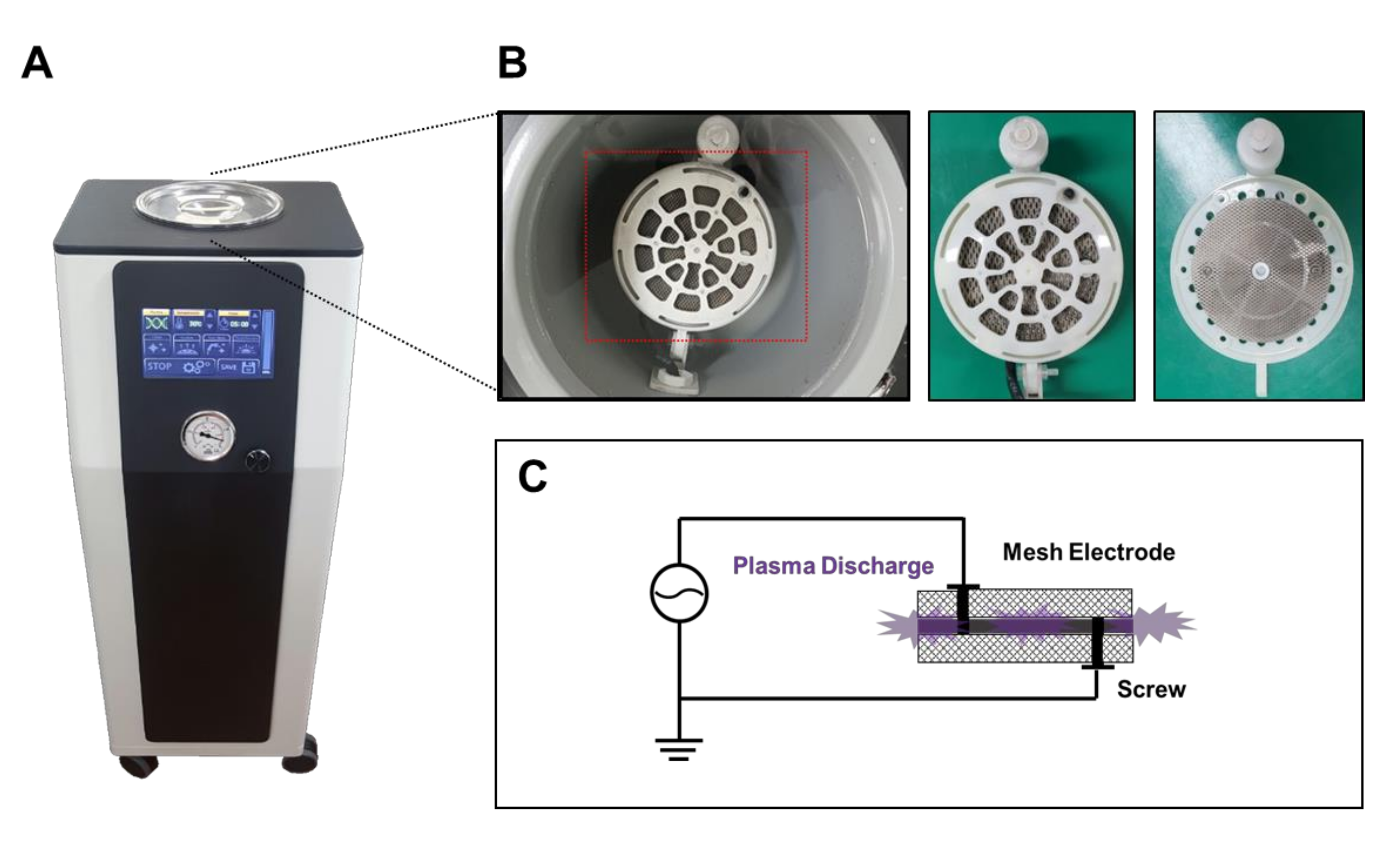
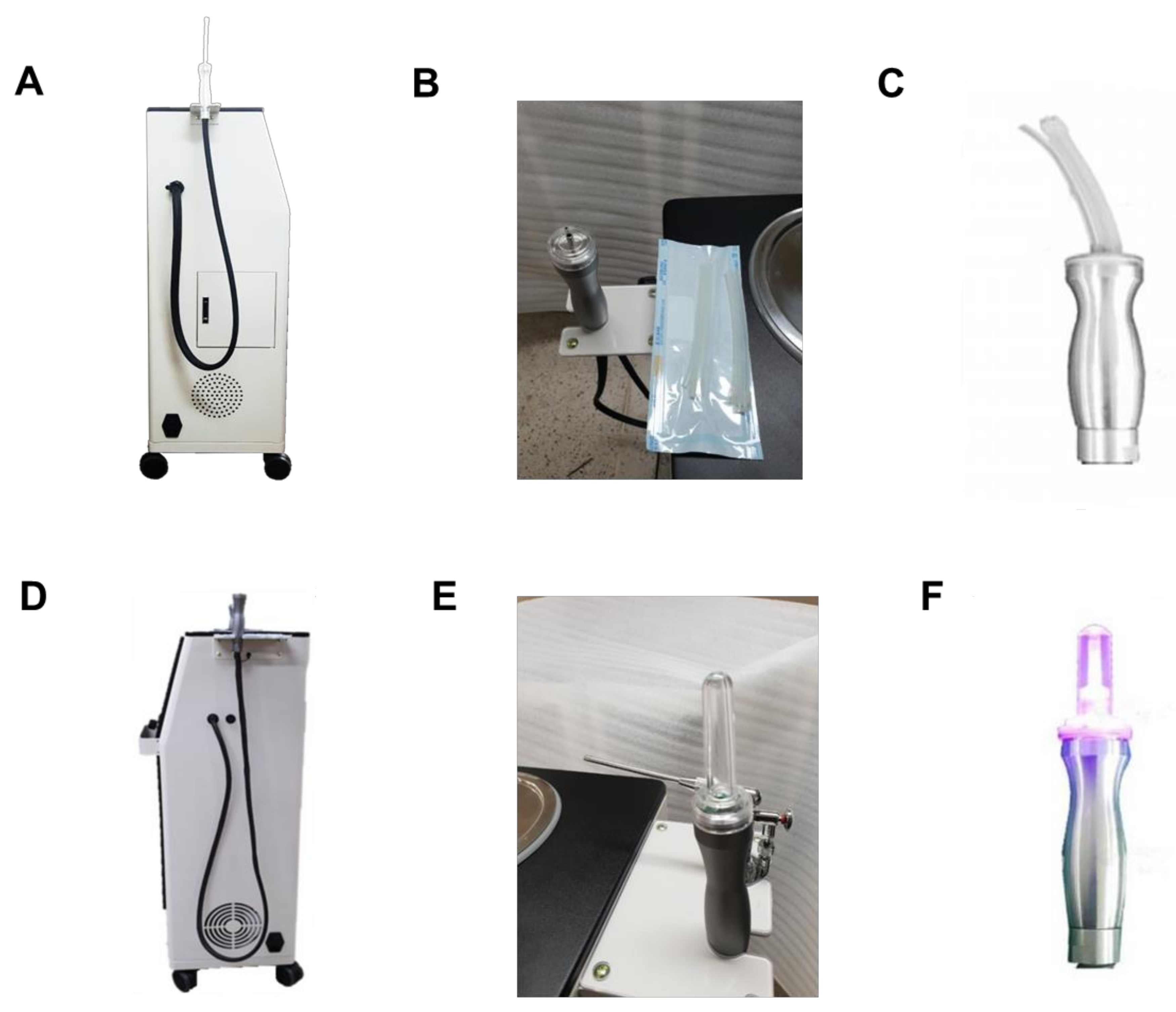
2.1. Device Design for Bacterial Vaginosis

Traditionally, warm water or medicine-treated water has been used to manually wash the contaminated vaginal tract, but this method has frequently resulted in infections. Accordingly, previous vaginal cleaning devices were provided for the user to dilute cleaning medicines in tap water and inject them into vaginal cleaning devices, but disinfecting the vaginal cleaning devices for reuse was inconvenient. In addition, previous devices were complex in structure, bulky and difficult to manufacture and install because they required a filtering part and temperature-controlling part in the device. Thus, the current device was intended to maximize the performance of vaginal cleaning by combining technologies that do not use separate filtration devices, are particularly suitable for sterilization, and can produce sterile water that is harmless to the human body. To achieve this purpose, the device was designed to effectively sterilize the infectious bacteria in the vaginal tract using plasma-activated water with small plasma modules. Furthermore, a nozzle was equipped with a laser light and air pump for a second sterilization and drying after washing with plasma-activated water.

2.2. Device Construction for Bacterial Vaginosis

Figure 1A shows the cleaning solution container in which the plasma-activated water is generated, accommodated, and kept warm. As shown in Figure 1B, the bottom of the cleaning solution container has a hole on the upper surface, and the plasma electrode of the plasma generation module is exposed to the hole to generate the sterilization water from the underwater plasma discharge. A heat-line heater was also prepared on the surface to regulate the temperature in the water. The plasma generation modules include a pair of plasma electrodes, an insulating frame placed between the plasma electrodes, and a conductive screw applying different polarity power to each plasma electrode (Figure 1C). The plasma electrode is a disc-shaped grid electrode with a diameter of 77 mm and a thickness of 0.5 mm, and titanium is used as the electrode material to prevent corrosion due to plasma. In the disc-shaped grid electrodes, the diameter and pitch of the grid were 1.07 mm and 3.92 mm, respectively. According to the dimension of the grid, the percentage of open space was 50%. In addition, a platinum thin film with a thickness of 300 nm was deposited on the grid surface using electroless plating in order to function as a catalyst for increasing the amount of plasma generated. The plasma is generated between two electrodes of different polarities, and the distance between the two electrodes was kept constant by a 2 mm thick insulating frame.
As shown in Figure 2A, a spraying nozzle that is connected to the left side of the device separately contains both a cleaning tube and a suction tube. The cleaning tube is connected to the cleaning solution container, allowing the inlet to spray plasma-activated water. In addition, the suction subnozzle is designed to recover the water that has cleaned the vaginal tract. The disposable silicone tube can be connected to the end of each tube of the spray nozzle and is inserted into the vagina for cleaning and suction (Figure 2B). Because the silicone tube is flexible and disposable, it is harmless and hygienic to the vagina. Remarkably, the suction tube is longer than the cleaning tube and is bent in a direction away from the cleaning tube, which makes it easier to collect wastewater without interfering with the release of sterilized water (Figure 2C).
The light-generating nozzle is incorporated into the handpiece for secondary sterilization. In addition, the nozzle can release air to dry the vagina, as the inlet is connected to an air pump (Figure 2D,E). The light-generating module contains an ultraviolet light emitting diode with wavelengths of 250 to 290 nm, which can be controlled not to exceed 300 nm as low-power diodes to protect against vaginal tissue damage [16]. The light-generating nozzle can be removed from the disposable cap that the hollow is formed in the axial direction, allowing light and air to be applied and released (Figure 2F).

3. Results and Discussion

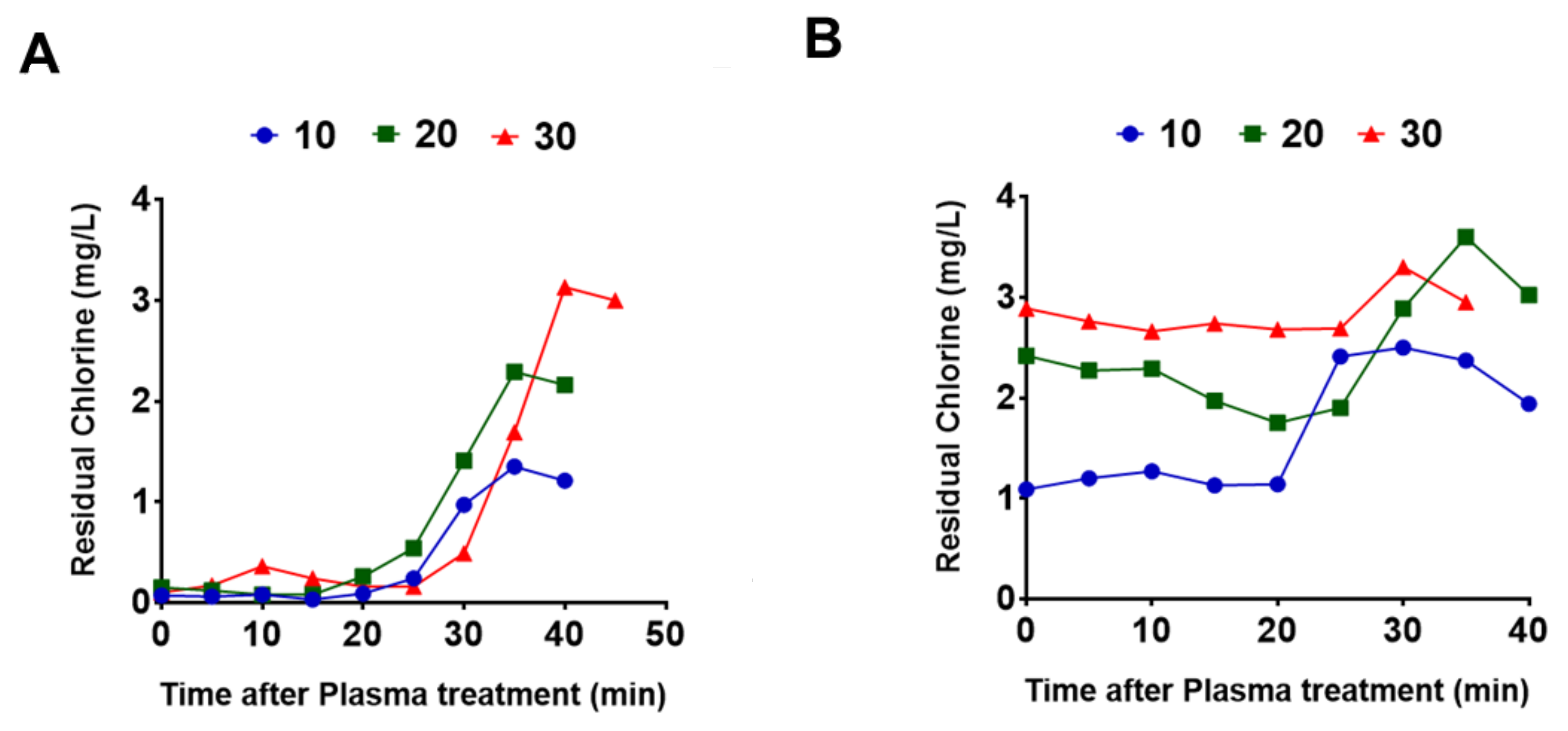
3.1. Performance Test

Previous studies demonstrated that underwater plasma discharge increases the concentration of hypochlorous acid in water, which is known to exhibit microbicidal activity [12,13]. In fact, the hypochlorous acid produced by the plasma has been known to sterilize more than 99.9% of bacteria within 15 s [12,13]. Transmission electron microscope image analysis demonstrated damaged outer walls of the bacteria after the bacteria were exposed to plasma-activated water [12,13]. Thus, we first examined the change in the free chlorine concentration after plasma discharge in the water using our device. The concentration of residual chlorine in water is determined by colorimetric method based on N,N-diethyl-p-phenylenediamine by using colorimeter instrument (Sinsche Technology, Q-CL501P) according to manufacturer’s instruction. As shown in Figure 3A, the graphs indicate the changes in the chlorine concentration after plasma treatment at 10 (circle, blue), 20 (rectangular, green), and 30 (triangle, red) minutes. As a result, it was discovered that the residual chlorine concentration after plasma discharge initially showed no increase and then gradually increased from 25 or 30 min to a peak at 35 or 40 min. The peak chlorine concentration was dependent on the plasma discharge time. Figure 3B shows the changes in the chlorine concentration according to secondary discharge an hour after the first plasma treatment. Interestingly, secondary plasma stimulation additionally increased the chlorine concentration from the residual amounts. Collectively, these findings showed an increased free chlorine concentration through underwater plasma discharge using our device, which suggests the potential to exhibit antimicrobial activity in patients with bacterial vaginosis. To clarify antibacterial effect of our plasma-activated water, further studies are needed how to increase active free chlorine concentrations by the plasma discharge in our system.

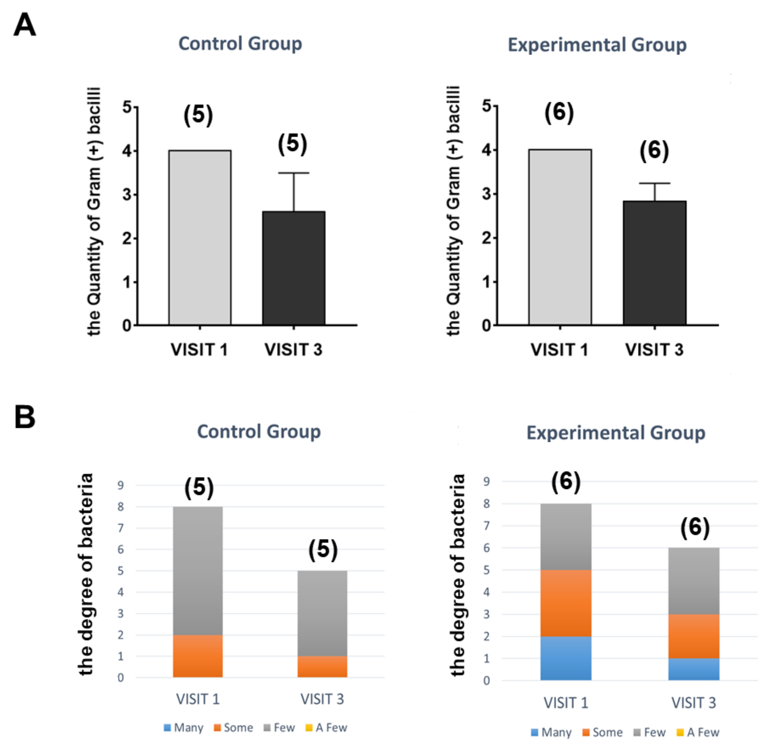
3.2. Clinical Design and Results to Assess the Safety and Effectiveness of the Device

To further test the safety and effectiveness of our device, we performed a clinical trial in patients with acterial vaginosis. The criteria for selecting clinical trial participants included people who tested positive for Gardnerella vaginalis or Mycoplasma hominis in a PCR test, who had not undergone a hysterectomy, and who were willing to voluntarily participate in the clinical trial and comply with the clinical trial plan. In contrast, the following patients were excluded: (1) those who had been treated for vaginitis within four weeks prior to the screening visit; (2) those who required medication immediately after the examiner judged that the quality of life of the patient was significantly reduced due to vaginitis accompanied by severe seizures, itching, etc.; (3) those suspected of having adnexitis, endometriosis, or pelvic inflammation; (4) those who had taken antibiotics or anti-viral drugs during the clinical test; (5) those who had an intrauterine device; and (6) pregnant or breastfeeding women.
The gram-positive test and bacterial culture test were examined from vaginal secretion of clinical trial participants with vaginitis (44 patients) at the first visit (VISIT 1). After one week, they were randomly divided into the control group (22 patients) and experimental group (20 patients). Our vaginal cleaning device was utilized for vaginal cleaning in the experimental group, and a topical antiseptic, betadine, was applied in the control group. For using plasma-activated water, the plasma discharged for 10 min in the cleaning solution container with 3 L of tap water. The plasma-activated water sprayed for 30~60 s (approximately 150~200 mL) to the patients of the experimental group through spraying nozzle, and subsequently exposed an ultraviolet light inside the vagina for 5 s through light-generating nozzle. Thereafter, the gram-positive test and bacterial culture tests were performed in the patients (VISIT 2). After 2 days, we examined the gram-positive test and bacterial culture test (VISIT 3) and additionally conducted the tests after 2 days (VISIT 4).
Consequently, a total of 20 patients in the experimental group and 22 patients in the control group were examined in this study. A cervix culture was performed 4 times in total for each patient, and the effects of cleaning were analyzed as the result of the culture at VISIT 1 and VISIT 3. The results are shown in Table 1 (control group) and Table 2 (experimental group). As shown in Figure 4A, 6 out of 20 patients in the experimental group showed a quantitative decrease in gram (+) bacilli between VISIT 1 (4.0 ± 0.0) and VISIT 3 (2.6 ± 0.9), and 5 out of 22 in the control group showed a quantitative decrease in gram (+) bacilli between VISIT 1 (4.0 ± 0.0) and VISIT 3 (2.8 ± 0.4). The quantity of gram (+) bacilli is indicated by the number of times that gram-stained bacilli was visible in the field when the microscopic field moved at random 10 times. For example, 4+ means that the fields with gram-stained bacilli were 4 times in a total of 10 fields. In addition, we classified the degree of bacterial contamination into four stages (many, some, a few, and few) according to the degree of spreading bacterial colony in the bacterial culture test. If bacteria grow in three parts when the petri dish is quartered, they are classified as many, some in two parts, a few in one part, and few if they grow little. As a result, we found a decrease in the degree of bacterial contamination in both the control and experimental groups (Figure 4B). This finding indicates that the treatment of plasma-activated water from our device in the patients with vaginitis exhibited a similar therapeutic effect as it did in the control patients treated with betadine antiseptic. However, the therapeutic effects shown in both the control and experimental groups were partially rediscovered on VISIT 4, which is a common symptom of recurrent bacterial vaginosis [17]. Therefore, several repetitive treatments of plasma-activated water are likely required to protect against the recurrence of vaginitis.
In addition, the above clinical trials revealed no adverse effects due to our device. There was no case of symptoms such as itching, flaring, and warmth of the vulva as possible adverse effects on the skin [18,19,20]. Therefore, this clinical test confirmed the stability of our device, and the above results established its validity.
Taken together, betadine is a common antiseptic used for skin disinfection, and its effect on killing the bacteria has well known in the patients with bacterial vaginosis. However, some side effects such as skin irritation have reported. In the present study, we show that plasma-activated water has an antiseptic effect on vaginal bacterial contamination similar to betadine. Even if it is similar to the effect of betaine or a little less, if plasma-activated water can replace the chemical betadine, it is believed that it has a great advantage in treating the patients with vaginitis.

4. Conclusions

In summary, we provided a novel vaginal cleaning device using plasma-activated water with antibacterial activity. The device is composed of a container with a plasma module and two nozzles. The water generated from the plasma module in the container is sprayed on and suctioned from the cervix via a spray nozzle. The other nozzle, which has a laser light and air pump, provides secondary sterilization and drying after washing. Because the flexible and disposable silicone tube is connected to the end of both nozzles, it is harmless and hygienic to the vagina and does not require sterilization for reuse. In fact, plasma activation increases free chlorine chemicals with antibacterial activity in the water. More importantly, the clinical safety and effectiveness of our device was demonstrated by the clinical test in which the device was used in patients with bacterial vaginosis. Therefore, it seems possible to use this simple and reliable treatment for bacterial vaginosis using plasma-activated water from the vaginal cleaning device in the hospital.
Plasma-activated media or water has largely known to produce reactive oxygen and nitrogen species containing solutions, which is believed to exhibit the antimicrobial activity when exposed to infected bacteria [21]. In addition, it has known that chlorine-based active species are created by the plasma treatment. These free active chlorines such as hypochlorous acid and hypochlorite are commonly considered as the active ingredient of the deactivation of pathogens in drinking water, swimming pool water and wastewater. Indeed, numerous studies have been supporting the antimicrobial activity of chlorine-base active species including hypochlorous acid as a reactive chlorine species [22]. Because of highly reactive nature as a strong oxidant, these chlorine-base active species have known to interact with most cellular macromolecules, lipids, and nucleotides. For example, hypochlorous acid rapidly reacts with sulfur-containing residues (cysteine, methionine, or glutathione) and amines of the protein. In the present study, we found an increased residual free chlorine concentration after plasma discharge. To more clarify the antibacterial effect of our plasma-activated water, further studies are additionally warranted whether reactive oxygen and nitrogen species are created in plasma-activated water produced from our device.

Author Contributions

Conceptualization, Y.H., H.J., and J.-H.K.; formal analysis, G.Y.W. and J.S.C.; data curation, H.K.K. and D.K.A.; writing and visualization, Y.J. All authors have read and agreed to the published version of the manuscript.

Funding

This research was funded by Hanyang University (HY-2018, 201800000003221).

Conflicts of Interest

Y.H. is a founder of SJ Global. The remaining authors have no financial conflict to disclose.

References

  1. Coudray, M.S.; Madhivanan, P. Bacterial vaginosis-A brief synopsis of the literature. Eur. J. Obstet. Gynecol. Reprod. Biol. 2020, 245, 143–148. [Google Scholar] [CrossRef] [PubMed]
  2. Bagnall, P.; Rizzolo, D. Bacterial vaginosis: A practical review. JAAPA 2017, 30, 15–21. [Google Scholar] [CrossRef] [PubMed]
  3. Haefner, H.K. Current evaluation and management of vulvovaginitis. Clin. Obstet. Gynecol. 1999, 42, 184–195. [Google Scholar] [CrossRef] [PubMed]
  4. Onderdonk, A.B.; Delaney, M.L.; Fichorova, R.N. The Human Microbiome during Bacterial Vaginosis. Clin. Microbiol. Rev. 2016, 29, 223–238. [Google Scholar] [CrossRef] [Green Version]
  5. Kaambo, E.; Africa, C.; Chambuso, R.; Passmore, J.S. Vaginal Microbiomes Associated With Aerobic Vaginitis and Bacterial Vaginosis. Front Public Health 2018, 6, 78. [Google Scholar] [CrossRef]
  6. Mendling, W. Vaginal Microbiota. Adv. Exp. Med. Biol. 2016, 902, 83–93. [Google Scholar] [CrossRef]
  7. Tomas, M.; Palmeira-de-Oliveira, A.; Simoes, S.; Martinez-de-Oliveira, J.; Palmeira-de-Oliveira, R. Bacterial vaginosis: Standard treatments and alternative strategies. Int. J. Pharm. 2020, 587, 119659. [Google Scholar] [CrossRef]
  8. Wewalka, G.; Stary, A.; Bosse, B.; Duerr, H.E.; Reimer, K. Efficacy of povidone-iodine vaginal suppositories in the treatment of bacterial vaginosis. Dermatology 2002, 204 (Suppl. S1), 79–85. [Google Scholar] [CrossRef]
  9. Bernhardt, T.; Semmler, M.L.; Schafer, M.; Bekeschus, S.; Emmert, S.; Boeckmann, L. Plasma Medicine: Applications of Cold Atmospheric Pressure Plasma in Dermatology. Oxid. Med. Cell Longev. 2019, 2019, 3873928. [Google Scholar] [CrossRef] [Green Version]
  10. Setsuhara, Y. Low-temperature atmospheric-pressure plasma sources for plasma medicine. Arch. Biochem. Biophys. 2016, 605, 3–10. [Google Scholar] [CrossRef]
  11. Brany, D.; Dvorska, D.; Halasova, E.; Skovierova, H. Cold Atmospheric Plasma: A Powerful Tool for Modern Medicine. Int. J. Mol. Sci. 2020, 21, 2932. [Google Scholar] [CrossRef] [PubMed] [Green Version]
  12. Lee, S.J.; Ma, S.H.; Hong, Y.C.; Choi, M.C. Effects of pulsed and continuous wave discharges of underwater plasma on Escherichia coli. Sep. Purif. Technol. 2018, 193, 351–357. [Google Scholar] [CrossRef]
  13. Hong, Y.C.; Park, H.J.; Lee, B.J.; Kang, W.S.; Uhm, H.S. Plasma formation using a capillary discharge in water and its application to the sterilization of E. coli. Phys. Plasmas 2010, 17, 053502. [Google Scholar] [CrossRef]
  14. Khlyustova, A.; Labay, C.; Machala, Z.; Ginebra, M.P.; Canal, C. Important parameters in plasma jets for the production of RONS in liquids for plasma medicine: A brief review. Front Chem. Sci. Eng. 2019, 13, 238–252. [Google Scholar] [CrossRef]
  15. Privat-Maldonado, A.; Schmidt, A.; Lin, A.; Weltmann, K.D.; Wende, K.; Bogaerts, A.; Bekeschus, S. ROS from Physical Plasmas: Redox Chemistry for Biomedical Therapy. Oxid. Med. Cell Longev. 2019, 2019, 9062098. [Google Scholar] [CrossRef] [Green Version]
  16. Mori, M.; Hamamoto, A.; Takahashi, A.; Nakano, M.; Wakikawa, N.; Tachibana, S.; Ikehara, T.; Nakaya, Y.; Akutagawa, M.; Kinouchi, Y. Development of a new water sterilization device with a 365 nm UV-LED. Med. Biol. Eng. Comput. 2007, 45, 1237–1241. [Google Scholar] [CrossRef]
  17. Faught, B.M.; Reyes, S. Characterization and Treatment of Recurrent Bacterial Vaginosis. J. Womens Health 2019, 28, 1218–1226. [Google Scholar] [CrossRef]
  18. Brosseau, G.; Page, N.; de Jaham, C.; Del Castillo, J.R.E. Medical honey for canine nasal intertrigo: A randomized, blinded, placebo-controlled, adaptive clinical trial to support antimicrobial stewardship in veterinary dermatology. PLoS ONE 2020, 15, e0235689. [Google Scholar] [CrossRef]
  19. Jang, Y.; Lee, W.J.; Hong, G.S.; Shim, W.S. Red ginseng extract blocks histamine-dependent itch by inhibition of H1R/TRPV1 pathway in sensory neurons. J. Ginseng Res. 2015, 39, 257–264. [Google Scholar] [CrossRef] [Green Version]
  20. Jang, Y.; Kim, M.; Hwang, S.W. Molecular mechanisms underlying the actions of arachidonic acid-derived prostaglandins on peripheral nociception. J. Neuroinflamm. 2020, 17, 30. [Google Scholar] [CrossRef]
  21. Kaushik, N.K.; Ghimire, B.; Li, Y.; Adhikari, M.; Veerana, M.; Kaushik, N.; Jha, N.; Adhikari, B.; Lee, S.J.; Masur, K.; et al. Biological and medical applications of plasma-activated media, water and solutions. Biol. Chem. 2018, 400, 39–62. [Google Scholar] [CrossRef] [PubMed]
  22. Gray, M.J.; Wholey, W.Y.; Jakob, U. Bacterial responses to reactive chlorine species. Annu. Rev. Microbiol. 2013, 67, 141–160. [Google Scholar] [CrossRef] [PubMed] [Green Version]
Figure 1. (A,B) Main body of the device showing the cleaning solution container with the plasma module and (C) the schematic principle for underwater plasma discharge.
Plasma 03 00016 g001
Figure 2. (A) A spraying nozzle with cleaning and suction functions, (B) the top view of the spraying nozzle and the disposable silicone tube, and (C) an image of the silicon tube-connected spraying nozzle. (D) The light-generating nozzle with an air pump, (E) the top view of the light-generating nozzle and disposable silicone cap, and (F) an image of the light-generating nozzle with a silicon cap.
Plasma 03 00016 g002
Figure 3. (A) Changes in the concentration of free chlorine chemicals in the water after plasma discharge and (B) changes in the concentration of chlorine chemicals after secondary plasma discharge.
Plasma 03 00016 g003
Figure 4. Therapeutic effects of plasma-activated water from our device (experimental group) and betadine antiseptic (control group) on bacterial infection in patients with vaginitis. (A,B) The patients in experimental group showed a quantitative decrease in gram (+) bacilli (A) and the degree of bacterial contamination (B) similar to those of control group when compared between VISIT 1 and VISIT 3.
Plasma 03 00016 g004
Table 1. Information and test results of control group.
PatientsSTD (VISIT 1)Culture (VISIT 1)Culture (VISIT 2)Culture (VISIT 3)Culture (VISIT 4)
1KJY
01880498
M. hominis (-)
G. vaginali (-)
Gram(+) bacilli 4+
Gram(-) bacilli 2+
Gram (+) bacilli 1+Gram (+) bacilli 1+
Gram (+) cocci pairs & chains 3+
Gram (-) bacilli 1+
Gram (+) bacilli 1+
Gram (+) cocci pairs & chains 1+
Gram (-) bacilli 2+
Enterobacter cloacae complex SomeNo gowth for 48hrsEscherichia coli fewEscherichia coli Many
2JSY
02331373
M. hominis (-)
G. vaginali (+)
C. trachoma(+)
C. albicans(+)
Gram(+) bacilli 4+
Gram (+) cocci pairs & chains 2+
Yeast-like organism 1+
Gram(+) bacilli 1+
Gram (+) cocci pairs & chains 1+
Yeast-like organism 1+
Gram(+) bacilli 3+
Gram (+) cocci pairs & chains 4+
Yeast-like organism 1+
Gram(+) bacilli 4+
Gram (+) cocci pairs & chains 2+
Yeast-like organism 1+
Candida albicans Few
Gram positive cocci Few
Lactobacillus sp. Few
Yeast Few
Non spore-forming Gram (+) bacilli Few
Candida albicans SomeYeast Few
Gram positive cocci Few
Non spore-forming Gram (+) bacilli Some
3HYS
02702448
M. hominis (-)
G. vaginali (-)
Gram(-) bacilli 4+
Gram(+) bacilli 4+
Gram(+) bacilli 3+Gram(+) bacilli 3+Gram (+) bacilli 4+
Excherichia coli SomeLactobacillus sp. FewLactobacillus sp. FewNon spore-forming Gram (+) bacilli Few
4HMJ
02720811
M. hominis (-)
G. vaginali (-)
Gram (+) bacilli 4+
Yeast-like organism 2+
Gram (+) bacilli 3+Gram (+) bacilli 3+
Gram (+) cocci pairs & chains 1+
Gram (+) bacilli 2+
Gram (+) cocci pairs & chains 4+
Gram (-) bacilli 4+
Non spore forming Gram (+) bacilli Few
Candida albicans Few
Lactobacillus sp. FewLactobacillus sp. FewEnterobacter aerogenes Many
5KKM
01398837
M. hominis (-)
G. vaginali (-)
Gram (+) bacilli 4+
Gram (+) cocci pairs & chains 1+
Gram (+) bacilli 4+Gram (+) bacilli 3+Gram (+) bacilli 4+
Streptococcus-agalactiae-(Group B) FewNon spore-forming Gram (+) bacilli FewNon spore-forming Gram (+) bacilli FewNon spore-forming Gram (+) bacilli Few
Table 2. Information and test results of experimental group.
PatientsSTD (VISIT 1)Culture (VISIT 1)Culture (VISIT 2)Culture (VISIT 3)Culture (VISIT 4)
1NTI
02759285
M. hominis (-)
G. vaginali (+)
Gram (+) bacilli 4+
Yeast-like organism 1+
Gram (+) bacilli 4+Gram (+) bacilli 3+Gram (+) bacilli 4+
Gram (+) cocci pairs & chains 1+
Gram (-) bacilli 4+
Candida albicans SomeYeast Few
Non spore-forming Gram(+) bacilli Few
Non spore-forming Gram(+) bacilli FewEscherichia coli Some
2PKH
01652805
M. hominis (-)
G. vaginali (+)
C. trachoma weakly (+)
C. albicans (+)
Gram (+) bacilli 4+
Gram (+) cocci pairs & chains 2+
Gram (-) bacilli 1+
Yeast-like organism 1+
Gram (+) bacilli 4+
Gram (+) cocci pairs & chains 2+
Gram (+) bacilli 3+
Gram (+) cocci pairs & chains 2+
Gram (+) bacilli 3+
Gram (+) cocci pairs & chains 1+
Yeast-like organism 1+
Candida albicans FewCandida albicans Few
Lactobacillus sp. Some
Gram positive cocci Many
Non spore-forming Gram(+) bacilli SomeYeast Few
Gram positive cocci Few
Non spore-forming Gram (+) cilli Some
Yeast Some
3SSY
01464025
M. hominis (-)
G. vaginali (+)
Gram(+) bacilli 4+
Gram(-) bacilli 4+
Gram(+) colli pairs & chains 1+
Gram(+) bacilli 1+Gram(+) bacilli 3+Gram(+) bacilli 4+
Escherichia coli ManyNon spore-forming Gram (+) bacillli FewLactobacillus sp. someLactobacillus sp. some
4JSS
01653482
M. hominis (-)
G. vaginali (+)
Gram (+) bacilli 4+Gram (+) bacilli 4+Gram (+) bacilli 3+Gram (+) bacilli 3+
Lactobacillus sp. Some
Gram positive cocci Few
Lactobacillus sp. FewLactobacillus sp. FewLactobacillus sp. Few
5LHJ
02727501
M. hominis (-)
G. vaginali (+)
C. trahoma (+)
Gram (+) bacilli 4+
Gram (+) cocci pairs & chains 1+
Gram (+) bacilli 4+
Gram (+) cocci pairs & chains 1+
Gram (+) bacilli 3+
Gram (+) cocci pairs & chains 1+
Gram (+) bacilli 3+
Streptococcus agalactiae-(Group B) SomeStreptococcus agalactiae-(Group B) FewStreptococcus agalactiae-(Group B) FewNon spore-forming Gram(+) bacilli Few
6HJY
01617842
M. hominis (-)
G. vaginali (-)
Gram (+) biacilli 4+
Gram (+) cocci pairs & chains 3+
Gram (+) biacilli 3+
Gram (+) cocci pairs & chains 1+
Gram (+) biacilli 2+Gram (+) biacilli 3+
Gram negative bacilli Few
Gram positive cocci Many
Lactobacillus sp. FewLactobacillus sp. FewLactobacillus sp. Some
Gram positive cocci Few
Publisher’s Note: MDPI stays neutral with regard to jurisdictional claims in published maps and institutional affiliations.

