The History of the Decline and Fall of the Glial Numbers Legend


1
Faculty of Biology, Medicine and Health, University of Manchester, Manchester M13 9PT, UK
2
Center for Basic and Translational Neuroscience, Faculty of Health and Medical Sciences, University of Copenhagen, 2200 Copenhagen, Denmark
3
Institute of Biomedical and Biomolecular Sciences, School of Pharmacy and Biomedical Science, University of Portsmouth, Portsmouth PO1 2UP, UK
*
Authors to whom correspondence should be addressed.
Neuroglia 2018, 1(1), 188-192; https://0-doi-org.brum.beds.ac.uk/10.3390/neuroglia1010013
Submission received: 2 July 2018
/
Accepted: 4 July 2018
/
Published: 17 July 2018
Abstract
:In the field of neuroscience and, more specifically glial cell biology, one of the most fundamentally intriguing and enduring questions has been “how many neuronal cells—neurones and glia—are there in the human brain?”. From the outset, the driving force behind this question was undoubtedly the scientific quest for knowledge of why humans are more intelligent than even our nearest relatives; the ‘neuronal doctrine’ dictated we must have more neurones than other animals. The early histological studies indicated a vast space between neurones that was filled by ‘nervenkitt’, later identified as neuroglia; arguably, this was the origin of the myth that glia massively outnumber neurones in the human brain. The myth eventually became embedded in ideology when later studies seemed to confirm that glia outnumber neurones in the human cortex—the seat of humanity—and that there was an inevitable rise in the glia-to-neurone ratio (GNR) as we climbed the evolutionary tree. This could be described as the ‘glial doctrine’—that the rise of intelligence and the rise of glia go hand-in-hand. In many ways, the GNR became a mantra for working on glial cells at a time when the neuronal doctrine ruled the world. However, the work of Suzana Herculano-Houzel which she reviews in this first volume of Neuroglia has led the way in demonstrating that neurones and glia are almost equal in number in the human cortex and there is no inexorable phylogenetic rise in the GNR. In this commentary we chart the fall and decline of the mythology of the GNR.
- “The various models of worship which prevailed … were all considered by the people as equally true; by the philosopher as equally false”
Edward Gibbon (1776), The Decline and Fall of the Roman Empire Volume 1, Chapter 2.
One of the most enduring and fundamentally important misconceptions in neuroscience is the number of glial cells in the human brain, purportedly outnumbering neurones by a factor of 10 or even 50 [1,2,3,4]. Unfortunately, the notion of a glia-to-neurone ratio (GNR) in the human brain of 10 or more became ‘general knowledge’ and commonplace in glial literature over several decades. In a way, this myth became a cornerstone of glial research, and, regrettably, the authors of this commentary used the same statement in our textbook in 2007 [5]. Like other glial cell biologists before us, we fell into the trap of blind belief of the accepted doctrine. In our defence, we amended this error in our second, more comprehensive book on glial cell physiology and pathology [6]. What was most surprising to us was that the perception of the high GNR of the human brain was not based on direct counts and experimental observations; rather, it was the result of several conjectures based on limited observation. Interestingly, the very first calculation of GNR made in 1936 defined it as 1.5 [7], which is very far from subsequent speculations and very close to the numbers accepted today. The GNR legend was challenged only about a decade ago by the efforts of the Brazilian neuroanatomist Suzana Herculano-Houzel, who was armed with a powerful cell counting tool known as the ‘isotropic fractionator’ [8,9,10,11]. This technique counts numbers of cell nuclei from homogenised nervous tissue and was initially developed in the middle of 20th century [12,13,14], and subsequently greatly improved by Suzana Herculano-Houzel; she recalls seeing a paper from the 1970s on attempts to isolate DNA from whole brains and thought “don’t count DNA, count nuclei!” This issue of Neuroglia contains a personal account of the dramatic history of glial numbers written by Suzana Herculano-Houzel [15].
The early concept of a high glial content of the human brain had wide repercussions. First and foremost, the high GNR of the human brain has been widely believed to reflect the evolutionary progress of the nervous system (this idea was initially suggested by Franz Nissl [16]), whereby the advance in glial numbers was considered to be directly linked to the rise in intelligence [17,18,19]. These ideas led to even further extremes, when some neuroscientists comprehended glia as the main element of information processing. The most outspoken was Robert Galambos who was convinced that neuroglia represent the primary seat of intelligence, and by extension ‘humanity’:
Glia is … conceived as genetically charged to organize and program neuron activity so that the best interests of the organism will be served; the essential product of glia action is visualized to be what we call innate and acquired behavioural responses. In this scheme, neurons in large part merely execute the instructions glia give them.[20]
Galambos also authored the most popular misquote of Fritjof Nancen, “glia is the seat of intelligence as it increases in size from the lower to the higher forms of animal”, which never occurred in Nancen’s original book [21].
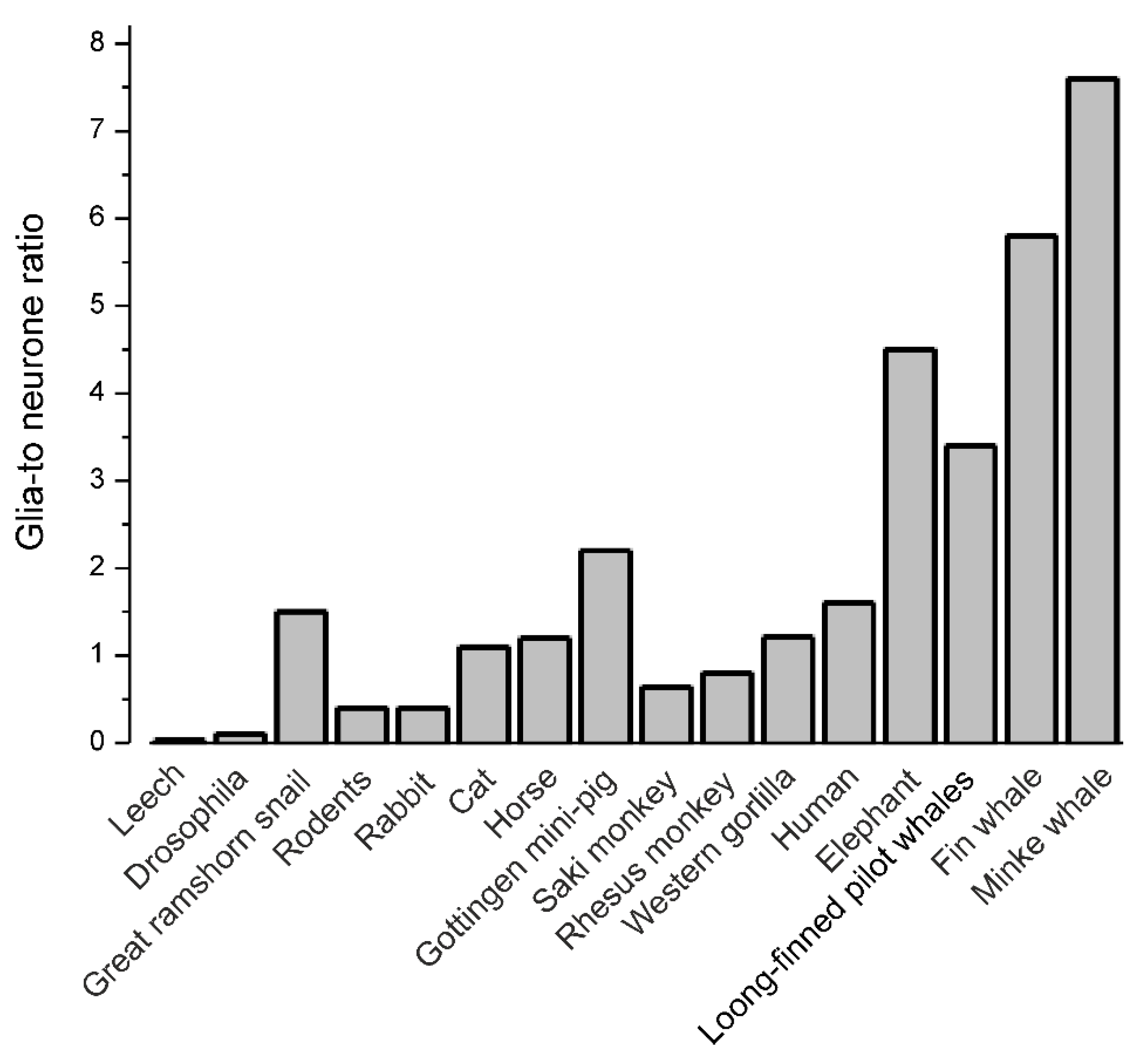
The evolutionary advance of glia is far more complicated, and humans are by no means in possession of the largest GNR (Figure 1). The number of glia varies substantially between different species and the GNR does not simply increase with increasing brain size. The nervous system of invertebrates has, as a rule, relatively smaller numbers of glial cells, with a GNR between 0.001 and 0.1 (56 glia per 302 neurones in Caenorhabditis elegans [22,23]; 10 glial cells per 400–700 neurones in every ganglion of the leech [24]; ~9000 glial cells per 90,000 neurones in the central nervous system (CNS) of Drosophila [25,26]). At the same time, the buccal ganglia of the great ramshorn snail Planorbis corneus contains 298 neurones and 391 glial cells [27], thus having a GNR of 1.3, very similar to that of humans. Similarly, in vertebrates there is no hard-and-fast increase in the GNR with increasing brain size; for example, in the cortex, the GNR is about 0.3–0.4 in rodents, ~1.1 in cat; ~1.2 in horse, 0.5–1.0 in rhesus monkey, 2.2 in Göttingen minipig, ~1.5 in humans, and as high as 4–8 in elephants and the fin whale [28,29,30,31,32,33,34]. The largest absolute number of glial cells has been counted in the neocortex of whales [31,35]); sterological cell counts in the neocortex of the long-finned pilot whale (Globicephala melas) brain determined there are approximately 37.2 × 10⁹ neurons and 127 × 109 glial cells [35], and this gives a high GNR of 3.4 which is as expected from neuronal density [36]. In the human brain, the total number of glia is comparable (or even slightly less) with the number of neurones—i.e., the neuronal counts are in a range of ~80 billions—whereas neuroglia are ~60 billions with substantial regional differences. However, it should be noted that the variance in numbers is remarkably high, with numbers of neurones in the neocortex, for example, varying between 20 and 30 billions in cognitively preserved individuals [37]. Remarkably, when compared to the evolutionary new brain, the more primitive parts of the human brain have a higher proportion of neuroglia, with a GNR approaching 7–10 in the brainstem, or even more according to some studies [10,38]; very recent observations, using both stereology and isotropic fractionation, gave a GNR of ~5 in the spinal cord of cynomolgus monkey and almost 7 in humans [39]. These trends argue against the concept that a high GNR reflects evolutionary advance and increased intelligence. Nonetheless, it is important to be aware that evolution brought with it substantial changes in the morphology and complexity of astroglia in the human cortex, which also contains several idiosyncratic types of glial cell [40,41,42]. However, astrocyte three-dimensional morphology has been systematically studied in too few species so far and none in brains larger than humans. How large and complex astrocytes of whales might be remains unknown. Possibly (though not necessarily), the differences between astrocytes are related to brain size, not clade, but that is a different story.
Another unexpected consequence of the erroneous perceptions arising from the GNR in the human brain concerns the relative number of the main glial cell types—astrocytes, oligodendrocytes and microglia. Again, the numerical prevalence of astroglia is repeatedly stated in many papers, but this appears to be based on no observations whatsoever (see Table 6 in [10]). Seemingly the most numerous glia are oligodendrocytes (40–60%), with astrocytes accounting for 20–40% and microglia for ~10%, although there is of course considerable variability between brain regions, developmental stage and species. For example, in area 17 of young monkeys, astrocytes account for 40% of total glia, oligodendrocytes for 53% and microglia for ~7%. In cortical layers 1 to 3 of adult monkey astrocytes were calculated at 57%, oligodendrocytes at 36% and microglia at 7%, whereas in the layer 4 (which has higher degree of myelination) 30% of glia are astrocytes, 62% are oligodendroglia and the remaining 8% are microglia [43].
In conclusion, legends, myths and superstitions are arguably the most long-lasting and resilient forms of conserving and passing on false knowledge. Without doubt, this has greatly affected the evolution of humanity and continues to shape the progress of natural science. It is hoped that the work highlighted in this commentary and Suzana Herculano-Houzel’s review article [15] will help reverse one of the most enduring misconceptions that glial cells massively outnumber neurones in the human brain. We opened this commentary with a quote from Gibbon’s The Decline and Fall of the Roman Empire; we think it is fitting to end with a quote from Darwin on the vicissitudes of ignorance and false knowledge.
“Ignorance more frequently begets confidence than does knowledge: it is those who know little, and not those who know much, who so positively assert that this or that problem will never be solved by science.”.Charles Darwin (1871), The Descent of Man, Volume 1 (Introduction)
Author Contributions
All authors participated equally in writing this commentary.
Conflicts of Interest
The authors declare no conflict of interest.
References
- Hyden, H. Dynamic aspects of the neuron-glia relationship—A study with microchemical methods. In The Neuron; Hyden, H., Ed.; Elsevier: Amsterdam, The Netherlands, 1967; pp. 179–217. [Google Scholar]
- Kandel, E.R.; Schwartz, J.H.; Jessell, T.M. Principles of Neural Science; McGrawhill: New York, NY, USA, 2000. [Google Scholar]
- Bear, M.F.; Connors, B.W.; Paradiso, M.A. Exploring the Brain; Lippincott Williams & Wilkins: Philadelphia, PA, USA, 2007. [Google Scholar]
- Darlington, C.L. The Female Brain; CRC Press: Boca Raton, FL, USA, 2009. [Google Scholar]
- Verkhratsky, A.; Butt, A. Glial Neurobiology: A Textbook; John Wiley & Sons: Chichester, UK, 2007. [Google Scholar]
- Verkhratsky, A.; Butt, A.M. Glial Physiology and Pathophysiology; Wiley-Blackwell: Chichester, UK, 2013; p. 560. [Google Scholar]
- Mühlmann, M. About the question of glia formation. Zur neurogliabildungsfrage. Beitr. Pathol. Anat. Allg. Pathol. 1936, 96, 361–374. [Google Scholar]
- Herculano-Houzel, S.; Lent, R. Isotropic fractionator: A simple, rapid method for the quantification of total cell and neuron numbers in the brain. J. Neurosci. 2005, 25, 2518–2521. [Google Scholar] [CrossRef] [PubMed]
- Azevedo, F.A.; Carvalho, L.R.; Grinberg, L.T.; Farfel, J.M.; Ferretti, R.E.; Leite, R.E.; Jacob Filho, W.; Lent, R.; Herculano-Houzel, S. Equal numbers of neuronal and nonneuronal cells make the human brain an isometrically scaled-up primate brain. J. Comp. Neurol. 2009, 513, 532–541. [Google Scholar] [CrossRef] [PubMed]
- Von Bartheld, C.S.; Bahney, J.; Herculano-Houzel, S. The search for true numbers of neurons and glial cells in the human brain: A review of 150 years of cell counting. J. Comp. Neurol. 2016, 524, 3865–3895. [Google Scholar] [CrossRef] [PubMed] [Green Version]
- Andrade-Moraes, C.H.; Oliveira-Pinto, A.V.; Castro-Fonseca, E.; da Silva, C.G.; Guimaraes, D.M.; Szczupak, D.; Parente-Bruno, D.R.; Carvalho, L.R.; Polichiso, L.; Gomes, B.V.; et al. Cell number changes in Alzheimer's disease relate to dementia, not to plaques and tangles. Brain 2013, 136, 3738–3752. [Google Scholar] [CrossRef] [PubMed] [Green Version]
- Nurnberger, J.I.; Gordon, M.W. The cell density of neural tissues: Direct counting method and possible applications as a biologic referent. Prog. Neurobiol. 1957, 2, 100–128. [Google Scholar] [PubMed]
- Brizzee, K.R.; Vogt, J.; Kharetchko, X. Postnatal changes in glia/neuron index with a comparison of methods of cell enumeration in the white rat. Prog. Brain Res. 1964, 4, 136–149. [Google Scholar]
- Zamenhof, S. Final number of purkinje and other large cells in the chick cerebellum influenced by incubation temperatures during their proliferation. Brain Res. 1976, 109, 392–394. [Google Scholar] [CrossRef]
- Herculano-Houzel, S.; Dos Santos, S. You don’t mess with the glia. Neuroglia 2018, 1, 13. [Google Scholar]
- Nissl, F. Nervenzellen und graue substanz. Munch. Med. Wochenschr. 1898, 45, 988–992. [Google Scholar]
- Friede, R. Der quantitative anteil der glia and der cortexentwicklung. Acta Anat. (Basel) 1954, 20, 290–296. [Google Scholar] [CrossRef] [PubMed]
- Pfrieger, F.W.; Barres, B.A. What the fly’s glia tell the fly’s brain. Cell 1995, 83, 671–674. [Google Scholar] [CrossRef]
- Fields, R.D. The Other Brain; Simon & Schuster: New York, NY, USA, 2009. [Google Scholar]
- Galambos, R. A glia-neural theory of brain function. Proc. Natl. Acad. Sci. USA 1961, 47, 129–136. [Google Scholar] [CrossRef] [PubMed]
- Nansen, F. The Structure and Combination of the Histological Elements of the Central Nervous System; John Grieg: Bergen, Norway, 1886. [Google Scholar]
- Stout, R.F., Jr.; Verkhratsky, A.; Parpura, V. Caenorhabditis elegans glia modulate neuronal activity and behavior. Front. Cell. Neurosci. 2014, 8, 67. [Google Scholar] [CrossRef] [PubMed]
- Oikonomou, G.; Shaham, S. The glia of caenorhabditis elegans. Glia 2011, 59, 1253–1263. [Google Scholar] [CrossRef] [PubMed]
- Deitmer, J.W.; Rose, C.R.; Munsch, T.; Schmidt, J.; Nett, W.; Schneider, H.P.; Lohr, C. Leech giant glial cell: Functional role in a simple nervous system. Glia 1999, 28, 175–182. [Google Scholar] [CrossRef]
- Edwards, T.N.; Meinertzhagen, I.A. The functional organisation of glia in the adult brain of Drosophila and other insects. Prog. Neurobiol. 2010, 90, 471–497. [Google Scholar] [CrossRef] [PubMed] [Green Version]
- Kremer, M.C.; Jung, C.; Batelli, S.; Rubin, G.M.; Gaul, U. The glia of the adult Drosophila nervous system. Glia 2017, 65, 606–638. [Google Scholar] [CrossRef] [PubMed] [Green Version]
- Pentreath, V.W.; Radojcic, T.; Seal, L.H.; Winstanley, E.K. The glial cells and glia-neuron relations in the buccal ganglia of Planorbis corneus (L.): Cytological, qualitative and quantitative changes during growth and ageing. Philos. Trans. R. Soc. Lond. B Biol. Sci. 1985, 307, 399–455. [Google Scholar] [CrossRef] [PubMed]
- Christensen, J.R.; Larsen, K.B.; Lisanby, S.H.; Scalia, J.; Arango, V.; Dwork, A.J.; Pakkenberg, B. Neocortical and hippocampal neuron and glial cell numbers in the rhesus monkey. Anat. Rec. (Hoboken) 2007, 290, 330–340. [Google Scholar] [CrossRef] [PubMed] [Green Version]
- Lidow, M.S.; Song, Z.M. Primates exposed to cocaine in utero display reduced density and number of cerebral cortical neurons. J. Comp. Neurol. 2001, 435, 263–275. [Google Scholar] [CrossRef] [PubMed]
- Pakkenberg, B.; Gundersen, H.J. Neocortical neuron number in humans: Effect of sex and age. J. Comp. Neurol. 1997, 384, 312–320. [Google Scholar] [CrossRef]
- Eriksen, N.; Pakkenberg, B. Total neocortical cell number in the mysticete brain. Anat. Rec. (Hoboken) 2007, 290, 83–95. [Google Scholar] [CrossRef] [PubMed] [Green Version]
- Hawkins, A.; Olszewski, J. Glia/nerve cell index for cortex of the whale. Science 1957, 126, 76–77. [Google Scholar] [CrossRef] [PubMed]
- Jelsing, J.; Nielsen, R.; Olsen, A.K.; Grand, N.; Hemmingsen, R.; Pakkenberg, B. The postnatal development of neocortical neurons and glial cells in the gottingen minipig and the domestic pig brain. J. Exp. Biol. 2006, 209, 1454–1462. [Google Scholar] [CrossRef] [PubMed]
- Tower, D.B. Structural and functional organization of mammalian cerebral cortex; the correlation of neurone density with brain size; cortical neurone density in the fin whale (Balaenoptera Physalus L.) with a note on the cortical neurone density in the indian elephant. J. Comp. Neurol. 1954, 101, 19–51. [Google Scholar] [CrossRef] [PubMed]
- Mortensen, H.S.; Pakkenberg, B.; Dam, M.; Dietz, R.; Sonne, C.; Mikkelsen, B.; Eriksen, N. Quantitative relationships in delphinid neocortex. Front. Neuroanat. 2014, 8, 132. [Google Scholar] [CrossRef] [PubMed]
- Kazu, R.S.; Maldonado, J.; Mota, B.; Manger, P.R.; Herculano-Houzel, S. Cellular scaling rules for the brain of Artiodactyla include a highly folded cortex with few neurons. Front. Neuroanat. 2014, 8, 128. [Google Scholar] [CrossRef] [PubMed]
- Pelvig, D.P.; Pakkenberg, H.; Stark, A.K.; Pakkenberg, B. Neocortical glial cell numbers in human brains. Neurobiol. Aging 2008, 29, 1754–1762. [Google Scholar] [CrossRef] [PubMed]
- Pakkenberg, B.; Gundersen, H.J. Total number of neurons and glial cells in human brain nuclei estimated by the disector and the fractionator. J. Microsc. 1988, 150, 1–20. [Google Scholar] [CrossRef] [PubMed]
- Bahney, J.; von Bartheld, C.S. The cellular composition and glia-neuron ratio in the spinal cord of a human and a nonhuman primate: Comparison with other species and brain regions. Anat. Rec. (Hoboken) 2018, 301, 697–710. [Google Scholar] [CrossRef] [PubMed]
- Verkhratsky, A.; Oberheim Bush, N.A.; Nedergaard, M.; Butt, A. The special case of human astrocytes. Neuroglia 2018, 1, 4. [Google Scholar] [CrossRef]
- Oberheim, N.A.; Takano, T.; Han, X.; He, W.; Lin, J.H.; Wang, F.; Xu, Q.; Wyatt, J.D.; Pilcher, W.; Ojemann, J.G.; et al. Uniquely hominid features of adult human astrocytes. J. Neurosci. 2009, 29, 3276–3287. [Google Scholar] [CrossRef] [PubMed]
- Verkhratsky, A.; Nedergaard, M. Physiology of astroglia. Physiol. Rev. 2018, 98, 239–389. [Google Scholar] [CrossRef] [PubMed]
- Peters, A.; Verderosa, A.; Sethares, C. The neuroglial population in the primary visual cortex of the aging rhesus monkey. Glia 2008, 56, 1151–1161. [Google Scholar] [CrossRef] [PubMed]
Figure 1.
Glia-to-neurone ratio in invertebrates and vertebrates. The glia-to-neurone ratio (GNR) is not clearly related to phylogeny; the great ramshorn snail has a GNR equivalent to the human cortex, and the GNR is over five-times greater in the Minke whale.
Figure 1.
Glia-to-neurone ratio in invertebrates and vertebrates. The glia-to-neurone ratio (GNR) is not clearly related to phylogeny; the great ramshorn snail has a GNR equivalent to the human cortex, and the GNR is over five-times greater in the Minke whale.

© 2018 by the authors. Licensee MDPI, Basel, Switzerland. This article is an open access article distributed under the terms and conditions of the Creative Commons Attribution (CC BY) license (http://creativecommons.org/licenses/by/4.0/).
Share and Cite
MDPI and ACS Style
Verkhratsky, A.; Butt, A.M. The History of the Decline and Fall of the Glial Numbers Legend. Neuroglia 2018, 1, 188-192. https://0-doi-org.brum.beds.ac.uk/10.3390/neuroglia1010013
AMA Style
Verkhratsky A, Butt AM. The History of the Decline and Fall of the Glial Numbers Legend. Neuroglia. 2018; 1(1):188-192. https://0-doi-org.brum.beds.ac.uk/10.3390/neuroglia1010013
Chicago/Turabian StyleVerkhratsky, Alexei, and Arthur M. Butt. 2018. "The History of the Decline and Fall of the Glial Numbers Legend" Neuroglia 1, no. 1: 188-192. https://0-doi-org.brum.beds.ac.uk/10.3390/neuroglia1010013