Article

Mediation of FoxO1 in Activated Neuroglia Deficient for Nucleoside Diphosphate Kinase B during Vascular Degeneration

1
Experimental Pharmacology, European Center of Angioscience, Medical Faculty Mannheim, Heidelberg University, 68167 Mannheim, Germany
2
5th Med, Medical Faculty Mannheim, Heidelberg University, 68167 Mannheim, Germany
*
Author to whom correspondence should be addressed.
Y.Q. and H.H. share first authorship.
Submission received: 4 July 2018 / Revised: 18 August 2018 / Accepted: 3 September 2018 / Published: 7 September 2018

Abstract

:
The pathogenesis of diabetic retinopathy is closely associated with the breakdown of the neurovascular unit including the glial cells. Deficiency of nucleoside diphosphate kinase B (NDPK-B) results in retinal vasoregression mimicking diabetic retinopathy. Increased retinal expression of Angiopoietin-2 (Ang-2) initiates vasoregression. In this study, Müller cell activation, glial Ang-2 expression, and the underlying mechanisms were investigated in streptozotocin-induced diabetic NDPK-B deficient (KO) retinas and Müller cells isolated from the NDPK-B KO retinas. Müller cells were activated and Ang-2 expression was predominantly increased in Müller cells in normoglycemic NDPK-B KO retinas, similar to diabetic wild type (WT) retinas. Diabetes induction in the NDPK-B KO mice did not further increase its activation. Additionally, cultured NDPK-B KO Müller cells were more activated and showed higher Ang-2 expression than WT cells. Müller cell activation and Ang-2 elevation were observed upon high glucose treatment in WT, but not in NDPK-B KO cells. Moreover, increased levels of the transcription factor forkhead box protein O1 (FoxO1) were detected in non-diabetic NDPK-B KO Müller cells. The siRNA-mediated knockdown of FoxO1 in NDPK-B deficient cells interfered with Ang-2 upregulation. These data suggest that FoxO1 mediates Ang-2 upregulation induced by NDPK-B deficiency in the Müller cells and thus contributes to the onset of retinal vascular degeneration.

1. Introduction

The pathogenesis of diabetic retinopathy (DR) is closely associated with the disturbance in the interplay between the retinal microvasculature, neurons, and glial cells [1]. Diabetic retinopathy is a common complication of diabetes and one of the leading causes of blindness in working-age adults [2]. The first morphological sign of DR is the loss of pericyte coverage in the microvasculature and the formation of acellular capillaries [1]. Several lines of evidence have indicated a pivotal role of angiopoietin-2 (Ang-2) in initiating the loss of pericytes [3,4,5].
Among the retinal neuroglia, i.e., astrocytes and Müller cells, Müller cells are the principal glial cells of the retina and play an important role in the maintenance of the retinal microenvironment [6]. Müller cells are now considered crucial players in the development of DR. For instance, Müller cells become activated during diabetes, which is indicated by the strong upregulation of the intermediate filament protein glial fibrillary acidic protein (GFAP) [6,7,8]. They also contribute to the neurotoxicity of glutamate during diabetes [9,10]. Furthermore, Müller cell-derived vascular endothelial growth factor (VEGF) stimulates retinal neovascularization [11]. Müller cells are also connected to DR because they are, besides endothelial cells, an important source of Ang-2 [12,13].
Nucleoside diphosphate kinase B (NDPK-B) is a ubiquitously expressed enzyme required for the synthesis of nucleoside triphosphates [14,15]. It is, besides NDPK-A, the second of the two major isoforms of the NDPK family [16]. NDPK-B has been reported to play multiple roles in cellular functions, including cell proliferation, migration, apoptosis [17,18,19], and signal transduction [20,21,22]. NDPK-B also plays a role in retinopathies [23,24]. Our previous data showed that the deficiency of NDPK-B resulted in increased level of Ang-2 in the retina and cultured endothelial cells, which is likely the cause of pericyte loss in NDPK-B deficient mice [24]. As the observed pathology in the eye is similar to DR, NDPK-B deficiency apparently mimics the effects of hyperglycemia at normoglycemic conditions.
In this study, we therefore investigated the role of Müller cells, glial Ang-2 expression, and the underlying mechanisms in the regulation of Ang-2 in the NDPK-B deficient retina. We found that NDPK-B deficiency induced an activation of retinal neuroglia during vascular degeneration, and Ang-2 expression was strongly upregulated in the activated Müller cells. Using Müller cells isolated from NDPK-B deficient retinas, we demonstrated that the transcription factor FoxO1 mediates the Ang-2 upregulation in the NDPK-B deficient retinas.

2. Materials and Methods

2.1. Animals

All animal studies were approved by the local ethics committee (Regierungspraesidium Karlsruhe, Germany), approval number 35-9185.81/G-203/10, date of approval 7 April 2011. The care and experimental use of animals were in accordance with institutional guidelines and in compliance with the Association for Research in Vision and Ophthalmology statement. The generation of NDPK-B−/− mice were as described previously [25]. Diabetes induction was achieved by intraperitoneal (i.p.) injection of streptozotocin (STZ, 145 mg/kg body weight; Roche, Mannheim, Germany) dissolved in citrate buffer (pH 4.5) in 2-month-old male mice. Age-matched mice injected with citrate buffer served as non-diabetic controls. Successful induction of diabetes was confirmed by blood glucose measurements over 250 mg/dL at one week after STZ treatment. Postnatal mice were killed at one week for Müller cell isolation or at three months after diabetes induction for the analysis of retinas.

2.2. Immunofluorescence and Quantification

The eyes were fixed with 4% formalin for 48 h at 4 °C and were dehydrated, paraffinized, and subsequently embedded in paraffin blocks. Sections of 6 µm were made and collected on silanized glass object slides. The sections were initially deparaffinized with incubation at 60 °C. The slides were then cooled and further de-paraffinized with Roti-Histol (Roth, Karlsruhe, Germany) and hydrated with ethanol solution of decreasing concentrations. Antigen retrieval treatment was performed by heating the slides in citrate buffer. After cooling down, the sections were washed and then blocked/permeabilized with 2.5% bovine serum albumin (BSA) and 0.3% Triton for 1 h at room temperature. Afterwards, the sections were incubated in primary antibodies at 4 °C overnight, then with corresponding secondary antibodies conjugated with fluorescein isothyocyanate (FITC) or tetramethylrhodamine (TRITC) for 1 h. Nuclear staining was done with 4′,6-diamidino-2-phenylindole (DAPI) for 15 min at room temperature. The sections were mounted with Roti-Mount FluorCare (Roth, Karlsruhe, Germany). The primary antibodies were rabbit-anti-GFAP (Dako, Glostrup, Denmark), rabbit-anti-Ang-2 (Acris, Herford, Germany), mouse-anti-Cellular retinaldehyde binding protein (CRALBP) (Abcam, Cambridge, UK), and mouse-anti-glutamine synthetase (GS) (Merck Millipore, Darmstaft, Germany). The secondary antibodies were swine-anti-rabbit FITC (Dako, Glostrup, Denmark), swine-anti-rabbit TRITC (Dako, Glostrup, Denmark), and goat-anti-mouse FITC (Sigma-Aldrich, Saint Louis, MO, USA). Images were taken with a confocal laser scanning microscope (Leica Microsystems, Wetzlar, Germany). Immunofluorescence staining of the paraffin sections was quantified by measuring the mean gray value of images. The expression pattern for GFAP and Ang-2 were measured using Image J software (NIH, Bethesda, MD, USA).
For Müller cell characterization, the cells were fixed in 4% paraformaldehyde for 10 min at room temperature. After washing steps, the cells were incubated in blocking/permeabilization buffer with 2.5% BSA and 0.3% Triton for 1 h. Then, the cells were stained with primary antibodies against GFAP, CRALBP, or GS overnight and corresponding secondary antibodies were used on the second day. Nuclei were counterstained with DAPI. Finally, photos were taken with a fluorescence microscope (Olympus, BX-51, Hamburg, Germany).

2.3. Cell Culture and High Glucose Stimulation

Müller cells were isolated from 8–10-day-old NDPK-B deficient and wild type (WT) mice as previously described [26,27]. The cells were cultured at 37 °C, 5% CO2 in a humidified incubator in DMEM containing 10% FCS, 200 mM Glutamine, which was replenished every 3–4 days. Cells until passage 4 were used in the experiments. The cells were seeded onto 6-well plates and serum-starved (0.5% FCS) overnight followed by stimulation with 30 mM D-glucose (high glucose, HG) or 5.5 mM D-glucose (normal glucose, NG) for 24 h as described [24].

2.4. siRNA Mediated FoxO1 Knockdown in Müller Cells

siRNA transfection was performed using lipofectamine RNAiMax (Life Technologies, Darmstadt, Germany) according to the manufacturer’s protocol. FoxO1-specific siRNA (GGU UCU AAU UUC CAG AUA ATT) and control siRNA (Qiagen, Hilden, Germany) were used. Forty-eight hours after transfection, the cells were collected and used for Western blot analysis.

2.5. Western Blot

Western blot was performed using Müller cell proteins extracted with the radioimmunoprecipitation assay buffer (RIPA buffer) as previously described [23]. The proteins were separated by SDS-PAGE and transferred onto nitrocellulose membranes. After blocking with Roti-block (Roth), the membranes were incubated with primary antibodies overnight and then with corresponding secondary antibodies and visualized using a chemiluminescent peroxidase substrate (Roche; or Thermo Scientific, Rockford, IL, USA). Protein expression was quantified using Image J (NIH, USA). Specific primary antibodies were mouse-anti-NDPK-B (Kamiya Biomedical, Seattle, WA, USA), rabbit-anti-Ang-2 (Acris, Herford, Germany), rabbit-anti-FoxO1 (Cell Signaling Technology, Beverly, MA, USA), rabbit-anti-GFAP (Dako, Glostrup, Denmark), and mouse-anti-Tubulin (Sigma-Aldrich, Munich, Germany). The secondary antibodies were rabbit-anti-mouse, goat-anti-rabbit or rabbit-anti-goat from Sigma-Aldrich.

2.6. Quantitative Real Time PCR

Quantitative real time PCR was performed as described previously [5]. In brief, RNA was isolated from Müller cells homogenized in 1 mL Trizol reagent (Invitrogen, Karlsruhe, Germany) at 4 °C according to the manufacturer’s instructions. RNA was then reverse transcribed with Superscript VILO cDNA synthesis kit (Thermo Fischer Scientific, Darmstadt, Germany) and subjected to Taqman analysis using the Taqman 2 × PCR master Mix (Applied Biosystems, Weiterstadt, Germany). The expression of genes was analysed by the 2−ΔΔCT method using β-Actin as housekeeping control. All primers and probes labeled with MGB-FAM for amplification were purchased from Thermo Fisher Scientific, β-Actin: Mm00607939_s1; Ang-2: Mm00545822_m1.

2.7. Statistical Analysis

Data are represented as mean ± standard error of the mean (SEM). One-way ANOVA with Bonferroni post-test was performed using GraphPad Prism 5 (GraphPad Software, La Jolla, CA, USA). p-values < 0.05 were considered significant.

3. Results

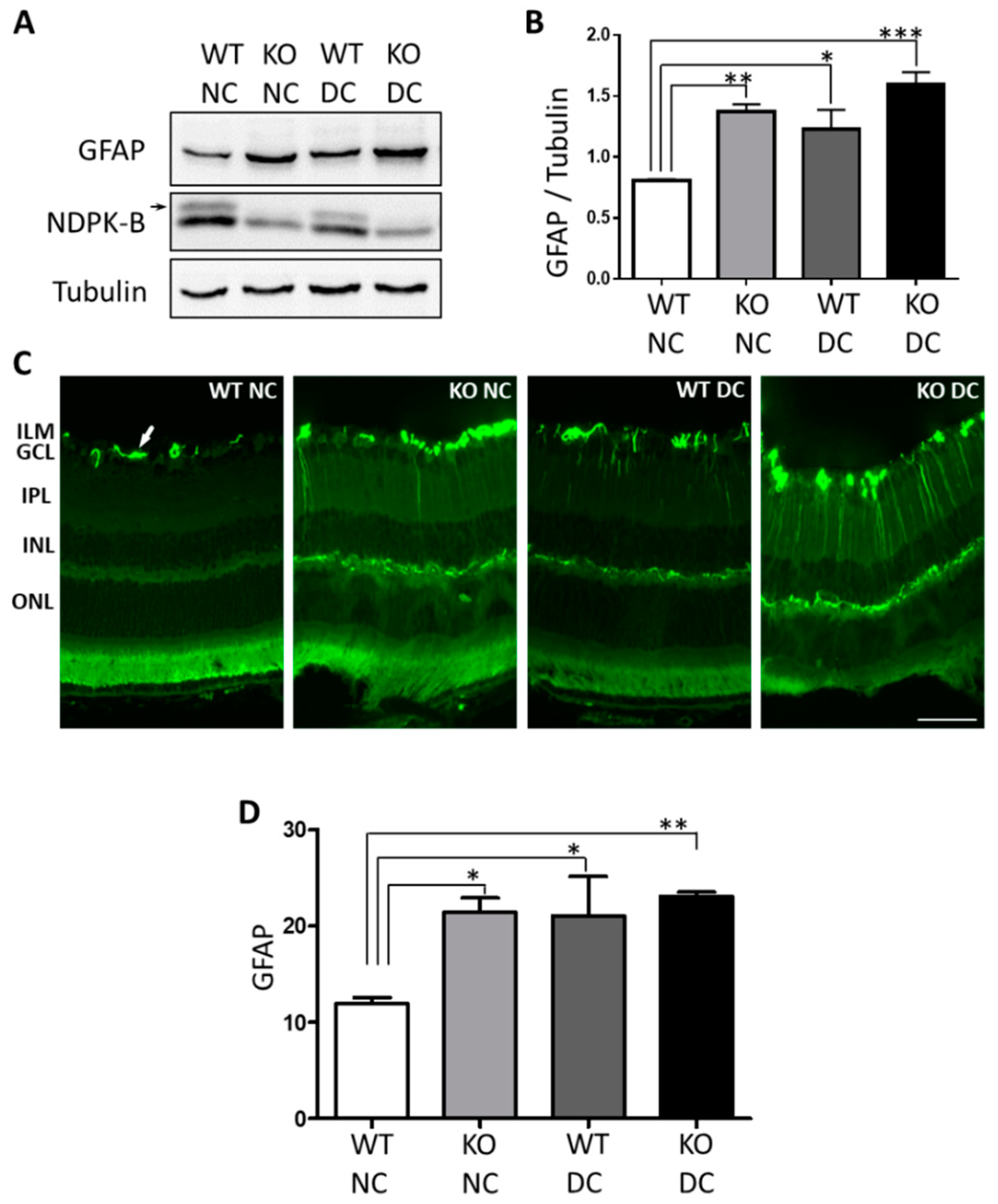
3.1. Müller Cells Are Activated in NDPK-B Deficient Retinas

Müller cell activation is an important process in the development of diabetic retinopathy [6]. Vascular degeneration occurring in NDPK-B deficient retinas without hyperglycemia mimics vascular pathology in DR [24]. Therefore, we investigated firstly, if glial cells were activated in the NDPK-B knockout (KO) retinas by detecting the expression of GFAP using immunoblotting and immunofluorescence. As shown in Figure 1A,B, in NDPK-B KO non-diabetic (NC) retinas, the expression of GFAP increased significantly in comparison with WT non-diabetic retinas (WT NC) (p < 0.01 vs. WT NC). Diabetes (DC) significantly enhanced GFAP expression in WT retinas as expected (p < 0.05, WT DC vs. WT NC). GFAP expression in diabetic NDPK-B KO (KO DC) retinas were also significantly enhanced compared to WT NC (p < 0.001), but diabetes induction in KO animals did not further enhance GFAP expression as compared to WT DC and KO NC retinas.
Additionally, we assessed the localization of elevated GFAP expression in the NDPK-B deficient retinas by staining the retinal paraffin sections with GFAP. As shown in Figure 1C, GFAP in WT NC retinas was predominantly localized in the astrocytes. Diabetes induced increased GFAP expression not only in the astrocytes but also a fiber-like staining pattern across the entire WT retina, indicating an enhanced expression of GFAP in the Müller cells. NDPK-B deficient retinas showed GFAP staining pattern similar to WT DC retinas, exhibiting markedly enhanced GFAP expression in astrocytes and Müller cells. There was significant increase in GFAP expression in KO NC/DC and WT DC compared to WT NC, but no difference between WT/KO DC as well as KO NC/DC. In the negative control staining, no signal was detected in the astrocytes or in the Müller cells (data not shown). GFAP expression by astrocytes in WT NC and by astrocyte and Müller cells in WT DC and KO retinas were confirmed by co-staining GFAP with the Müller cell marker GS (Supplementary Figure S1). Taken together, the elevated levels of GFAP in both the NDPK-B deficient and diabetic retinas verify the activation of retinal astrocytes/Müller cells, confirming the similarity between these two models [24].

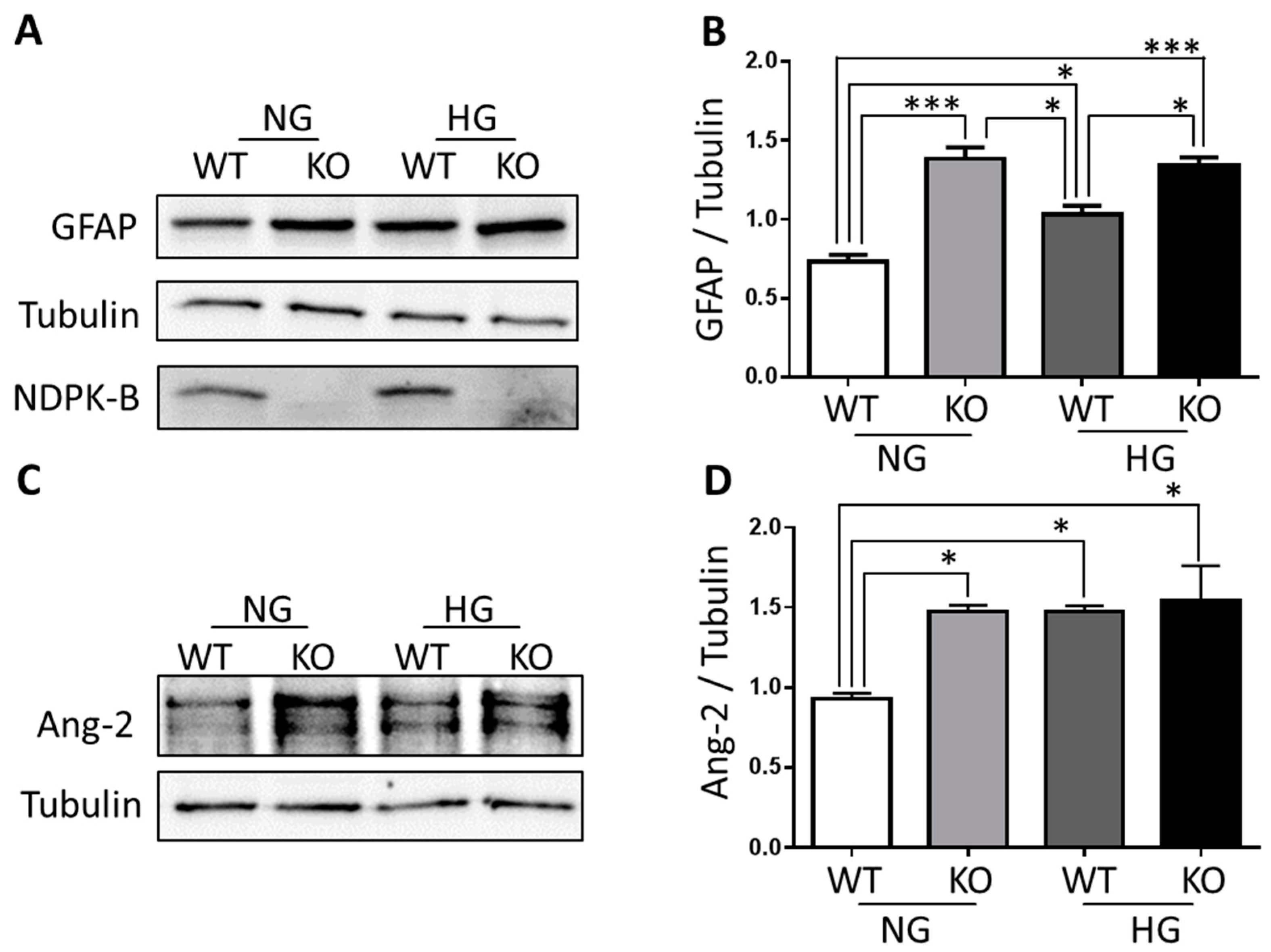
3.2. Ang-2 Is Preferentially Upregulated in Müller Cells in the Retina during Vascular Degeneration

Our previous data have shown that diabetes as well as the loss of NDPK-B caused a significant increase in Ang-2 levels in the retina [24]. To identify the source of upregulated Ang-2 in the NDPK-B retinas, we stained Ang-2 in retinal paraffin sections and investigated its localization. As shown in Figure 2, Ang-2 prominently localized in fiber-like structure spanning across the entire thickness of the retina, mostly in the inner retina. When the sections were co-stained against CRALBP, a Müller cell marker, the majority of Ang-2 co-localized with CRALBP, identifying retinal Müller cells as the major source of retinal Ang-2. In WT NC retinas Ang-2 was only detected at modest levels, whereas under NDPK-B deficient and diabetic conditions, Ang-2 was expressed abundantly in Müller cells. The Ang-2 staining intensity in diabetic NDPK-B KO retinas was similar to NDPK-B KO NC and WT DC retinas. Ang-2 levels were significantly enhanced in KO NC, WT DC, and KO DC groups compared to WT NC. These results confirm our previously published data showing that Ang-2 levels are elevated in hyperglycemia and in NDPK-B deficient retinas [24]. The main source of the upregulated Ang-2 in hyperglycemia and NDPK-B deficient retinas appears to be the Müller cells.

3.3. NDPK-B Deficiency Caused Enhancement of Ang-2 in Isolated Müller Cells

To verify Müller cells as a source of Ang-2 expression in NDPK B-deficient retinas and to further investigate the underlying mechanisms, we isolated Müller cells from NDPK-B KO and WT mice. The Müller cells were characterized by the positive staining for GFAP, GS, and CRALBP (Supplementary Figure S2). Firstly, we examined the GFAP levels of the WT and KO cells using Western blotting to determine the activation status of the isolated cells. As shown in Figure 3A,B, a basal GFAP expression was detectable in WT cells incubated in NG condition. In WT cells stimulated with HG, GFAP levels increased significantly compared with WT NG cells (p < 0.05). In NDPK-B deficiency GFAP levels were even higher (p < 0.001 vs. WT NG). GFAP levels in NDPK-B KO NG are higher than WT HG cells, indicating KO NG Müller cells are more active than the WT HG cells. In KO cells stimulated with HG, GFAP levels were significantly higher compared with WT cells stimulated with HG (p < 0.05), but were not further increased compared to KO NG cells. The deficiency of NDPK-B in the KO cells was confirmed by immunoblotting. These data confirm the previously stated results from the retinal immunofluorescence and demonstrate that NDPK-B deficiency induces the Müller cell activation, thereby mimicking the effect of hyperglycemia/high glucose.
Subsequently, we examined Ang-2 expression in the isolated Müller cells by Western blotting (Figure 3C,D). HG treatment significantly increased the Ang-2 expression (p < 0.05 vs. WT NG), which is in agreement with previously published data [28]. Similar to the whole retina lysates, NDPK-B deficiency increased the Ang-2 content in cultured Müller cells to a similar level observed in WT cells stimulated with HG (p < 0.05 vs. WT NG). However, when NDPK-B KO cells were treated with HG, no further elevation of Ang-2 was detected. There was no difference in Ang-2 levels between KO NG, WT HG and KO HG. The data are in agreement with those on Ang-2 regulation in the diabetic NDPK-B deficient retinas we published before [24]. To assess the transcriptional regulation of Ang-2, we performed quantitative PCR in Müller cells isolated from KO and WT retinas with and without HG stimulation. Neither NDPK-B deficiency nor high glucose stimulation regulated Ang-2 expression in Müller cells (Supplementary Figure S3).

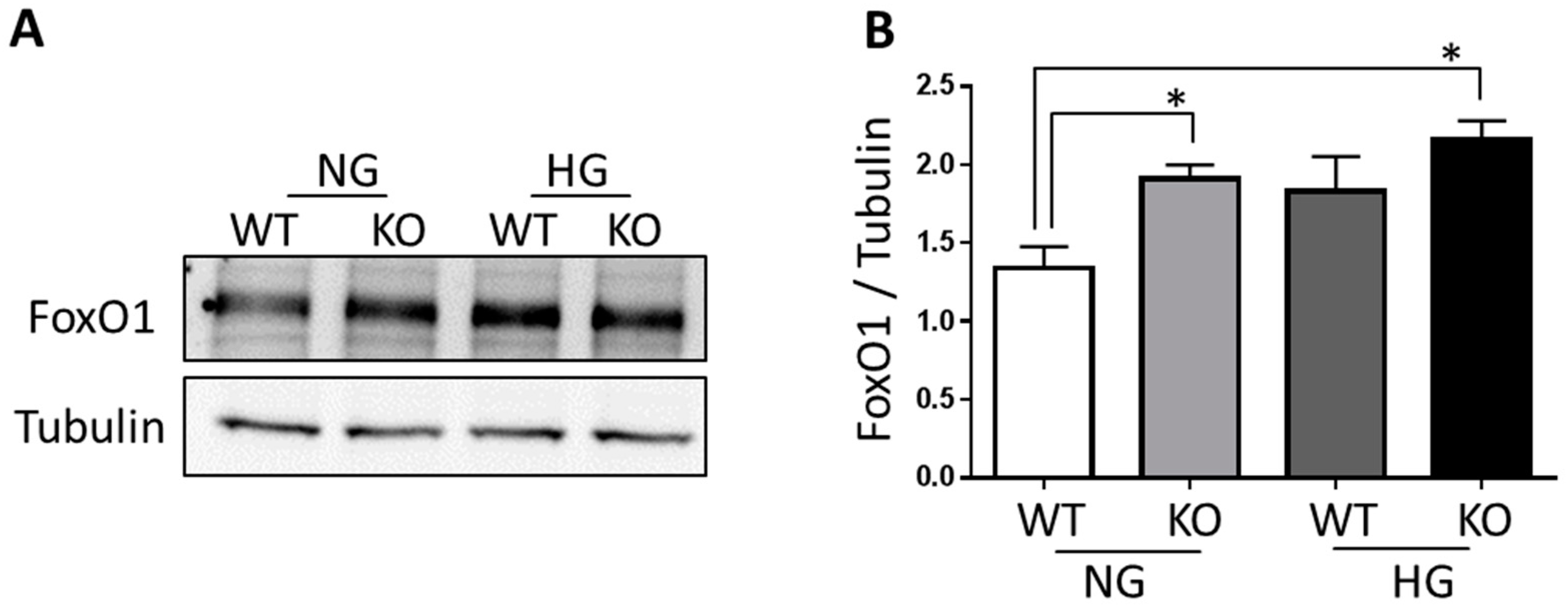
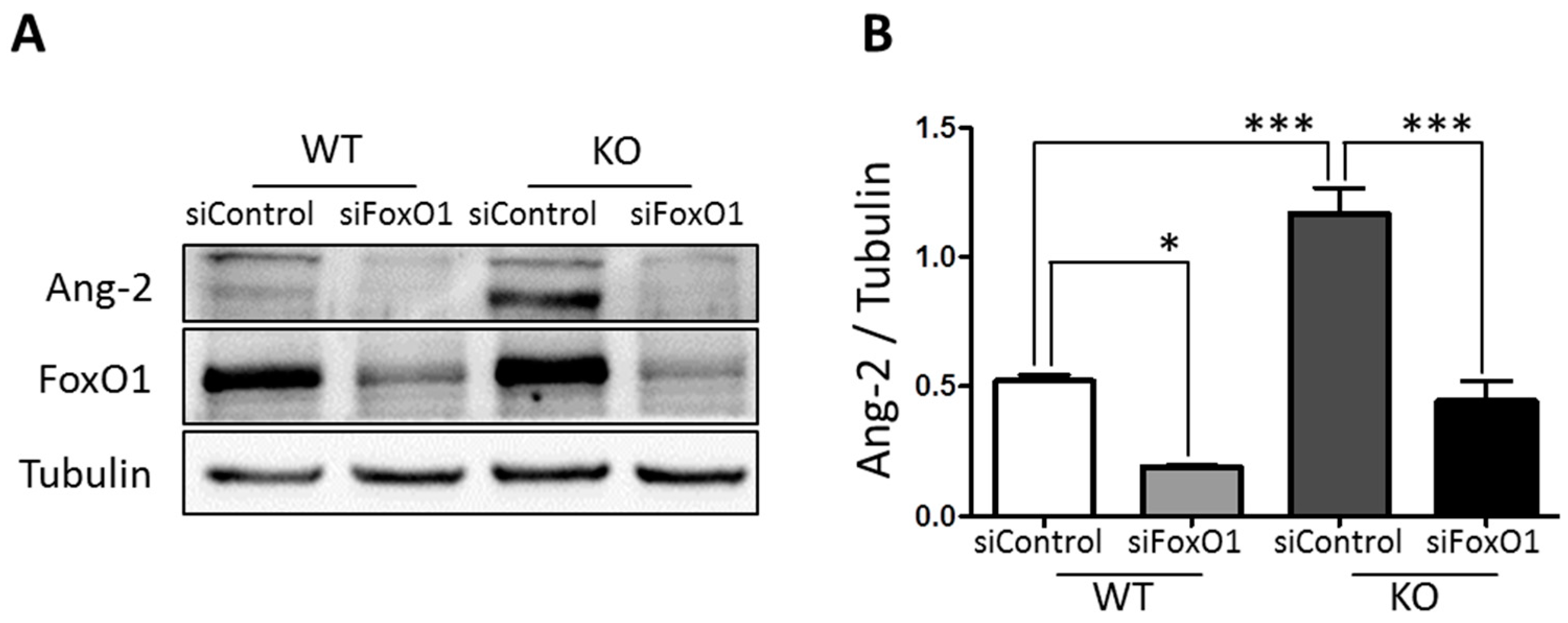
3.4. FoxO1 Is Required for NDPK-B Deficiency Induced Ang-2 Upregulation in Müller Cells

The transcription factor FoxO1 has been shown to regulate Ang-2 expression [29,30]. To examine whether FoxO1 is involved in the NDPK-B deficiency-induced Ang-2 upregulation, we estimated the level of FoxO1 in isolated NDPK-B KO Müller cells. As shown in Figure 4A,B, in NDPK-B KO cells, FoxO1 expression increased significantly compared to WT Müller cells under NG condition (p < 0.05); FoxO1 level did not further increase when KO Müller cells were stimulated with HG. In WT Müller cells stimulated with HG, a similar increase in the FoxO1 level was observed, which however did not reach statistical significance. Nevertheless, the data imply a possible role for FoxO1 in the regulation of Ang-2 in NDPK-B deficient Müller cells.
To examine whether FoxO1 controls the enhancement of Ang-2 induced by NDPK-B deficiency in Müller cells, we performed siRNA-mediated knockdown of FoxO1 and quantified the expression of Ang-2. As shown in Figure 5, FoxO1 depletion was successfully achieved by siRNA-mediated gene knockdown. In WT Müller cells, FoxO1 knockdown resulted in a decrease in Ang-2 levels (WT siControl vs. WT siFoxO1: p < 0.05). In KO Müller cells, NDPK-B deficiency significantly enhanced Ang-2 (KO siControl vs. WT siControl: p < 0.001). This increase of Ang-2 in NDPK-B KO Müller cells was suppressed by FoxO1 knockdown (KO siFoxO1 vs. KO siControl: p < 0.001). Taken together, these data argue for FoxO1 as an important mediator in NDPK-B deficiency-induced Ang-2 upregulation in Müller cells during vascular degeneration.

4. Discussion

In this study, we demonstrated that NDPK-B deficiency led to an activation of neuroglia in the retina during vascular degeneration, and that Ang-2 expression was strongly and preferentially upregulated in cells stained by the retinal Müller cell marker CRALBP. We confirmed, by isolation and culture of Müller cells, that enhanced Ang-2 expression due to NDPK-B deficiency indeed occurred in Müller cells and required the transcription factor FoxO1 as mediator for the Ang-2 upregulation. Importantly, hyperglycemia or HG treatment of WT Müller cells caused a similar activation of Müller cells and upregulation of Ang-2 expression upon NDPK-B deficiency.
We have previously demonstrated that NDPK-B deficiency is a risk factor for development of DR and showed that the retinal level of Ang-2 is elevated in NDPK-B deficiency-related vascular degeneration in the eyes [24]. Ang-2 is normally produced and released by endothelial cells [31,32]. In the retina, however, Müller cells are considered to be another important source for Ang-2 [11,12]. Although in our previous study, a similar upregulation of Ang-2 was found in endothelial cells [24], the immunofluorescence staining of Ang-2 in the retina performed herein fully supports that Müller cells are a major source of Ang-2 in the retina thus at least partially responsible for driving the vasoregressive pathology. In accordance to this, transgenic mOpsinhAng-2 mice with overexpression of human Ang-2 in the photoreceptor cells exhibited reduced pericyte coverage [33] and intravitreal injection of recombinant Ang-2 led to pericyte dropout [5]. These data show that Ang-2 secretion in the retina leads to pericyte loss independent of the source of Ang-2. As Müller cells and endothelial cells synergistically overproduce Ang-2 in the retina, both cell types are likely responsible for the DR-like pathology occurring under NDPK-B deficiency. How NDPK-B regulates Ang-2 remains elusive. Berberich et al. reported that NDPK-B may regulate gene transcriptional elements through the NDPK-B/PuF binding site [34]. Our data demonstrated that NDPK-B likely regulates Ang-2 through translational but not transcriptional levels, although NDPK-B may act as a co-transcriptional regulator. Ang-2 might be regulated by NDPK-B in an indirect manner.
We found that the presence and expression level of the transcription factor FoxO1 is important for the NDPK-B deficiency-induced Ang-2 in activated Müller cells isolated from NDPK-B deficient and littermate WT retinas. Whether the observed increase in FoxO1 content occurs also in other cells of the retina is currently not known. Due to lack of antibodies detecting FoxO1 in retinal paraffin sections and cryosections, we were not able to address this question directly. Nevertheless, we recently found that siRNA-mediated depletion of NDPK-B in endothelial cells increased the FoxO1 content, and like HG treatment, induced the upregulation of Ang-2 [24]. These data indicate that FoxO1 might be associated with the upregulation of Ang-2 in retinal endothelial cells as well as Müller cells.
Interestingly, FoxO1 might also be the regulatory factor where the effects of NDPK-B deficiency and hyperglycemia/HG treatment converge. Levels of FoxO1 tended to be increased in HG treated Müller cells, and the NDPK-B deficiency-induced upregulation of Ang-2 was attenuated by FoxO1 depletion. Indeed, a transcriptional regulation of Ang-2 by FoxO1 has been reported previously [29,30]. FoxO1 plays an important role in the insulin pathway [35,36], and is an apoptotic factor in retinal endothelial cells and pericytes [37,38]. The activity of FoxO1 can be regulated by multiple pathways, such as the insulin pathway though IRS-1 and Akt, the ROS through c-Jun N-terminal kinase (JNK) signaling [35]. Furthermore, O-GlcNAc modification of FoxO1 increases its activity in hepatocytes [39,40]. How NDPK-B deficiency upregulates FoxO1 in Müller cells remains unclear, but taking into account that high glucose levels also enhance protein O-GlcNAcylation of proteins, this might be an interesting hypothesis. On the other hand, HG also upregulates Ang-2 through enhanced O-GlcNAc and methylglyoxal modification of the transcription factors Sp3 [28]. Thus, other possibilities have to be considered as well. Therefore, more work is needed to identify how NDPK-B-deficiency and hyperglycemia regulate the activity and content of FoxO1 in endothelial and Müller cells.

Supplementary Materials

The following are available online at https://0-www-mdpi-com.brum.beds.ac.uk/2571-6980/1/1/19/s1, Figure S1: GFAP is expressed in astrocytes and Müller cells in the retina, Figure S2: Characterization of Müller cells in vitro, Figure S3: Ang-2 RNA expression is unaltered in NDPK-B KO NG/HG and WT NG/HG Müller cells.

Author Contributions

Conceptualization, H.-P.H., T.W., and Y.F.; Formal analysis, Y.Q., H.H., and A.C.; Funding acquisition, Y.F.; Investigation, Y.Q., H.H., L.D.T., F.S.B., and A.C.; Methodology, Y.Q., H.H., L.D.T., F.S.B., and A.C.; Project administration, T.W. and Y.F.; Supervision, H.-P.H., T.W., and Y.F.; Validation, H.H.; Writing—original draft, Y.Q. and Y.F.; Writing—review & editing, H.H., A.C., H.-P.H., T.W., and Y.F.

Funding

This research was funded by the European Foundation for the Study of Diabetes (EFSD, Novartis-2014, Y.F.), the Deutsche Forschungsgemeinschaft (DFG, FE 969/2-1) and the Deutsche Diabetes Gesellschaft (DDG, A.C.).

Acknowledgments

The authors thank Heike Rauscher for her excellent technical assistance. We are grateful to Michael Potente from Max Planck Institute for Heart and Lung Research, Bad Nauheim, for his support.

Conflicts of Interest

The authors declare no conflict of interest. The funders had no role in the design of the study; in the collection, analyses, or interpretation of data; in the writing of the manuscript, and in the decision to publish the results.

References

  1. Hammes, H.P.; Feng, Y.; Pfister, F.; Brownlee, M. Diabetic retinopathy: Targeting vasoregression. Diabetes 2011, 60, 9–16. [Google Scholar] [CrossRef] [PubMed]
  2. Frank, R.N. Diabetic retinopathy. N. Engl. J. Med. 2004, 350, 48–58. [Google Scholar] [CrossRef] [PubMed]
  3. Feng, Y.; Vom Hagen, F.; Wang, Y.; Beck, S.; Schreiter, K.; Pfister, F.; Hoffmann, S.; Wagner, P.; Seeliger, M.; Molema, G. The absence of angiopoietin-2 leads to abnormal vascular maturation and persistent proliferative retinopathy. Thromb. Haemost. 2009, 102, 120–130. [Google Scholar] [CrossRef] [PubMed]
  4. Hammes, H.-P.; Lin, J.; Wagner, P.; Feng, Y.; Vom Hagen, F.; Krzizok, T.; Renner, O.; Breier, G.; Brownlee, M.; Deutsch, U. Angiopoietin-2 causes pericyte dropout in the normal retina: Evidence for involvement in diabetic retinopathy. Diabetes 2004, 53, 1104–1110. [Google Scholar] [CrossRef] [PubMed]
  5. Pfister, F.; Wang, Y.; Schreiter, K.; Vom Hagen, F.; Altvater, K.; Hoffmann, S.; Deutsch, U.; Hammes, H.-P.; Feng, Y. Retinal overexpression of angiopoietin-2 mimics diabetic retinopathy and enhances vascular damages in hyperglycemia. Acta Diabetol. 2010, 47, 59–64. [Google Scholar] [CrossRef] [PubMed]
  6. Bringmann, A.; Pannicke, T.; Grosche, J.; Francke, M.; Wiedemann, P.; Skatchkov, S.N.; Osborne, N.N.; Reichenbach, A. Müller cells in the healthy and diseased retina. Prog. Retin. Eye Res. 2006, 25, 397–424. [Google Scholar] [CrossRef] [PubMed]
  7. Bringmann, A.; Iandiev, I.; Pannicke, T.; Wurm, A.; Hollborn, M.; Wiedemann, P.; Osborne, N.N.; Reichenbach, A. Cellular signaling and factors involved in Müller cell gliosis: Neuroprotective and detrimental effects. Prog. Retin. Eye Res. 2009, 28, 423–451. [Google Scholar] [CrossRef] [PubMed]
  8. Zong, H.; Ward, M.; Madden, A.; Yong, P.; Limb, G.; Curtis, T.; Stitt, A. Hyperglycaemia-induced pro-inflammatory responses by retinal Müller glia are regulated by the receptor for advanced glycation end-products (rage). Diabetologia 2010, 53, 2656–2666. [Google Scholar] [CrossRef] [PubMed]
  9. Bringmann, A.; Pannicke, T.; Biedermann, B.; Francke, M.; Iandiev, I.; Grosche, J.; Wiedemann, P.; Albrecht, J.; Reichenbach, A. Role of retinal glial cells in neurotransmitter uptake and metabolism. Neurochem. Int. 2009, 54, 143–160. [Google Scholar] [CrossRef] [PubMed]
  10. Reichelt, W.; Stabel-Burow, J.; Pannicke, T.; Weichert, H.; Heinemann, U. The glutathione level of retinal Müller glial cells is dependent on the high-affinity sodium-dependent uptake of glutamate. Neuroscience 1997, 77, 1213–1224. [Google Scholar] [CrossRef]
  11. Bai, Y.; Ma, J.X.; Guo, J.; Wang, J.; Zhu, M.; Chen, Y.; Le, Y.Z. Müller cell-derived VEGF is a significant contributor to retinal neovascularization. J. Pathol. 2009, 219, 446–454. [Google Scholar] [CrossRef] [PubMed]
  12. Hammes, H.-P.; Porta, M. Subject Index. In Experimental Approaches to Diabetic Retinopathy; Karger Publishers: Basel, Switzerland, 2010; Volume 20, pp. 229–232. [Google Scholar]
  13. Matsumura, T.; Hammes, H.-P.; Thornalley, P.J.; Edelstein, D.; Brownlee, M. Hyperglycemia increases angiopoietin-2 expression in retinal muller cells through superoxide-induced overproduction of [Alpha]-Oxoaldehyde age precursors. Diabetes 2000, 49, A55. [Google Scholar]
  14. Gilles, A.-M.; Presecan, E.; Vonica, A.; Lascu, I. Nucleoside diphosphate kinase from human erythrocytes. Structural characterization of the two polypeptide chains responsible for heterogeneity of the hexameric enzyme. J. Biol. Chem. 1991, 266, 8784–8789. [Google Scholar] [PubMed]
  15. Lascu, I.; Gonin, P. The catalytic mechanism of nucleoside diphosphate kinases. J. Bioenerg. Biomembr. 2000, 32, 237–246. [Google Scholar] [CrossRef] [PubMed]
  16. Janin, J.; Dumas, C.; Moréra, S.; Xu, Y.; Meyer, P.; Chiadmi, M.; Cherfils, J. Three-dimensional structure of nucleoside diphosphate kinase. J. Bioenerg. Biomembr. 2000, 32, 215–225. [Google Scholar] [CrossRef] [PubMed]
  17. Fancsalszky, L.; Monostori, E.; Farkas, Z.; Pourkarimi, E.; Masoudi, N.; Hargitai, B.; Bosnar, M.H.; Deželjin, M.; Zsákai, A.; Vellai, T. NDK-1, the homolog of NM23-H1/H2 regulates cell migration and apoptotic engulfment in C. Elegans. PLoS ONE 2014, 9, e92687. [Google Scholar] [CrossRef] [PubMed] [Green Version]
  18. Fournier, H.-N.; Albigès-Rizo, C.; Block, M.R. New insights into NM23 control of cell adhesion and migration. J. Bioenerg. Biomembr. 2003, 35, 81–87. [Google Scholar] [CrossRef] [PubMed]
  19. Snider, N.T.; Altshuler, P.J.; Omary, M.B. Modulation of cytoskeletal dynamics by mammalian nucleoside diphosphate kinase (NDPK) proteins. Naunyn-Schmiedeberg’s Arch. Pharmacol. 2015, 388, 189–197. [Google Scholar] [CrossRef] [PubMed]
  20. Hippe, H.-J.; Luedde, M.; Lutz, S.; Koehler, H.; Eschenhagen, T.; Frey, N.; Katus, H.A.; Wieland, T.; Niroomand, F. Regulation of cardiac camp synthesis and contractility by nucleoside diphosphate kinase b/g protein βγ dimer complexes. Circ. Res. 2007, 100, 1191–1199. [Google Scholar] [CrossRef] [PubMed]
  21. Hippe, H.-J.; Lutz, S.; Cuello, F.; Knorr, K.; Vogt, A.; Jakobs, K.H.; Wieland, T.; Niroomand, F. Activation of heterotrimeric G proteins by a high energy phosphate transfer via nucleoside diphosphate kinase (NDPK) B and Gβ subunits specific activation of Gsα by an NDPK B·Gβγ complex in H10 cells. J. Biol. Chem. 2003, 278, 7227–7233. [Google Scholar] [CrossRef] [PubMed]
  22. Hippe, H.-J.; Wolf, N.M.; Abu-Taha, H.I.; Lutz, S.; Le Lay, S.; Just, S.; Rottbauer, W.; Katus, H.A.; Wieland, T. Nucleoside diphosphate kinase B is required for the formation of heterotrimeric G protein containing caveolae. Naunyn-Schmiedeberg’s Arch. Pharmacol. 2011, 384, 461–472. [Google Scholar] [CrossRef] [PubMed] [Green Version]
  23. Feng, Y.; Gross, S.; Wolf, N.M.; Butenschön, V.M.; Qiu, Y.; Devraj, K.; Liebner, S.; Kroll, J.; Skolnik, E.Y.; Hammes, H.-P. Nucleoside diphosphate kinase B regulates angiogenesis through modulation of vascular endothelial growth factor receptor type 2 and endothelial adherens junction proteins. Arterioscler. Throm. Vasc. Biol. 2014, 34, 2292–2300. [Google Scholar] [CrossRef] [PubMed]
  24. Qiu, Y.; Zhao, D.; Butenschön, V.-M.; Bauer, A.T.; Schneider, S.W.; Skolnik, E.Y.; Hammes, H.-P.; Wieland, T.; Feng, Y. Nucleoside diphosphate kinase B deficiency causes a diabetes-like vascular pathology via up-regulation of endothelial angiopoietin-2 in the retina. Acta Diabetol. 2016, 53, 81–89. [Google Scholar] [CrossRef] [PubMed]
  25. Di, L.; Srivastava, S.; Zhdanova, O.; Sun, Y.; Li, Z.; Skolnik, E.Y. Nucleoside diphosphate kinase B knock-out mice have impaired activation of the k+ channel KCa3. 1, resulting in defective T cell activation. J. Biol. Chem. 2010, 285, 38765–38771. [Google Scholar] [CrossRef] [PubMed]
  26. Hicks, D.; Courtois, Y. The growth and behaviour of rat retinal Müller cells in vitro 1. An improved method for isolation and culture. Exp. Eye Res. 1990, 51, 119–129. [Google Scholar] [CrossRef]
  27. Hu, J.; Popp, R.; Frömel, T.; Ehling, M.; Awwad, K.; Adams, R.H.; Hammes, H.-P.; Fleming, I. Müller glia cells regulate Notch signaling and retinal angiogenesis via the generation of 19,20-dihydroxydocosapentaenoic acid. J. Exp. Med. 2014, 211, 281–295. [Google Scholar] [CrossRef] [PubMed]
  28. Yao, D.; Taguchi, T.; Matsumura, T.; Pestell, R.; Edelstein, D.; Giardino, I.; Suske, G.; Rabbani, N.; Thornalley, P.J.; Sarthy, V.P. High glucose increases angiopoietin-2 transcription in microvascular endothelial cells through methylglyoxal modification of mSin3A. J. Biol. Chem. 2007, 282, 31038–31045. [Google Scholar] [CrossRef] [PubMed]
  29. Daly, C.; Wong, V.; Burova, E.; Wei, Y.; Zabski, S.; Griffiths, J.; Lai, K.-M.; Lin, H.C.; Ioffe, E.; Yancopoulos, G.D. Angiopoietin-1 modulates endothelial cell function and gene expression via the transcription factor FKHR (FOXO1). Genes Dev. 2004, 18, 1060–1071. [Google Scholar] [CrossRef] [PubMed] [Green Version]
  30. Potente, M.; Urbich, C.; Sasaki, K.-I.; Hofmann, W.K.; Heeschen, C.; Aicher, A.; Kollipara, R.; DePinho, R.A.; Zeiher, A.M.; Dimmeler, S. Involvement of Foxo transcription factors in angiogenesis and postnatal neovascularization. J. Clin. Investig. 2005, 115, 2382–2392. [Google Scholar] [CrossRef] [PubMed] [Green Version]
  31. Augustin, H.G.; Koh, G.Y.; Thurston, G.; Alitalo, K. Control of vascular morphogenesis and homeostasis through the angiopoietin-Tie system. Nat. Rev. Mol. Cell Biol. 2009, 10, 165–177. [Google Scholar] [CrossRef] [PubMed]
  32. Hanahan, D. Signaling vascular morphogenesis and maintenance. Science 1997, 277, 48–50. [Google Scholar] [CrossRef] [PubMed]
  33. Feng, Y.; Vom, H.F.; Pfister, F.; Djokic, S.; Hoffmann, S.; Back, W.; Wagner, P.; Lin, J.; Deutsch, U.; Hammes, H.P. Impaired pericyte recruitment and abnormal retinal angiogenesis as a result of angiopoietin-2 overexpression. Thromb. Haemost. 2007, 97, 99–108. [Google Scholar] [CrossRef] [PubMed]
  34. Berberich, S.; Postel, E. PuF/NM23-H2/NDPK-B transactivates a human c-myc promoter-CAT gene via a functional nuclease hypersensitive element. Oncogene 1995, 10, 2343–2347. [Google Scholar] [PubMed]
  35. Eijkelenboom, A.; Burgering, B.M.T. Foxos: Signalling integrators for homeostasis maintenance. Nat. Rev. Mol. Cell Biol. 2013, 14, 83–97. [Google Scholar] [CrossRef] [PubMed]
  36. Nakae, J.; Kitamura, T.; Silver, D.L.; Accili, D. The forkhead transcription factor Foxo1 (Fkhr) confers insulin sensitivity onto glucose-6-phosphatase expression. J. Clin. Investig. 2001, 108, 1359–1367. [Google Scholar] [CrossRef] [PubMed] [Green Version]
  37. Alikhani, M.; Roy, S.; Graves, D.T. FoxO1 plays an essential role in apoptosis of retinal pericytes. Mol. Vis. 2010, 16, 408–415. [Google Scholar] [PubMed]
  38. Behl, Y.; Krothapalli, P.; Desta, T.; Roy, S.; Graves, D.T. FoxO1 plays an important role in enhanced microvascular cell apoptosis and microvascular cell loss in type 1 and type 2 diabetic rats. Diabetes 2009, 58, 917–925. [Google Scholar] [CrossRef] [PubMed]
  39. Housley, M.P.; Rodgers, J.T.; Udeshi, N.D.; Kelly, T.J.; Shabanowitz, J.; Hunt, D.F.; Puigserver, P.; Hart, G.W. O-GlcNAc regulates FoxO activation in response to glucose. J. Biol. Chem. 2008, 283, 16283–16292. [Google Scholar] [CrossRef] [PubMed]
  40. Kuo, M.; Zilberfarb, V.; Gangneux, N.; Christeff, N.; Issad, T. O-glycosylation of FoxO1 increases its transcriptional activity towards the glucose 6-phosphatase gene. FEBS Lett. 2008, 582, 829–834. [Google Scholar] [CrossRef] [PubMed] [Green Version]
Figure 1. Müller cells are activated in NDPK-B deficient retinas. (A,B): representative Western blot and quantification of GFAP (glial fibrillary acidic protein) expression in the retina. In both KO NC and WT DC retina, GFAP levels significantly increased compared to WT NC retinas; GFAP level in KO DC retinas is similar to KO NC and WT DC retinas. (C,D): Immunofluorescence staining and quantification of GFAP in the retina. GFAP localized in astrocytes in WT NC retinas (arrow). Diabetes increased a radial expression pattern of GFAP immunoreactivity similar to Müller cells. NDPK-B deficiency induced a staining pattern similar to WT DC. KO: NDPK-B knockout; DC: diabetic; NC: non-diabetic; WT: wild type; ILM: inner limiting membrane; GCL: ganglion cell layer; IPL: inner plexiform layer; INL: inner nuclear layer; ONL: outer nuclear layer. Scale bar: 50 μm. * p < 0.05, ** p < 0.01, n = 3–4 in each group.
Neuroglia 01 00019 g001
Figure 2. Ang-2 is upregulated in Müller cells in the retina. Immunofluorescence staining (A) and quantification (B) of Ang-2. Ang-2 (red) and the Müller cell marker CRALBP (green) were stained in the retina. Significantly enhanced levels of Ang-2 were detected in KO NC, WT DC, and KO DC conditions compared to WT NC. As shown the merged images, the majority of Ang-2 was detected in the CRALBP positive Müller cells. ILM: inner limiting membrane; GCL: ganglion cell layer; IPL: inner plexiform layer; INL: inner nuclear layer; ONL: outer nuclear layer. Scale bar: 50 μm. ** p < 0.01, *** p < 0.001, n = 4–5 in each group.
Neuroglia 01 00019 g002
Figure 3. NDPK-B deficiency enhanced Ang-2 content in isolated Müller cells. (A,B): Representative Western blot and quantification of GFAP expression in the Müller cells, respectively. In both KO normal glucose (NG) cells and WT high glucose (HG) cells, the level of GFAP increased significantly compared to WT NG cells. GFAP in KO HG cells is significantly higher than in WT HG cells. * p < 0.05, *** p < 0.001, n = 4. (C,D): Representative Western blot and quantification of Ang-2 expression in the isolated NDPK-B KO Müller cells. Both NDPK-B deficiency and HG induced a significant increase in Ang-2 levels in the Müller cells. The level of Ang-2 in KO HG cells was similar as in KO NG cells and WT HG cells. * p < 0.05, n = 4.
Neuroglia 01 00019 g003
Figure 4. FoxO1 is increased in NDPK-B KO Müller cells. (A,B): Representative Western blot and quantification of FoxO1 content in the Müller cells, respectively. NDPK-B deficiency significantly increased FoxO1 in the Müller cells compared to WT NG cells. The FoxO1 level in KO HG cells is similar to KO NG cells. * p < 0.05, n = 5.
Neuroglia 01 00019 g004
Figure 5. FoxO1 is required for Ang-2 upregulation induced by NDPK-B deficiency in Müller cells. (A,B): Representative Western blot and quantification of Ang-2 expression in Müller cells isolated from WT and NDPK-B deficient mice 48 h after siRNA transfection. FoxO1 knockdown suppressed the inhibited basal as well as NDPK-B deficiency induced Ang-2 content. siControl: control siRNA, siFoxO1: FoxO1 siRNA. * p < 0.05, *** p < 0.001, n = 4.
Neuroglia 01 00019 g005

