
Early Detection of Post-Endarterectomy Complication by Point-of-Care Ultrasound


Department of Emergency Medicine, National Cheng Kung University Hospital, College of Medicine, National Cheng Kung University, Tainan 70403, Taiwan
*
Author to whom correspondence should be addressed.
Reports 2021, 4(3), 21; https://doi.org/10.3390/reports4030021
Submission received: 11 June 2021
/
Revised: 3 July 2021
/
Accepted: 6 July 2021
/
Published: 8 July 2021
Abstract
:Endarterectomy is an effective intervention to remove the atheromatous plaque in the inner lining of the artery, aiming to revascularize the occluded/stenosed vessel in patients with peripheral arterial occlusive disease (PAOD). The most common wound-related complication is postoperative bleeding, followed by infection, hematoma, and seroma. However, hematoma complications with air surrounded have rarely been reported in clinical cases. Case presentation: A 90-year-old female patient visited our emergency department because of a rapidly growing hematoma with pulsatile bleeding over her right groin area. She had received bilateral percutaneous transluminal angioplasty with endarterectomy for PAOD one month prior. A point-of-care ultrasound revealed a large hypoechoic mass, with a dirty shadow on the right groin area. Computed tomography angiography showed a hematoma over her right femoral region, with free air surrounding the right femoral artery. Angiography revealed an irregular shaped lesion on the right femoral artery without contrast extravasation. The patient was diagnosed with right-femoral post-endarterectomy infection with infected hematoma, with the inclusion of air. She underwent urgent excision and repair of the right femoral artery infectious lesion, debridement of the infectious hematoma and stenting of the right external iliac artery, common femoral artery and superficial femoral artery.
1. Introduction
Peripheral arterial occlusive disease (PAOD) is a manifestation of atherosclerosis that affects blood perfusion to the distal portion of the arteries, followed by partial or complete blood perfusion impairment and tissue hypoperfusion. The management of PAOD includes the reduction in cardiovascular risk factors, pharmacotherapy of vasoactive drugs, and arterial revascularization via endovascular or open surgical treatment. Nevertheless, the precise therapeutic approach for PAOD patients depends on the disease stage, patient background, underlying disease, invasiveness, risk, prognosis and possible complications.
Endarterectomy is an effective intervention to remove the atheromatous plaque in the inner lining of the artery, aiming to revascularize the occluded/stenosed vessel in patients with PAOD. The most common wound-related complication is postoperative bleeding, followed by infection, hematoma, and seroma. However, emphysematous hematoma complications have rarely been reported in clinical cases. Here, we report a case of post-endarterectomy infection with hematoma and emphysema along the right femoral artery.
2. Case Presentation
A 90-year-old female patient visited our emergency department because of a rapidly growing hematoma, with pulsatile bleeding over her right groin area. She had received bilateral femoral endarterectomy with patched common femoral artery and superficial femoral artery for peripheral arterial occlusive disease in another hospital one month prior. The initial vital signs at triage were stable, with blood pressure of 145/78 mmHg, heart rate of 88 beats/min and respiratory rate of 19 breaths/min. The complete blood count revealed leukocytosis with segment predominance (white blood cell count: 14,900/mm3, Seg: 90%), a decreased hemoglobin level of 7.6 g/dL, and an elevated CRP level of 144 mg/dL. Her coagulation profile was normal, with APTT: 25.1 secs, PT: 12.1 secs, and PT (INR): 1.1
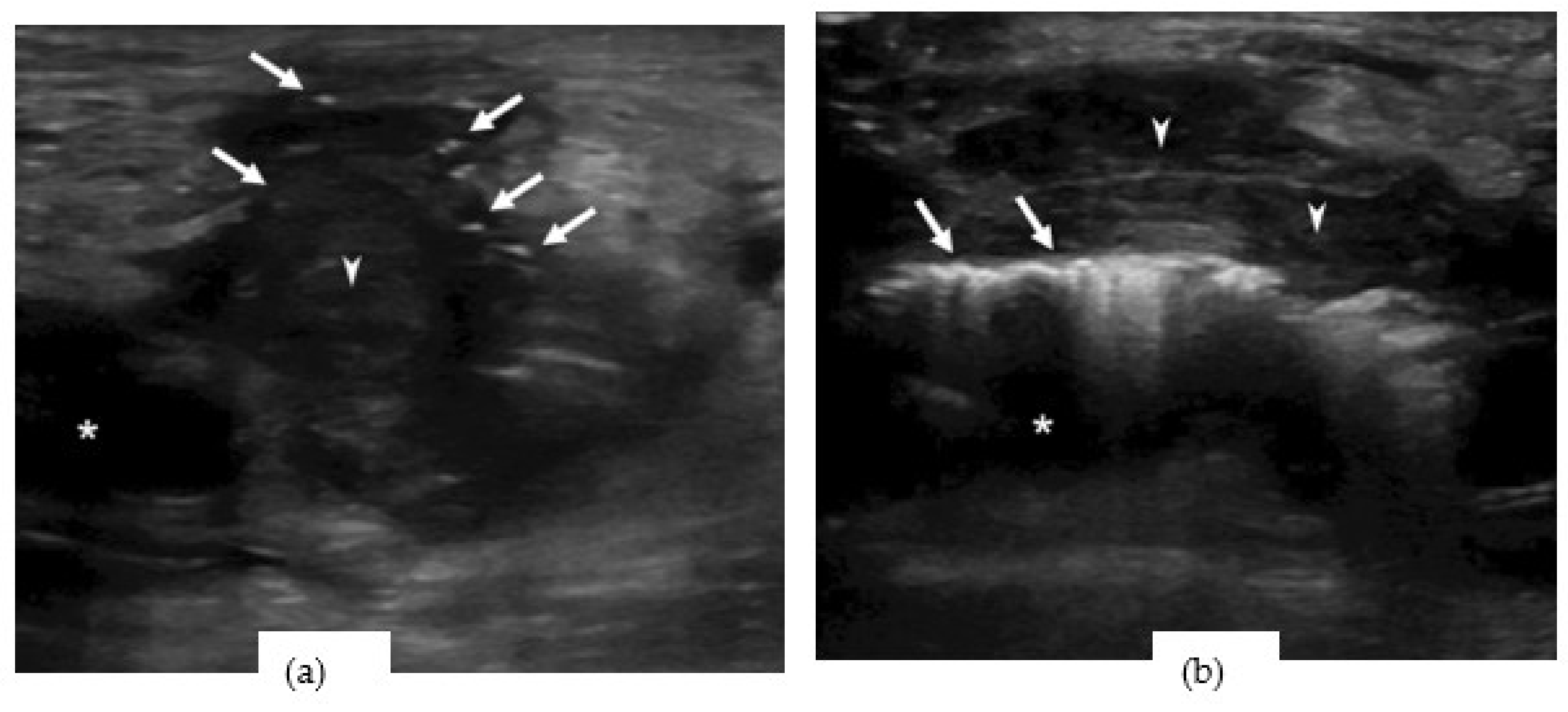
A point-of-care ultrasound (POCUS) revealed a large hypoechoic mass with a dirty shadow on the right groin area (Figure 1a). Dirty shadowing is thought to be produced by sound-reflecting materials such as subcutaneous free air [1]. An echogenic rim with dirty posterior shadowing along the pulsatile femoral artery was found next to the hematoma (Figure 1b). A video recording of the soft tissue echogram (Video S1 and Video S2) was obtained.
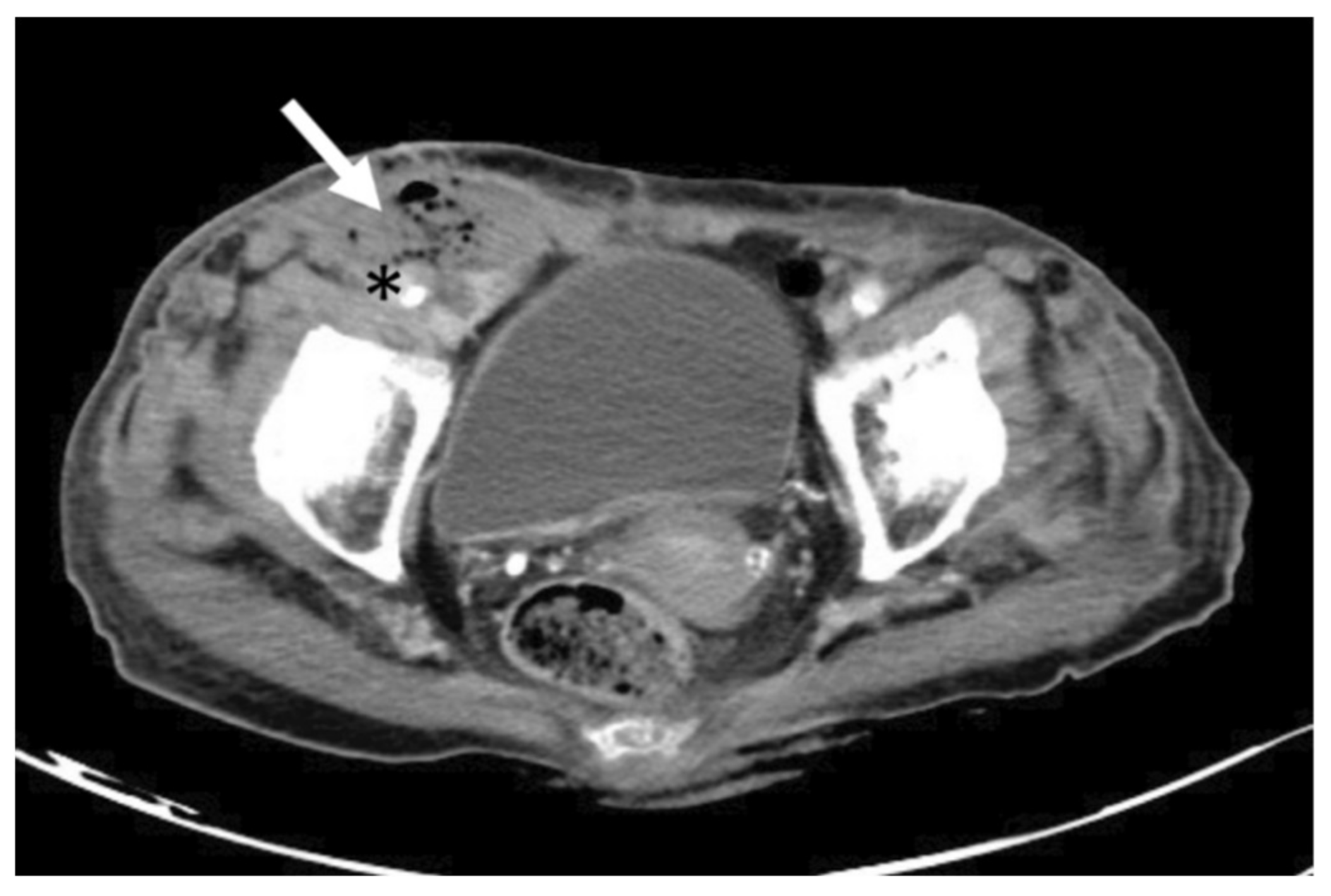
Computed tomography angiography (CTA) showed a hematoma over her right femoral region, with free air surrounding the right femoral artery (Figure 2).
The patient was then admitted to the cardiovascular surgery ward with a scheduled operation. However, emergency angiography and surgery were arranged the following morning, due to the patient’s unstable hemodynamic status and hemorrhagic shock. Initial angiography revealed a right femoral artery, irregular-shape lesion, without contrast extravasation and suspected hematoma compression (Figure 3). The irregular shape of the patched vessel wall was due to infection.
The patient was diagnosed with post-endarterectomy infection with hematoma along the right femoral artery. She underwent urgent excision and repair of the right femoral artery infectious vessel wall. A massive contrast extravasation from the patched, right, common femoral artery was noted after the infectious hematoma was evacuated. Due to the hematoma compression effect, the signs of active bleeding were not detected in the angiography. The infectious vessel wall was repaired smoothly after stenting of the right external iliac artery, common femoral artery, and superficial femoral artery. The patient has been hospitalized since the operation.
3. Discussion
Endarterectomy is an effective intervention for removing atheromatous plaque from the inner lining of the artery. Common femoral endarterectomy (CFE) is able to provide adequate blood supply via revascularization of the occluded/stenosed vessel, and is recognized as the preferred treatment for occlusive disease of the common femoral artery [2]. However, some studies have shown that CFE is not a low-risk invasive surgery. A study of 1843 cases of CFE reported a 3.4% mortality rate, and a wound-related complication rate of 8%, indicating that the risk of death and wound complications is not negligible [3]. Derksen et al. reported that 14% of CFE patients have surgical-site infections [4].
Although CTA scans are considered as the optimal diagnostic tool for vascular lesions, POCUS, which is able to detect specific signs, and is useful for early diagnosis in the emergency department [5]. POCUS has been advocated and widely used in emergency departments due to its noninvasiveness, rapid deployment, repeatability, real-time ability, lack of exposure to radiation, and high accuracy. Many ultrasound-based protocols have been established for clinical scenarios and different topics, such as an extended, focused assessment with sonography in trauma (eFAST), rapid ultrasound in shock (RUSH), bedside lung ultrasound in emergency (BLUE) and fluid administration, limited by lung sonography (FALLS) [6]. An ultrasonography with a high-frequency linear transducer (5–10 MHz) is suitable for imaging soft tissue and vascular lesion detection. A study comparing medical resource utilization and cost between the 135 patients who received point-of-care, limited ultrasonography and 127 control patients who received usual care in the emergency department found that patients who received point-of-care ultrasound administration had significantly reduced time to operative care, fewer complications, and lower medical expenses [7]. This case study reports the benefits of POCUS for imaging in superficial hematoma patients and emphasizes its implications for diagnosis.
4. Conclusions
Post-endarterectomy infection and hematoma are common. However, infected hematoma with the inclusion of air rarely appears after surgery. Computer tomography angiography is considered the optimal tool for diagnosing vascular lesions. POCUS with a high-frequency linear transducer (5–10 MHz) can rapidly and accurately detect infection signs (free air artifacts along vessels and hematoma).
Supplementary Materials
The following are available online at https://0-www-mdpi-com.brum.beds.ac.uk/article/10.3390/reports4030021/s1, Video S1: Infected hematoma with inclusion of air; Video S2: Echogenic rim with dirty posterior shadowing along the femoral artery.
Author Contributions
B.-K.C. and P.-W.C. contributed to acquisition of data. B.-K.C., P.-W.C. and C.-H.L. drafted the manuscript. P.-W.C. is the corresponding author who takes responsibility for the paper as a whole. All authors have read and agreed to the published version of the manuscript.
Funding
This research received no external funding.
Institutional Review Board Statement
This study fulfilled the requirement for a waiver of the review of the institutional review board.
Informed Consent Statement
Verbal consent was obtained from the patient.
Data Availability Statement
Not applicable.
Conflicts of Interest
The authors declare that they have no known competing financial interests or personal relationships that could have appeared to influence the work reported in this paper.
References
- Rubin, J.M.; Adler, R.S.; O Bude, R.; Fowlkes, J.B.; Carson, P. Clean and dirty shadowing at US: A reappraisal. Radiology 1991, 181, 231–236. [Google Scholar] [CrossRef] [PubMed]
- Kang, J.L.; Patel, V.I.; Conrad, M.F.; LaMuraglia, G.M.; Chung, T.; Cambria, R.P. Common femoral artery occlusive disease: Contemporary results following surgical endarterectomy. J. Vasc. Surg. 2008, 48, 872–877.e1. [Google Scholar] [CrossRef] [PubMed] [Green Version]
- Nguyen, B.-N.; Amdur, R.L.; Abugideiri, M.; Rahbar, R.; Neville, R.F.; Sidawy, A.N. Postoperative complications after common femoral endarterectomy. J. Vasc. Surg. 2015, 61, 1489–1494.e1. [Google Scholar] [CrossRef] [PubMed] [Green Version]
- Derksen, W.J.M.; Verhoeven, B.A.N.; Van De Mortel, R.H.W.; Moll, F.L.; De Vries, J.-P.P.M. Risk Factors for Surgical-Site Infection Following Common Femoral Artery Endarterectomy. Vasc. Endovasc. Surg. 2008, 43, 69–75. [Google Scholar] [CrossRef] [PubMed]
- Ijaz, M.; Shiloh, A.; Eisen, L. Point of Care Ultrasound: A Tool for Rapid Diagnosis of a Life-Threatening Hematoma. Chest 2016, 150, 239A. [Google Scholar] [CrossRef]
- Lee, L.; Decara, J.M. Point-of-Care Ultrasound. Curr. Cardiol. Rep. 2020, 22, 1–10. [Google Scholar] [CrossRef] [PubMed]
- Melniker, L.A.; Leibner, E.; McKenney, M.G.; Lopez, P.; Briggs, W.M.; Mancuso, C.A. Randomized Controlled Clinical Trial of Point-of-Care, Limited Ultrasonography for Trauma in the Emergency Department: The First Sonography Outcomes Assessment Program Trial. Ann. Emerg. Med. 2006, 48, 227–235. [Google Scholar] [CrossRef] [PubMed]
Figure 1.
Point-of-care ultrasound revealed (a) A hypoechoic mass (arrowhead) surrounding with a dirty shadow (arrows) around the femoral artery (asterisk). (b) A hypoechoic mass (arrowhead) and an echogenic rim with dirty posterior shadowing (arrows) along the femoral artery (asterisk).
Figure 1.
Point-of-care ultrasound revealed (a) A hypoechoic mass (arrowhead) surrounding with a dirty shadow (arrows) around the femoral artery (asterisk). (b) A hypoechoic mass (arrowhead) and an echogenic rim with dirty posterior shadowing (arrows) along the femoral artery (asterisk).

Figure 2.
The transverse view of Computed tomography angiography revealed an emphysematous hematoma (arrow) close to right femoral artery (asterisk).
Figure 2.
The transverse view of Computed tomography angiography revealed an emphysematous hematoma (arrow) close to right femoral artery (asterisk).

Figure 3.
Angiography revealed right femoral artery, irregular-shape lesion (arrow)with infection. No contrast extravasation due to hematoma compression effect.
Figure 3.
Angiography revealed right femoral artery, irregular-shape lesion (arrow)with infection. No contrast extravasation due to hematoma compression effect.

Publisher’s Note: MDPI stays neutral with regard to jurisdictional claims in published maps and institutional affiliations. |
© 2021 by the authors. Licensee MDPI, Basel, Switzerland. This article is an open access article distributed under the terms and conditions of the Creative Commons Attribution (CC BY) license (https://creativecommons.org/licenses/by/4.0/).
Share and Cite
MDPI and ACS Style
Chen, B.-K.; Chiu, P.-W.; Lin, C.-H. Early Detection of Post-Endarterectomy Complication by Point-of-Care Ultrasound. Reports 2021, 4, 21. https://0-doi-org.brum.beds.ac.uk/10.3390/reports4030021
AMA Style
Chen B-K, Chiu P-W, Lin C-H. Early Detection of Post-Endarterectomy Complication by Point-of-Care Ultrasound. Reports. 2021; 4(3):21. https://0-doi-org.brum.beds.ac.uk/10.3390/reports4030021
Chicago/Turabian StyleChen, Bo-Ku, Po-Wei Chiu, and Chih-Hao Lin. 2021. "Early Detection of Post-Endarterectomy Complication by Point-of-Care Ultrasound" Reports 4, no. 3: 21. https://0-doi-org.brum.beds.ac.uk/10.3390/reports4030021