Influence of Environment on Microbial Colonization of Historic Stone Buildings with Emphasis on Cyanobacteria

Hon Researcher, Department of Microbiology and Plant Biology, University of Oklahoma, Norman, OK 73019, USA
Heritage 2020, 3(4), 1469-1482; https://0-doi-org.brum.beds.ac.uk/10.3390/heritage3040081
Submission received: 5 November 2020
/
Revised: 23 November 2020
/
Accepted: 27 November 2020
/
Published: 30 November 2020
(This article belongs to the Special Issue Built Heritage Conservation and Climate Change)
Abstract
:Microbial cells that produce biofilms, or patinas, on historic buildings are affected by climatic changes, mainly temperature, rainfall and air pollution, all of which will alter over future decades. This review considers the colonization of stone buildings by microorganisms and the effects that the resultant biofilms have on the degradation of the structure. Conservation scientists require a knowledge of the potential effects of microorganisms, and the subsequent growth of higher organisms such as vascular plants, in order to formulate effective control strategies. The vulnerability of various structural materials (“bioreceptivity”) and the ways in which the environmental factors of temperature, precipitation, wind-driven rain and air pollution influence microbial colonization are discussed. The photosynthetic microorganisms, algae and cyanobacteria, are acknowledged to be the primary colonizers of stone surfaces and many cyanobacterial species are able to survive climate extremes; hence special attention is paid to this group of organisms. Since cyanobacteria require only light and water to grow, can live endolithically and are able to survive most types of stress, they may become even more important as agents of stone cultural property degradation in the future.
1. Introduction
Climate change is now an established fact. Global temperatures, precipitation, wind patterns and other weather phenomena are altering. Like all objects exposed to the atmosphere, cultural heritage buildings must face the effects of these changes. There may be direct results on the materials used in construction (chemical and physical degradation), but environmental factors also influence the microorganisms in the surroundings, and these can also cause weathering (biodeterioration and biodegradation). Microbial cells that impinge upon buildings, colonize them and produce biofilms (sometimes called “patinas”) on their surfaces are encouraged or inhibited by changes in local climate. The changes most likely to affect microbial growth are increased temperature, rainfall and air pollution, all of which are set to rise over the next decades [1], and references therein]. Most deteriorating organisms will benefit from the warmer and wetter environment, leading to faster and less predictable deterioration. Airborne pollutants associated with climate change may act as additional food sources [2], or, inversely, may inhibit microbial growth [3]. Conservation scientists require a knowledge of the potential effects of microorganisms, and the resultant growth of macroorganisms such as vascular plants, in order to formulate effective control strategies.
Colonization of Stone by Microorganisms
Bioreceptivity, the ability of a material to be colonized by living organisms [4] defines the properties of a building that attract and support biological colonization. It is a measure of the inherent vulnerability of the material to biological weathering. Chemical composition, porosity, and roughness of the building materials determine bioreceptivity and recently it has been shown that the color of the stone surface can also influence biofilm formation; a red-tinged granite (Rosa Porriño) was found to develop more algal growth than a grey-tinged Grissal granite in controlled trials [5].
The stone properties most likely to be influenced by future climate changes are porosity and roughness, both of which will increase with higher precipitation and wind levels. Changes in these properties will affect the vulnerability of the stone to microbial colonization. Vazquez-Nion et al. [6] suggested that granite has lower bioreceptivity than marbles, limestones, lime mortars, and many sandstones because of their lower porosity and Caneva et al. [7] also considered porosity the most important factor for stone bioreceptivity. Rougher textured surfaces such as brick are more vulnerable to microbial colonization because they present surface pits and depressions to accommodate and protect adhering cells [8].
Ortega-Morales et al. [9], however, in a meta-analysis of data from a variety of publications, determined that stone type (siliceous or calcareous) was a much less important factor in the colonization of historic buildings than environmental factors. Environmental factors like humidity and temperature obviously play a part in the colonization process. Clearly, the bioreceptivity of the surface will alter with time. As the stone weathers its surface will change because of environmental effects; physical and chemical actions will almost certainly increase the roughness of the surface, for instance, while the formation of the initial biofilm may either increase or decrease vulnerability to further colonization, depending on factors such as species present and whether large quantities of extracellular polymeric materials, which act as glues, are produced by the sessile cells.
To produce effective conservation strategies for preventing the biodeterioration of culturally important buildings, it is important to know which microorganisms are present on the surfaces, the energy sources they consume and the effects they have, not only on the stone itself, but also on further colonization by other microorganisms, higher plants and animals.
There have been a number of reviews describing the microbial biofilms present on cultural heritage buildings [9,10,11,12]. The first organisms to colonize a stone surface to produce the so-called “subaerial biofilms” [13] are those that do not require organic foodstuffs - the phototrophic algae and cyanobacteria (see, for example, [14,15,16]). Autotrophic bacteria, such as those that utilize inorganic nitrogen or sulfur as energy sources, may also colonize at this time. This new surface can then be colonized by heterotrophic bacteria and fungi that add to the production of corrosive acids. As the stone surface breaks down to form a “protosoil”, it becomes susceptible to the deposition and growth of spores and seeds from other, higher, organisms, finally leading to the formation of the typical overgrown appearance of neglected historic properties.
2. Deterioration of Stone Buildings by Microorganisms
There are a number of ways in which microorganisms can affect the weathering of buildings exposed to the outdoor environment. The earliest obvious changes are probably discoloration of the surfaces. With the primary photosynthetic colonizers come a range of colors associated with their pigments (Table 1). These can confer colorations to the surfaces that were not intended by the architects.
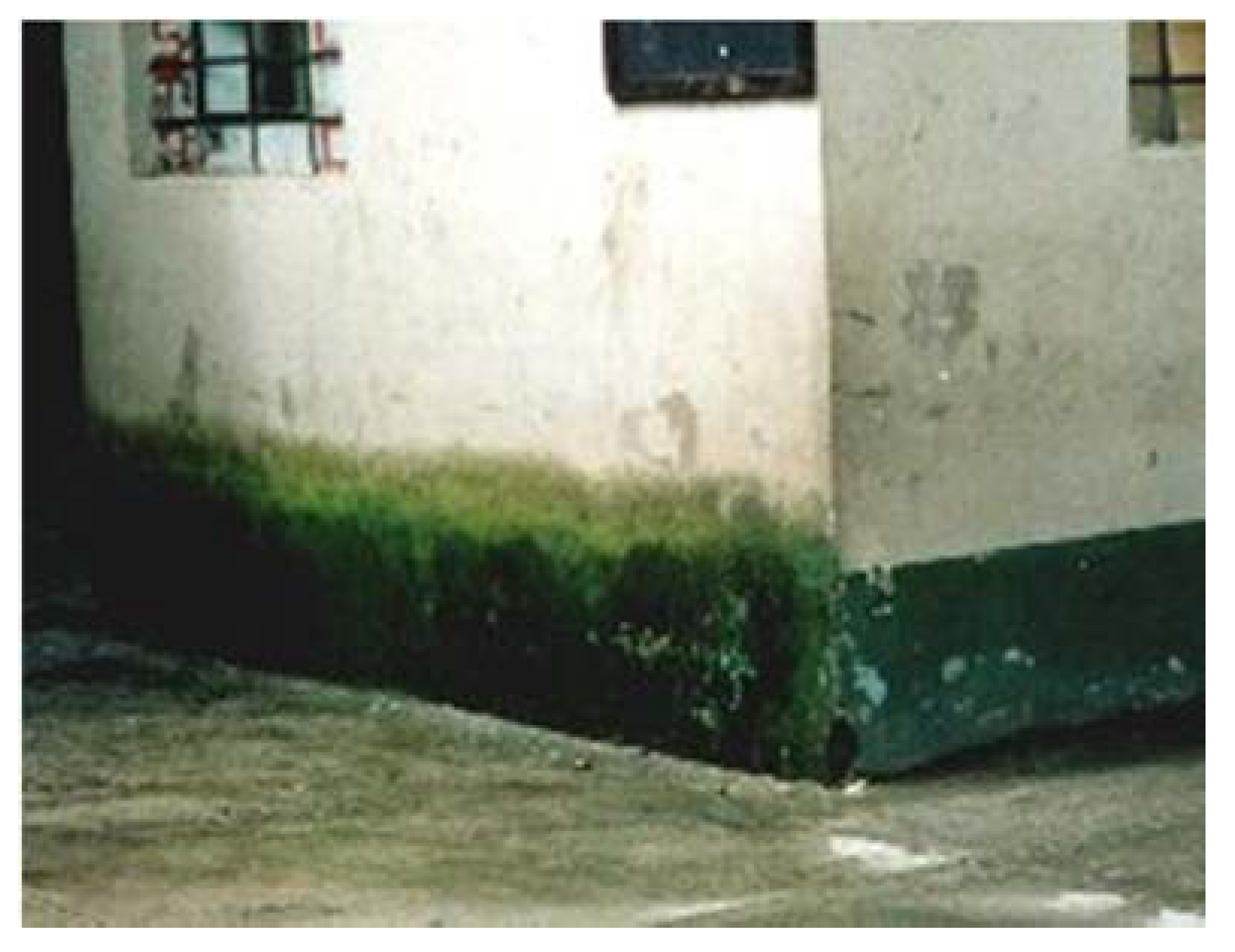
Both algae and cyanobacteria can produce thick green biofilms in areas of high humidity (Figure 1), but in less wet areas the colors may vary, especially in the case of cyanobacteria (Figure 2 and Figure 3).
The level of humidity may be considered one of the most important factors determining the colonization of buildings. A study of microbial biofilms on two areas of the same historic fort in Niteroi, Rio de Janeiro, Brazil, showed that colonization below a leaking pipe was very different from that in a dry position protected from rain [17]. The former biofilm was dark green/brown and contained Chloroflexi, photosynthetic filamentous bacteria, as the preponderant group, while the dry biofilm was grey/light green and composed mainly of non-photosynthetic Proteobacteria. The relative lack of algal genera at the two sites was attributed to the absence of liquid water and the relative humidity of less than 100%, either of which has been said to be necessary for optimum algal growth [18].
The other factor of major importance in determining microbial colonization is temperature, which, of course, also affects the humidity of the surface. In the case of external surfaces, heating will be solar and thus its effects cannot be separated from those of UV irradiation. All microorganisms have their own preferred temperature ranges for growth; hence the composition of an external, mature, biofilm will vary according to the temperatures to which the surface is subjected. Generally, however, lower temperatures will lead to the development of thinner biofilms because of slower microbial growth rates, and very high temperatures will inhibit or even kill the cells, similarly leading to thinner biofilms. Some colonizing species are able to protect themselves from damaging UV in sunlight by the production of protective pigments (see Table 1). For culturally important buildings in the tropics and subtropics, these organisms are often particularly important as surface discoloring agents. Figure 2 shows a thick black subaerial biofilm composed mainly of the filamentous cyanobacterium, Scytonema. Its sheath contains the UV-protective brown pigment, scytonemin. Under conditions where there is little UV, the sheath remains colorless and the cells green, leading to a green biofilm even though the same genus is present. Another, more complex, example is seen in the dark surface patina from a parapet of the church of Nossa Senhora do Rosário in the sunny, elevated town of Ouro Preto, Brazil [9]. The biofilm was shown to consist entirely of the dark-sheathed filamentous cyanobacterium Stigonema ocellatum and the dark red encapsulated spherical genus Gloeocapsa. Gloeocapsa species with dark purple capsules have been identified in biofilms on other churches in the same state of Minas Gerais, in the Brazilian highlands, subject to high UV levels (author’s unpublished observations).
Not only cyanobacteria, but also some algae, can be responsible for non-green discolorations. An excellent example is the filamentous alga Trentepohlia. Although classified with the green algae, these cells are frequently identified when growing on external walls by their red coloration (Figure 4), caused by carotene-containing oil droplets within the cells.
On the Rio Bec style Mayan buildings at Becán, Chicanná and Hormiguero (Campeche state, Mexico) it was found that red colorations caused by these algae were were predominantly associated with North- and East-facing walls (Figure 5), or on otherwise orientated sites protected by tree canopies or architectural elements [19]. On more exposed sites, grey-black discoloration composed of dehydration- and UV-resistant cyanobacteria (the most common being the coccoid genera Gloeocapsa and Chroococcidiopsis) was dominant.
Dark surface coatings can have physical, as well as aesthetic, effects. Biofilmed areas absorb more sunlight, thus increasing the physical stresses of temperature changes that lead to expansion and contraction of the stone surface. The darkening of the stone surface decreases its albedo, so that it experiences greater stresses through not only heating/cooling, but also wetting/drying cycles [20].
From the surface biofilm, microorganisms can penetrate within the stone, becoming endolithic. Many microorganisms have been detected, growing within all types of stone, with accompanying chemical and physical degradation; this includes phototrophs [21], other autotrophic and chemoheterotrophic bacteria [22], fungi [21] and lichens [23]. They are mostly associated with adverse environments, with extreme temperatures and droughts [[24], and references therein]; endolithotrophy may be a microbial survival mechanism under such conditions. The alga Trentepohlia, mentioned above, has been shown to penetrate the surface of the limestone monuments of the Mayan buildings at Edzna, Campeche, Mexico, where UV levels are high and temperatures can be up to 36 °C; the black algal colonies caused pitting of the surface [25].
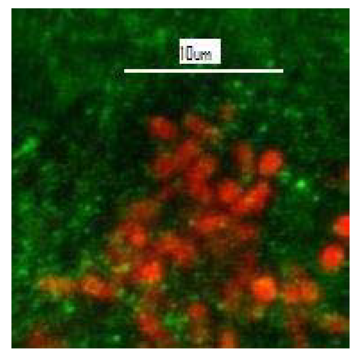
Gaylarde and Gaylarde [26] discuss the mechanisms whereby endolithic microorganisms induce the degradation of siliceous stone, citing historic buildings in Latin America where they have been detected, and Gaylarde et al. [27] report coccoid and filamentous cyanobacteria and algae, including Cyanidiales, growing endolithically in the facades of historic buildings of limestone, sandstone, granite, basalt and soapstone, as well as in some natural rocks. Figure 6 shows red autofluorescent cyanobacterial cells visualized by confocal laser scanning microscopy within a sample of soapstone from the church of São Francisco de Paula in Tiradentes, Minas Gerais, Brazil.
Endolithic organisms disrupt stone structure physically and by production of organic and inorganic acids [28], as well as by dissolution and remobilization of ions such as iron and manganese from the rock [29]. The arenitous sandstone of the Argentine missions was shown to have an outer layer enriched in Fe and Mn, produced by these biogenic processes [30], while the augen gneiss facades of churches in the city of Rio de Janeiro, Brazil, were found to contain endolithic filamentous cyanobacteria encrusted with neogypsum, redeposited after dissolution from the deteriorated crust [31]. Gypsum is one of the most highly damaging salts for rock structure [32]. It is only slightly soluble in water, but at higher pH values, as produced by actively photosynthesising phototrophs, it becomes even less soluble; thus gypsum solubilized at night by bacterial and fungal acids at the stone surface or near surface will be later redeposited as neogypsum around endolithic phototrophic cells. As these solubilization and reprecipitation cycles are repeated, the gypsum particles become larger and denser, disrupting the stone structure even more and increasing the possibility of spalling.
Lichens (Figure 7), symbiotic associations of fungi and phototrophic microorganisms (algae or cyanobacteria) with associated non-photosynthetic bacteria [33], are perhaps the stone biofilms most obvious to the general public and are often regarded as aesthetically pleasing. However, their small roots (rhizines) penetrate into the stone with obvious destructive effects. They also produce acids, sometimes called “lichenic acids”, the main one being oxalic acid. This is particularly damaging to carbonate stones, but can also affect siliceous rocks ([34], and references therein).
Lichens are poikilohydric organisms, able to increase or reduce their metabolism depending on water availability, and so are able to resist periods of drought, similar to cyanobacteria ([2], and references therein). It has been suggested that lichens may protect stone surfaces from chemical and atmospheric attack [35], but this potential must be balanced against the inherently destructive nature of lichen growth.
3. Black Crusts: A Special Type of Biofilm?
The black discolorations seen on buildings in heavily polluted cities are known as “black crusts” and are composed principally of gypsum and entrapped pollutants. Carbonaceous particles (such as fly ash) from power plants and road traffic act as catalysts for the transformation of calcium carbonate into gypsum [36] and the trapped particulates are the main reason for the blackening. Even siliceous stones can carry this disfiguring layer, incorporating calcium from outside sources.
Black crusts of different origin can also be seen, however, on buildings in unpolluted environments. On historic baluartes (forts) and the cathedral in Campeche, Yucatan peninsula, Mexico (Figure 8), these black crusts were found to be composed almost entirely of Subsection IV and V filamentous cyanobacteria (as defined by [37]). The cells did not penetrate into the limestone, but remained as readily detached surface layers, leading to degradation by differential heating and water retention effects [38].
4. Effects of Environment/Climate
Ortega-Morales et al. [9], in a meta-analysis of cyanobacterial colonization of historic buildings in six tropical and subtropical countries, determined that Köppen climate class influenced cyanobacterial colonization; the more temperate areas of class C were particularly separated in the statistical analysis from the more tropical areas, with class Cfa (the Argentine missions) forming a compact group on their own. Class Aw (towns in Minas Gerais, Brazil) grouped separately from the much more geographically diverse tropical monsoon (Am) class. Similar, although less widespread, results on the influence of climate on stone colonization were found by Louati et al. [40] in North Africa and Keshari and Adhikary [41] in India. Tropical wet climates, with high water and light availability, clearly offer ideal conditions for the growth of primary colonizers, which, in turn, pave the way for the potentially more destructive heterotrophic fungi and higher plants. As Liu et al. [2] point out, monuments in tropical regions, such as the sandstone temples of Angkor Wat in Cambodia and limestone buildings in the ancient Mayan cities of Calakmul and Uxmul in Mexico, are more degraded than those in cold and arid regions like the Mogao and Maijishan grottoes on the Silk Road in west China.
Gaylarde and Gaylarde [42], analyzing microbial biofilms from a wide variety of buildings with different surfaces and in various parts of the world, showed that cyanobacteria (mostly coccoid colonial types), were the most frequent major biomass on Latin American buildings composed of stone or composites, with an average 66.1% of total biomass, followed by fungi (17.7%) and algae (12.9%), while in Europe algae were most common at 46.32% total biomass, followed by cyanobacteria (35.2%). Fungi were more common on painted (20.8% total biomass on this substrate) than stone surfaces overall, perhaps because paint can provide a nutrient source for heterotrophs. The differences in biofilm composition between the two geographical areas were attributed to the climates prevailing at the sampling sites, with Latin American countries (Northern Argentina, Bolivia, Brazil, Colombia, Ecuador, Mexico and Peru) being generally hotter and subject to more torrential rains than those in Europe (Czech Republic, England, France, Italy, Poland, Portugal and Spain).
5. Effect of Air Pollution
Although now improving in some countries, air pollution has increased considerably over the centuries. Traffic fumes and industrial and domestic wastes, accompanying urbanization and more intense agricultural production procedures, have considerably added to air pollution. Nitrogen and sulfur compounds, as well as volatile organic compounds, are of particular interest, as they may serve as nutrients for epilithic biodeteriogens [2].
Ortega-Morales et al. [9], in the same analysis as cited in above sections, determined that there were significant differences between the cyanobacterial populations on the stone surfaces of buildings in highly polluted cities (Rio de Janeiro, Brazil, and Belize City) and on those in more rural situations. This difference resided in the lower biodiversity in polluted environments, also shown by Auras et al. [43] in Germany and discussed by Cutler and Viles [44] and Mihajlovski et al. [45]. The more resistant cyanobacteria will be able to survive increasing pollution levels better than other primary colonizers. Scytonemin, for example (see Table 1), is produced not only in high UV environments, but also in areas with high levels of air pollution; it has been suggested that it is, in fact, a general cell protectant, indicative of stressful environments [46].
Basu et al. [47], writing about the preservation of important heritage buildings in London, UK, point out that it is necessary to consider how the stone could be affected by changing air pollution, added to the rising average rainfall and temperatures, with hotter, drier summers and warmer, wetter winters. Indeed, air pollution does act synergistically with weather and climatic changes [48] to increase both non-biological and general biodegradation of stone. However, the converse effect of air pollution, its reduction of biodegradation of historic buildings, becomes apparent when considering lichen growth. These microbial symbioses are inhibited in polluted environments and have actually been used as indicators of good air quality [49,50]. Their inherently aggressive action on stone means that a reduction in their growth will also reduce stone attrition, but their potentially protective action against atmospheric erosion [35] may, possibly, cancel out this effect. Regardless of the effects on lichens, fungi and other heterotrophic organisms may become prevalent in biofilms in polluted areas and in extreme environmental conditions; this group of organisms may even utilize the polluting chemicals as nutrient sources [2,15,51], leading to increased stone biodeterioration. Hence the effects of changing levels of environmental pollution on biodegradation of stone buildings are difficult to predict.
6. Potential Effects of Climate Change
It is obvious from the effects of climate on microbial biofilms discussed in the above section that climate change will affect the degradation of historic properties caused by microorganisms, even though the subaerial biofilm is a complex consortium and thus able to overcome many types of environmental stress [52]. Nevertheless, future increases in temperature and rainfall will doubtless result in alterations in biofilm formation [53]. Water availability at the surface may be the most important factor. Considering the primary colonizers, the body of work on cyanobacteria indicates considerable dependence of biofilm biodiversity on the level of humidity. Miller et al. [54] found that high values of water absorption by capillarity and open porosity of various types of limestone determined their bioreceptivity for the primary phototrophic colonizers. Subaerial biofilms on monuments in Eastern India varied greatly in their biodiversity between the hot tropical summer and the rainy season [55]. The only phototrophs detected during the former were cyanobacteria of the genera Gloeocapsa, Gloeocapsopsis, Porphyrosiphon, Leptolyngbya, Lyngbya, Phormidium, Nostoc, Scytonema, Tolypothrix, Hassallia and Stigonema. During the rainy season, however, the biofilms were more diverse, also containing Cyanosarcina, Gloeocapsopsis, Phormidium, Pseudophormidium, Schizothrix, Lyngbya, Plectonema, Nostoc, Scytonema, Tolypothrix, Dichothrix and Calothrix, together with the green alga Chlorella sphaerica. Ortega-Morales et al. [9], in their meta-analytical study, found that precipitation level was more important for overall phototroph growth, rather than for phototroph diversity. Thickest biofilms were present at the highest rainfall sites, Palenque, Mexico (Figure 9) and Laos, with precipitation levels of 2697 mm/y and 2069.5 mm/y, respectively, but the highest level of diversity was found at the Argentine missions, with an annual rainfall of 1281.7 mm/y and a rather low mean annual temperature of 19.2 °C. Biofilms at the latter site were, however, relatively thin.
Becan, Campeche, Mexico (962 mm/y) had the lowest rainfall of the sites studied and the temperature range there is from 17−36 °C; this was a site where considerable Trentepohlia growth was seen, especially on NNE-facing surfaces. These algae are common phycobionts in lichen symbioses and there is increasing evidence that lichens are responding to climate change in Western Europe [56]. Epiphytic species (growing on higher plants) are increasing with warmer weather, while terricolous species (on soil and buildings) are declining. Some of the different species most rapidly increasing contain Trentepohlia as the phycobiont, suggesting that Trentepohlia is the main lichen constituent affected by global warming.
Prieto et al. [57] consider that CO2 levels are also important for colonization of granite buildings, but showed that the effects of increased levels were counteracted by increased water availability.
Not only annual rainfall, but also the amount of wind-driven rain (WDR) is predicted to increase in the future. Orr et al. [58], pointing out that WDR is an important environmental risk to built cultural heritage, calculate that WDR spells will be more frequent and more intense in the UK towards the end of this century, and in 2020, Orr and Cassar [59] proposed indices to represent the exposure of built cultural heritage to WDR. The ISO standard (no. 1,5927-3), which was only tested in the UK, has recently been challenged by Pérez-Bella et al. [60]. This particular aspect of climate change will also affect the colonization of exposed surfaces. This was demonstrated in the current climate at the UNESCO archaeological site of Pompeii, Italy [61]. Microbiological growth consisted mainly of cyanobacteria, algae and lichens, and was shown to be more prominent on N and W exposed walls, where the West wind influenced WDR.
A certain level of light is obviously also needed for the growth of most cyanobacteria, although some are able to grow heterotrophically [62,63]. Caneva et al. [7], investigating the temples in Angkor Wat, found that light and water together are the most critical factors in explaining the distribution of the colonizing communities. This interaction has been confirmed by various authors ([64], and references therein). Li et al. [15], using a metagenomic approach, detected cyanobacteria as the principal colonizers (> 50% of total bacterial OTUs) on six samples taken from four different buildings at three locations around West Lake, Hangzhou, China. They stated that cyanobacterial colonization was correlated with light intensity and humidity and, indeed, Andre et al. [65] provided further evidence for the influence of light when they examined the historical evidence on changes around Angkor Wat and found that that removal of forest from around the temples resulted in increased stone attrition. Although some cyanobacteria can grow (slowly) in the absence of light, many of them can survive without free water, or even under almost completely dry conditions. Nevertheless, as with all other living organisms, water is necessary for their growth.
Predictions of climate change in Europe vary across the European continent. Greater warming is probable in summer for Southern Europe, while In North Eastern Europe this is likely to be the winter scenario [1]. Gómez-Bolea et al. [66] produced a mathematical model to predict how temperature and humidity changes would affect the build-up of microbial biomass on historic stone buildings. They used the equation B = exp (–0.964 + 0.003P–0.01T), where B = biomass accumulation (mg cm−2), P = annual precipitation (mm) and T = annual mean temperature (°C) of projected climate data, to calculate that increased temperature and precipitation in the years 2070−2099 would lead to increased biomass in Northern Europe, and that increased temperature but reduced precipitation would have the opposite effect in Southern Europe. Obviously, increased temperature with lower rainfall would lead to much lower availability of liquid water on exposed stone surfaces and thus reduced biofilm growth. This model was designed with reference to horizontal surfaces of hard acid stones in nonurban, unpolluted environments in a temperate climate (siliceous rocks in Spain), but, nevertheless, it can give an indication of the potential for change in biological deterioration of stone cultural heritage throughout the world.
Identification of climate as an important factor in microbial colonization of built structures has important implications for future biodeterioration problems and their control. Viles and Cutler [53] predicted that, in areas likely to experience more climatic disturbances, a shift from bioprotective to biodeterioration conditions could occur. They considered that areas with increased biological stresses (such as decreased rainfall) would show reduced rates of biofilm formation, with a switch to organisms more able to tolerate and survive stress and a concomitant decrease in biodeterioration. They also considered that changes from stressed to disturbed conditions could produce no net change in biofilm growth but a switch to more deteriorative colonizers, based on the fact that opportunist organisms, arriving rapidly after any disturbance and able both to utilize readily available materials as nutrients and to live endolithically, are more likely to cause deterioration. Cyanobacteria and fungi are likely to fit within this opportunist pattern. Subsequent disturbance to the surface through weathering would favor continuing colonization by ruderal organisms (the first to colonize disturbed areas). In contrast, stress-tolerant species should lead to less deterioration because they do not allow new, potentially more aggressive species, to colonize. Cyanobacteria, but not fungi, fit into both these classifications; they require only light and water to grow and can live endolithically, but are also able to survive most types of stress and, indeed, have been reactivated from air-dried laboratory cultures after 20 years of ambient storage (author’s unpublished observations). They may become even more important as agents of stone cultural property degradation in the future.
7. Conclusions
Cyanobacteria are probably the most important colonizers of stone buildings, since they are not dependent on any organic source of carbon and are very resistant to environmental changes. They can be directly responsible for stone degradation, through their ability to grow endolithically, but can also act as an organic food material for the growth of other biodeteriogens, such as fungi. It is impossible to prevent the growth of microorganisms on surfaces exposed to the open air, and regular cleaning with substances that do not damage the stone is recommended. Many potential treatments to prevent or retard growth have been tested over the years, but the discussion of these would require another review.
Funding
No funding was received for this contribution.
Conflicts of Interest
The author declares no conflict of interest.
References
- Mahdjoubi, L.; Hawas, S.; Fitton, R.; Dewidar, K.; Nagy, G.; Marshall, A.; Alzaatreh, A.; Abdelhady, E. A Guide for Monitoring the Effects of Climate Change on Heritage Building Materials and Elements. Report Prepared for the Funded Research Project: ‘Heritage Building Information Modelling and Smart Heritage Buildings Performance Measurements for Sustainability’. 2017. Available online: https://uwe-repository.worktribe.com/output/882571 (accessed on 16 October 2020).
- Liu, X.; Koestler, R.J.; Warscheid, T.; Katayama, Y.; Gu, J.-D. Microbial deterioration and sustainable conservation of stone monuments and buildings. Nat. Sustain. 2020. [Google Scholar] [CrossRef]
- Mitchell, R.; Gu, J.-D. Changes in the biofilm microflora of limestone caused by atmospheric pollutants. Int Biodeterior. Biodegrad. 2000, 46, 299–303. [Google Scholar] [CrossRef]
- Guillitte, O. Bioreceptivity: A new concept for building ecology studies. Sci. Total Environ. 1995, 167, 215–220. [Google Scholar] [CrossRef]
- Sanmartín, P.; Grove, R.; Carballeira, R.; Viles, H. Impact of colour on the bioreceptivity of granite to the green alga Apatococcus lobatus: Laboratory and field testing. Sci. Total Environ. 2020, 745, 141179. [Google Scholar] [CrossRef]
- Vazquez-Nion, D.; Silva, B.; Prieto, B. Influence of the properties of granitic rocks on their bioreceptivity to subaerial phototrophic biofilms. Sci. Total Environ. 2018, 610–611, 44–54. [Google Scholar] [CrossRef]
- Caneva, G.; Di Stefano, D.; Giampaolo, C.; Ricci, S. Stone cavity and porosity as a limiting factor for biological colonisation: The travertine of Lungotevere (Rome). In Proceedings of the 10th International Congress on Deterioration and Conservation of Stone, Stockholm, Sweden, 27 June–2 July 2004; Kwiatkowski, D., Löfvendahl, R., Eds.; ICOMOS: Stockholm, Sweden, 2004; Volume 1, pp. 227–232. [Google Scholar]
- Del Mondo, A.; Pinto, G.; Carbone, D.A.; Pollio, A.; De Natale, A. Biofilm architecture on different substrates of an Oculatella subterranea (Cyanobacteria) strainisolated from Pompeii archaeological site (Italy). Environ. Sci. Pollut. Res. 2018. [Google Scholar] [CrossRef] [PubMed]
- Ortega-Morales, O.; Montero-Muñoz, J.L.; Baptista Neto, J.A.; Beech, I.B.; Sunner, J.; Gaylarde, C. Deterioration and cyanobacterial colonization of cultural heritage stone buildings in polluted and unpolluted tropical and subtropical climates: A meta-analysis. Int. Biodeterior. Biodegrad. 2019, 143, 104734. [Google Scholar] [CrossRef]
- Ortega-Morales, B.O.; Narvaéz-Zapata, J.A.; Schmalenberger, A.; Sosa-López, A.; Tebbe, C.C. Biofilms fouling ancient limestone Mayan monuments in Uxmal, Mexico: A cultivation-independent analysis. Biofilms 2004, 1, 79–90. [Google Scholar] [CrossRef]
- Li, Q.; Zhang, B.; Yang, X.; Ge, Q. Deterioration-associated microbiome of stone monuments: Structure, variation, and assembly. Appl. Environ. Microbiol. 2018, 84. [Google Scholar] [CrossRef] [PubMed] [Green Version]
- Morillas, H.; Maguregui, M.; Gallego-Cartagena, E.; Huallparimachi, G.; Marcaida, I.; Salcedo, I.; Silva, L.F.; Astete, F. Evaluation of the role of biocolonizations in the conservation state of Machu picchu (Peru): The sacred rock. Sci. Total Environ. 2019, 654, 1379–1388. [Google Scholar] [CrossRef] [PubMed]
- Gorbushina, A.A. Life on the rocks. Environ. Microbiol. 2007, 9, 1613–1631. [Google Scholar] [CrossRef] [PubMed]
- Crispim, C.A.; Gaylarde, C.C. Cyanobacteria and biodeterioration of cultural heritage: A review. Microb. Ecol. 2005, 49, 1–9. [Google Scholar] [CrossRef] [PubMed]
- Li, Q.; Zhang, B.; He, Z.; Yang, X. Distribution and diversity of bacteria and fungi colonization in stone monuments analyzed by high-throughput sequencing. PLoS ONE 2016, 11, e0163287. [Google Scholar] [CrossRef] [PubMed] [Green Version]
- Becerra, J.; Zaderenko, A.P.; Gómez-Morón, M.A.; Ortiz, P. Nanoparticles applied to stone buildings. Int. J. Architect. Herit. 2019. [Google Scholar] [CrossRef]
- Ogawa, A.; Celikkol-Aydin, S.; Gaylarde, C.; Baptista-Neto, J.A.; Beech, I. Microbial communities on painted wet and dry external surfaces of a historic fortress in Niteroi, Brazil. Int. Biodeterior. Biodegrad. 2017, 123, 164–173. [Google Scholar] [CrossRef]
- Häubner, N.; Schumann, R.; Karsten, U. Aeroterrestrial algae growing in biofilms on facades e response to temperature and water stress. Microb. Ecol. 2006, 51, 285–293. [Google Scholar] [CrossRef]
- Ortega-Morales, B.O.; Gaylarde, C.; Anaya-Hernandez, A.; Chan-Bacab, M.J.; De la Rosa-García, S.C.; Arano-Recio, D.; Montero-M, J. Orientation affects Trentepohlia-dominated biofilms on Mayan monuments of the Rio Bec style. Int. Biodeterior. Biodegrad. 2013, 84, 351–356. [Google Scholar] [CrossRef]
- Warscheid, T.; Braams, J. Biodeterioration of stone: A review. Int. Biodeterior. Biodegrad. 2000, 46, 343–368. [Google Scholar] [CrossRef]
- Golubic, S.; Perkins, R.D.; Lukas, K.J. Boring microorganisms and microborings in carbonate substrates. In The Study of Trace Fossils; Frey, R.W., Ed.; Springer: Berlin/Heidelberg, Germany, 1975. [Google Scholar] [CrossRef]
- Wierzchos, J.; Casero, M.C.; Artieda, O.; Ascaso, C. Endolithic microbial habitats as refuges for life in polyextreme environment of the Atacama Desert. Curr. Opin. Microbiol. 2018, 43, 124–131. [Google Scholar] [CrossRef]
- Ascaso, C.; Wierzchos, J.; Castello, R. Study of the biogenic weathering of calcareous litharenite stones caused by lichen and endolithic microorganisms. Int. Biodeterior. Biodegrad. 1998, 42, 29–38. [Google Scholar] [CrossRef]
- Qu, E.B.; Omelon, C.R.; Oren, A.; Meslier, V.; Cowan, D.A.; Maggs-Kölling, G.; DiRuggiero, J. Trophic selective pressures organize the composition of endolithic microbial communities from global deserts. Front. Microbiol. 2020, 10, 2952. [Google Scholar] [CrossRef] [PubMed] [Green Version]
- Gaylarde, P.; Englert, G.; Ortega-Morales, O.; Gaylarde, C. Lichen-like colonies of pure Trentepohlia on limestone monuments. Int. Biodeterior. Biodegrad. 2006, 58, 119–123. [Google Scholar] [CrossRef]
- Gaylarde, P.; Gaylarde, C. Deterioration of siliceous stone monuments in Latin America: Microorganisms and mechanisms. Corros. Revs. 2004, 22, 395–415. [Google Scholar] [CrossRef]
- Gaylarde, C.C.; Gaylarde, P.M.; Neilan, B.A. Endolithic phototrophs in built and natural stone. Curr. Microbiol. 2012, 65, 183–188. [Google Scholar] [CrossRef]
- Zhang, G.; Gong, C.; Gu, J.; Katayama, Y.; Someya, T.; Gu, J.D. Biochemical reactions and mechanisms involved in the biodeterioration of stone world cultural heritage under the tropical climate conditions. Int. Biodeterior. Biodegrad. 2019, 143, 104723. [Google Scholar] [CrossRef]
- Esposito, A.; Borruso, L.; Rattray, J.E.; Brusetti, L.; Ahmed, E. Taxonomic and functional insights into rock varnish microbiome using shotgun metagenomics. FEMS Microbiol. Ecol. 2019, 95, fiz180. [Google Scholar] [CrossRef] [PubMed]
- Barrionuevo, M.R.E.; Hidalgo, G.E.; Gaylarde, C.C. Physical and microbiological analysis of sandstone deterioration in the Argentine Jesuit missions. Geomicrobiol. J. 2016. [Google Scholar] [CrossRef]
- Gaylarde, C.; Baptista-Neto, J.A.; Ogawa, A.; Kowalski, M.; Celikkol-Aydin, S.; Beech, I. Epilithic and endolithic microorganisms and deterioration on stone church facades subject to urban pollution in a sub-tropical climate. Biofouling 2017, 33, 113–127. [Google Scholar] [CrossRef]
- Duffy, A.P.; Cooper, T.P.; Perry, S.H. Repointing mortars for conservation of a historic stone building in Trinity College, Dublin. Mater. Struct. 1993, 26, 302–306. [Google Scholar] [CrossRef] [Green Version]
- Grube, M.; Cernava, T.; Soh, J.; Fuchs, S.; Aschenbrenner, I.; Lassek, C.; Wegner, U.; Becher, D.; Riedel, K.; Sensen, C.W.; et al. Exploring functional contexts of symbiotic sustain within lichen-associated bacteria by comparative omics. ISME J. 2015, 9, 412–424. [Google Scholar] [CrossRef] [Green Version]
- Scheerer, S.; Ortega-Morales, O.; Gaylarde, C. Microbial deterioration of stone monuments—An updated overview. Adv. Appl. Microbiol. 2009, 66, 97–139. [Google Scholar]
- Garcia-Vallès, M.; Topal, T.; Vendrell-Saz, M. Lichenic growth as a factor in the physical deterioration or protection of Cappadocian monuments. Environ. Geol. 2003, 43, 776–781. [Google Scholar] [CrossRef]
- Ausset, P.; Del Monte, M.; Lefèvre, R.A. Embryonic sulphated black crusts on carbonate rocks in atmospheric simulation chamber and in the field: Role of carbonaceous fly-ash. Atmos. Environ. 1999, 33, 1525–1534. [Google Scholar] [CrossRef]
- Castenholz, R.W.; Wilmotte, A.; Herdman, M.; Rippka, R.; Waterbury, J.B.; Iteman, I.; Hoffmann, L. Phylum BX. Cyanobacteria. In Bergey’s Manual® of Systematic Bacteriology; Boone, D.R., Castenholz, R.W., Garrity, G.M., Eds.; Springer: New York, NY, USA, 2001. [Google Scholar] [CrossRef]
- Gaylarde, C.C.; Ortega-Morales, B.O.; Bartolo-Perez, P. Biogenic black crusts on buildings in unpolluted environments. Curr. Microbiol. 2007, 54, 162–166. [Google Scholar] [CrossRef] [PubMed]
- García de Miguel, J.M.; Sánchez-Castillo, L.; Ortega-Calvo, J.J.; Gil, J.A.; Saiz-Jimenez, C. Deterioration of building materials from the Great Jaguar Pyramid at Tikal, Guatemala. Build. Environ. 1995, 30, 591–598. [Google Scholar] [CrossRef]
- Louati, M.; Ennis, N.J.; Ghodhbane-Gtari, F.; Hezbri, K.; Sevigny, J.L.; Fahnestock, M.F.; Cherif-Silini, H.; Bryce, J.G.; Tisa, L.S.; Gtari, M. Elucidating the ecological networks in stone-dwelling microbiomes. Environ. Microbiol. 2019. [Google Scholar] [CrossRef] [PubMed]
- Keshari, N.; Adhikary, S.P. Characterization of cyanobacteria isolated from biofilms on stone monuments at Santiniketan, India. Biofouling 2013, 29. [Google Scholar] [CrossRef] [PubMed]
- Gaylarde, C.C.; Gaylarde, P.M. A comparative study of the major microbial biomass of biofilms on exteriors of buildings in Europe and Latin America. Int. Biodeterior. Biodegrad. 2005, 55, 131–139. [Google Scholar] [CrossRef]
- Auras, M.; Bundschuh, P.; Eichhorn, J.; Kirchner, D.; Mach, M.; Seewald, B.; Scheuvens, D.; Snethlage, R. Traffic-induced emissions on stone buildings. In Future for Stone, Proceedings of the 13th International Congress on the Deterioration and Conservation of Stone, Paisley, Scotland, 6–10 September 2016; Hughes, J., Howind, T., Eds.; University of the West of Scotland: Paisley, Scotland, 2016; Volume 1, pp. 3–12. ISBN 978-1-903978-57-3. [Google Scholar]
- Cutler, N.; Viles, H. Eukaryotic microorganisms and stone biodeterioration. Geomicrobiol. J. 2010, 27, 630–646. [Google Scholar] [CrossRef]
- Mihajlovski, A.; Seyer, D.; Benamara, H.; Bousta, F.; Di Martino, P. An overview of techniques for the characterization and quantification of microbial colonization on stone monuments. Ann. Microbiol. 2015, 65, 1243–1255. [Google Scholar] [CrossRef] [Green Version]
- Ibarrondo, I.; Prieto-Taboada, N.; Martinez-Arkarazo, I.; Madariaga, J.M. Resonance Raman imaging as a tool to assess the atmospheric pollution level: Carotenoids in Lecanoraceae lichens as bioindicators. Environ. Sci. Pollut. Res. 2016, 23, 6390–6399. [Google Scholar] [CrossRef] [PubMed]
- Basu, S.; Orr, S.A.; Aktas, Y.D. A geological perspective on climate change and building stone deterioration in London: Implications for urban stone-built heritage research and management. Atmosphere 2020, 11, 788. [Google Scholar] [CrossRef]
- Pinheiro, A.C.; Mesquita, N.; Trovão, J.; Soares, F.; Tiago, I.; Coelho, C.; de Carvalho, H.P.; Gil, F.; Catarino, L.; Piñar, G.; et al. Limestone biodeterioration: A review on the Portuguese cultural heritage scenario. J. Cult. Herit. 2019, 36, 275–285. [Google Scholar] [CrossRef]
- Matos, P.; Vieira, J.; Rocha, B.; Branquinho, C.; Pinho, P. Modeling the provision of air-quality regulation ecosystem service provided by urban green spaces using lichens as ecological indicators. Sci. Total Environ. 2019, 665, 521–530. [Google Scholar] [CrossRef] [PubMed]
- De Wit, T. Lichens as indicators for air quality. In Ecological Indicators for the Assessment of the Quality of Air, Water, Soil, and Ecosystems; Best, E.P.H., Haeck, J., Eds.; Springer: Dordrecht, The Netherlands, 1983. [Google Scholar] [CrossRef]
- Nuhoglu, Y.; Oguz, E.; Uslu, H.; Ozbek, A.; Ipekoglu, B.; Ocak, I.; Hasenekoglu, I. The accelerating effects of the microorganisms on biodeterioration of stone monuments under air pollution and continental-cold climatic conditions in Erzurum, Turkey. Sci. Total Environ. 2006, 364, 272–283. [Google Scholar] [CrossRef] [Green Version]
- Villa, F.; Stewart, P.S.; Klapper, I.; Jacob, J.M.; Cappitelli, F. Subaerial biofilms on outdoor stone monuments: Changing the perspective toward an ecological framework. BioScience 2016, 66, 285–294. [Google Scholar] [CrossRef] [Green Version]
- Viles, H.A.; Cutler, N.A. Global environmental change and the biology of heritage structures. Glob. Chang. Biol. 2012, 18, 2406–2418. [Google Scholar] [CrossRef]
- Miller, A.Z.; Dionísio, A.; Macedo, M.F.; Sáiz-Jiménez, C. Primary bioreceptivity of limestones to phototrophic microorganisms: A laboratory-based stone colonization experiment. Technoheritage 2017, 97. Available online: http://hdl.handle.net/10261/155200 (accessed on 30 October 2020).
- Adhikary, S.P.; Keshari, N.; Urzi, C.; De Phillipis, R. Cyanobacteria in biofilms on stone temples of Bhubaneswar, Eastern India. Algol. Stud. 2015, 147, 67–93. [Google Scholar] [CrossRef] [Green Version]
- Aptroot, A.; van Herk, C.M. Further evidence of the effects of global warming on lichens, particularly those with Trentepohlia phycobionts. Environ. Poll. 2007, 146, 293–298. [Google Scholar] [CrossRef]
- Prieto, B.; Vázquez-Nion, D.; Fuentes, E.; Durán-Román, A.G. Response of subaerial biofilms growing on stone-built cultural heritage to changing water regime and CO2 conditions. Int. Biodeterior. Biodegrad. 2020, 148, 104882. [Google Scholar] [CrossRef]
- Orr, S.A.; Young, M.; Stelfox, D.; Curran, J.; Viles, H. Wind-driven rain and future risk to built heritage in the United Kingdom: Novel metrics for characterising rain spells. Sci. Total Environ. 2018, 640–641, 1098–1111. [Google Scholar] [CrossRef]
- Orr, S.A.; Cassar, M. Exposure indices of extreme wind-driven rain events for Built Heritage. Atmosphere 2020, 11, 163. [Google Scholar] [CrossRef] [Green Version]
- Pérez-Bella, J.M.; Domínguez-Hernández, J.; Cano-Suñén, E.; Alonso-Martínez, M.; del Coz-Díaz, J.J. Equivalence between the methods established by ISO 15927-3 to determine wind-driven rain exposure: Reanalysis and improvement proposal. Build. Environ. 2020, 174, 106777. [Google Scholar] [CrossRef]
- Traversetti, L.; Bartoli, F.; Caneva, G. Wind-driven rain as a bioclimatic factor affecting the biological colonization at the archaeological site of Pompeii. Italy. Int. Biodeterior. Biodegrad. 2018, 134, 31–38. [Google Scholar] [CrossRef]
- Lee, C.G.; Watanabe, T.; Fujita, Y.; Asakawa, S.; Kimura, M. Heterotrophic growth of cyanobacteria and phage-mediated microbial loop in soil: Examination by stable isotope probing (SIP) method. Soil Sci. Plant Nutrit. 2012, 58, 161–168. [Google Scholar] [CrossRef]
- Meireles dos Santos, A.; Vieira, K.R.; Basso, S.R.; Meireles dos Santos, A.; Queiroz, M.I.; Queiroz, Z.L.; Jacob-Lopes, E. Heterotrophic cultivation of cyanobacteria: Study of effect of exogenous sources of organic carbon, absolute amount of nutrients, and stirring speed on biomass and lipid productivity. Front. Bioeng. Biotechnol. 2017, 5, 12. [Google Scholar] [CrossRef] [Green Version]
- Miller, A.Z.; Sanmartín, P.; Pereira-Pardo, L.; Dionísio, A.; Saiz-Jimenez, C.; Macedo, M.F.; Prieto, B. Bioreceptivity of building stones: A review. Sci. Total Environ. 2012, 426, 1–12. [Google Scholar] [CrossRef]
- André, M.-F.; Vautier, F.; Voldoire, O.; Roussel, E. Accelerated stone deterioration induced by forest clearance around the Angkor temples. Sci. Total Environ. 2014, 493, 98–108. [Google Scholar] [CrossRef]
- Gómez-Bolea, A.; Llop, E.; Ariño, X.; Saiz-Jiménez, C.; Bonazza, A.; Messina, P.; Sabbioni, C. Mapping the impact of climate change on biomass accumulation on stone. J. Cult. Herit. 2012, 13, 254–258. [Google Scholar] [CrossRef]
Figure 1.
Heavy algal growth on the wet pediment of a building in Aguas Caliente, Peru.

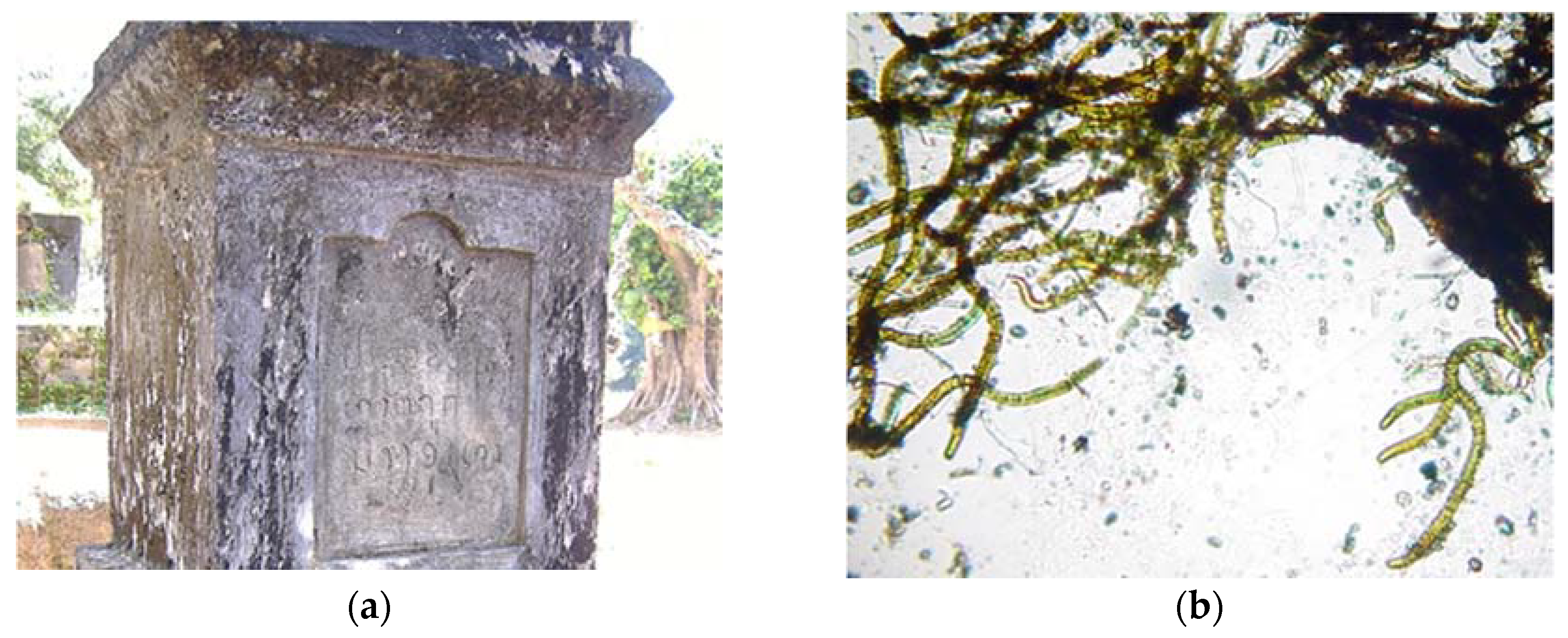
Figure 2.
(a) Black discoloration on a gravestone in Dom Khom, Laos, (b) the green/brown cyanobacteria (genus Scytonema) mainly responsible.
Figure 2.
(a) Black discoloration on a gravestone in Dom Khom, Laos, (b) the green/brown cyanobacteria (genus Scytonema) mainly responsible.

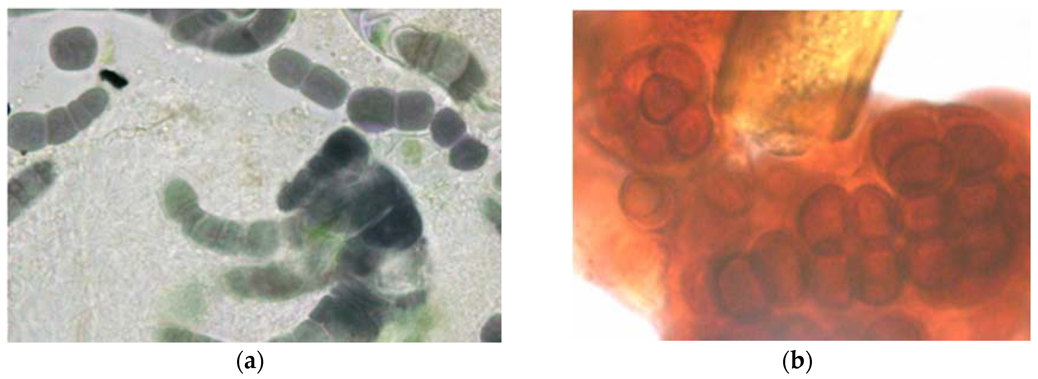
Figure 3.
Deeply colored cyanobacteria from external stone walls (a) Purple filamentous species, probably Fischerella genus, (b) Deep red coccoid species, probably Gloeocapsa, growing around a greenish brown sheathed cyanobacterial filament.
Figure 3.
Deeply colored cyanobacteria from external stone walls (a) Purple filamentous species, probably Fischerella genus, (b) Deep red coccoid species, probably Gloeocapsa, growing around a greenish brown sheathed cyanobacterial filament.

Figure 4.
Trentepohlia algae forming deep red discoloration on the painted wall of a public building in Ponta Delgada, Azores, Portugal.
Figure 4.
Trentepohlia algae forming deep red discoloration on the painted wall of a public building in Ponta Delgada, Azores, Portugal.

Figure 5.
Stone altar in the architectural zone of Becan, Campeche state, Mexico, showing red staining (Trentepohlia) on North and East facing sides.
Figure 5.
Stone altar in the architectural zone of Becan, Campeche state, Mexico, showing red staining (Trentepohlia) on North and East facing sides.

Figure 6.
Unstained CLSM image showing red autofluorescent single-celled cyanobacteria (mixed Synechococcus and Synechocystis) within the green soapstone background of a flake removed from the church of São Francisco de Paula, Tiradentes, Minas Gerais, Brazil.
Figure 6.
Unstained CLSM image showing red autofluorescent single-celled cyanobacteria (mixed Synechococcus and Synechocystis) within the green soapstone background of a flake removed from the church of São Francisco de Paula, Tiradentes, Minas Gerais, Brazil.

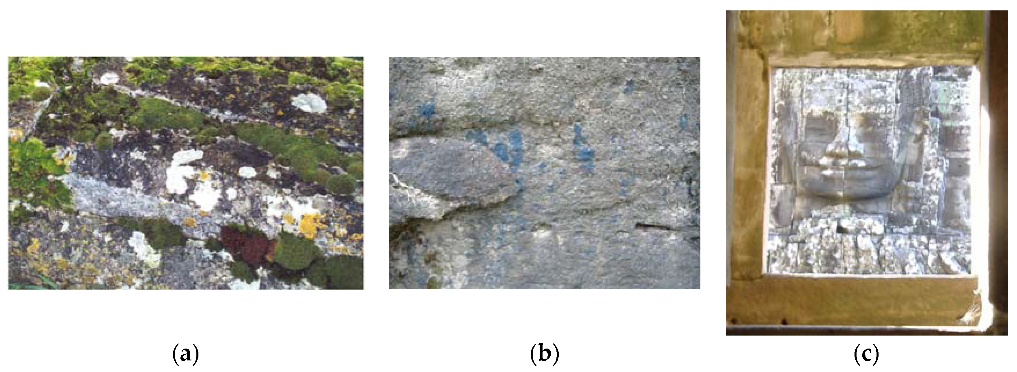
Figure 7.
(a) White and yellow lichens (and green moss) growing on a tombstone in a country churchyard in the UK, (b) blue lichen growing into a rock close to aboriginal art in Australia, (c) white lichens growing on a stone head viewed through a sandstone window with a green biofilm above it, Bayon temple, Angkor Thom, Cambodia.

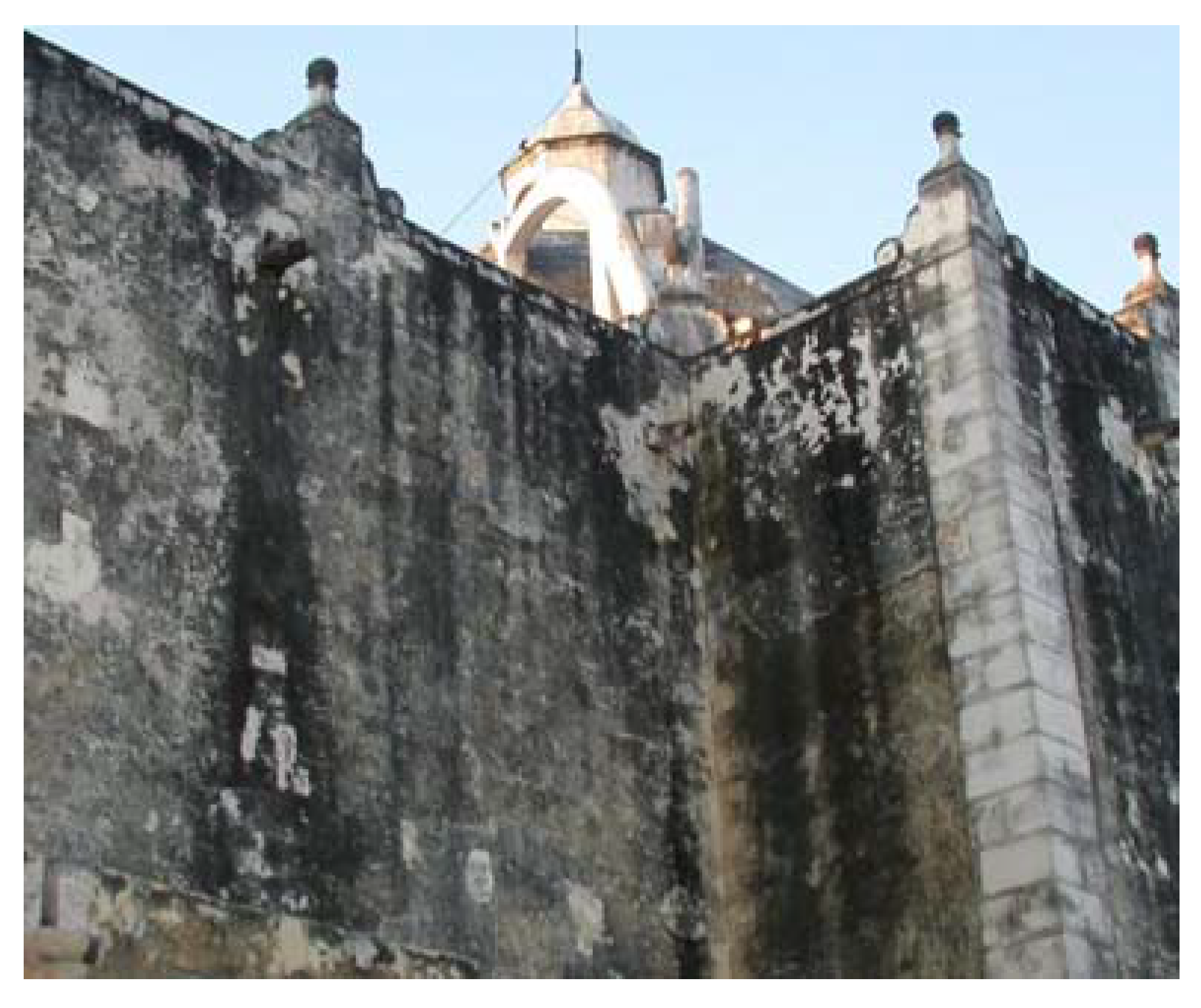
Figure 8.
Black cyanobacterial crusts on the walls of the Our Lady of the Immaculate Conception cathedral in the center of Campeche, Campeche state, Mexico.
Figure 8.
Black cyanobacterial crusts on the walls of the Our Lady of the Immaculate Conception cathedral in the center of Campeche, Campeche state, Mexico.

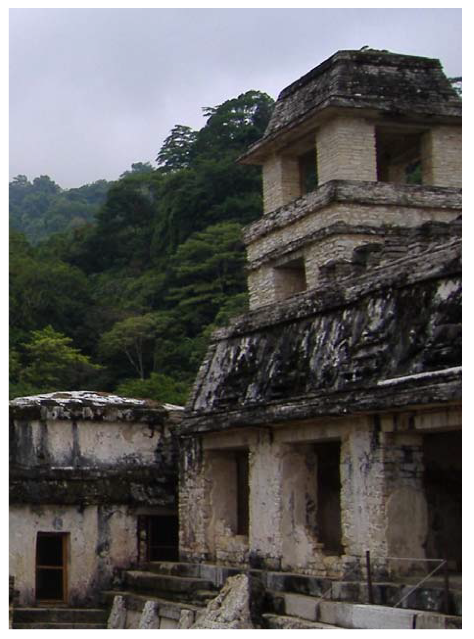
Figure 9.
Thick black biofilms on buildings in the patio of the Captives, Palenque, Mexico.


Table 1.
Pigments associated with algae and cyanobacteria.
| Pigment | Biological Function | Color(s) | Comments |
|---|---|---|---|
| Chlorophylls | Light absorption for energy production−photosynthesis | Green | Cyanobacteria contain only chlorophyll a |
| Carotenoids | Accessory pigments, light absorption, photoprotection | Orange, red, yellow, brown | Mainly membrane-bound |
| Phycobilins | Accessory pigments, light harvesting | Blue, red | Water-soluble, absorb green/red wavelengths |
| Mycosporine-like amino acids (MAAs) | Protective “sunscreen” | Dark brown | Found in cyanobacteria and some algae |
| Scytonemin | UV (perhaps general) protection | Dark brown/red | Only found in sheathed cyanobacteria |
Publisher’s Note: MDPI stays neutral with regard to jurisdictional claims in published maps and institutional affiliations. |
© 2020 by the author. Licensee MDPI, Basel, Switzerland. This article is an open access article distributed under the terms and conditions of the Creative Commons Attribution (CC BY) license (http://creativecommons.org/licenses/by/4.0/).
Share and Cite
MDPI and ACS Style
C. Gaylarde, C. Influence of Environment on Microbial Colonization of Historic Stone Buildings with Emphasis on Cyanobacteria. Heritage 2020, 3, 1469-1482. https://0-doi-org.brum.beds.ac.uk/10.3390/heritage3040081
AMA Style
C. Gaylarde C. Influence of Environment on Microbial Colonization of Historic Stone Buildings with Emphasis on Cyanobacteria. Heritage. 2020; 3(4):1469-1482. https://0-doi-org.brum.beds.ac.uk/10.3390/heritage3040081
Chicago/Turabian StyleC. Gaylarde, Christine. 2020. "Influence of Environment on Microbial Colonization of Historic Stone Buildings with Emphasis on Cyanobacteria" Heritage 3, no. 4: 1469-1482. https://0-doi-org.brum.beds.ac.uk/10.3390/heritage3040081