
Roman Wall Paintings: Characterisation of Plaster Coats Made of Clay Mud


1
Institute for Heritage Science (ISPC), National Research Council (CNR), Via Roberto Cozzi 53, 20125 Milan, Italy
2
Department of Human Sciences and Innovation for the Territory, Università degli Studi dell’Insubria, Via Sant’Abbondio 12, 22100 Como, Italy
*
Author to whom correspondence should be addressed.
Heritage 2021, 4(2), 889-905; https://0-doi-org.brum.beds.ac.uk/10.3390/heritage4020048
Submission received: 29 April 2021
/
Revised: 17 May 2021
/
Accepted: 19 May 2021
/
Published: 20 May 2021
(This article belongs to the Special Issue Earth Architecture: Culture, Issues, and Solution for Its Conservation)
Abstract
:This paper reports on the mineralogical characterisation of samples of wall paintings from various Roman sites in Lombardy (Italy), revealing recurrent types of stratigraphy. One of the stratigraphic samples analysed was found to be a particular kind of plaster: a three-coat work featuring two coats made of clay mud, found in the site of Santa Maria alla Porta (area of the Imperial Palace of Milan—first century CE). The fragments were analysed using optical microscopy on thin sections, X-ray diffraction, scanning electron microscopy with an energy-dispersive spectrometer and infrared spectroscopy, also in non-invasive external reflection mode (7500–375 cm−1). The most interesting feature found was the finish coat made of clay mud (illite, chlorite, kaolinite and fine quartz) with a few coarse clasts and linear cavities. This clay coat was the first example ever detected in Roman Lombardy and was used in combination with a thin painted coat made of clay mud with coarse clasts together with a blue pigment (Egyptian blue) and a render coat made of lime associated with lithic clasts (sand). Our findings brought to light a particular construction technique, since in the historical sources clay is only recommended for daubing on reeds and as a render coat.
1. Introduction
Roman painted plasters made up of overlying coats are reported by Vitruvius and by Pliny in a sequence containing: a render coat, made of lime as binder together with coarse sand as aggregate (sand from river deposits—harenatum); a finish coat, made of lime as the binder with fine marble powder as the aggregate (crushed calcite crystals—marmoratum); and a painted coat as the top layer [1,2,3]. A significant feature of these recipes is the change in aggregate composition between the render coat and the finish coat.
Archaeometric studies on Roman archaeological sites of the present-day region of Lombardy (corresponding to the eastern part of X Regio Venetia and to the western part of XI Regio Transpadana) have reported on various plasters that match to some extent the guidelines of Vitruvius and Pliny [4]. The difference in the composition between the render coats (silicate sand) and the finishing coats (calcite powder) was very often in accordance with the guidelines; however, the number of coats was significantly reduced (two or, rarely, three coats) [5]. Other kinds of aggregate were also employed (i.e., a marmoratum made of crystals of quartz instead of crystals of calcite).
We carried out a systematic campaign of mineralogical analyses on numerous samples of wall paintings from various Roman sites in Lombardy (northern Italy). The results, along with the description and comparison of the mortars, led to the identification of recurrent types of stratigraphy. An excavation recently made in the area of the former Palatium (Imperial Palace) in Mediolanum (Milan, Italy) (Figure 1) unearthed erratic fragments coming from the painted plaster showing two anomalous coats made of clay mud. Despite the extensive literature, even in recent years, describing analyses conducted on Roman wall paintings [6,7,8,9,10,11,12,13], to the best of our knowledge nothing similar has ever been characterised before. Using microscopic and chemical investigations, we focused on these particular fragments in order to characterise a Roman building technique never described so far in the literature.
In this paper, we report our analysis of the wall paintings from various Roman sites and describe our analytical campaign conducted on the particular samples from Milan, comparing it with our previous analyses [4,5,14]. The analyses were carried out with optical microscopy on thin sections, X-ray diffraction on powders and on fragments specifically treated for clay analysis, scanning electron microscopy equipped with an energy-dispersive spectrometer and infrared spectroscopy. Infrared measurements were also made in external reflection mode (FTIR-ER), typically used for non-invasive characterisation of painted surfaces, but rarely for wall paintings [9,11,12,15,16,17,18,19,20].
The archaeological site
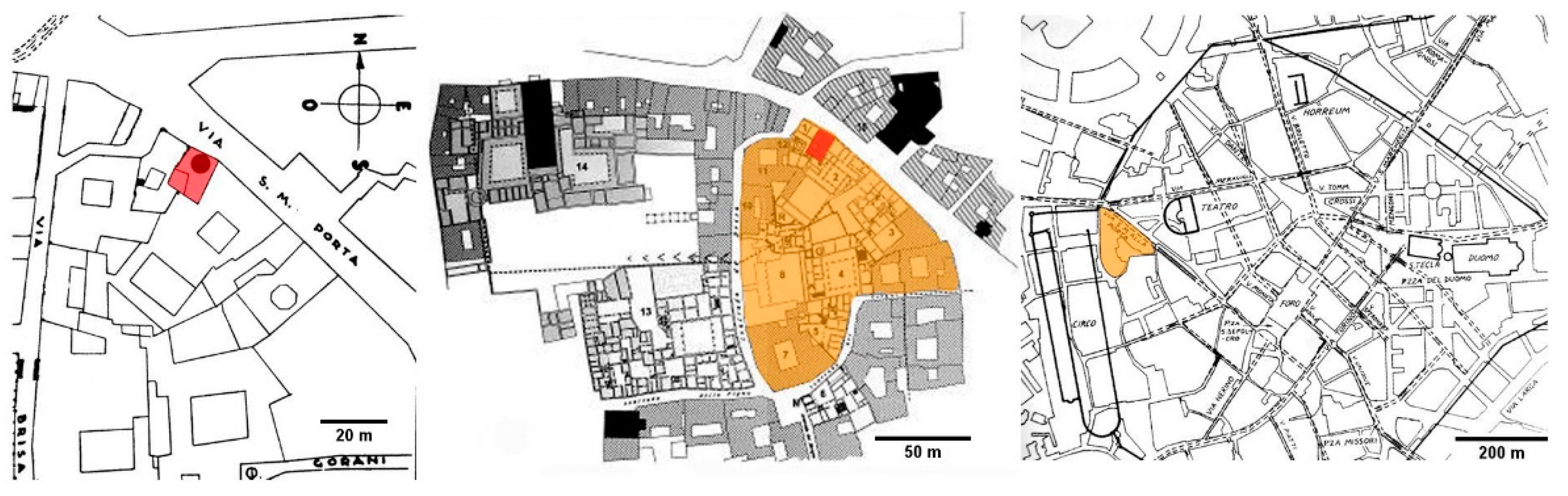
A district of the Roman town of Mediolanum (Milan, Italy) was occupied by a vast complex of buildings, known as the Palatium, which was built in the first century CE and encircled by the urban walls on the northern side and bordered by the circus on the western side (Figure 1). Very little is left of the Palatium: the most significant remains are located in via Brisa and consist of a circular colonnaded peristyle surrounded by some rooms of different shapes. The excavations carried out in 2015 in the northern part of the Imperial Palace area (via Santa Maria alla Porta 3, near via Brisa), unearthed a rubbish heap containing fragments of painted plasters mixed with other materials. These remains were from the first century CE, and their destruction was caused by the work on the Imperial building. The site was unearthed, cleaned out, studied and then covered because it was not possible to musealise the archaeological remains. The samples here analysed were labelled by the archaeologists as erratic fragments of the wall paintings present at the site.
Our main interest was on some fragments of wall paintings featuring a three-coat work together with a blue pigment (Figure 2).
2. Materials and Methods
Samples of each plaster coat were analysed as thin sections using optical microscopy (polarised light), as powders and fragments with X-ray diffraction and infrared spectroscopy, as polished cross sections with scanning electron microscopy and optical microscopy, ‘as is’ with external reflection infrared spectroscopy and digital portable microscopy.
Optical microscopy
The painted layer was observed with digital portable microscope MAOZUA USB001 and images were acquired using the software MicroCapture Plus (Mustech Electronics Co., Ltd., Shenzhen, China).
Thin sections were prepared according to the standard method. Nikon Eclipse E400Pol equipped with Nikon Pol objectives was used for the observation.
A fragment of sample was embedded in an epoxy resin, cross-cut with a diamond saw and then mechanically polished. The polished cross section was then observed with an optical microscope, Nikon Eclipse LV150, equipped with a Nikon DS-FI1 digital image acquisition system. Images were acquired and elaborated using the NIS-elements F software.
Scanning Electron Microscopy and Energy Dispersive X-ray spectroscopy
A sample prepared as a polished cross section was observed with a FEI/Philips XL30 ESEM (low vacuum mode (1 torr), 20 kV, BSE detector) [21,22]. The elemental analyses were carried out using an X-ray energy dispersive spectrometer, EDAX AMETEK Element, coupled to SEM.
X-ray diffraction
The samples were analysed as fine ground powders using a Rigaku MiniFlex 300 diffractometer (Rigaku Americas Corporation, The Woodlands, TX, USA) (30 kV, 10 mA, Cu-Kα radiation (λ = 1.5418 Å), 5–55° Theta/2-Theta, step scan 0.02°, scan speed 3°/min). PDXL2 software supporting ICDD (International Centre for Diffraction Data) PDF2 databases were used to identify the phases.
Some fragments were processed for clay mineral analysis too [23,24] as follows: dilution in water (30 mL); coarse grains removal by wet sieving (71 micrometres sieve); first analysis of dried specimens; exposition (two hours) to ethylene glycol (about 60 °C); cooling; exposition (two hours) in a muffle furnace (about 550 °C); cooling and second analysis; treatment with hydrochloric acid; third analysis. The instrument was a PANalytical X’Pert PRO MPD: generator settings 40 mA and 40 kV; radiation Cu-Ka λ = 1.5406 Å; scan range 3–35° 2θ; step size 0.017 2θ; scan step time 10.3376 s; continuous scan type; software PANalytical X’Pert HighScore.
Infrared spectroscopy
FTIR-ATR spectra were acquired by means of a Thermo Scientific Nicolet iS10 instrument (Thermo Fisher Scientific, Waltham, MA, USA), in the range between 4000 and 600 cm−1, 4 cm−1 resolution, 32 scans. The background was periodically registered.
FTIR measurements in external reflection mode (FTIR-ER) were carried out on the surface of the fragments with an Alpha Bruker FTIR portable spectrophotometer, equipped with a DTGS detector. The optimal distance was achieved by checking the focus via the on-board camera. Subsequently, a finer tuning was achieved via software, looking for the maximum signal directly in the interferogram. The average working distance from the surface was about 1–1.5 cm. Spectra were collected between 7500 and 375 cm−1, with 4 cm−1 resolution and 200 scans. The background was periodically acquired using a flat gold mirror.
3. Results
Table 1 reports the results of our systematic campaign of mineralogical analysis of wall paintings from Roman sites in Lombardy.
With regards to the site at Santa Maria alla Porta, the features of the samples are described, starting with the painted coat, i.e., the external layer.
(i) Painted coat
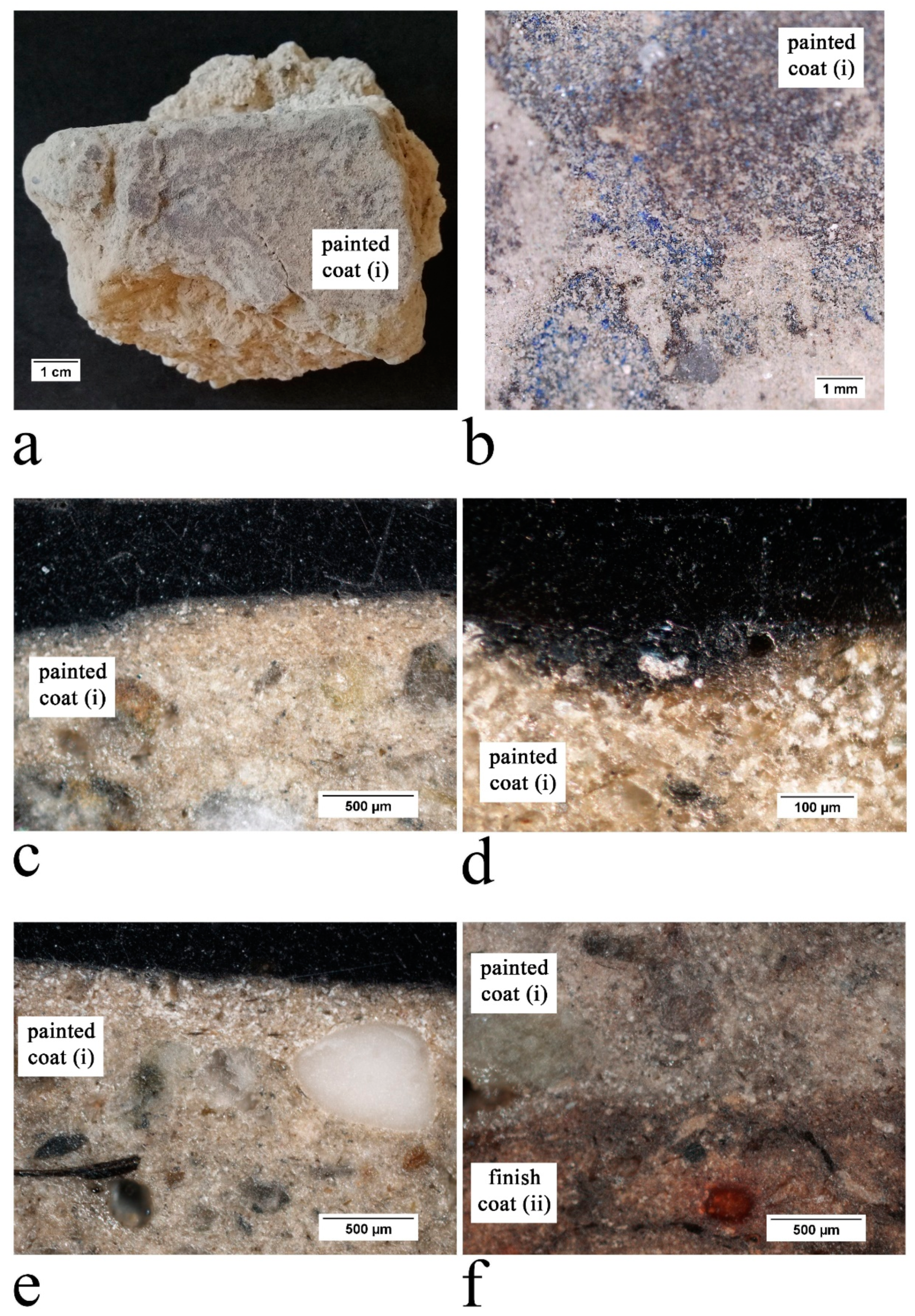
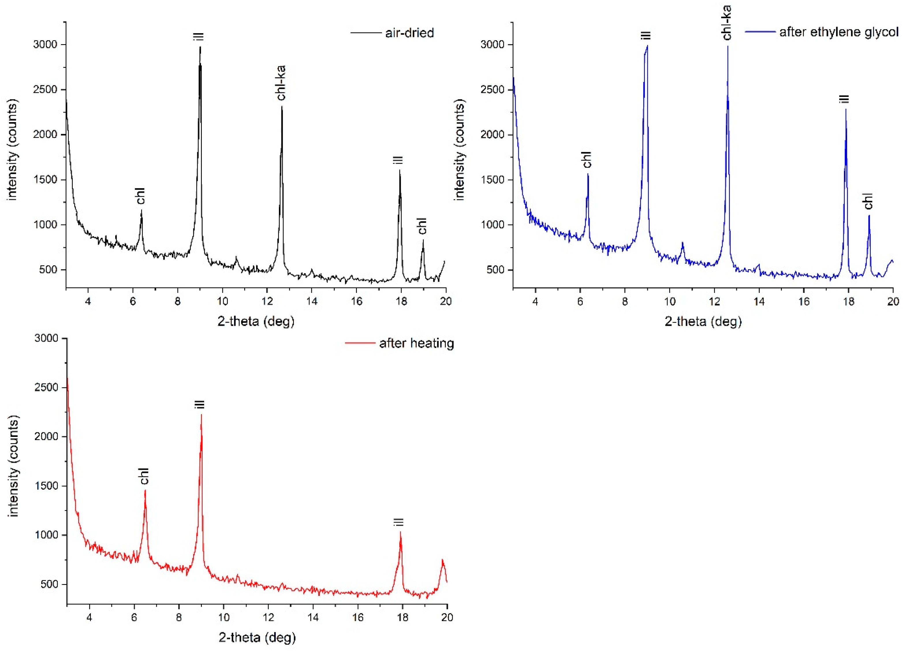
Based on the observation of thin sections, the paint layer contains a pigment made of Egyptian blue (CaCuSi4O10, calcium copper silicate—synthetic analogous of cuprorivaite). This layer lies on a coat (thickness 4 mm) containing clay mud with a homogeneous texture and small irregular cavities (size 0.2–0.4 till 1.0 mm) (Figure 3). Figure 4 shows the XRD analyses of the samples processed for clay mineral analysis. Kaolinite is destroyed upon heating, while as for illite and chlorite no change at all in the peak position and shapes is observed, except for the disappearance of the chlorite signal at 4.68 Å [23,24]. Thus, the clay is made up of these minerals. Angular quartz (size 0.05 mm) is present as well. There are also many coarse clasts of quartz, gneiss, limestone and marble (sub-angular corners, low sorting, size from 0.2 to 1.5 mm).
The FTIR spectrum in ATR mode of the same layer (Figure 5) shows an intense peak at 997 cm−1 and a smaller one at 1161 cm−1, which resemble the antisymmetric Si-O-Si stretching modes of Egyptian blue [27]. The signals at 760 and 671 cm−1 are likely due to the symmetric stretching of the same group, and thus are confirmatory. The absorbance peaks at 1418, 874 and 713 cm−1 (C-O asymmetric stretching, out-of-plane bending and in-plane bending vibrations, respectively) show the presence of calcium carbonate from the substrate [28,29]. The doublet at 795 and 778 cm−1 and the signal at 694 cm−1 (Si-O symmetrical stretching and bending vibrations, respectively) resemble quartz, present as an impurity of the pigment or in the aggregate fraction of the mortar [26].
The FTIR-ER analysis (Figure 5) confirmed the presence of calcium carbonate, as can be seen from the bands at 713 cm−1 (C-O symmetric in-plane bending), 875 cm−1 (C-O asymmetric out-of-plane bending), 1453 cm−1 (C-O asymmetric stretching), 1796 cm−1 (combination band), 2519 cm−1 and its shoulder at 2585 cm−1 (combination bands) and 4261 cm−1 (overtone) (Figure 5) [11,30]. There are quartz peaks at 469 cm−1 (Si-O asymmetric bending), 521 cm−1 (Si-O asymmetric bending), 695 and 793 cm−1 (Si-O symmetric stretching), 810 cm−1 (Si-O bending), 1158 cm−1 (Si-O asymmetric stretching), 1873 and 1990 cm−1 [30,31]. Kaolinite, found in the clay-based support, is particularly evident from peaks at 3697 and 3623 cm−1 (O-H group vibrations) and 911 cm−1 (Si-O stretching) [12,15]. The presence of gypsum at the surface was not detected either from ATR or XRD, but can be inferred from peaks at 2136 and 2236 cm−1 (overtone and combination bands) [15,30]. The peak at 1624 cm−1 is either gypsum or an unidentifiable organic substance (other peaks can be seen at 2987, 2930 and 2877 cm−1).
The identification of Egyptian blue pigment signals was complicated both by the small amounts of pigment on the surface and by the presence of other silicates in the substrate. The peaks at 1159 cm−1 (Si-O asymmetric stretching), 1047 cm−1 (Si-O asymmetric stretching), 998 cm−1 (Si-O asymmetric stretching), 751 and 673 cm−1, fit our Egyptian blue reference and other references reported in the literature [15]. The peak at 1283 cm−1 may be related either to an unidentified organic treatment or possibly to Egyptian blue [32] or gypsum [12].
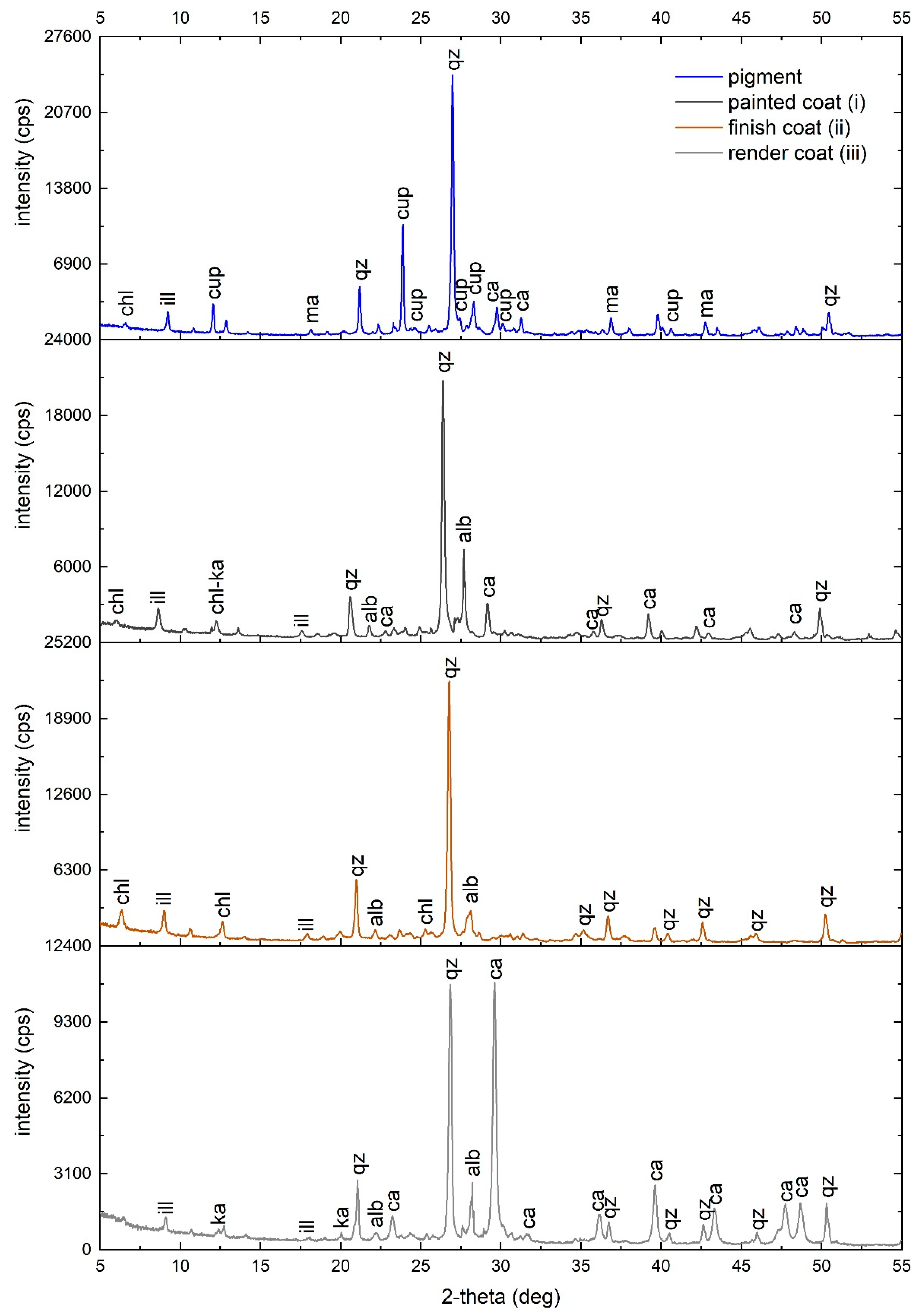
XRD analysis of powders detected quartz, calcite, illite, albite, kaolinite and chlorite. There are also some weak peaks that may be related to cuprorivaite (Egyptian blue) and magnetite (black earth) (Figure 6).
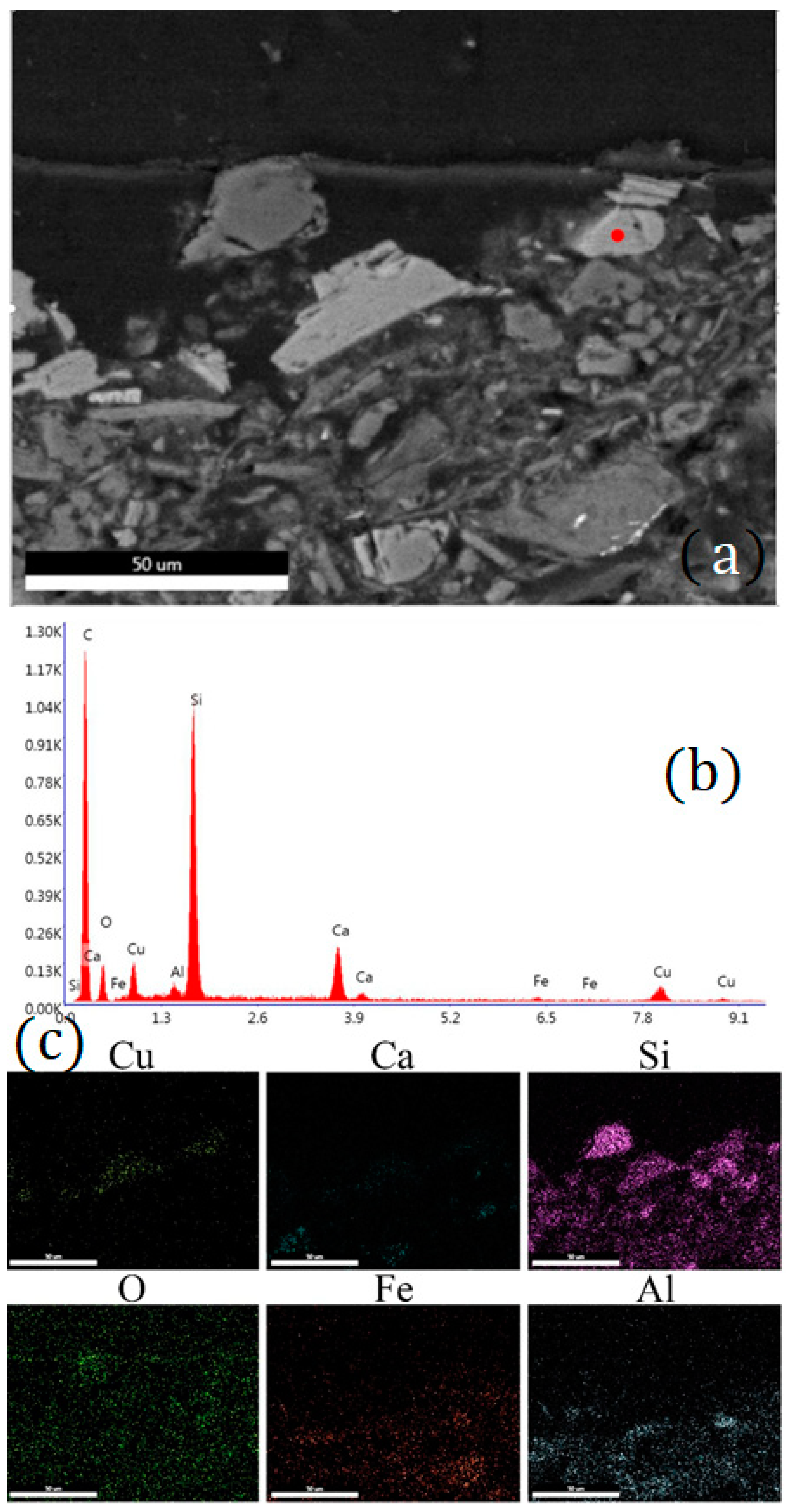
We also used SEM-EDX on a polished cross section of the layer in order to obtain information on the pigments used. Particles containing copper, calcium and silicon were detected. Their qualitative and semiquantitative elemental composition is compatible with that of Egyptian blue. The blue pigment particles are not uniformly distributed over the entire surface and vary in size between 15 and 40 µm. The absence of tin and zinc in the EDX analyses proves the use of pure copper-based compounds as precursors for the synthesis of Egyptian blue [11].
Figure 7 shows a typical EDX spectrum of these particles, together with their elemental distribution map. Iron is widely present, likely due to the substrate or to the black pigment, which was identified as a black earth as also hypothesised by X-ray diffraction analysis. Figure 8 highlights some particles of Egyptian blue on the surface, as well as the general composition of the clay substrate, with silicon and aluminium being the most diffuse elements.
(ii) Finish coat
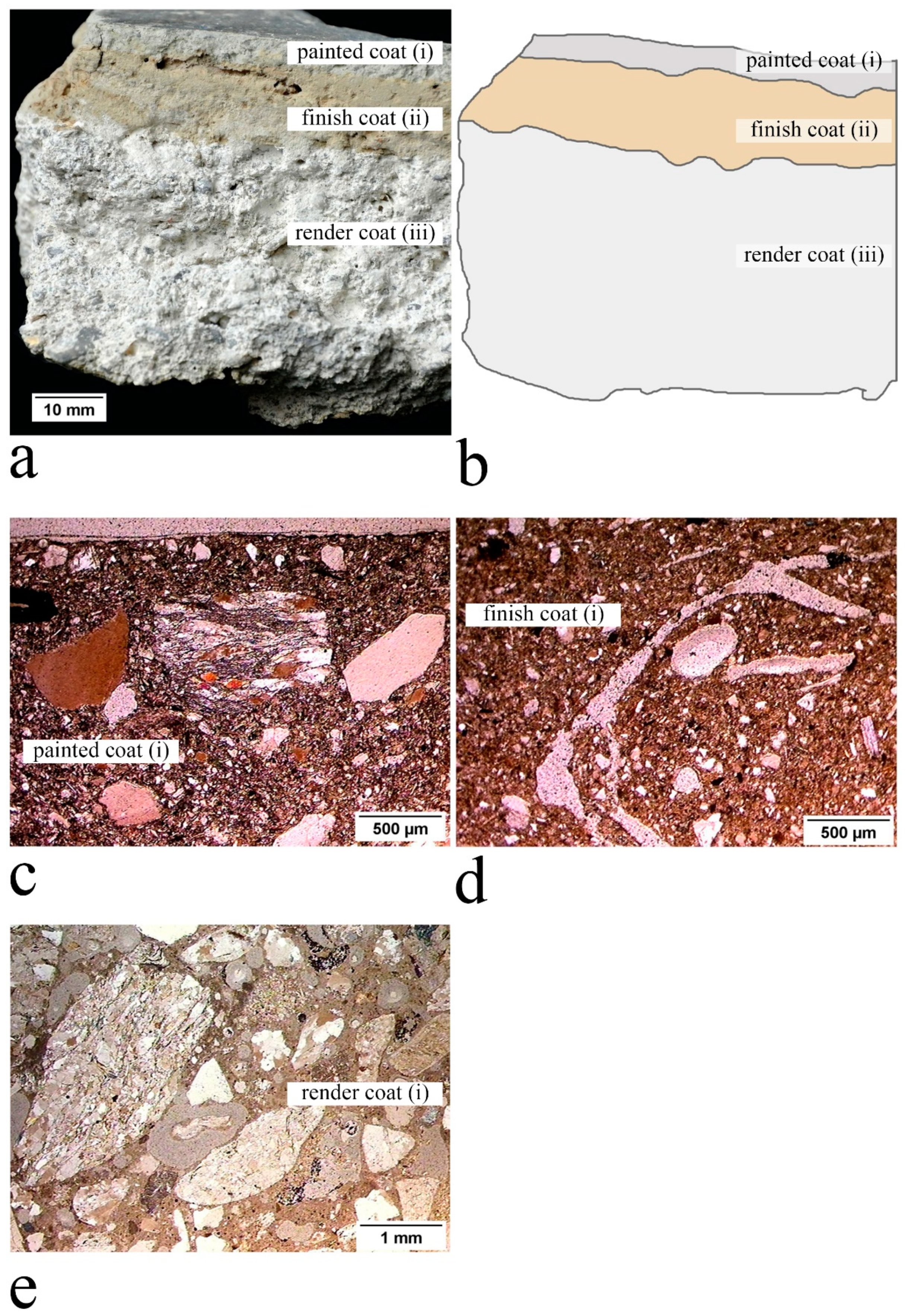
The thickness (11 mm) is almost homogeneous, the contact surface of the underlying coat is nearly flat, but it is marked by narrow cavities running parallel to the same surface.
The bulk is made of clay mud with many rounded (size 0.5–1 mm) and linear cavities (long and narrow—length about 10 mm) (Figure 3). The composition of the clay enlightened after the pre-treatment of the samples consists of: illite, chlorite and kaolinite together with angular quartz (size 0.05 mm) [23]. The XRD patterns are very similar to those shown in Figure 4. There are few signs of coarse clasts (gneiss with sub-angular corners, size 3.0 mm). The texture is similar to the texture of the painted coat (i), and the different shape of cavities and the different percentage of coarse clasts are worth noting.
Some of the signals observed in the FTIR spectrum are those already observed in the painted coat, which are the peaks already attributed to calcium carbonate (1408, 872 and 712 cm−1) and to quartz (797, 778 and 694 cm−1), which make up the mortar. There are peaks at 3694, 3651, 3627 and 3618 cm−1 (stretching of outer and inner hydroxyl ions), and 1084 and 1017 cm−1 (Si–O stretching bands of quartz and kaolinite). These peaks are probably related to clay minerals, such as illite [26]. XRD analysis on powders revealed the presence of quartz, illite, albite and chlorite (Figure 6).
(iii) Render coat
The thickness (30 mm) shows some irregularities, but the contact surface with the underlying coat is quite flat.
The coat contains a Mg lime binder associated with an aggregate made of sub-rounded clasts of quartz, limestone, gneiss, serpentine etc. (sand from Pre-Alpine river deposits, low sorting, size 0.5–5.0 mm); the texture is marked by many rounded cavities of different shapes and sizes (0.2–1.2 mm) (Figure 3). This layer has commonly been detected in Roman plasters in Lombardy [5]. The FTIR analysis suggested the same composition as with the finish coat. XRD on powder analysis confirmed the presence of calcite, quartz, albite, kaolinite and illite (Figure 6).
4. Discussion
Vitruvius and Pliny recommended using clayey earth only for daubing on reeds, and the use in plaster as a render coat (i.e., southern Gallia) [14,33]. However, the plaster found at the site of Santa Maria all Porta s hows the first detection in Roman Lombardy of a three-coat work (Figure 3), combining a finish coat and an intermediate coat, both containing clay with quartz and different proportions of coarse clasts (perhaps added to prevent cracking as the clay dries) and the proportion and shape of the cavities (possibly due to the presence of organic fibres?); these coats are lying on a render coat made, as usual, of lime and sandy aggregate.
Coats made of clay mud are extremely rare in the archaeological sites of Lombardy. A comparable case to date is a wall painting fragment (about 1.2–2 mm thick) pertaining to the wall plasters of the Republican Sanctuary of Brescia, built in the first half of the first century BCE. However, this differs greatly in the thickness of the painted layer and in the morphological features of the finish coat layer [14]. The painted coat of Brescia in fact supports a pigmented layer made of green earth and Egyptian blue and matches the features (composition, texture, presence of coarser clasts) of the painted coat found in Milan. A noticeable difference is the coat thickness (the coat from Milan is about two- or three-fold thick: 4 mm versus 1.2–2 mm). The finish coat shows a comparable texture as the previous one, however, with a considerably lower percentage of coarse clasts, and with the presence of elongated cavities. This is the first time that this kind of finish coat has been detected here in Roman Lombardy [14]. The render coat shows the same lithological composition and the same texture as already detected in equivalent coats of Roman Lombardy [5]. The features of the aggregate match those of the sands of the Pre-Alpine river deposits, diffused around Milan [5].
Regarding the provenance of the clay, the identification is uncertain as clay deposits are dispersed in the river Po alluvial plain in central and southern Lombardy: two areas near Milan, pertaining to this plain, were historically used to supply this raw material, mainly to make bricks. The first area is located in the north-western area of Milan and consists of a loessic-colluvial cover (2–3 m thick, clay) on glacio-fluvial deposits (weathered gravel) of middle Pleistocene. The second area is located near the southern border of the present-day metropolitan area and is made up of glacio-fluvial deposits of low energy (sand, silt and clayey silt) of upper Pleistocene [34].
With regard to the pigment determined in the fragments from Milan, Egyptian blue is the most common inorganic blue used by the Romans and is referred to as caerulem aegyptium by both Pliny and Vitruvius [35,36]. The painting layer also contains magnetite (black earth), as highlighted by both the XRD and EDX results. The mixture of Egyptian blue with black, such as carbon or iron pigments, has already been identified in Roman painted surface samples [37] for example, at Paestum [38]. FTIR-ER analyses revealed organic compounds on the external surface, probably originally used as a surface treatment [14], and gypsum, likely due to decay phenomena [29].
5. Conclusions
The wall painting fragments excavated in the Palatium of Mediolanum show a stratigraphy combining a painted and a finish coat both containing clay mud and fine quartz, lying on a render coat made of lime and silicate sand. The pigment is a mixture of Egyptian blue and magnetite (black earth), which was a regular artistic practice as attested in the literature.
A similar painted coat was found in the wall plasters of the Republican Sanctuary of Brixia; however, a finish coat made of clay was detected here for the first time in terms of the plasters of Roman Lombardy. In particular, given the position of the coat in the plaster sequence from the site of Santa Maria alla Porta, it would seem to be an alternative to the coat called marmoratum, which was normally made of crushed calcite crystals and is almost always present in painted plasters throughout the Roman world.
Unfortunately, the plaster found in Milan was excavated from rubble: it is thus impossible to know the exact dating, the original location or the role of this particular kind of mortar in rendering the walls.
Finally, we believe this work provides an important contribution to the study of the entire sequence of creating plaster; a study frequently neglected in comparison with the emphasis on painted surfaces. The palette of pigments used in Roman wall paintings is widely documented, but the characteristics (texture and composition) of the mortar layers supporting the painting only derive from a few archaeological sites. The expansion of the knowledge of these characteristics to a great number of Roman sites, together with the relationships among different layers and different pigments, may help to understand the processes involved in painted plaster.
Author Contributions
Conceptualisation, R.B. and L.F.; investigation, R.B., L.F., C.C. and L.R.; writing—original draft preparation, R.B., L.F., C.C. and L.R.; writing—review and editing, R.B., L.F., C.C. and L.R. All authors have read and agreed to the published version of the manuscript.
Funding
This research received no specific grant from any funding agency in the public, commercial or not-for-profit sectors.
Data Availability Statement
The data presented in this study are available on request from the corresponding author.
Acknowledgments
The authors heartily acknowledge the Fondazione Banca del Monte di Lombardia and Norberto Masciocchi for support.
Conflicts of Interest
The authors declare no conflict of interest.
References
- Vitruvius. On Architecture; Schofield, R., Ed.; Penguin classics; Penguin Books Limited: London, UK, 2009; ISBN 9780141931951. [Google Scholar]
- Pliny. Natural History; Eichholz, D.E., Ed.; Loeb Classical Library; Heinemann: Cambridge, UK, 1962. [Google Scholar]
- Laurie, A.P. Greek and Roman Methods of Painting: Some Comments on the Statements Made by Pliny and Vitruvius about Wall and Panel Painting; Cambridge University Press: Cambridge, UK, 1910. [Google Scholar]
- Bugini, R.; Folli, L.; Biondelli, D. Grain morphology of aggregates in Roman plasters. In Proceedings of the 14th Euroseminar on Microscopy on Applied to Building Materials, Helsingør, Denmark, 10–14 June 2013; Danish Technological Institute: Taastrup, Denmark, 2013; pp. 25–28. [Google Scholar]
- Bugini, R.; Folli, L. Critères pour la comparaison des enduits peints romains de la Lombardie. ArcheoSciences 2013, 37, 41–50. [Google Scholar] [CrossRef]
- Ergenç, D.; La Russa, M.F.; Ruffolo, S.A.; Fort, R.; Sánchez Montes, A.L. Characterization of the wall paintings in La Casa de los Grifos of Roman city Complutum. Eur. Phys. J. Plus 2018, 133, 355. [Google Scholar] [CrossRef]
- Mateos, L.D.; Esquivel, D.; Cosano, D.; Jiménez-Sanchidrián, C.; Ruiz, J.R. Micro-Raman analysis of mortars and wall paintings in the Roman villa of Fuente Alamo (Puente Genil, Spain) and identification of the application technique. Sens. Actuators A Phys. 2018, 281, 15–23. [Google Scholar] [CrossRef]
- Giorgi, L.; Nevin, A.; Nodari, L.; Comelli, D.; Alberti, R.; Gironda, M.; Mosca, S.; Zendri, E.; Piccolo, M.; Izzo, F.C. In-situ technical study of modern paintings part 1: The evolution of artistic materials and painting techniques in ten paintings from 1889 to 1940 by Alessandro Milesi (1856–1945). Spectrochim. Acta Part A Mol. Biomol. Spectrosc. 2019, 219, 530–538. [Google Scholar] [CrossRef]
- Nodari, L.; Ricciardi, P. Non-invasive identification of paint binders in illuminated manuscripts by ER-FTIR spectroscopy: A systematic study of the influence of different pigments on the binders’ characteristic spectral features. Herit. Sci. 2019, 7, 7. [Google Scholar] [CrossRef]
- La Nasa, J.; Moretti, P.; Maniccia, E.; Pizzimenti, S.; Colombini, M.P.; Miliani, C.; Modugno, F.; Carnazza, P.; De Luca, D. Discovering Giuseppe Capogrossi: Study of the Painting Materials in Three Works of Art Stored at Galleria Nazionale (Rome). Heritage 2020, 3, 52. [Google Scholar] [CrossRef]
- Pronti, L.; Romani, M.; Viviani, G.; Stani, C.; Gioia, P.; Cestelli-Guidi, M. Advanced methods for the analysis of Roman wall paintings: Elemental and molecular detection by means of synchrotron FT-IR and SEM micro-imaging spectroscopy. Rend. Lincei Sci. Fis. Nat. 2020, 31, 485–493. [Google Scholar] [CrossRef]
- Sbroscia, M.; Cestelli-Guidi, M.; Colao, F.; Falzone, S.; Gioia, C.; Gioia, P.; Marconi, C.; Mirabile Gattia, D.; Loreti, E.M.; Marinelli, M.; et al. Multi-analytical non-destructive investigation of pictorial apparatuses of “Villa della Piscina” in Rome. Microchem. J. 2020, 153, 104450. [Google Scholar] [CrossRef]
- Cortea, I.M.; Ghervase, L.; Țentea, O.; Pârău, A.C.; Rădvan, R. First Analytical Study on Second-Century Wall Paintings from Ulpia Traiana Sarmizegetusa: Insights on the Materials and Painting Technique. Int. J. Archit. Herit. 2020, 14, 751–761. [Google Scholar] [CrossRef]
- Bugini, R.; Corti, C.; Folli, L.; Rampazzi, L. Unveiling the Use of Creta in Roman Plasters: Analysis of Clay Wall Paintings From Brixia (Italy). Archaeometry 2017, 59, 84–95. [Google Scholar] [CrossRef]
- Germinario, C.; Francesco, I.; Mercurio, M.; Langella, A.; Sali, D.; Kakoulli, I.; De Bonis, A.; Grifa, C. Multi-analytical and non-invasive characterization of the polychromy of wall paintings at the Domus of Octavius Quartio in Pompeii. Eur. Phys. J. Plus 2018, 133, 359. [Google Scholar] [CrossRef]
- Biron, C.; Mounier, A.; Arantegui, J.P.; Bourdon, G.L.; Servant, L.; Chapoulie, R.; Roldán, C.; Almazán, D.; Díez-de-Pinos, N.; Daniel, F. Colours of the «images of the floating world». Non-invasive analyses of Japanese ukiyo-e woodblock prints (18th and 19th centuries) and new contributions to the insight of oriental materials. Microchem. J. 2020, 152, 104374. [Google Scholar] [CrossRef]
- Daveri, A.; Malagodi, M.; Vagnini, M. The Bone Black Pigment Identification by Noninvasive, In Situ Infrared Reflection Spectroscopy. J. Anal. Methods Chem. 2018, 2018, 6595643. [Google Scholar] [CrossRef] [Green Version]
- Izzo, F.; Germinario, C.; Grifa, C.; Langella, A.; Mercurio, M. External reflectance FTIR dataset (4000–400 cm−1) for the identification of relevant mineralogical phases forming Cultural Heritage materials. Infrared Phys. Technol. 2020, 106, 103266. [Google Scholar] [CrossRef]
- Zuena, M.; Buemi, L.P.; Stringari, L.; Legnaioli, S.; Lorenzetti, G.; Palleschi, V.; Nodari, L.; Tomasin, P. An integrated diagnostic approach to Max Ernst’s painting materials in his Attirement of the Bride. J. Cult. Herit. 2020, 43, 329–337. [Google Scholar] [CrossRef]
- Rosi, F.; Miliani, C.; Delaney, J.; Dooley, K.; Stringari, L.; Subelyte, G.; Buemi, L.P. CHAPTER 1. Jackson Pollock’s Drip Paintings: Tracing the Introduction of Alkyds Through Non-invasive Analysis of Mid-1940s Paintings. In Science and Art; The Royal Society of Chemistry: London, UK, 2020; pp. 1–18. ISBN 9781788016384. [Google Scholar]
- Ranalli, G.; Zanardini, E.; Andreotti, A.; Colombini, M.P.; Corti, C.; Bosch-Roig, P.; De Nuntiis, P.; Lustrato, G.; Mandrioli, P.; Rampazzi, L.; et al. Hi-tech restoration by two-steps biocleaning process of Triumph of Death fresco at the Camposanto Monumental Cemetery (Pisa, Italy). J. Appl. Microbiol. 2018, 125, 800–812. [Google Scholar] [CrossRef]
- Goldstein, J.I.; Newbury, D.E.; Michael, J.R.; Ritchie, N.W.M.; Scott, J.H.J.; Joy, D.C. Scanning Electron Microscopy and X-ray Microanalysis; Springer: New York, NY, USA, 2017; ISBN 9781493966769. [Google Scholar]
- Thorez, J. Phyllosilicates and Clay Minerals: A Laboratory Handbook for Their X-ray Diffraction Analysis; Lelotte: Dison, Belgium, 1975. [Google Scholar]
- Moore, D.M.; Reynolds, R.C., Jr. X-ray Diffraction and the Identification and Analysis of Clay Minerals; Oxford University Press: Oxford, UK, 1997; ISBN 9780195087130. [Google Scholar]
- Farmer, V.C. The Infrared Spectra of Minerals; Mineralogical Society monograph; Mineralogical Society: London, UK, 1974; ISBN 9780903056052. [Google Scholar]
- Wilson, M.J. Clay Mineralogy: Spectroscopic and Chemical Determinative Methods; Wilson, M.J., Ed.; Chapman & Hall: London, UK, 1994; ISBN 9780412533808. [Google Scholar]
- Mirti, P.; Appolonia, L.; Casoli, A.; Ferrari, R.P.; Laurenti, E.; Amisano Canesi, A.; Chiari, G. Spectrochemical and structural studies on a roman sample of Egyptian blue. Spectrochim. Acta Part A Mol. Biomol. Spectrosc. 1995, 51, 437–446. [Google Scholar] [CrossRef]
- Rampazzi, L.; Andreotti, A.; Bressan, M.; Colombini, M.P.; Corti, C.; Cuzman, O.; D’Alessandro, N.; Liberatore, L.; Palombi, L.; Raimondi, V.; et al. An interdisciplinary approach to a knowledge-based restoration: The dark alteration on Matera Cathedral (Italy). Appl. Surf. Sci. 2018, 458, 529–539. [Google Scholar] [CrossRef]
- Ranalli, G.; Zanardini, E.; Rampazzi, L.; Corti, C.; Andreotti, A.; Colombini, M.P.; Bosch-Roig, P.; Lustrato, G.; Giantomassi, C.; Zari, D.; et al. Onsite advanced biocleaning system for historical wall paintings using new agar-gauze bacteria gel. J. Appl. Microbiol. 2019, 126, 1785–1796. [Google Scholar] [CrossRef]
- Brunello, V.; Corti, C.; Sansonetti, A.; Tedeschi, C.; Rampazzi, L. Non-invasive FTIR study of mortar model samples: Comparison among innovative and traditional techniques. Eur. Phys. J. Plus 2019, 134, 270. [Google Scholar] [CrossRef]
- Arrizabalaga, I.; Gomez-Laserna, O.; Carrero, J.A.; Bustamante, J.; Rodriguez, A.; Arana, G.; Madariaga, J.M.; Antonio Carrero, J.; Bustamante, J.; Rodriguez, A.; et al. Diffuse reflectance FTIR database for the interpretation of the spectra obtained with a handheld device on built heritage materials. Anal. Methods 2015, 7, 1061–1070. [Google Scholar] [CrossRef]
- Bruni, S.; Cariati, F.; Casadio, F.; Toniolo, L. Spectrochemical characterization by micro-FTIR spectroscopy of blue pigments in different polychrome works of art. Vib. Spectrosc. 1999, 20, 15–25. [Google Scholar] [CrossRef]
- Ramjoue, E. Quelques particularites techniques des fresques romaines de Vandoeuvres dans le Canton de Geneve. In Roman Wall Painting; Béarat, H., Ed.; Fribourg University, Institute of Mineralogy and Petrography: Fribourg, Switzerland, 1997; pp. 167–179. [Google Scholar]
- Carta Geologica d’Italia (CARG) 1:50.000—Foglio Milano n. 118, Servizio Geologico d’Italia (Piacenza, Italia). Available online: https://www.isprambiente.gov.it/Media/carg/lombardia.html (accessed on 28 April 2021).
- Gettens, R.J.; Stout, G.L. Painting Materials: A Short Encyclopaedia; Dover Publications: New York, NY, USA, 1966; ISBN 0486215970. [Google Scholar]
- Riederer, J. Egyptian blue. In Artists’ Pigments. A Handbook of Their History and Characteristics—Vol. 3; West FitzHugh, E., Ed.; National Gallery of Art, Washington and Oxford University Press: Oxford, UK, 1997; pp. 23–45. ISBN 9782970013204. [Google Scholar]
- Edreira, M.C.; Feliu, M.J.; Fernández-Lorenzo, C.; Martín, J. Spectroscopic Study of Egyptian Blue Mixed with Other Pigments. Helv. Chim. Acta 2003, 86, 29–49. [Google Scholar] [CrossRef]
- Alberghina, M.F.; Germinario, C.; Bartolozzi, G.; Bracci, S.; Grifa, C.; Izzo, F.; La Russa, M.F.; Magrini, D.; Massa, E.; Mercurio, M.; et al. Non-invasive characterization of the pigment’s palette used on the painted tomb slabs at Paestum archaeological site. IOP Conf. Ser. Mater. Sci. Eng. 2020, 949, 012002. [Google Scholar] [CrossRef]
Figure 1.
The location of the site of Santa Maria alla Porta (Milan, Italy). The investigated site and the Palatium are marked in red and orange colour, respectively. The map is shown at different levels of magnification (see scale bars) to help to situate the site in the Roman city structure.
Figure 1.
The location of the site of Santa Maria alla Porta (Milan, Italy). The investigated site and the Palatium are marked in red and orange colour, respectively. The map is shown at different levels of magnification (see scale bars) to help to situate the site in the Roman city structure.

Figure 2.
(a) Painted surface of the sample and (b) digital microscope image of the painted surface: a mixture of blue and black particles can be seen; (c–e) optical microscope image of layer (i) (polished cross section); (f) optical microscope image of layer (ii) and layer (i) (polished cross section).

Figure 3.
(a,b) A sample from the site of Santa Maria alla Porta, showing the sequence of the plaster coats: (i) painted (top, thin); (ii) finish (middle); (iii) render (bottom, thick). (c) Thin cross sections of the painted coat (i) with clay mud and coarse clasts. (d) Finish coat (ii) with clay mud and linear cavities. (e) Render coat (iii) with lime and lithic clasts (optical microscopy plane-polarised light).

Figure 4.
XRD analysis of a sample from the painted coat (i) processed for clay mineral analysis. Diffractograms of the air-dried sample (top, left), after exposition to ethylene glycol (top, right) and after heating (bottom, left). Legend: chl: chlorite; ill: illite; ka: kaolinite.
Figure 4.
XRD analysis of a sample from the painted coat (i) processed for clay mineral analysis. Diffractograms of the air-dried sample (top, left), after exposition to ethylene glycol (top, right) and after heating (bottom, left). Legend: chl: chlorite; ill: illite; ka: kaolinite.

Figure 5.
ATR spectrum (top) of the painted layer, showing the presence of Egyptian blue (1161, 997, 760 and 671 cm−1), calcite (1418, 874 and 713 cm−1) and quartz (795, 778 and 694cm−1); FTIR-ER spectrum (bottom) of the painted coat, with the most interesting peaks. Peaks of Egyptian blue (1159, 1047, 998, 751, 673 cm−1—see also an excerpt of FTIR-ER spectrum of Kremer 10060 Egyptian blue standard pigment), gypsum (2136, 2236 cm−1) and organic substance (2987, 2939, 2877 cm−1) were shown.

Figure 6.
XRD analysis on powders of the different layers. From the top, the diffractograms of the pigment, the painted coat (i), the finish coat (ii) and the render coat (iii) can be seen, showing the main signals of the identified minerals. Legend: chl: chlorite; ill: illite; cup: cuprorivaite; ma: magnetite; qz: quartz; ca: calcite; ka: kaolinite; alb: albite.

Figure 7.
(a) SEM image in BSE mode of the painted layer; (b) EDX spectrum of a particle of Egyptian blue; (c) EDX elemental distribution maps (scale bar: 50 µm).
Figure 7.
(a) SEM image in BSE mode of the painted layer; (b) EDX spectrum of a particle of Egyptian blue; (c) EDX elemental distribution maps (scale bar: 50 µm).

Figure 8.
(a) SEM image in BSE mode of the painted layer and its substrate; (b) EDX elemental distribution maps (scale bar: 100 µm).
Figure 8.
(a) SEM image in BSE mode of the painted layer and its substrate; (b) EDX elemental distribution maps (scale bar: 100 µm).


Table 1.
Description and main mineralogical features of the analysed plasters coming from Roman archaeological sites in Milan and Brescia.
Table 1.
Description and main mineralogical features of the analysed plasters coming from Roman archaeological sites in Milan and Brescia.
| Site | Number of Samples with the Same Stratigraphy | Chronology (Century *) | Plaster Thickness (mm) | Render Coat: Aggregate Composition | Finish Coat | Pigment | ||
|---|---|---|---|---|---|---|---|---|
| Binder Composition | Aggregate Composition | Crystal Size (mm) | ||||||
| Milano | ||||||||
| Università Cattolica | 6 | 1st | 12–30 | quartz, silicates, limestone | Mg lime | quartz, limestone, brick | 0.04–3.0 | not examined |
| Università Cattolica | 17 | 3rd | 18–35 | quartz, silicates, brick | Mg lime | calcite, quartz, silicates | 0.04–2.7 0.1–3.0 | not examined |
| piazza Fontana | 6 | mid 1st | 20–35 | quartz, silicates | Mg lime | quartz, silicates | 1.0–3.0 | cinnabar, carbon black |
| piazza Fontana | 2 | early 1st | 25 | quartz, silicates | Mg lime | quartz | 0.1–3.0 | carbon black |
| piazza Meda | 2 | mid 1st | 25–30 | quartz, silicates, brick | Mg lime | quartz | 0.4 | red ochre |
| piazza Meda | 1 | early 4th | 28 | quartz, silicates, brick | Mg lime | quartz | 0.4 | green earth |
| via Correnti | 9 | 1st | 15–25 | quartz, silicates, limestone | Mg lime | quartz, silicates, limestone | 0.1–1.2 | green earth |
| via Correnti | 25 | 2nd | 15–20 | quartz, silicates, limestone | Mg lime | quartz, limestone, calcite | 0.05–2.5 0.04–2.0 | green earth, yellow ochre |
| via Broletto | 9 | 3rd | 15–22 | quartz, silicates | Mg lime | quartz, silicates, calcite | 0.05–0.4 | cinnabar, red ochre, green earth |
| corso Magenta (Monastero Maggiore) | 10 | 3rd | 25–55 | quartz, silicates, brick | Mg lime | quartz, calcite | 0.1–2.0 | red ochre |
| corso Magenta (palazzo Litta) | 6 | 3rd | 40–45 | quartz, silicates | Mg lime | quartz | 1.0–3.0 | Egyptian blue, green earth, red ochre, carbon black |
| via S. Maria Porta | 3 | 3rd | 15–60 | quartz, silicates | Mg lime | calcite | 0.2–1.5 | cinnabar, yellow ochre, Egyptian blue |
| Brescia | ||||||||
| Under the Sanctuary | 6 | 2nd BCE | 2–5 | limestone, gneiss | Mg lime | quartz, limestone | 0.4 | red earth, carbon black, chalk |
| Sanctuary | 34 | early 1st BCE | 5–10 | quartz, limestone, dolomite | Ca lime | dolomite | 0.3–3.0 | yellow ochre, red earth, Egyptian blue + green earth, cinnabar, carbon black |
| Sanctuary | 18 | early 1st BCE | 20–25 | limestone, quartz, brick | Ca lime | clay + quartz | 0.1–0.3 | Egyptian blue, red ochre |
| Sanctuary | 1 | early 1st | 30 | limestone, flint | Ca lime | dolomite, limestone | 0.3–3.0 | not examined |
| via Trieste | 6 | mid 1st | 2–3 | quartz, limestone | Mg lime | calcite | 1.5 | red earth, green earth, yellow ochre |
| palazzo Martinengo | 7 | early 1st | 4–5 | limestone | Mg lime | dolomite | 0.3–3.5 | red earth, Egyptian blue + green earth |
| palazzo Martinengo | 14 | late 1st | 4–13 | quartz, limestone | Mg lime | dolomite | 0.3–4.0 | Egyptian blue + green earth, red earth |
| Liceo Arnaldo | 19 | mid 1st–early 2nd | 4–8 | dolomite | Mg lime | dolomite | 0.5–1.5 | red earth, green earth, yellow ochre |
| Santa Giulia | 42 | late 2nd–early 3rd | 3–11 | dolomite | Mg lime | dolomite | 0.3–2.5 | green earth, yellow ochre, Egyptian blue |
| Brescia Province | ||||||||
| Cividate Camuno | ||||||||
| via Palazzo | 9 | 1st | 10–20 | quartz, silicates | Mg lime | calcite, limestone | 0.1–3.5 | red ochre, yellow ochre, Egyptian blue |
| Theatre | 5 | late 1st | 5 | quartz, limestone, brick | Mg lime | calcite | 0.1–1.0 | red earth |
| domus | 7 | mid 1st | 10–20 | quartz, limestone | Mg lime | calcite | 0.3–4.0 | not examined |
| Amphitheatre | 10 | early 1st | 5–15 | quartz, limestone | Mg lime | calcite | 0.3–4.0 | red ochre, green earth |
| Sanctuary of Minerva-Breno | 18 | 1st | 10–20 | quartz, limestone | Mg lime | calcite | 0.1–3.5 | red ochre, yellow ochre, Egyptian blue |
| Sirmione-Villa Grotte di Catullo | 19 | 2nd | 15–50 | limestone | Ca lime | calcite, limestone | 0.2–4.0 0.2–0.6 | Egyptian blue, cinnabar, red and yellow ochre, minium (lead), green earth, carbon back |
| Bedriacum | ||||||||
| domus | 20 | 1st–2nd | 15–20 | quartz, limestone | Ca lime | brick, calcite, limestone | 0.4–5.0 0.1–0.8 | yellow ochre, red earth, green earth |
| Rubble pit | 8 | 1st–5th | 10–20 | quartz, limestone | Mg lime | quartz, limestone, brick | 0.5–5.0 | red ochre, red earth, chalk |
Legend: Ca lime: calcitic lime; Mg lime: magnesian lime; * site chronology is intended to be CE unless otherwise indicated.
Publisher’s Note: MDPI stays neutral with regard to jurisdictional claims in published maps and institutional affiliations. |
© 2021 by the authors. Licensee MDPI, Basel, Switzerland. This article is an open access article distributed under the terms and conditions of the Creative Commons Attribution (CC BY) license (https://creativecommons.org/licenses/by/4.0/).
Share and Cite
MDPI and ACS Style
Bugini, R.; Corti, C.; Folli, L.; Rampazzi, L. Roman Wall Paintings: Characterisation of Plaster Coats Made of Clay Mud. Heritage 2021, 4, 889-905. https://0-doi-org.brum.beds.ac.uk/10.3390/heritage4020048
AMA Style
Bugini R, Corti C, Folli L, Rampazzi L. Roman Wall Paintings: Characterisation of Plaster Coats Made of Clay Mud. Heritage. 2021; 4(2):889-905. https://0-doi-org.brum.beds.ac.uk/10.3390/heritage4020048
Chicago/Turabian StyleBugini, Roberto, Cristina Corti, Luisa Folli, and Laura Rampazzi. 2021. "Roman Wall Paintings: Characterisation of Plaster Coats Made of Clay Mud" Heritage 4, no. 2: 889-905. https://0-doi-org.brum.beds.ac.uk/10.3390/heritage4020048