
Paper Foxing Stains on a Historic Manuscript from the Early Qajar Era: Abiotic or Biotic Foxing?


Faculty of Cultural Materials Conservation, Tabriz Islamic Art University, Tabriz 5164736931, Iran
*
Author to whom correspondence should be addressed.
Heritage 2021, 4(3), 1366-1374; https://0-doi-org.brum.beds.ac.uk/10.3390/heritage4030074
Submission received: 1 June 2021
/
Revised: 15 July 2021
/
Accepted: 15 July 2021
/
Published: 18 July 2021
(This article belongs to the Collection Feature Papers)
Abstract
:The aim of this study was to identify the nature and cause of foxing spots in a historical manuscript. This manuscript was a Holy Quran from the beginning of the Qajar period and the end of the 18th century. Samples were incubated for 14 days and were evaluated for the presence of fungal activity. UV fluorescence photography, micro X-ray fluorescence spectroscopy and Fourier transform infrared spectroscopy were also used to investigate the characteristics and causes of foxing spots. The results showed that there was no fungal activity in the foxing spots of this manuscript. Based on the morphology of the stain in UV fluorescence photography, these foxing stains are of the Bullseye type, usually associated with metal ions. µXRF spectroscopy also showed a high accumulation of iron and copper at the site of these spots. This indicates abiotic foxing in this manuscript. Based on FTIR spectroscopy and peak deconvolution and fitting by Gaussian function, abiotic foxing increases the cellulose oxidation rate. Intensification of cellulose oxidation in foxing stains can be considered as one of the reasons for paper discoloration.
1. Introduction
Paper is one of the most commonly used materials in historical objects. Today, numerous paper-based artifacts can be found in museums, libraries and archives. However, paper is a material that easily undergoes chemical, mechanical and biodegradation, especially if not stored under controlled environmental conditions. The reddish-brown, brown or yellowish irregular spots are one important aspect of deterioration observed on historical papers [1]. The formation of these stains, called foxing, has been reported on historical paper materials for decades [2]. These stains can be seen on many types of historical papers produced after the 16th century [3]. In the foxing area, the paper becomes weaker, friable, brittle and more acidic than the unaffected part [4]. Foxing spots spread on paper over time, which can be monitored using photography and image processing techniques [5]. In addition to paper materials, foxing stains are also common in textile collections [6].
The first use of the term foxing dates back to 1848 and originated from the rusty red color of fox [7]. The term foxing refers to roundish small stains of yellowish or reddish-brown color, which are found on paper or other cellulosic fiber materials. This vague definition sometimes makes it difficult to identify foxing [8]. In other words, spots with different formation mechanisms, but similar appearance, can be classified as foxing.
The study of foxing has been one of the topics of interest due to its complexity, destructiveness and related unknown mechanism. Research about foxing has been established since the 1930s [8]. This damage has been reported on old books, postage stamps, archive documents, the printed photo, drawings, pigment-coated papers, etc. [1,2,3,9,10]. Studies on foxing have been carried out from various points of view, but to date, its exact mechanism is not known to researchers [11].
Nonetheless, various hypotheses have been put forward about the reason for the formation of foxing in historical papers which are as follows:
- -
- -
- -
- According to reports, the simultaneous presence and activity of microorganisms and metal inclusions is another reason for the formation of foxing spots [8].
- -
Therefore, the study of foxing stains, due to their complex nature, is still a key issue in the preservation of historical papers. Thus, the aim of this study was to identify the cause and nature of foxing stains and the degree of degradation caused by them in the cellulose structure. For this purpose, a manuscript of the Quran related back to the early Qajar period was studied. Certainly, diagnosing the nature of the foxing spot and the damage it has caused can be helpful in choosing a suitable treatment method.
2. Materials and Methods
2.1. Sample Description
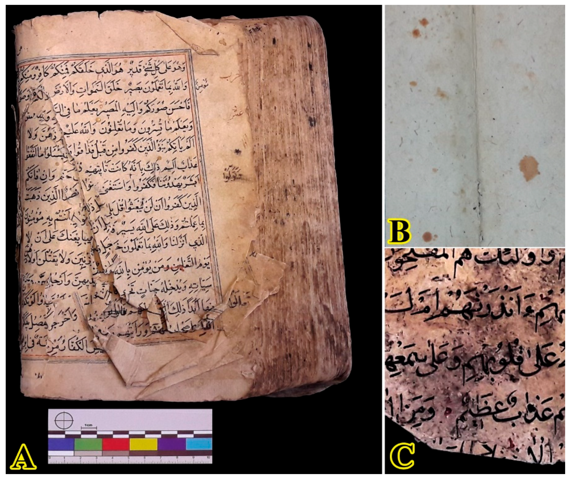
The investigation was focused on a historical manuscript of the Holy Quran belonging to a private collection. In some pages of this manuscript, a seal with the date 1210 HA (1795–1796 AD) can be seen. This date shows that this manuscript belongs to the first years of the Qajar era. Brown-orange spots, foxing, can be seen on some pages of this manuscript. Additionally, a page shows black spots that may be caused by the activity of microorganisms (Figure 1).
2.2. Isolation and Identification of Fungi
A sample of black spots and five samples of foxed spots were studied to investigate the possibility of the presence of fungi. Fungi were cultivated in sabouraud dextrose agar (SDA; Merck, Germany) medium which was autoclaved at 121 °C for 15 min to sterilize. Samples were incubated for 14 days at 28 ± 2 °C and 70 ± 5% RH. Cultivated fungi were characterized according to their macroscopic and microscopic properties, using an Olympus BX51 microscope.
2.3. µ-XRF Spectroscopy
The qualitative concentration of the elements in the two foxed and adjacent unfoxed areas was investigated using a micro XRF. An Unisantis 104 X-ray micro fluorescence spectrometer was used for XRF experiments. The excitation settings are 25 kV and 300 mA/120 s.
2.4. FTIR Spectroscopy
FTIR analysis was carried out using a Nicolet 680Plus FTIR spectrometer (Jasco, Japan) with KBr pellets. One sample of paper without foxing and two samples with foxing stains (S1 and S2) were examined. All Spectra were collected in the range of 400–4000 cm−1 at 4 cm−1 resolution with 32 numbers of scan. After modifying the baseline in the range of 850 to 1800 cm−1, the Gaussian function was used to peaks deconvolution and fitting.
2.5. UV Fluorescence Photography
The images were taken with a Nikon D750 DSLR camera equipped with an AF Nikkor 50 mm f/1.8D lens. UV fluorescence (UVF) photography was performed in a 45° setup by two xenon flash light with B + W UV-Pass filter, used as a radiation source, and a Baader UV/IR cut filter mounted on top of the camera lens.
3. Results and Discussion
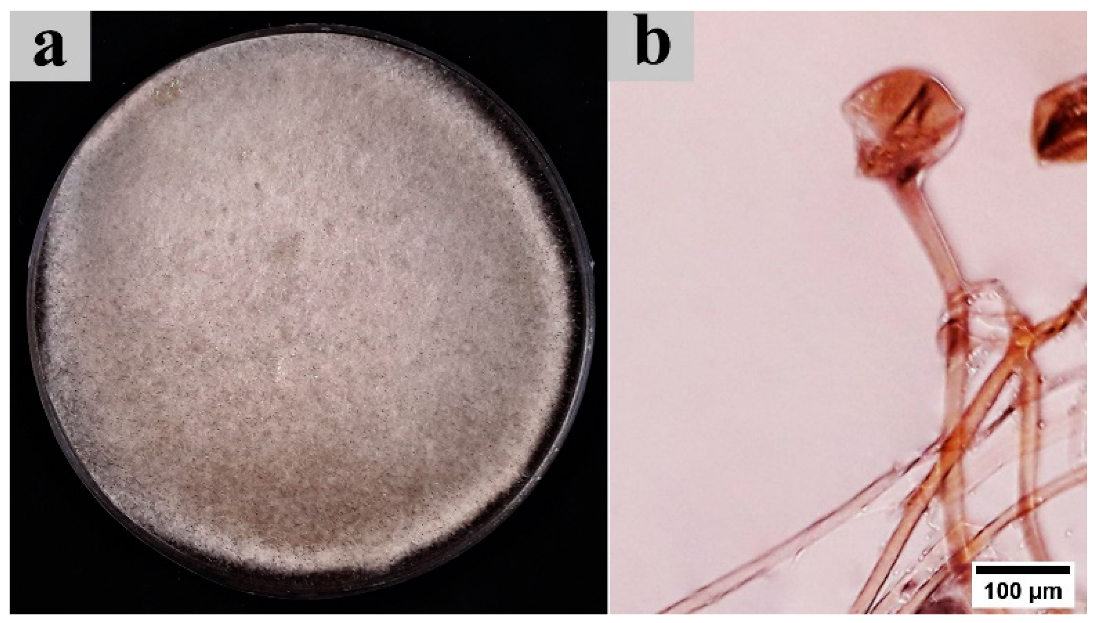
First, the presence of fungus activity in foxing spots was investigated using cultivation. After the incubation period, no fungal growth was observed in the foxing spots. In fact, it can be said that fungi are not the reason for the formation of foxing stains in this manuscript. However, in one of the manuscript sheets (Figure 1C), which was without foxing spots, the presence and growth of the fungus was observed. This fungus’ morphological features with fast-growing and deeply cottony texture of the colony having a white to gray color and dark pigment sporangium are shown in Figure 2a. Microscopic features show transparent and non-septate mycelium. The non-branched sporangiophores are long and terminate whit columella surrounded by a dark, round sporangium (Figure 2b). These are the characteristics of the genus Rhizopus [16].
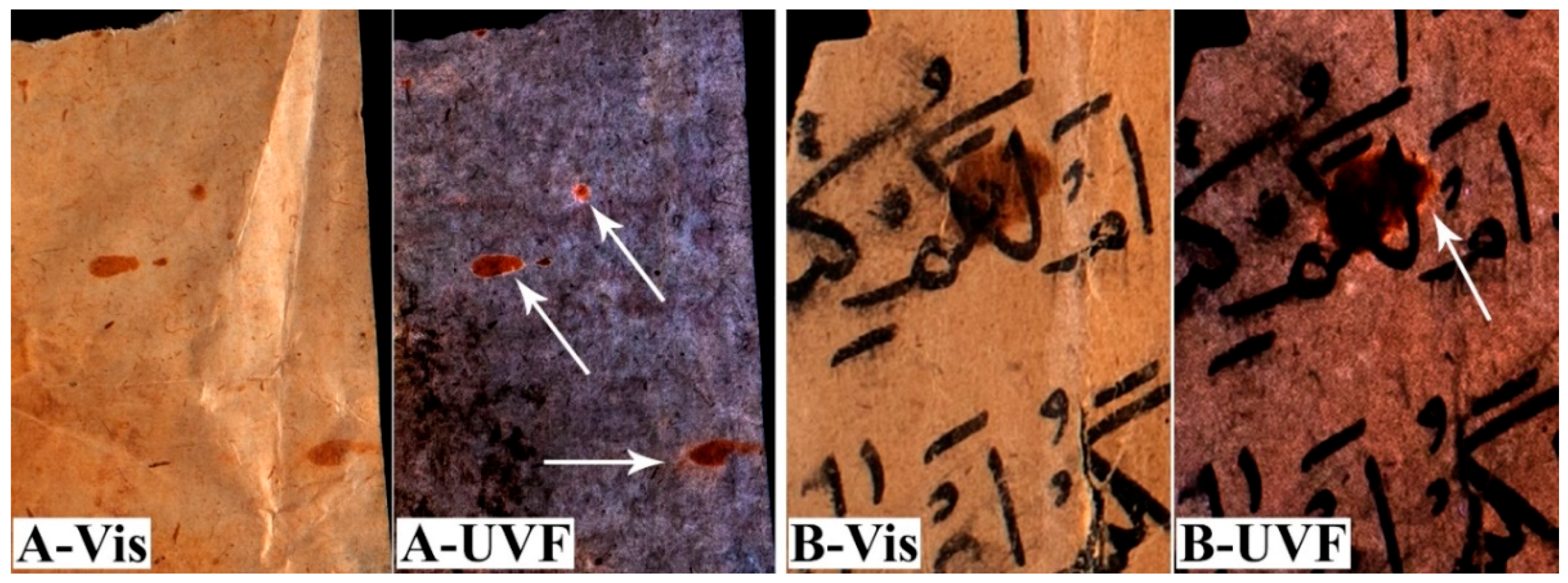
As mentioned, no fungal activity was observed in the foxing stains. Therefore, these stains should be considered abiotic foxing. One sign of foxing stains is their fluorescence under UV light [8,17,18]. The color of foxing spots of the manuscript, under reflected light, appear to be orange-brown. Similar to the report of Francisca et al. (2020) [17], under UV radiation, there is a round, well-delimited white fluorescent halo surrounding a non-fluorescent darker center. What is clear is that the halo extends beyond the visible stain (Figure 3). Cain and Miller (1984) have classified these spots as Bullseye [19]. This type of foxing always has metal cores, which do not fluoresce and appear dark [20].
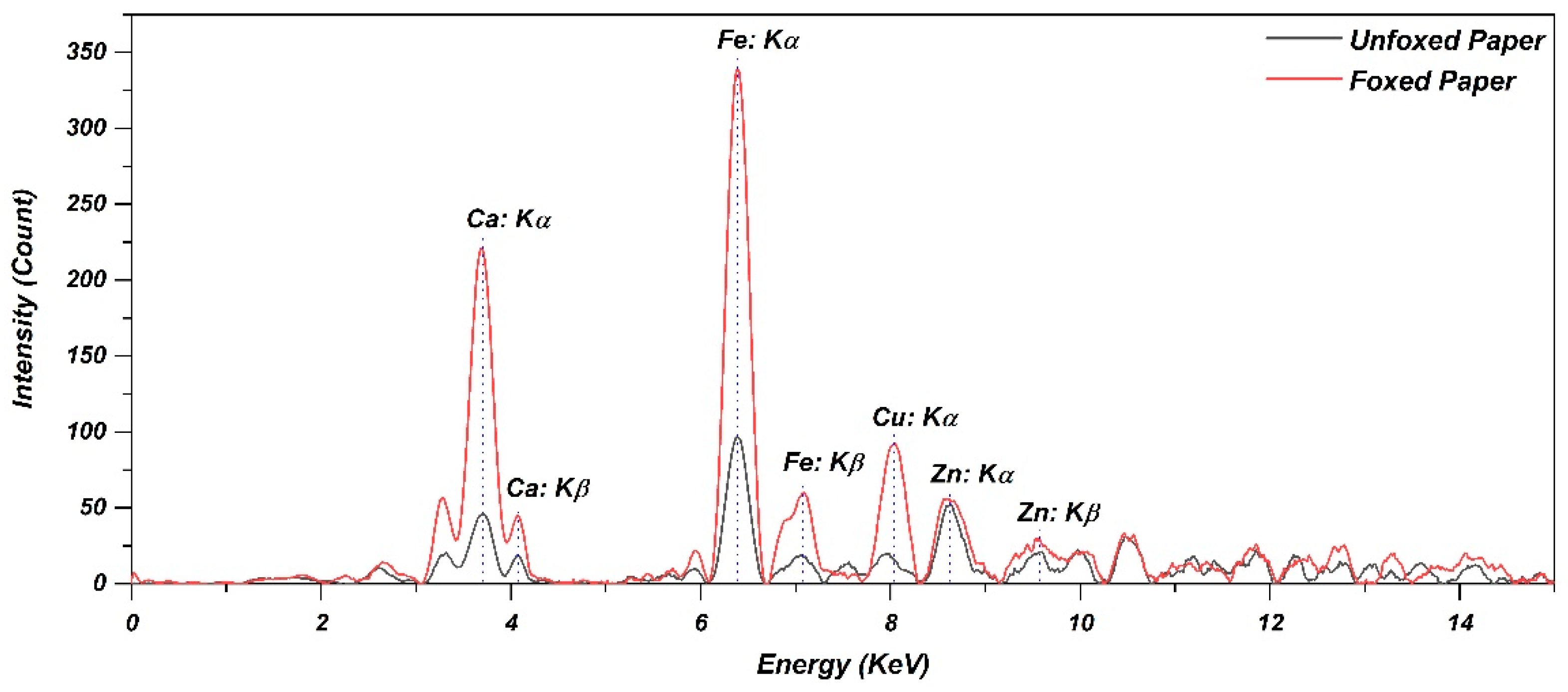
The qualitative concentration of metal elements on the foxed spots of the paper, and adjacent unfoxed areas, were checked by µXRF spectroscopy. The µXRF spectra are presented in Figure 4. These spectra show a high accumulation of iron, copper and calcium in the foxing spots compared to the adjacent section. In previous reports, the presence of iron, copper and calcium was documented in foxing spots [15,20,21,22]. These elements in the paper are believed to result from the papermaking process [22]. Metal contamination may also come from airborne dust. One of the main known sources of these metals in foxing stains is water [14]. Therefore, due to the shape and scattering of the foxings in this manuscript, the source of these metals is likely to be dirty water droplets. Meanwhile, the presence of copper and iron has been confirmed in almost all samples with abiotic foxing spots. Metal ions, iron (II) and copper (I) ions in particular, act as a catalyst and accelerator for cellulose free radical oxidative reaction in cellulose [4,23]. Furthermore, it seems that copper ions, due to their higher catalytic effect, are more capable in foxing formation than iron ions [8,23].
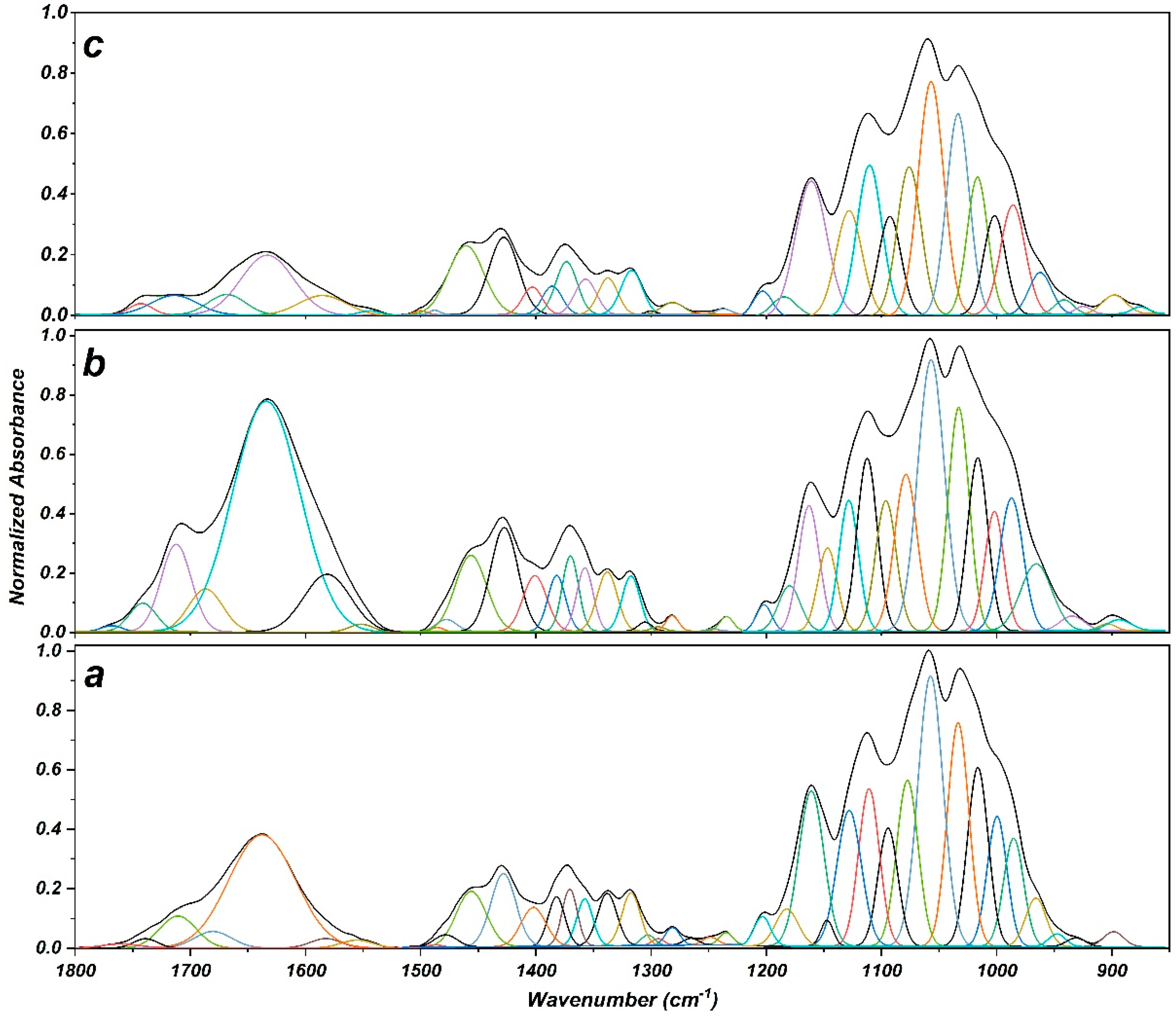
The oxidation rate at foxing stains was evaluated using FTIR spectroscopy. Figure 5 presents the Gaussian peak deconvolution of the FTIR spectra in the range of 850–1800 cm−1. Spectra changes under the influence of foxing show the increase in the absorbance peaks at 1700–1760 cm−1. This region corresponds to stretching vibrations of C=O groups and is known as the carbonyl index in the FTIR spectrum. The formation and intensity increase of these peaks can be considered as the oxidation index of cellulose in paper [24,25].
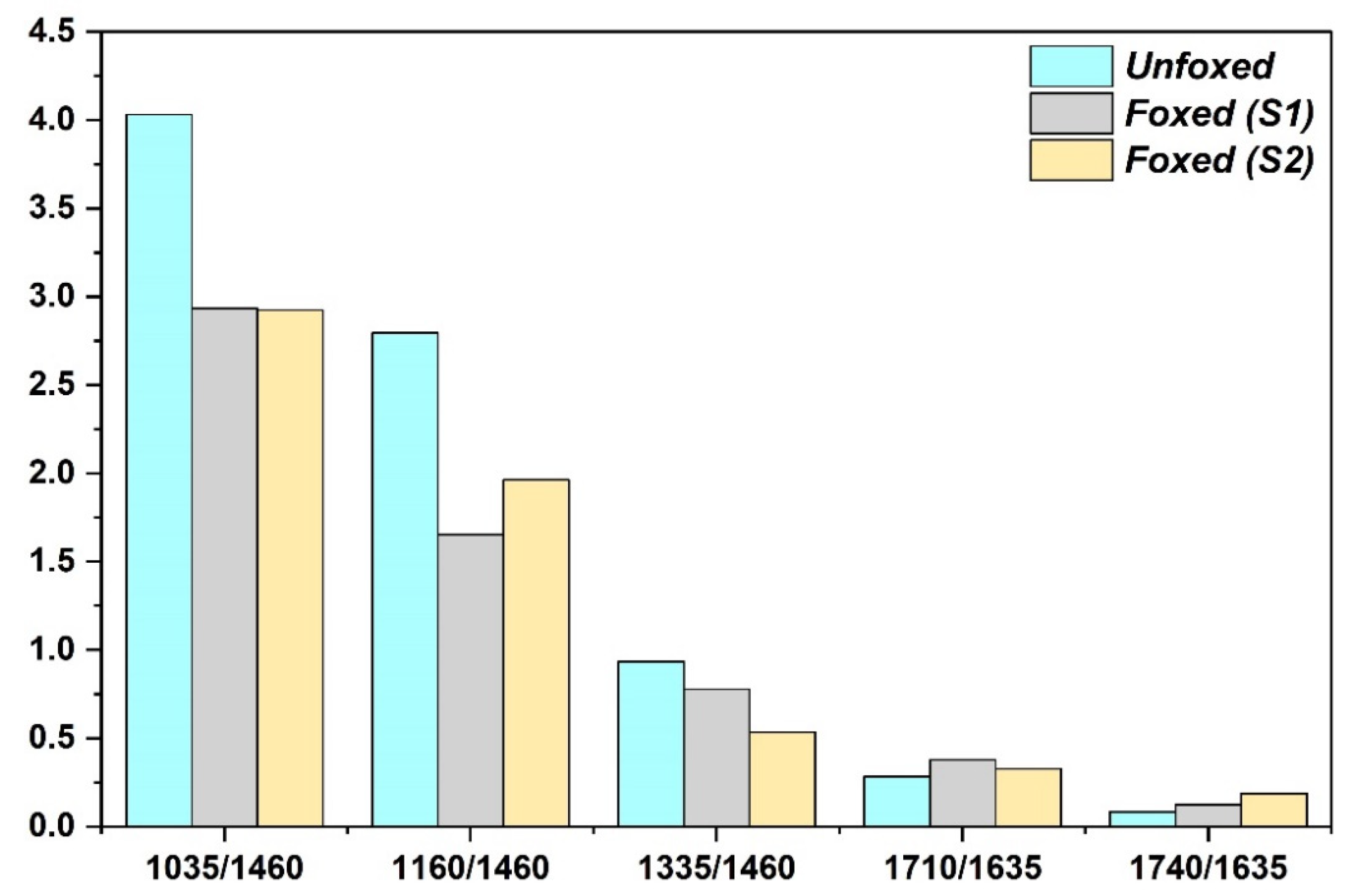
According to Lojewska et al. (2005) [26], cellulose oxidation index defined as the ratio of the intensity of absorbance bands of about 1720 to 1630 cm−1. Therefore, the ratio of these peaks was calculated based on the deconvoluted peaks presented in Table 1. The results show that the intensity ratio of peaks is increased in the foxing spots (Figure 6). In other words, it indicates more oxidation of cellulose in the foxed paper compared to the unfoxed paper.
In the FTIR spectra, the absorbance bands around 1710 and 1740 cm−1 correspond to C=O stretching vibrations in ketone and aldehyde groups, respectively [27]. The results show that foxing degradation in both samples has increased the C=O intensity ratio. Both S1 and S2 samples show an increase in both ketones and aldehydes. However, ketones and aldehydes have formed more in the S1 and S2 samples, respectively.
According to a previous study, cellulose oxidation is involved with C-OH and C-O-C vibrations, for secondary alcohol at C2 and C3, primary alcohol at C6, the C-O-C of glucopyranose ring and oxygen bridge [24]. In other words, the oxidation of cellulose leads to a decrease in the absorption peaks at 1035, 1160 and 1325 cm−1 which corresponds to the C-O-C glucopyranose ring stretching vibration [28], stretching vibration of the glycosidic oxygen bridge (C-O-C) [29] and C-OH stretching vibration [24], respectively.
The decreasing rate at 1035, 1160 and 1325 cm−1 has been evaluated and reported in Figure 6 with respect to the intensity corresponding to 1460 cm−1 as the internal standard peak. The internal standard peak was selected based on a previous study [24]. According to the results, foxing-induced oxidation has resulted in the breakdown of glycosidic bond, glucopyranose ring-opening and oxidation of C-OH groups. The breakdown of glycosidic bond has occurred more in the S1 than in the S2. This has led to the formation of more ketone groups in this sample. Conversely, in the S2 sample, more oxidation of the C-OH has led to the formation of more aldehydes.
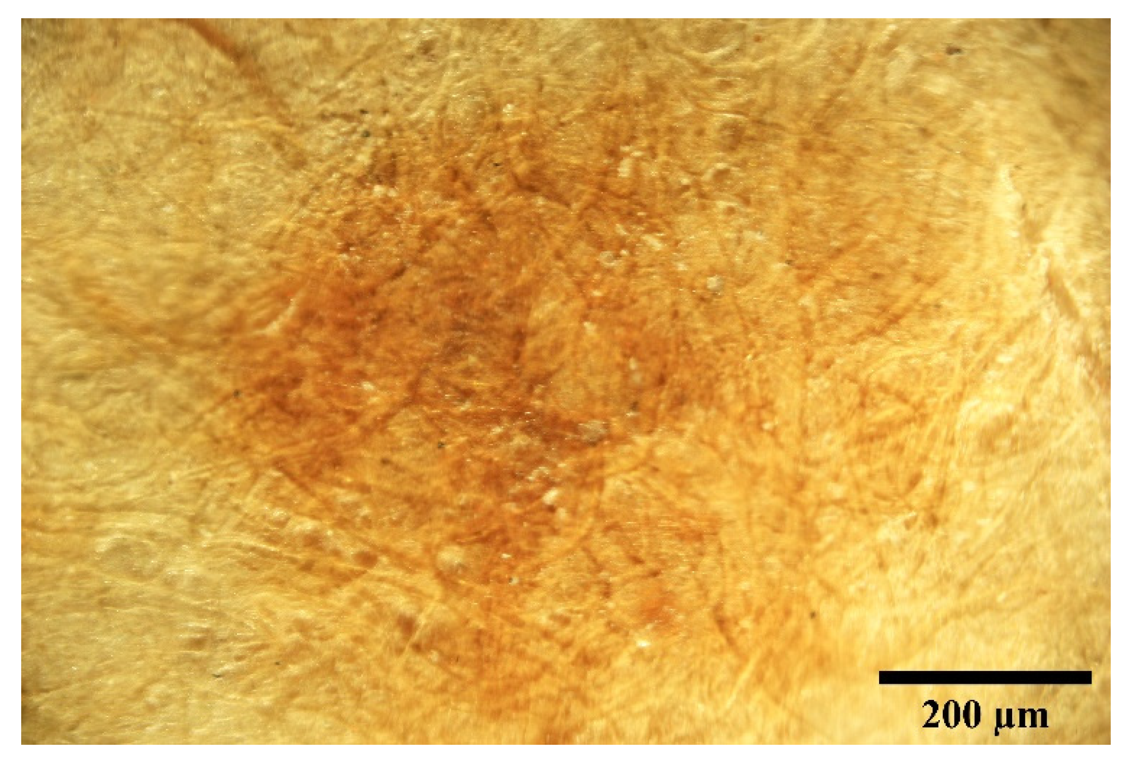
As mentioned, cellulose oxidation increases carbonyl groups. Carbonyl groups are precursors of chromophores that cause cellulose fibers to become yellow [30]. Previous studies have shown that ketones are the main cause of the yellowing of cellulosic pulps [31]. In addition, carboxyls can also be formed during the oxidation processes of cellulose [30]. However, carboxyls alone do not play a significant role in discoloration, but their concomitant presence with ketones and aldehydes exacerbates yellowing [30]. Therefore, oxidation of cellulose and formation of carbonyls can be considered as one of the main reasons for the formation of yellow-brown spots in foxings (Figure 7).
4. Conclusions
In this study, the foxing spots in a manuscript from the late 18th century were examined. Based on the results of this study, no fungal activity was observed on the foxing spots in this manuscript. The presence of a Rhizopus fungus was detected on only one page of the manuscript, and not in any of the foxing stains. Therefore, it can be concluded that these spots are abiotic foxing.
UV fluorescence examination also showed a bright halo around the foxing spots, which is known as Bullseye. It has been reported that this type of foxing is usually caused by metal ions. µ-XRF spectroscopy confirmed that iron and copper had accumulated in the foxing area. The accumulation of these metals intensifies the oxidation of cellulose by catalyzing radical reactions. In addition, cellulose oxidation in foxing stains was investigated using FTIR spectroscopy. The results show that the ratio of the intensity of bands about 1710 and 1740 cm−1 to that at 1635 cm−1 can increase in foxed area. In other words, ketone and aldehyde groups have increased as a result of cellulose oxidation due to foxing. Therefore, it is an appropriate indicator to study the oxidation intensity in abiotic foxing spots. The results show that the cellulose oxidation in foxing stains has been involved with the glucopyranose ring opening, oxygen bridges breaking and also hydroxyl groups. Cellulose oxidation can be considered as one of the main reasons for the color changing of paper to yellow-brown at foxing stains.
Author Contributions
A.K. designed the work process and analyzed and interpreted the data. S.A.G. participated in experiments. Both authors discussed the results and contributed to the final manuscript. Both authors have read and agreed to the published version of the manuscript.
Funding
This research received no external funding.
Institutional Review Board Statement
Not applicable.
Informed Consent Statement
Not applicable.
Data Availability Statement
The data presented in this study are available on request from the corresponding author.
Acknowledgments
The author would like to thank Mohsen Mohammadi Achachluei and Mehri Qobadi, from art university of Isfahan, for their advice and help in µ-XRF spectroscopy, and Behrooz Jelodarian, from Tabriz Islamic art university, for his advice and help in UV fluorescence photography.
Conflicts of Interest
The authors declare no conflict of interest. In addition, the funders had no role in the design of the study, in the collection, analyses or interpretation of data, in the writing of the manuscript or in the decision to publish the results.
References
- Fouda, A.; Abdel-Maksoud, G.; Abdel-Rahman, M.A.; Eid, A.M.; Barghoth, M.G.; El-Sadany, M.A.H. Monitoring the effect of biosynthesized nanoparticles against biodeterioration of cellulose-based materials by Aspergillus niger. Cellulose 2009, 26, 6583–6597. [Google Scholar] [CrossRef]
- Arai, H. Foxing caused by Fungi: Twenty-five years of study. Int. Biodeterior. Biodegrad. 2000, 46, 181–188. [Google Scholar] [CrossRef]
- Manso, M.; Pessanha, S.; Figueira, F.; Valadas, S.; Guilherme, A.; Afonso, M.; Rocha, A.C.; Oliveira, M.J.; Ribeiro, I.; Carvalho, M.L. Characterisation of foxing stains in eighteenth to nineteenth century drawings using non-destructive techniques. Anal. Bioanal. Chem. 2009, 395, 2029–2036. [Google Scholar] [CrossRef]
- Ciofini, D.; Osticioli, I.; Micheli, S.; Montalbano, L.; Siano, S. Laser Removal of Mold and Foxing Stains from Paper Artifacts: Preliminary Investigation. In Proceedings SPIE 9065, Fundamentals of Laser-Assisted Micro- and Nanotechnologies; Veiko, V.P., Vartanyan, T.A., Eds.; SPIE Press: Bellingham, WA, USA, 2013; p. 906512. [Google Scholar] [CrossRef]
- Kim, G.; Kim, J.G.; Kang, K.; Yoo, W.S. Image-Based Quantitative Analysis of Foxing Stains on Old Printed Paper Documents. Heritage 2019, 2, 2665–2677. [Google Scholar] [CrossRef] [Green Version]
- Mina, L. Foxy Underpants: Or the Use of Chelators and Enzymes to Reduce Foxing Stains on Early Nineteenth Century Men’s Linen Underpants. J. Am. Inst. Conserv. 2020, 59, 3–17. [Google Scholar] [CrossRef]
- Elkhial, M.M. Identification, Causes, Classification and Treatment of FOXING: A Literature Review. In Proceedings of the International Conference on Chemistry, Chem 06, Contemporary Chemistry and Environment, Cairo University, Cairo, Egypt, 1–4 March 2010. [Google Scholar]
- Choi, S. Foxing on Paper: A Literature Review. J. Am. Inst. Conserv. 2007, 46, 137–152. [Google Scholar] [CrossRef]
- Buzio, R.; Calvini, P.; Ferroni, A.; Valbusa, U. Surface analysis of paper documents damaged by foxing. Appl. Phys. A 2004, 79, 383–387. [Google Scholar] [CrossRef]
- De Paolis, M.R.; Lippi, D. Use of metabolic and molecular methods for the identification of a Bacillus strain isolated from paper affected by foxing. Microbiol. Res. 2008, 163, 121–131. [Google Scholar] [CrossRef] [PubMed]
- Rakotonirainy, M.S.; Dubar, P. Application of bioluminescence ATP measurement for evaluation of fungal viability of foxing spots on old documents. Luminescence 2013, 28, 308–312. [Google Scholar] [CrossRef] [PubMed]
- Corte, A.M.; Ferroni, A.; Salvo, V.S. Isolation of fungal species from test samples and maps damaged by foxing, and correlation between these species and the environment. Int. Biodeterior. Biodegrad. 2003, 51, 167–173. [Google Scholar] [CrossRef]
- Zotti, M.; Ferroni, A.; Calvini, P. Microfungal biodeterioration of historic paper: Preliminary FTIR and microbiological analyses. Int. Biodeterior. Biodegrad. 2008, 62, 186–194. [Google Scholar] [CrossRef]
- Szulc, J.; Otlewska, A.; Ruman, T.; Kubiak, K.; Karbowska-Berent, J.; Kozielec, T.; Gutarowska, B. Analysis of paper foxing by newly available omics techniques. Int. Biodeterior. Biodegrad. 2018, 132, 157–165. [Google Scholar] [CrossRef]
- Press, R.E. Observations on the foxing of paper. Int. Biodeterior. Biodegrad. 2001, 48, 94–97. [Google Scholar] [CrossRef]
- Dolatabadi, S.; Walther, G.; Gerrits van den Ende, A.H.G.; de Hoog, G.S. Diversity and delimitation of Rhizopus microspores. Fungal Divers. 2014, 64, 145–163. [Google Scholar] [CrossRef]
- Francisca, F.; Matos, M.; Nunes, A.; Afonso, M.; Rocha, A.C.; Campelo, J.; Ferreira, T. Considerations about foxing stains in three paper collections ranging from the 16th to the 20th century. Conserv. Património 2020, 35, 45–57. [Google Scholar] [CrossRef]
- Nitiu, D.S.; Mallo, A.C.; Saparrat, M.C.N. Fungal melanins that deteriorate paper cultural heritage: An overview. Mycologia 2020, 112, 859–870. [Google Scholar] [CrossRef] [PubMed]
- Cain, C.; Miller, B. Proposed Classification of Foxing. In Proceedings of the American Institute for Conservation 10th Annual Meeting, Milwaukee, WI, USA, 26–30 May 1982; AIC: Washington, DC, USA, 1982; pp. 29–30. [Google Scholar]
- Ardelean, E.; Melniciuc-Puid, N. Conservation of Paper Documents Damaged By Foxing. Eur. J. Sci. Theol. 2013, 9, 117–124. [Google Scholar]
- Cain, C.E.; Miller, B.A. Photographic, Spectral and Chromatographic Searches into the Nature of Foxing. In Proceedings of the American Institute for Conservation 10th Annual Meeting, Milwaukee, WI, USA, 26–30 May 1982; AIC: Washington, DC, USA, 1982; pp. 54–62. [Google Scholar]
- Krstic, D.; Schauperl, Z. Characterization of Foxing Stains in Eighteenth Century Books. HDKBR INFO Mag. 2013, 3, 32–39. [Google Scholar]
- Rebrikova, N.L.; Manturovskaya, N.V. Foxing—A New Approach to an Old Problem. Restaur. Int. J. Preserv. Libr. Arch. Mater. 2000, 21, 85–100. [Google Scholar] [CrossRef]
- Ahmadi, H.; Mallakpour, S.; Koochakzaei, A. Evaluating the Role of antioxidants in the stabilization of hydroxypropyl cellulose by ATR-FTIR Spectroscopy. Prog. Color Colorants Coat. 2018, 11, 93–101. [Google Scholar]
- Rakotonirainy, M.S.; Bénaud, O.; Vilmont, L.-B. Contribution to the characterization of foxing stains on printed books using infrared spectroscopy and scanning electron microscopy energy dispersive spectrometry. Int. Biodeterior. Biodegrad. 2015, 101, 1–7. [Google Scholar] [CrossRef]
- Łojewska, J.; Miśkowiec, P.; Łojewski, T.; Proniewicz, L.M. Cellulose oxidative and hydrolytic degradation: In situ FTIR approach. Polym. Degrad. Stab. 2005, 88, 512–520. [Google Scholar] [CrossRef]
- Nandiyanto, A.; Oktiani, R.; Ragadhita, R. How to Read and Interpret FTIR Spectroscope of Organic Material. Indones. J. Sci. Technol. 2019, 4, 97–118. [Google Scholar] [CrossRef]
- El-Sakhawy, M.; Kamel, S.; Salama, A.; Sarhan, H.A. Preparation and infrared study of cellulose based amphiphilic materials. Cellul. Chem. Technol. 2018, 52, 193–200. [Google Scholar]
- Lv, P.; Almeida, G.; Perré, P. TGA-FTIR analysis of torrefaction of lignocellulosic components (cellulose, xylan, lignin) in isothermal conditions over a wide range of time durations. Bioresources 2015, 10, 4239–4251. [Google Scholar] [CrossRef] [Green Version]
- Ahn, K.; Zaccaron, S.; Zwirchmayr, N.S.; Hettegger, H.; Hofinger, A.; Bacher, M.; Henniges, U.; Hosoya, T.; Potthast, A.; Rosenau, T. Yellowing and brightness reversion of celluloses: CO or COOH, who is the culprit? Cellulose 2019, 26, 429–444. [Google Scholar] [CrossRef] [Green Version]
- Perrin, J.; Pouyet, F.; Chirat, C.; Lachenal, D. Formation of Carbonyl and Carboxyl Groups on Cellulosic Pulps: Effect on Alkali Resistance. BioResources 2014, 9, 7299–7310. [Google Scholar] [CrossRef] [Green Version]
Figure 1.
(A) The manuscript of the Holy Quran from the late 18th century and the types of stains studied: (B) foxing spots, (C) fungal colony.
Figure 1.
(A) The manuscript of the Holy Quran from the late 18th century and the types of stains studied: (B) foxing spots, (C) fungal colony.

Figure 2.
(a) Fungal colony grown on SDA, (b) microscopic image.

Figure 3.
Photographic imaging of two foxing spot on two different papers (A and B) of manuscript; Vis: visible reflected light, UVF: Ultraviolet fluorescence.
Figure 3.
Photographic imaging of two foxing spot on two different papers (A and B) of manuscript; Vis: visible reflected light, UVF: Ultraviolet fluorescence.

Figure 4.
XRF spectra of paper with and without foxing stains that show increased levels of iron, copper and calcium in the foxed area.
Figure 4.
XRF spectra of paper with and without foxing stains that show increased levels of iron, copper and calcium in the foxed area.

Figure 5.
Gaussian peak deconvolution of FTIR spectra of foxed (b: S1, c: S2) and unfoxed (a) papers, baseline corrected in the region of 850–1800 cm−1.
Figure 5.
Gaussian peak deconvolution of FTIR spectra of foxed (b: S1, c: S2) and unfoxed (a) papers, baseline corrected in the region of 850–1800 cm−1.

Figure 6.
Peaks intensity ratios as oxidation indices of cellulose in the FTIR spectra of unfoxed and foxed papers.
Figure 6.
Peaks intensity ratios as oxidation indices of cellulose in the FTIR spectra of unfoxed and foxed papers.

Figure 7.
Discoloration of paper fibers to yellow-brown in foxing stains as a result of cellulose oxidation.
Figure 7.
Discoloration of paper fibers to yellow-brown in foxing stains as a result of cellulose oxidation.


Table 1.
The results of peak deconvolution and fitting Gaussian function of the spectra.
| Sample | Fitting Parameters | Deconvoluted Peaks | ||||||
|---|---|---|---|---|---|---|---|---|
| 1035 | 1160 | 1335 | 1635 | 1710 | 1740 | 1460 | ||
| Unfoxed paper | Center | 1033.4 | 1160.9 | 1338.1 | 1637.7 | 1710.7 | 1738.8 | 1456.1 |
| Height | 0.754 | 0.523 | 0.175 | 0.381 | 0.108 | 0.032 | 0.187 | |
| Area | 17.58 | 14.14 | 3.72 | 27.43 | 4.29 | 0.85 | 6.07 | |
| FWHH * | 21.92 | 25.41 | 19.95 | 67.66 | 37.13 | 24.94 | 30.53 | |
| Foxed paper (S1) | Center | 1033.0 | 1162.9 | 1338.1 | 1634 | 1712.2 | 1741.2 | 1456.4 |
| Height | 0.754 | 0.425 | 0.2 | 0.778 | 0.295 | 0.097 | 0.257 | |
| Area | 17.17 | 9.60 | 4.37 | 56.99 | 9.72 | 2.82 | 8.75 | |
| FWHH | 21.39 | 21.24 | 20.51 | 68.80 | 30.95 | 27.43 | 31.94 | |
| Foxed paper (S2) | Center | 1033.9 | 1161.2 | 1337.7 | 1635.1 | 1716.1 | 1742.8 | 1461 |
| Height | 0.716 | 0.487 | 0.132 | 0.232 | 0.064 | 0.041 | 0.247 | |
| Area | 19.09 | 16.48 | 3.28 | 18.15 | 3.11 | 1.05 | 9.51 | |
| FWHH | 25.05 | 31.78 | 23.29 | 73.41 | 45.55 | 24.31 | 36.15 | |
* Full Width at the Half-Height.
Publisher’s Note: MDPI stays neutral with regard to jurisdictional claims in published maps and institutional affiliations. |
© 2021 by the authors. Licensee MDPI, Basel, Switzerland. This article is an open access article distributed under the terms and conditions of the Creative Commons Attribution (CC BY) license (https://creativecommons.org/licenses/by/4.0/).
Share and Cite
MDPI and ACS Style
Koochakzaei, A.; Alizadeh Gharetapeh, S. Paper Foxing Stains on a Historic Manuscript from the Early Qajar Era: Abiotic or Biotic Foxing? Heritage 2021, 4, 1366-1374. https://0-doi-org.brum.beds.ac.uk/10.3390/heritage4030074
AMA Style
Koochakzaei A, Alizadeh Gharetapeh S. Paper Foxing Stains on a Historic Manuscript from the Early Qajar Era: Abiotic or Biotic Foxing? Heritage. 2021; 4(3):1366-1374. https://0-doi-org.brum.beds.ac.uk/10.3390/heritage4030074
Chicago/Turabian StyleKoochakzaei, Alireza, and Samane Alizadeh Gharetapeh. 2021. "Paper Foxing Stains on a Historic Manuscript from the Early Qajar Era: Abiotic or Biotic Foxing?" Heritage 4, no. 3: 1366-1374. https://0-doi-org.brum.beds.ac.uk/10.3390/heritage4030074