
Characterization of Natural Stone from the Archaeological Site of Pella, Macedonia, Northern Greece

 , ,
, ,  and
and 
Abstract
:1. Introduction
2. Materials and Methods
3. Results and Discussion
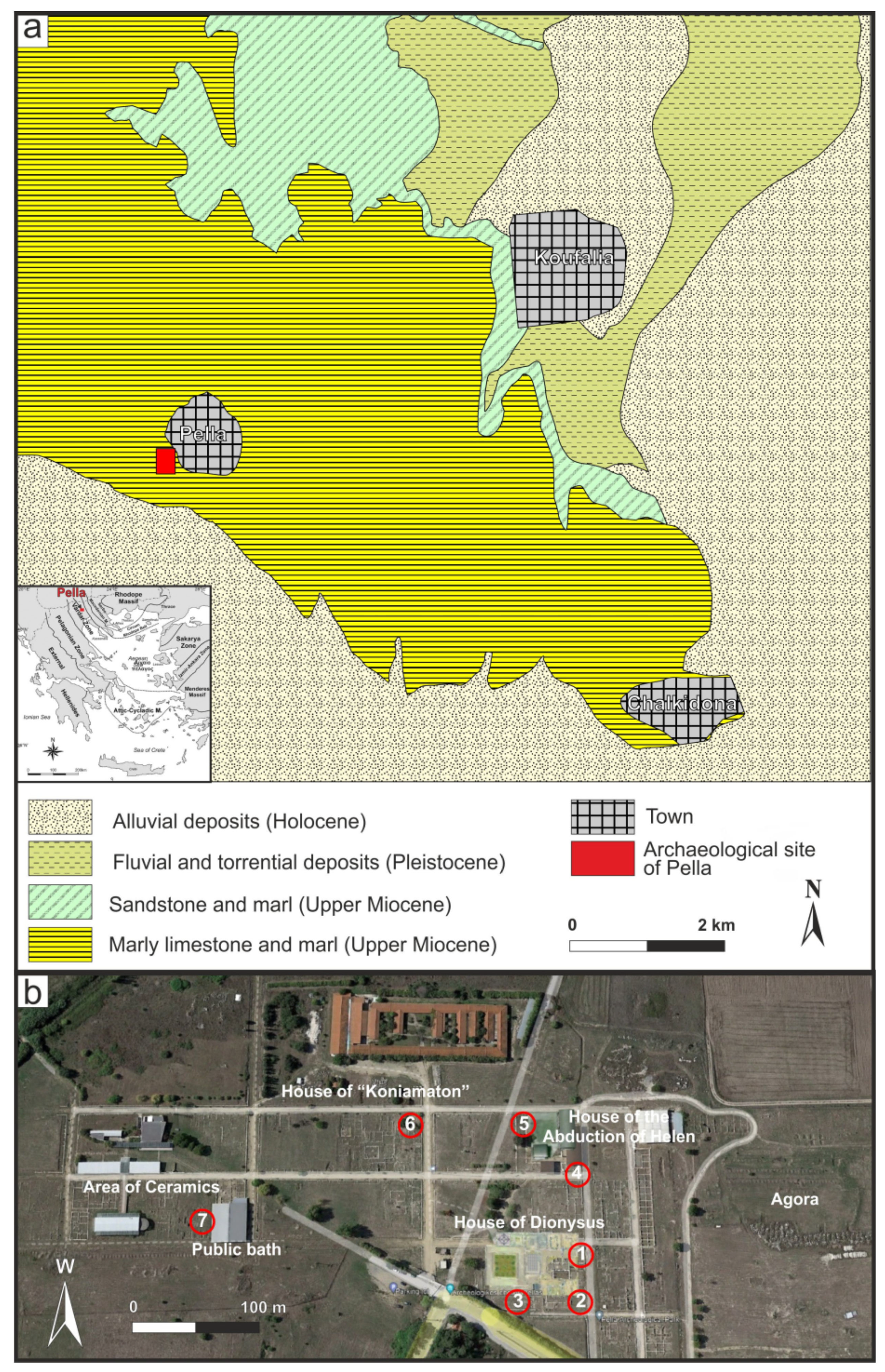
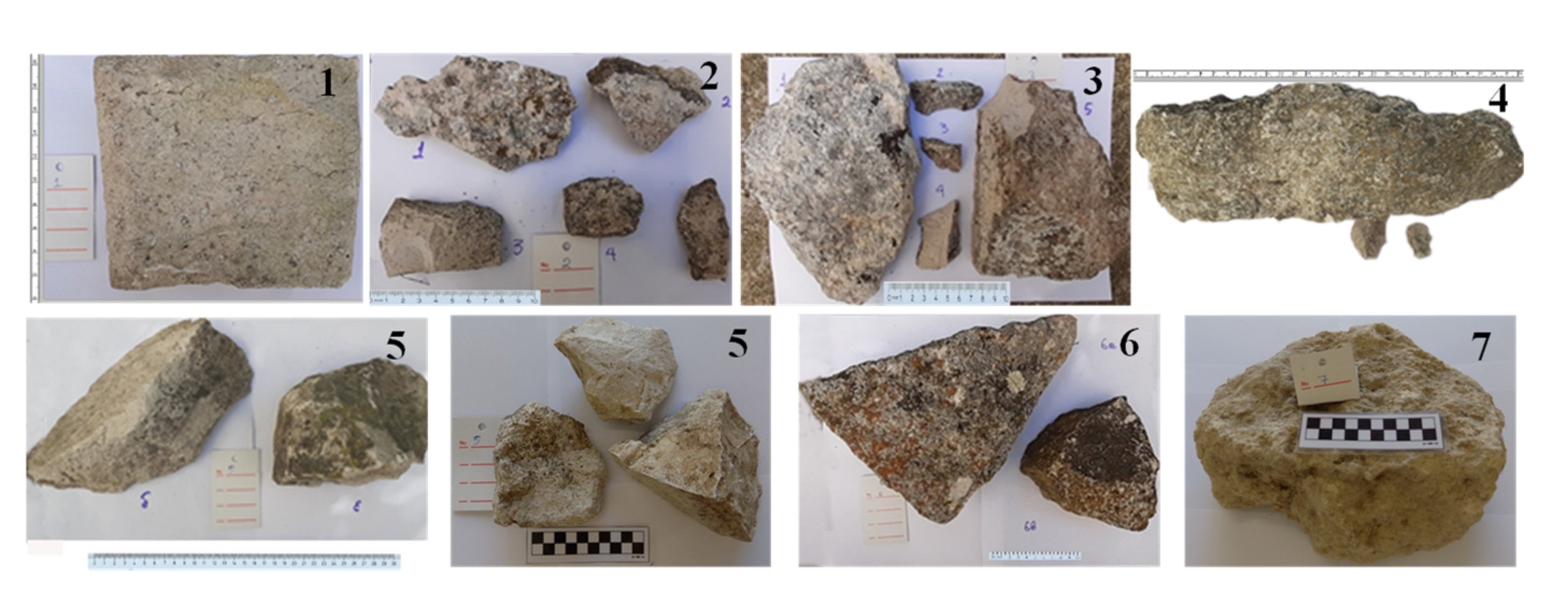
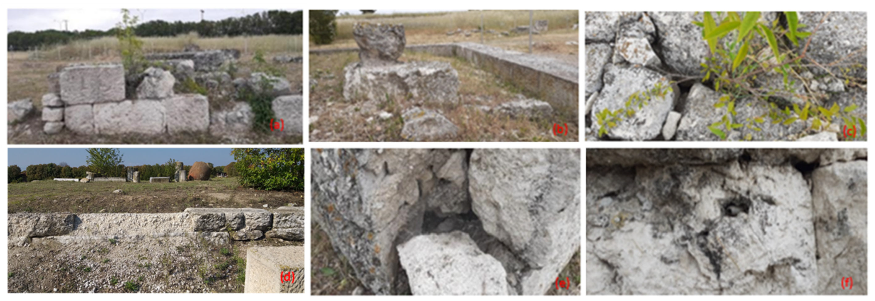
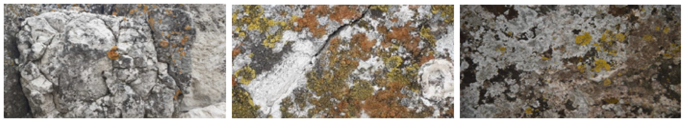
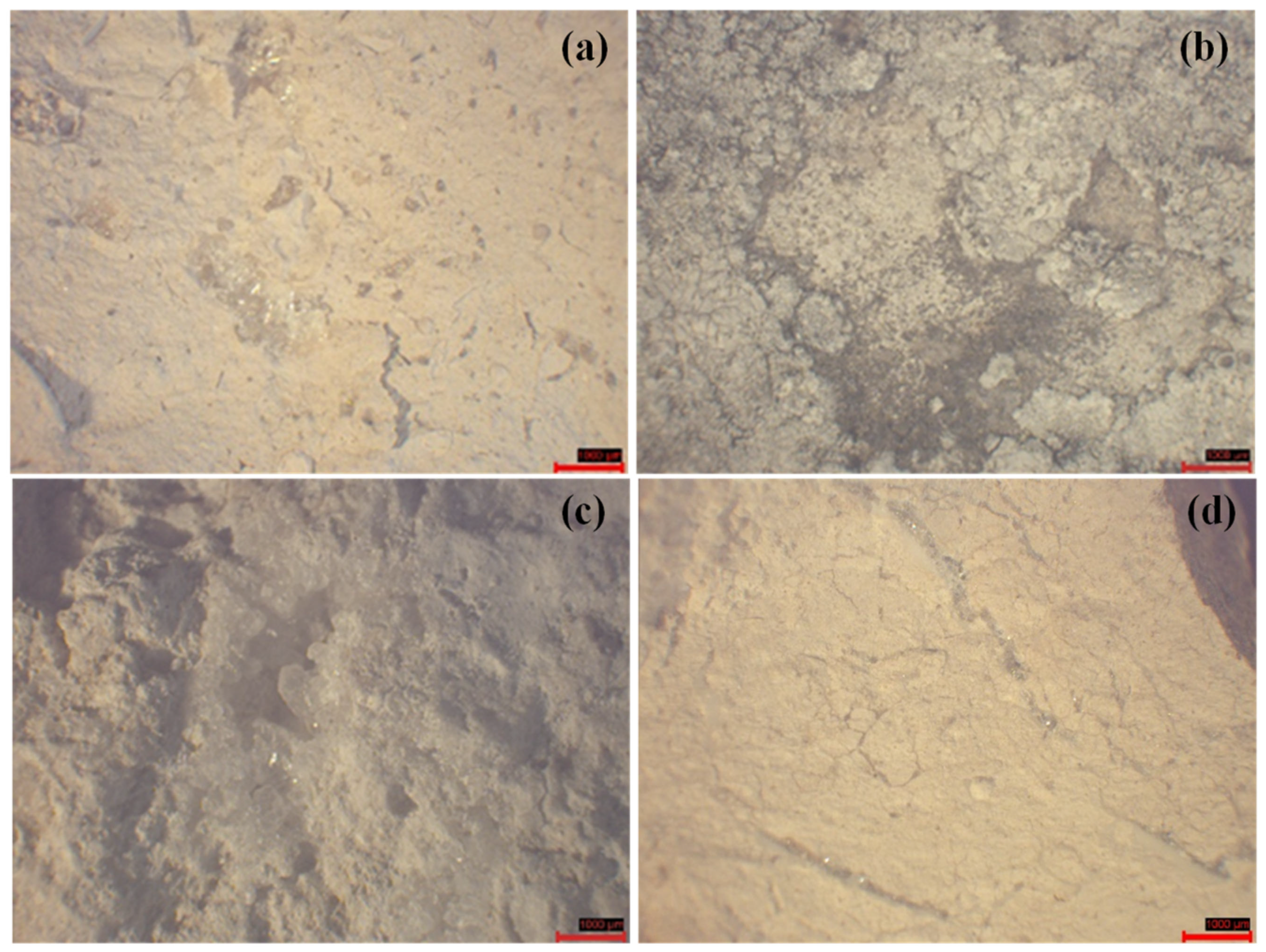
3.1. On-Site Macroscopic Observations
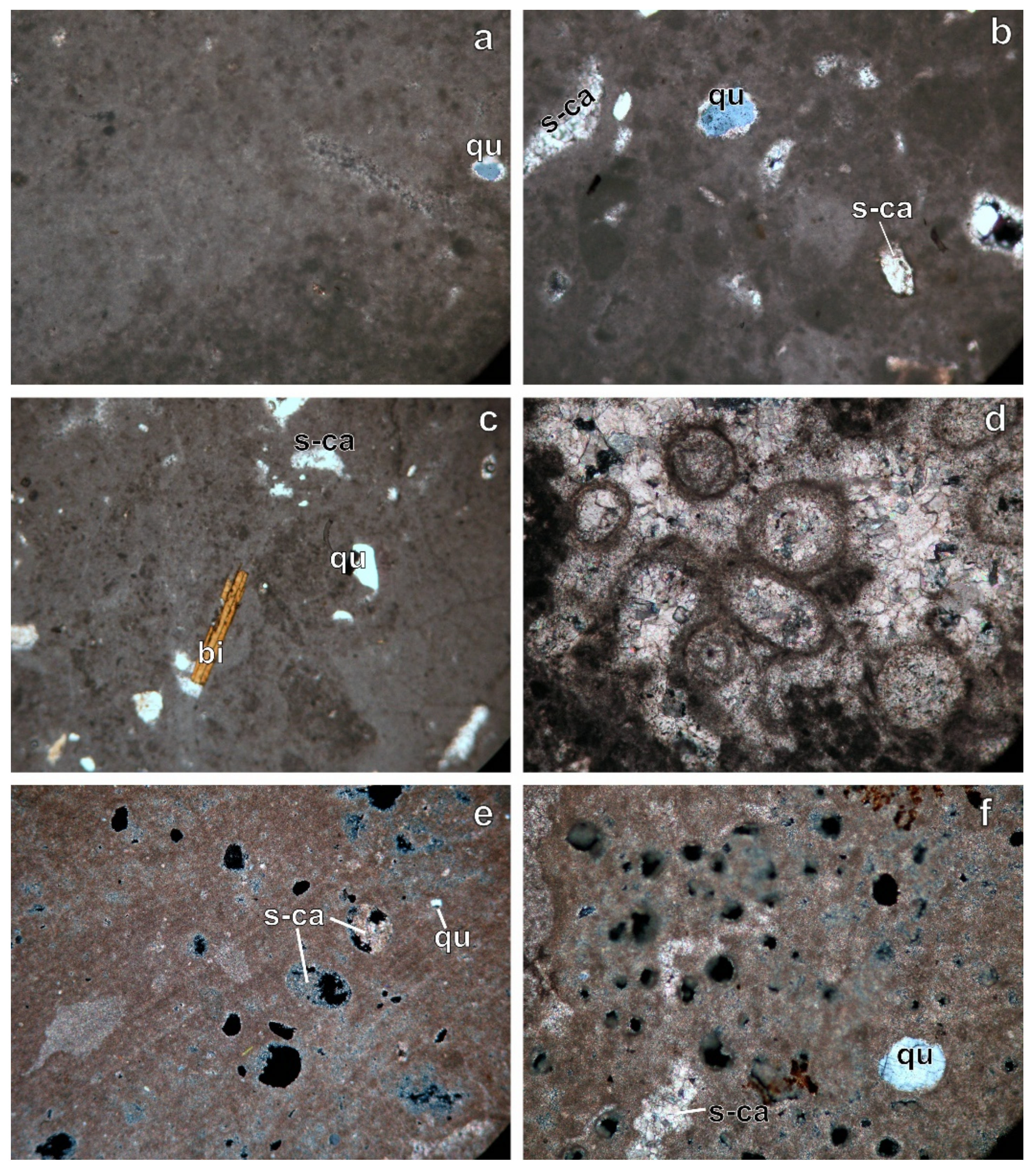
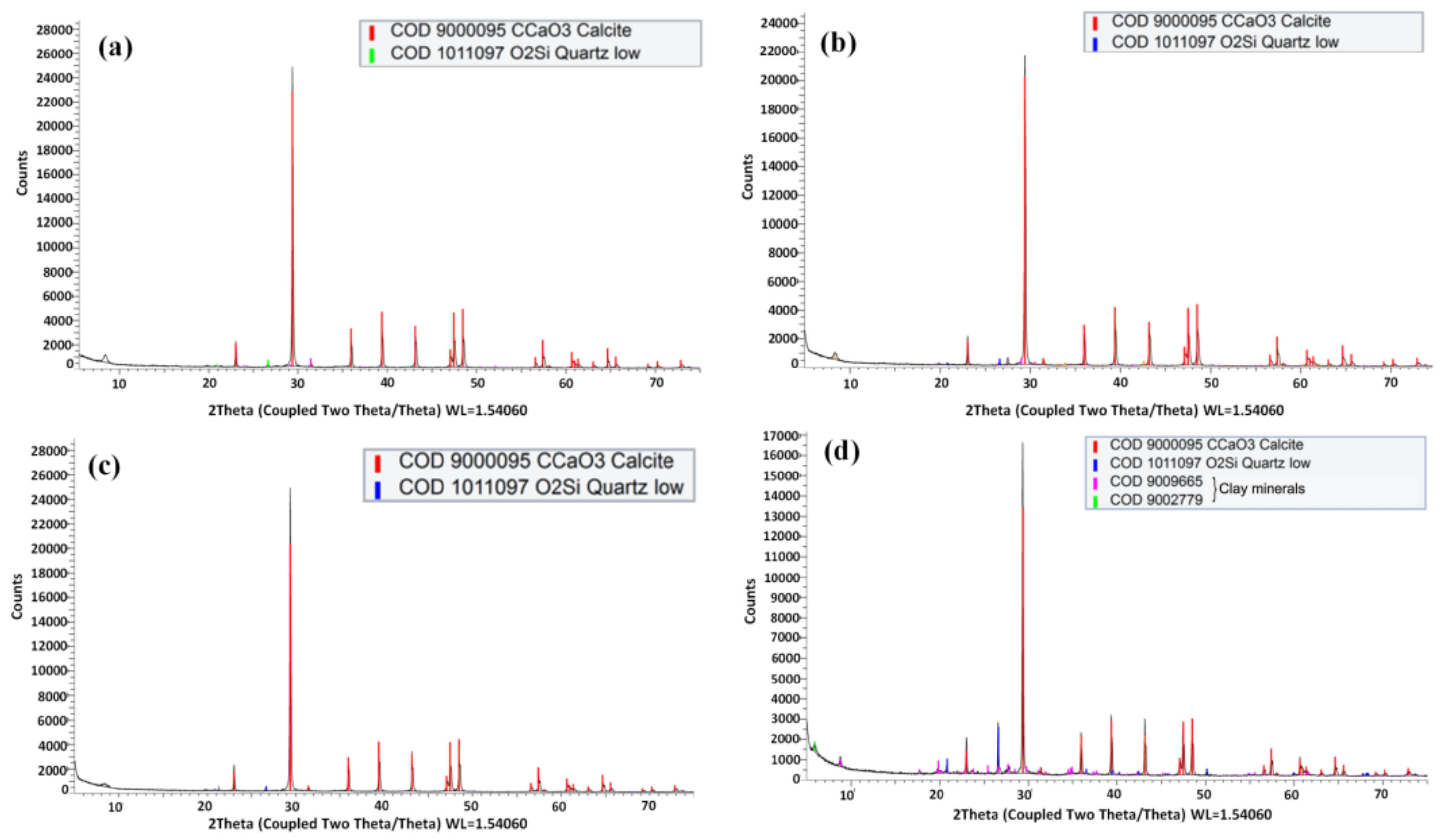
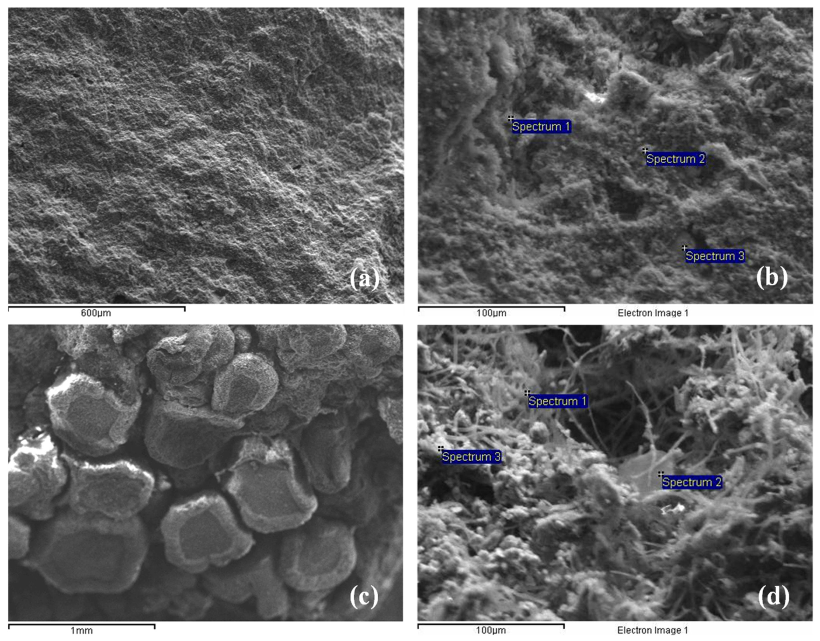
3.2. Petrographic, Mineralogical and Physicochemical Characteristics
3.3. Physicomechanical Characteristics
3.4. Thermographical Characteristics
4. Conclusions
Author Contributions
Funding
Institutional Review Board Statement
Informed Consent Statement
Acknowledgments
Conflicts of Interest
References
- Spathis, P.; Papanikolaou, E.; Melfos, V.; Samara, C.; Christaras, B.; Katsiotis, N. Characterization and Weathering of the Building Materials of Sanctuaries in the Archaeological Site of Dion, Greece. Trends J. Sci. Res. 2015, 2, 95–103. [Google Scholar]
- Liali, T.; Chrysostomou, E.; Karasarlidis, E. Maintenance and Restoration Study of the Propylon and the Building I of the Palace of Pella; Ephorate of Antiquities of Pella: Pella, Greece, 2018. (In Greek) [Google Scholar]
- Papayianni, I.; Stefanidou, M.; Pachta, V. Survey of Repaired and Artificial Stones of the Archaeological Site of Pella Five Years after Application; Springer: Singapore, 2014; pp. 431–442. [Google Scholar]
- Xydas, S.; Efstratides, G. Geological Map of Greece, Koufalia Sheet, 1:50,000; IGME: Athens, Greece, 1993. [Google Scholar]
- Kouzeli, K. Stone of the Archaeological Site of Pella—Special Features—Decay Mechanism—Conservation Proposals; Ministry of Culture, Stone Center: Athens, Greek, 2009. (In Greek) [Google Scholar]
- Grossi, C.M.; Brimblecombe, P.; Harris, I. Predicting long term freeze–thaw risks on Europe built heritage and archaeological sites in a changing climate. Sci. Total Environ. 2007, 377, 273–281. [Google Scholar] [CrossRef] [PubMed]
- Thomas, C.; Lombillo, I.; Setién, J.; Polanco, J.A.; Villegas, L. Characterization of materials with repellents and consolidants from a historic building. J. Mater. Civil Eng. 2013, 25, 1742–1751. [Google Scholar] [CrossRef]
- Herrera, L.K.; Videla, H.A. Surface analysis and materials characterization for the study of biodeterioration and weathering effects on cultural property. Int. Biodeterior. Biodegrad. 2009, 63, 813–822. [Google Scholar] [CrossRef]
- Ruedrich, J.; Kirchner, D.; Siegesmund, S. Physical weathering of building stones induced by freeze–thaw action: A laboratory long-term study. Environ. Earth Sci. 2010, 63, 1573–1586. [Google Scholar] [CrossRef] [Green Version]
- Török, Á. Surface strength and mineralogy of weathering crusts on limestone buildings in Budapest. Build. Environ. 2003, 38, 1185–1192. [Google Scholar] [CrossRef]
- Amer, O.; Aita, D.; Mohamed, E.K.; Torky, A.; Shawky, A. Experimental Investigations and Microstructural Characterization of Construction Materials of Historic Multi-Leaf Stone-Masonry Walls. Heritage 2021, 4, 135. [Google Scholar] [CrossRef]
- Curulli, A.; Montesperelli, G.; Ronca, S.; Cavalagli, N.; Ubertini, F.; Padeletti, G.; Ciprioti, S.V. A multidisciplinary approach to the mortars characterization from the Town Walls of Gubbio (Perugia, Italy). J. Therm. Anal. Calorim. 2020, 142, 1721–1737. [Google Scholar] [CrossRef]
- Recommendations, R. Absorption of water by immersion under vacuum. Mater. Struct. 1984, 101, 391–394. [Google Scholar]
- European Committee for Standardization. Natural Stone Test Methods—Determination of Water Absorption Coefficient by Capillarity; EN 1925; European Committee for Standardization: Brussels, Belgium, 1999. [Google Scholar]
- European Committee for Standardization. Natural Stone Test Methods—Determination of Uniaxial Compressive Strength; EN 1926; European Committee for Standardization: Brussels, Belgium, 2006. [Google Scholar]
- Dakal, T.C.; Cameotra, S.S. Microbially induced deterioration of architectural heritages: Routes and mechanisms involved. Environ. Sci. Eur. 2012, 24, 36. [Google Scholar] [CrossRef] [Green Version]
- Pinna, D.; Salvadori, O.; Tretiach, M. An anatomical investigation of calcicolous endolithic lichens from the Trieste karst (NE Italy). Plant Biosyst.-Int. J. Deal. All Asp. Plant Biol. 1998, 132, 183–195. [Google Scholar] [CrossRef]
- Scheerer, S.; Ortega-Morales, O.; Gaylarde, C. Microbial deterioration of stone monuments –An updated overview. Adv. Appl. Microbiol. 2009, 66, 97–139. [Google Scholar] [PubMed]
- Papakosta, V. Colonization with Mosses of Archaeological Sites and Monuments. Master’s Thesis, Aristotle University of Thessaloniki, Thessaloniki, Greek, 2012. (In Greek). [Google Scholar]
- Dotsika, E.; Psomiadis, D.; Poutoukis, D.; Raco, B.; Gamaletsos, P. Isotopic analysis for degradation diagnosis of calcite matrix in mortar. Anal. Bioanal. Chem. 2009, 395, 2227–2234. [Google Scholar] [CrossRef] [PubMed]
- Stefanidou, M.; Pachta, V.; Papayianni, I. Design and testing of artificial stone for the restoration of stone elements in monuments and historic buildings. Constr. Build. Mater. 2015, 93, 957–965. [Google Scholar] [CrossRef]
- Avdelidis, N.; Moropoulou, A.; Theoulakis, P. Detection of water deposits and movement in porous materials by infrared imaging. Infrared Phys. Technol. 2003, 44, 183–190. [Google Scholar] [CrossRef]
- Çanakci, H.; Demirboğa, R.; Karakoç, M.B.; Şirin, O. Thermal conductivity of limestone from Gaziantep (Turkey). Build. Environ. 2007, 42, 1777–1782. [Google Scholar] [CrossRef]
- Merriman, J.D.; Hofmeister, A.M.; Roy, D.J.; Whittington, A.G. Temperature-dependent thermal transport properties of carbonate minerals and rocks. Geosphere 2018, 14, 1961–1987. [Google Scholar] [CrossRef] [Green Version]
- Engineering ToolBox, Specific Heat of Solids. 2003. Available online: https://www.engineeringtoolbox.com/specific-heat-solids-d_154.html (accessed on 27 May 2021).


| Analysis/Technique | Sampling Locations | ||||||
|---|---|---|---|---|---|---|---|
| 1 | 2 | 3 | 4 | 5 | 6 | 7 | |
| Stereomicroscopy | √ | √ | √ | √ | √ | √ | √ |
| Isotopic | √ | ||||||
| Polarizing light microscopy | √ | √ | √ | ||||
| SEM–EDS | √ | √ | √ | √ | √ | √ | √ |
| XRD | √ | √ | √ | √ | √ | √ | |
| RILEM CPC 11.3 | √ | √ | √ | √ | √ | ||
| Capillary water absorption | √ | √ | √ | √ | √ | √ | |
| Compressive strength | √ | √ | √ | √ | |||
| Thermographical | √ | √ | |||||
| Spectrum | O | Mg | Al | Si | K | Ca |
|---|---|---|---|---|---|---|
| Spectrum 1 | 72.8 | 2.6 | 2.5 | 10.1 | - | 12.0 |
| Spectrum 2 | 69.5 | 1.0 | 1.1 | 3.4 | - | 25.0 |
| Spectrum 3 | 63.9 | 3.6 | 3.9 | 14.1 | 1.3 | 13.3 |
| Mean | 68.7 | 2.4 | 2.5 | 9.2 | 1.3 | 16.8 |
| Std. Deviation | 4.5 | 1.3 | 1.4 | 5.4 | - | 7.2 |
| Spectrum | O | Mg | Al | Si | S | K | Ca | Fe |
|---|---|---|---|---|---|---|---|---|
| Spectrum 1 | 70.8 | 1.3 | 2.4 | 12.9 | 1.0 | 0.9 | 10.8 | - |
| Spectrum 2 | 65.6 | 1.4 | 5.4 | 15.2 | 1.0 | 1.3 | 8.2 | 2.1 |
| Spectrum 3 | 57.9 | 2.0 | 8.9 | 21.0 | - | 1.8 | 4.5 | 3.8 |
| Mean | 64.8 | 1.6 | 5.6 | 16.3 | 1.0 | 1.3 | 7.8 | 3.0 |
| Std. Deviation | 6.5 | 0.4 | 3.3 | 4.2 | 0.0 | 0.5 | 3.2 | 1.2 |
| Parameter | Mean Value * | Min | Max |
|---|---|---|---|
| Porosity (%) | 18.4 ± 3.6 | 12.06 | 21.09 |
| Specific gravity | 2.1 ± 0.1 | 2.03 | 2.28 |
| Water absorption (%) | 8.8 ± 2.0 | 5.3 | 10.4 |
| Water absorption coefficient (g/m2·s0.5) | 27.2 ± 6.8 | 15.9 | 35.4 |
| Compressive strength (MPa) | 19.1 ± 7.8 | 11.3 | 27.7 |
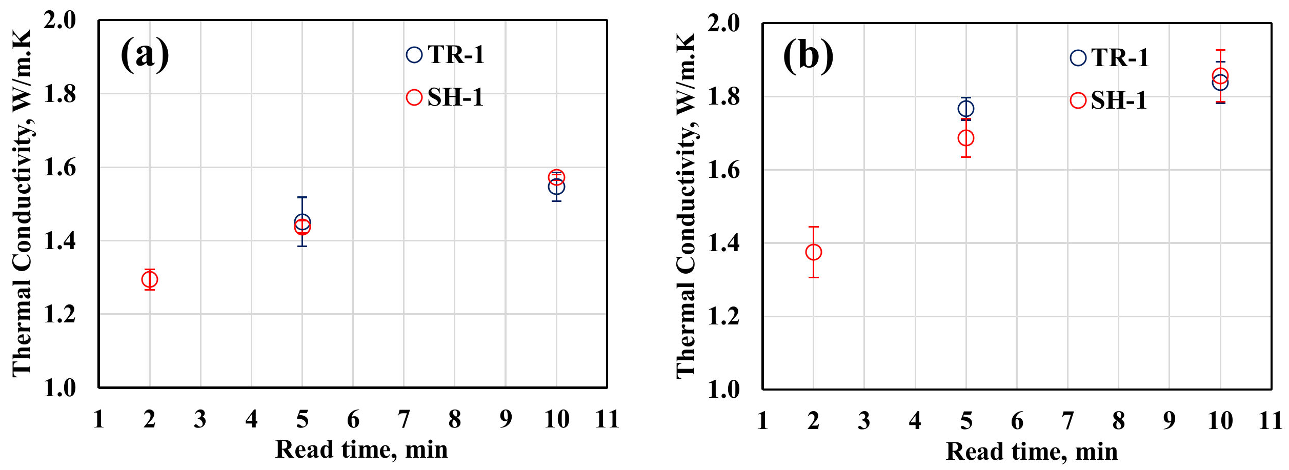
| Read Time, min | Thermal Conductivity, W/m·K | |||
|---|---|---|---|---|
| Stone from Sampling Point 1 | Stone from Sampling Point 2 | |||
| Sensor TR-1 | Sensor SH-1 | Sensor TR-1 | Sensor SH-1 | |
| 2 | - | 1.294 ± 0.028 | - | 1.375 ± 0.069 |
| 5 | 1.451 ± 0.067 | 1.437 ± 0.016 | 1.766 ± 0.031 | 1.687 ± 0.052 |
| 10 | 1.547 ± 0.040 | 1.572 ± 0.007 | 1.838 ± 0.057 | 1.856 ± 0.071 |
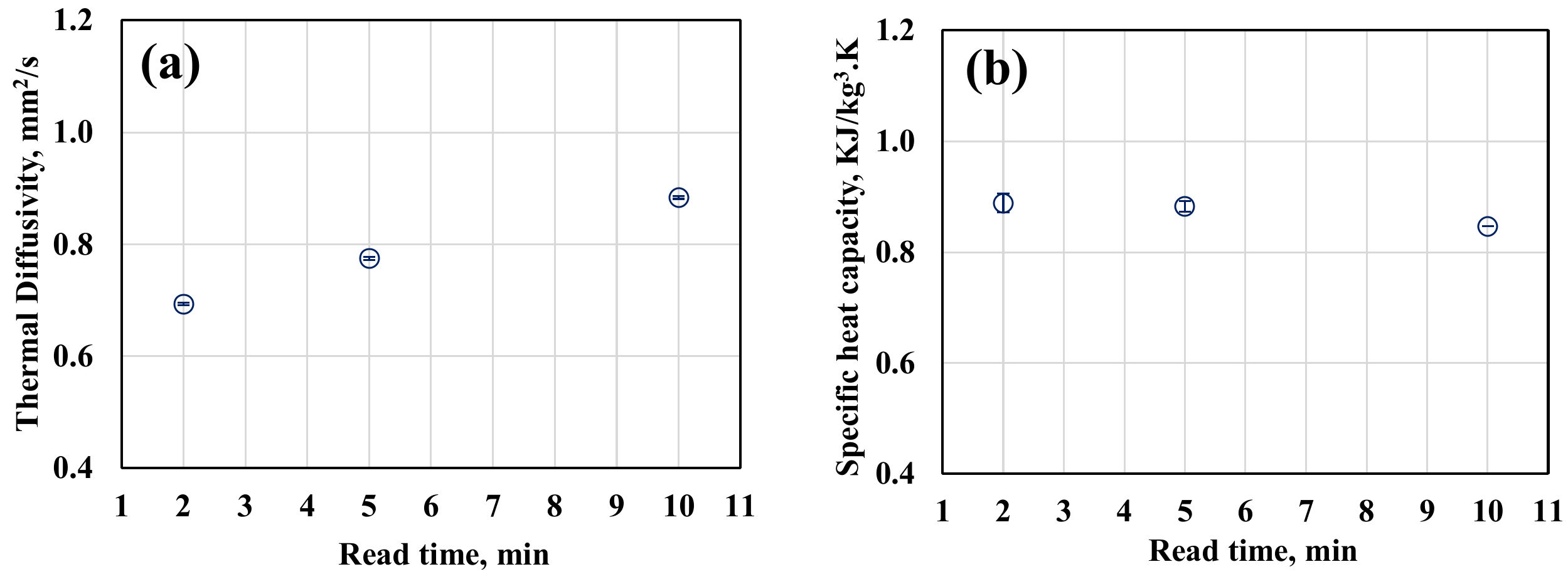
| Read Time, min | Stone from Sampling Point 1/Sensor SH-1 | |
|---|---|---|
| Thermal Diffusivity, mm2/s | Specific Heat Capacity, KJ/kg3·K | |
| 2 | 0.694 ± 0.002 | 0.888 ± 0.017 |
| 5 | 0.775 ± 0.003 | 0.883 ± 0.009 |
| 10 | 0.884 ± 0.003 | 0.847 ± 0.000 |
Publisher’s Note: MDPI stays neutral with regard to jurisdictional claims in published maps and institutional affiliations. |
© 2021 by the authors. Licensee MDPI, Basel, Switzerland. This article is an open access article distributed under the terms and conditions of the Creative Commons Attribution (CC BY) license (https://creativecommons.org/licenses/by/4.0/).
Share and Cite
Spathis, P.K.; Mavrommati, M.; Gkrava, E.; Tsiridis, V.; Evgenidis, S.P.; Karapanagiotis, I.; Melfos, V.; Karapantsios, T.D. Characterization of Natural Stone from the Archaeological Site of Pella, Macedonia, Northern Greece. Heritage 2021, 4, 4665-4677. https://0-doi-org.brum.beds.ac.uk/10.3390/heritage4040257
Spathis PK, Mavrommati M, Gkrava E, Tsiridis V, Evgenidis SP, Karapanagiotis I, Melfos V, Karapantsios TD. Characterization of Natural Stone from the Archaeological Site of Pella, Macedonia, Northern Greece. Heritage. 2021; 4(4):4665-4677. https://0-doi-org.brum.beds.ac.uk/10.3390/heritage4040257
Chicago/Turabian StyleSpathis, Panayotis K., Maria Mavrommati, Eirini Gkrava, Vasilios Tsiridis, Sotiris P. Evgenidis, Ioannis Karapanagiotis, Vasilios Melfos, and Thodoris D. Karapantsios. 2021. "Characterization of Natural Stone from the Archaeological Site of Pella, Macedonia, Northern Greece" Heritage 4, no. 4: 4665-4677. https://0-doi-org.brum.beds.ac.uk/10.3390/heritage4040257