1. Introduction
Diagnostic studies on ethnographic art objects can be fundamental to answering questions about the origin, dating and attribution of these complex and fragile artifacts, and to approach their restoration and conservation.
Non-invasive methods show significant advantages when studying fragile multi-material ethnographic artifacts. These kinds of multi-cultural artworks are generally characterized by complex combinations and stratifications of different materials—representing iconographic contents and production techniques less familiar to western culture, and often little studied, and usually they have received scarcely documented restoration treatments across their life. Changes of destination and use, coupled with dismemberments, uncertain storage and the possibility of cultural misrepresentation [
1] contribute to their frequently troubled conservative history.
The object of this work is the investigation of two ethnographic artifacts from Polynesia: a painted barkcloth (
Figure 1) and a feathered headdress (
Figure 2), both coming from the ethnographic collection of the museum of the Congregation of the Sacred Hearts of Jesus and Mary (Picpus) in Rome and never analyzed before the present work. The diagnostic approach combined imaging techniques with spectroscopy analyses in order to characterize the constitutive materials of the two artifacts, evaluate their state of conservation, and support the decision-making process for their restoration.
The first investigated artifact is a decorated barkcloth, also known as tapa, which is not a woven material, but made from the inner part of the bark that has been softened through a process of soaking and beating [
2]. The inner bark is taken from several types of trees or shrubs [
3], most of them from the
Moraceae family, such as paper mulberry (
Broussonetia papyrifera) and native species of fig (
Ficus prolixa,
Ficus bengalensis), and it is decorated—usually with geometric patterns or elements referring to nature—with paints and vegetable dyes of light brown, red, yellow and black. Polynesian barkcloth is manufactured for everyday needs such as room dividers, clothing, and floor mats, as well as ceremonial uses, or exchanged as a sign of respect within the community [
4]. Though there are a variety of local names, the word tapa, originally from Tahiti, is commonly used to refer to barkcloth made all over the world. While tapa cloth is most often recognized as a Polynesian craft, it has also been made in South America, Indonesia, New Guinea, Melanesia, and parts of Africa.
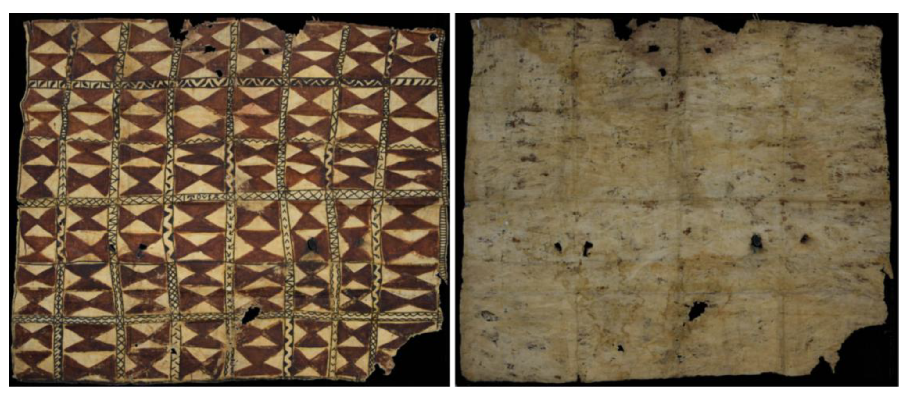
The example shown in this study is a tapa (135.5 × 147.5 cm) painted with hourglass geometric motifs in brown-red tones and a black inscription in the center with the word "Tovia”, considered by art historians as referring to the name of the artist or of the commissioner. It is listed in the catalog of the museum as “Ta262” from Tahiti, classified as “very rare”, having arrived in Europe in the first half of 19th century; despite being included in the collection from Tahiti, a note in the description of the object in the catalog suggests that the tapa would have come from Western Polynesia, according to its typical decoration. In fact, the ethnographic investigation confirmed the similarity of the tapa Ta262 to those from the archipelagos Western Polynesians of Samoa, Tonga and Fiji, which as the anthropologist A. Kaeppler recalls, due to the influence and exchanges between these islands over centuries, are usually very similar to each other [
5,
6].
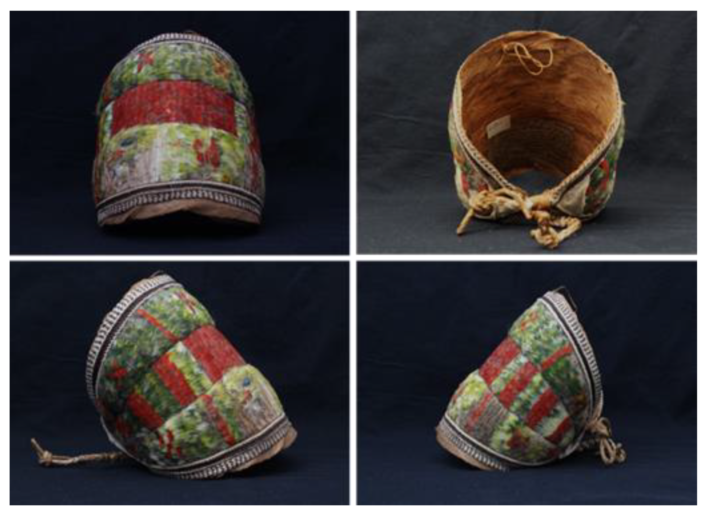
The second investigated artifact is a Polynesian headdress in feathers (18 × 16 cm), known by the indigenous word pa’e ku’a and identified with the code M098 in the
Catalogue des objets de la Polynésie et autres pays. Dans la Collection de la Congrégation des Sacrés-Coeurs (Picpus) in Rome. More specifically, it is a diadem made up of vegetable fibers and feathers of red, green and yellow color from the Marquesas Islands, in all likelihood from the island of Nuku Hiva. This type of headdress was reserved for the chiefs (both men and women) of the tribes, who used to wear it in ceremonies, associated with a number of further ornaments [
7]. It seems probable that the rarity of
pa’e ku’a specimens is due to the nature of the feathers that make up its decoration, though literary and archival sources report different versions. In fact, although many agree on the fact that the green feathers belong to a local turtle dove (kuku), there is some discrepancy regarding the red feathers. Some think they may have been taken from the head of the same turtle dove, but other scholars [
8] report that, according to the input of some Nuku Hiva natives, the red feathers belonged to a bird called
manu ku’a that once populated the desert lands of Nuku Hiva, but today is supposed to be extinct.
Historic surveys made during this research work towards the correct identification of the realization process and usage of this artifact have revealed that the current inhabitants of the Polynesian islands no longer have a memory of these craftsmanship practices, which gives higher importance to the scientific study of an ethnographic heritage that could be lost.
2. Materials and Methods
Due to the intrinsic fragility and conservation needs of both ethnographic objects, the diagnostic approach focused on non-invasive contactless surveys to evaluate their general state of conservation and provide a first identification and characterization of original materials and the eventual restoration and/or degradation products. The diagnostic multi-technique approach started with imaging surveys through ultraviolet fluorescence (UVF) photography and multispectral imaging, and later addressed analytic investigations for material characterization in order to understand the craftsmanship and decorative procedures, and reconstruct the conservative path of the two art objects.
2.1. UVF Photography and 3D UVF Photogrammetry
UV fluorescence images of the two artifacts were obtained with a Nikon D5300 digital SLR camera, with a multifocal 18/105 mm lens, in the following shooting conditions: aperture opening f/4.4, exposure time 15 s, sensitivity ISO-200, focal length 35 mm [
9]. For a proper acquisition of photographs in UV fluorescence, the acquisitions were done in a dark room to avoid contamination with the visible component of environmental light, which could affect the response of the constituent materials. Furthermore, specific filters were placed in front of the camera objective: a Kodak Wratten 2B and an 85B amber filter, in order to exclude the reflected UV component and the blue cast typical of UVF photos. Two UV LED lamps (emission peak at 365 nm) were used to stimulate UV fluorescence, positioned at 45° with respect to the artifact’s surface. In the case of the headdress, a 3D model under UV was also produced. To reach this goal, the headdress was placed on a rotating platform in order to acquire the frames without moving the camera or the LED lamps around the artifact, but simply rotating the object, as explained in a previous paper [
10]. The final 39 frames were saved in .jpg format, resolution 24 MP, and sRGB color with a depth of 24 bit. The three-dimensional UV model was obtained using a digital photogrammetric software providing a comprehensive Structure from Motion (SfM) approach, which integrates digital photogrammetry and computer vision facilities with the ability to process unsorted photographs into photorealistic, geometrically accurate, and georeferenced 3D models. The detailed procedure for obtaining the UVF 3D model has been previously explained in detail [
10]. The workflow may be reported as follows:
- (1)
Uploading of the acquired photos;
- (2)
Using the SIFT algorithm (Scale-Invariant Feature Transform) to calculate and detect the positions throughout the image set of homologous points (pixels) to sufficiently establish the spatial relationships within a relative XYZ coordinate system. Thus it arranges the photos according to the calculated parameters. The SIFT algorithm also allows one to connect common characteristics, even with variations in scale, points of view, partial occlusions and object brightness;
- (3)
Applying the bundle adjustment algorithm to control and limit errors during the transformation of the coordinates of the 3D points taken from a cloud of points that is more or less dense, depending on the number of detected key-points;
- (4)
Generating a dense cloud of points through the dense image-matching algorithms;
- (5)
Sizing the model in order to obtain millimeter precision.
2.2. Hypercolorimetric Multispectral Imaging (HMI)
Multispectral images were acquired through Hypercolorimetric Multispectral Imaging (HMI), a portable system consisting of a Nikon D800 36 Mpx camera (modified to obtain full-range spectral reflectance measurements from ultraviolet to near-infrared (300 to 1000 nm)), 2 filters selecting registered wavebands, standard white patches and a color-checker positioned in the scene, and an illuminating system based on flashes modified to be as wide as possible, as described previously [
11]. The two acquired images were then calibrated through SpectraPick software, which produced seven high-resolution spectral images in .tiff format from UV to NIR at 350, 450, 550, 650, 750, 850 and 950 nm. The calibration procedure, achieving a precision higher than 95% on the spectral reflectance images and a color error CIE ΔE2000 of less than 2, has been fully described in previous works [
11,
12,
13]. The native HMI digital image processing software, named Pickviewer, provides several diagnostic imaging and statistical tools to produce false color images, and applies principal component analysis (PCA) to different spectral bands to enhance hidden information and map materials based on their spectral reflectance or colorimetric values.
2.3. Scanning Electron Microscopy coupled with Energy Dispersive Spectrometer (SEM-EDS)
SEM-EDS analyses were conducted at the electron microscopy section of the Special Equipment Center (CGA) of the University of Tuscia with a scanning electron microscope Jeol JSM 6010LA model operating at 20 kV as the acceleration voltage of the electrons, coupled with an EDS probe, this last used to reveal the chemical elements in the sample from tiara. SEM investigations enabled the morphological study and identification of the plant samples used for the tapa and the headdress, placing samples on aluminum stubs, fixed with carbon tape and metallized with gold (sputtering) through a sputter coater Balsers MED10, operating under vacuum. The images obtained at different magnifications were compared with the available scientific data.
2.4. X-ray Fluorescence Spectroscopy (XRF)
XRF spectra were acquired with a Surface Monitor II (Assing™, Rome, Italy) spectrometer, which operated with the following settings: Ag tube at 40 kV, current 76 A, acquisition time equal to 60 s, distance from the analyzed surface equal to 94 mm, spot 2 mm. The instrument was equipped with an Amptek X-123 Si-PIN detector (Amptek, Bedford, US), resolution 145 to 260 eV at 5.9 keV, optimum energy range 1–40 keV. All spectra were collected by Gonio software by Assing™. XRF analyses were performed on the painted surface of the tapa in order to characterize pigments and the eventual restoration or degradation products, and on the vegetal fibrous support of the tiara to identify possible coloring treatments or consolidation materials.
2.5. Fourier Transform Infrared Spectroscopy (FTIR)
Infrared spectroscopic analyses were performed for the characterization of original and eventual restoration adhesive substances, both in the tiara and in the tapa. FTIR spectra were acquired using a Nicolet Avatar 360 spectrometer with a DTGS detector (deuterated triglycine sulfate), a Michelson interferometer, and an accessory for diffuse reflectance analysis (DRIFT). For each sample about 5 mg of ground KBr was used as a background. In total, 128 spectra were acquired in the mid-infrared region (400–4000 cm
−1) on each sample, with a resolution of 4 cm
−1. Spectra processing was performed in Thermo’s OMNIC version 8.0 software of Fisher Scientific Inc. The reading and interpretation of the spectra was based on the management tools available in the above-mentioned software, the scientific literature [
14,
15], on-line databases (IRUG Database, RRuff) and in-house libraries created by the Laboratory of Diagnostics and Materials Science of Tuscia University.
2.6. Surface-Enhanced Raman Spectroscopy (SERS)
In recent years, as reported in the literature, the SERS technique has been used with excellent results in the field of heritage diagnostics for the characterization of organic dyes of natural and synthetic origin, allowing for the overcoming of fluorescence problems that hinder the identification of these molecular species using conventional Raman analysis [
16,
17,
18]. In particular, with reference to the results of some previous case studies, a specific type of colloid reduced with hydroxylamine [
19] was used for this study because of its lower interference with the background of the spectrum (due to the reducing agent) compared to the more traditional citrate colloids [
20], as well as its improved reproducibility. In addition, SERS was applied via the fiber approach: the colloid is put in direct contact with the fiber, dyed or painted, and the spectra are acquired in correspondence with clusters of nanoparticles in close proximity to the sample.
In this study, the colloid was prepared with silver nitrate (AgNO
3, purity ≥99.0%), hydroxylamine hydrochloride (purity = 99.0%) and sodium hydroxide solution (NaOH 10 M BioUltra) from Sigma Aldrich. The colloid was prepared according to the protocol developed by Leopold and Lendl [
19]. A quantity of 200 μL of colloid was taken and placed in an Eppendorf
® tube; 20 μL of a solution of MgSO
4 was added to induce the aggregation of the nanoparticles. Subsequently, 20 μL of aggregated colloid was placed in contact with sample fragments. Once the water had evaporated, the SERS spectra were recorded not in direct correspondence with the fiber sample, but on clusters of silver nanoparticles close to it, for a better quality and lower background fluorescence.
SERS spectra were acquired with a Raman Horiba Jobin-Yvon HR Evolution spectrometer coupled with a microscope (objective used: 50X) using a He–Ne laser with a wavelength of 633 nm. The radiation used had an intensity of approximately 0.38 mW, while 60 scans of up to 6 seconds each were collected. For each sample, spectra were collected at three different points in the spectral range 100–2000 cm
−1, in order to consider the variability of the spectrum in correspondence with different clusters. The three spectra were averaged, in order to evaluate the most recurrent spectral features. The assignment of the peaks was carried out on the basis of the methods used in the literature [
21,
22,
23,
24].
3. Results and Discussion
3.1. UVF Photography and 3D UVF Photogrammetry
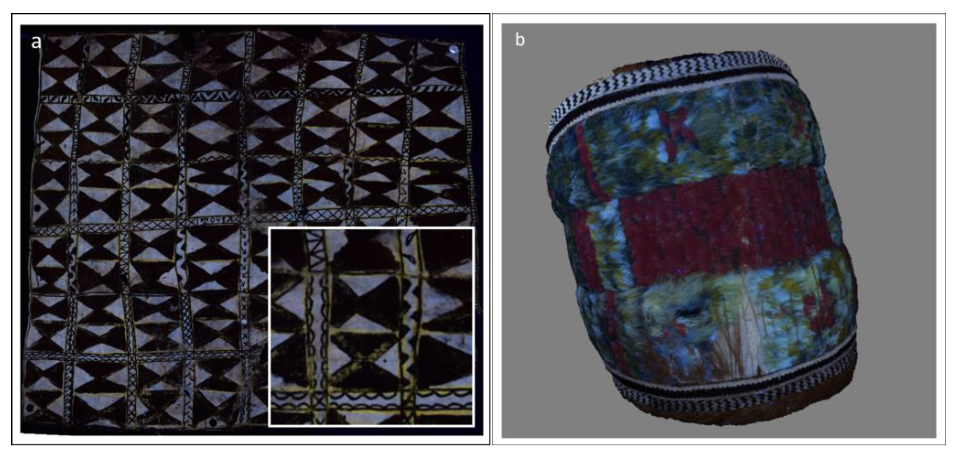
Ultraviolet fluorescence images of the tapa highlighted a strong yellow fluorescence in response to UV radiation, suggesting the usage of some organic substance (
Figure 3a). It was found mainly on the recto of the artifact, below the other colors, whose purpose seems to be compatible with the hypothesis that it is a preparatory drawing. There are traces also on the reverse of the artifact, a sign that the substance was probably transferred from the front during execution. Considering the scientific literature [
25,
26], it was preliminarily assumed that it could be a dye extracted from the roots of turmeric, which was widely used in the daily life of Polynesian communities [
27].
In birds, color is generally used as a signal through which females can select their mates. Unlike humans, birds have an expansive range of vision in the ultraviolet spectrum (300–400 nm), which assumes considerable importance for the transmission of signals between specimens.
The overall UV response from the feathers is the result of the incoherent backscattering of UV rays from the keratin surface, combined with their coherent dispersion, reinforced by the spongy structure of the flag.
Recent studies show that some bio-pigments, including psittacofulvins, which are more concentrated in the red and yellow feathers of the
Psittaciformes spp. (colored birds including parrots, parakeets, macaws and cockatoos), can be identified based on their response to UV radiation [
28]. The presence of violet fluorescence in the red regions and the increase in fluorescence in the yellow regions, as well as a darkening of the green areas, testifies to the presence of psittacofulvin in feather artifacts. The application of 3D UVF photogrammetry, starting from photographic images before ornithological recognition, was chosen as a non-invasive preliminary investigation approach to understand whether the plumage of the bird, which constitutes the decorative apparatus of the headdress, was affected by coloring based on this bio-pigment.
Compared to the visible photography, it was indeed possible to observe the slight purple fluorescence of the red feathers, the strong pale white fluorescence of the yellow feathers, the intense yellow fluorescence for other yellow feathers, and the weak darkening for the green and blue ones, suggesting the presence of psittacofulvin and the attribution to Psittaciformes spp. for all the feathers used in the tiara.
3.2. Hypercolorimetric Multispectral Imaging (HMI)
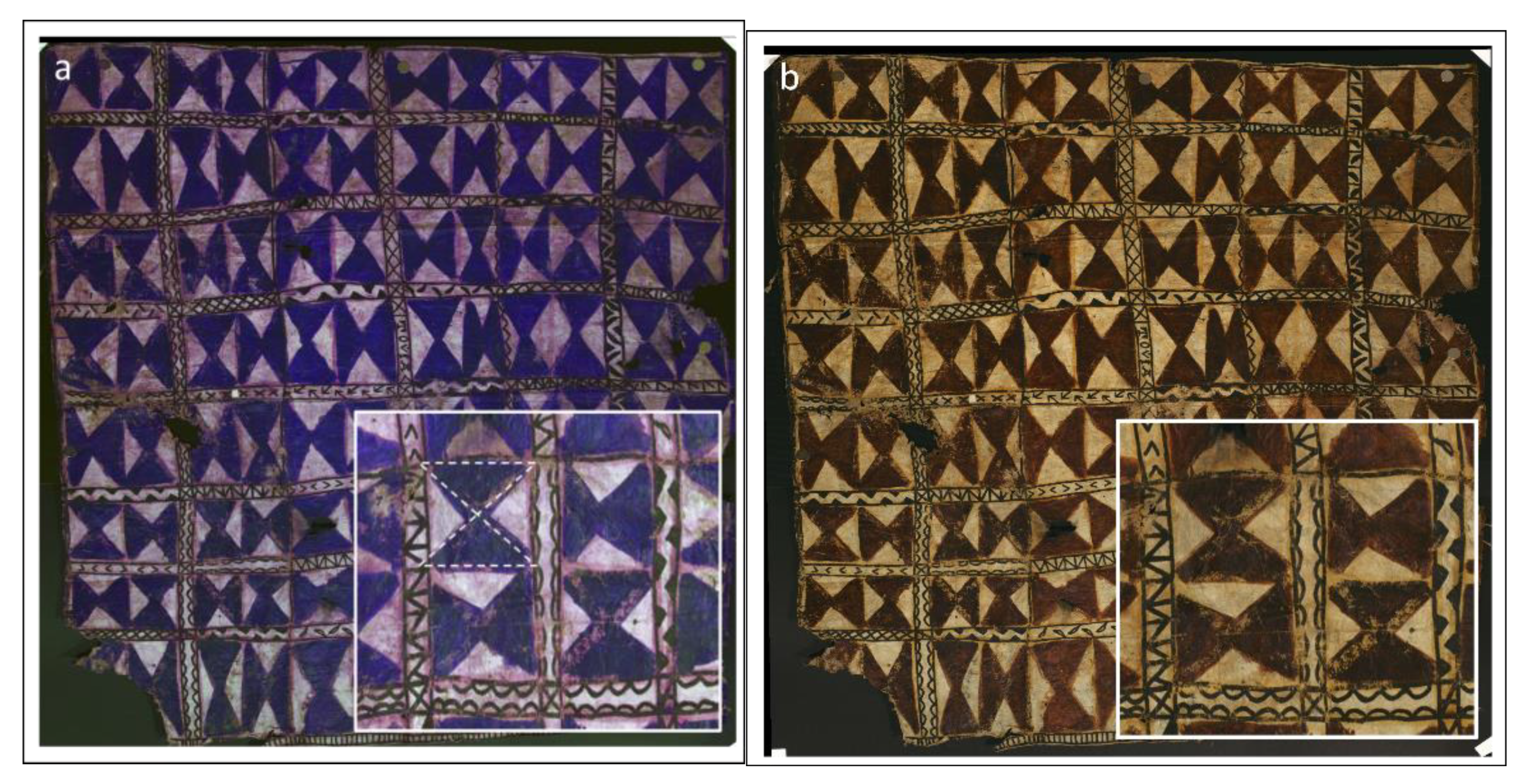
The seven spectral bands produced after the calibration step for both art objects were analyzed in the PickViewer
® digital image processing software to extract useful information on painting materials and the general state of conservation of the surface. In particular, the UV and IR calibrated bands were used to create false color images that enabled us to highlight different materials that have a similar appearance in visible light, but actually have different compositions, and therefore a different spectral behavior. The UV-false color (UVFC) image of the tapa (
Figure 4a) allowed us to visualize the color used to paint the hourglasses, here appearing in a deep violet shade, and the different pigment/dye used to draw the borders (already suggested in the UVF photography) that in this case appear in a bright pink color. Such spectral behavior corresponds with turmeric in the database created by the Center for Conservation and Restoration “La Venaria Reale” (Turin, Italy) in collaboration with the National Institute of Metrological Research (INRIM) and the Scientific Analysis Laboratory of the Autonomous Region of Valle d’Aosta (LAS) [
29].
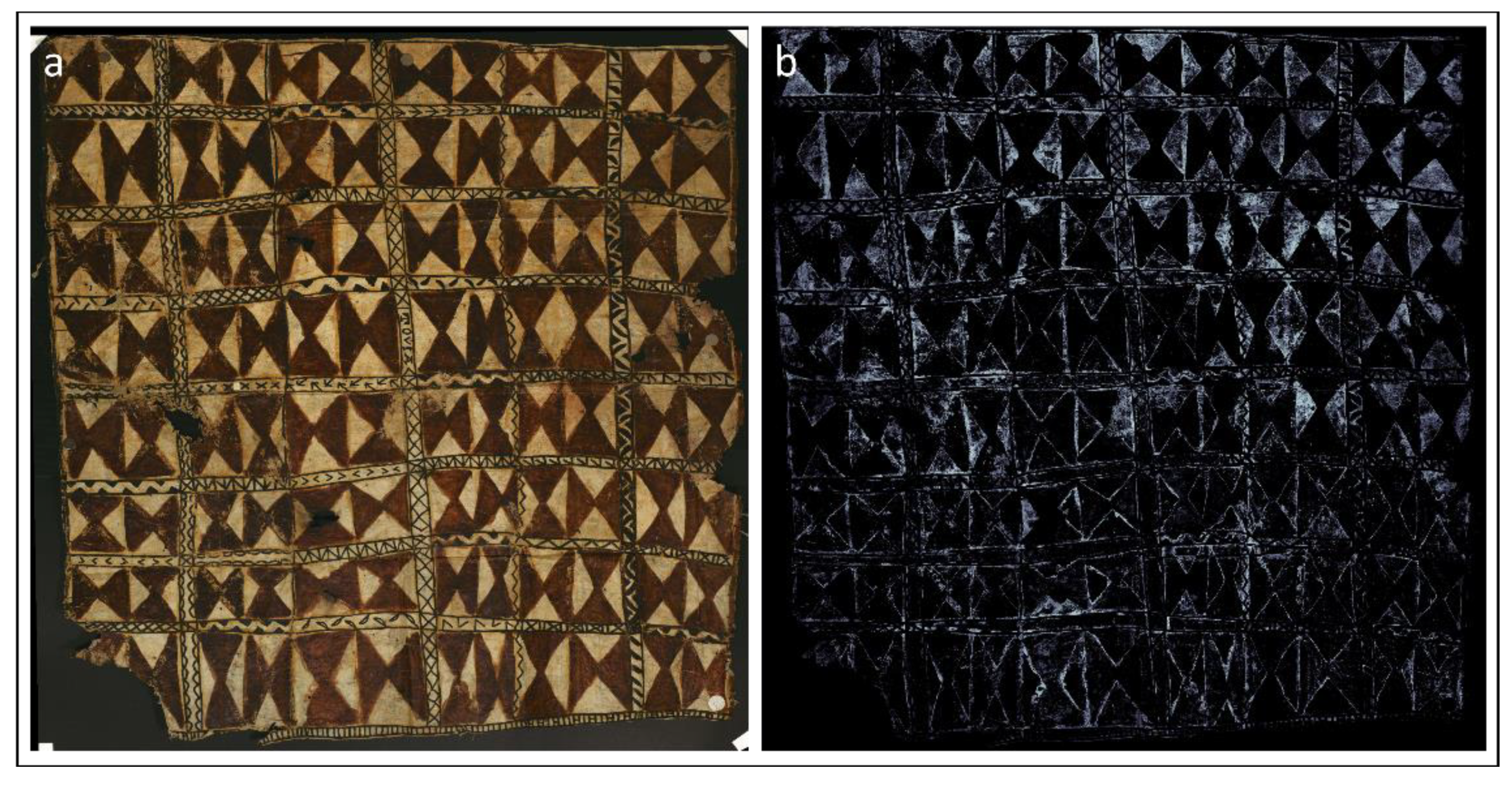
A further image processing approach, available in the HMI original software, is to build maps of similarity based on the multispectral curve or on the colorimetric CIE L*a*b* values of a selected area, representing the distribution of similar materials.
Figure 5b shows the chromatic distribution map of the yellow pigment used for the borders of the hourglass motif, which is not easy to distinguish in the visible image (
Figure 5a), highlighting its presence also in the background of the squared area containing the hourglasses and in the geometric grid painted in black [
30].
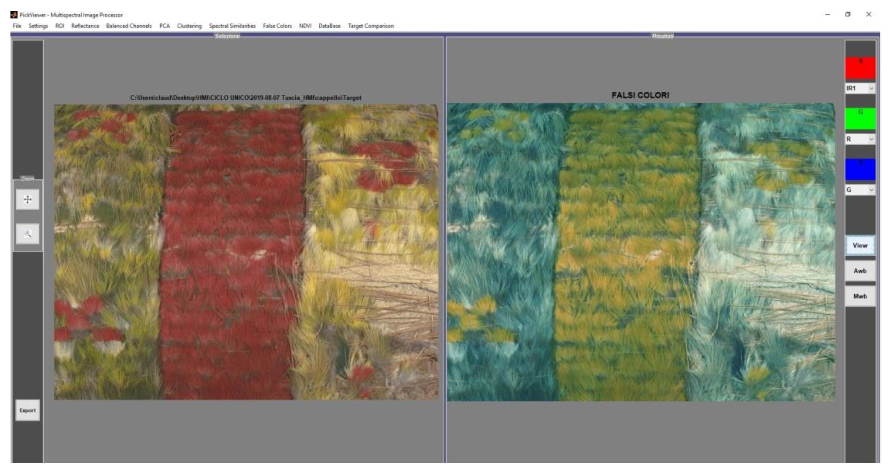
In the case of the tiara, processing through PickViewer was performed in order to investigate the similarities and differences between the feathers. The IR-false color (IRFC) image compared to the visible one shows that the red feathers exhibited two different false colors: yellow and dark green, this last being similar to that of the green feathers (
Figure 6).
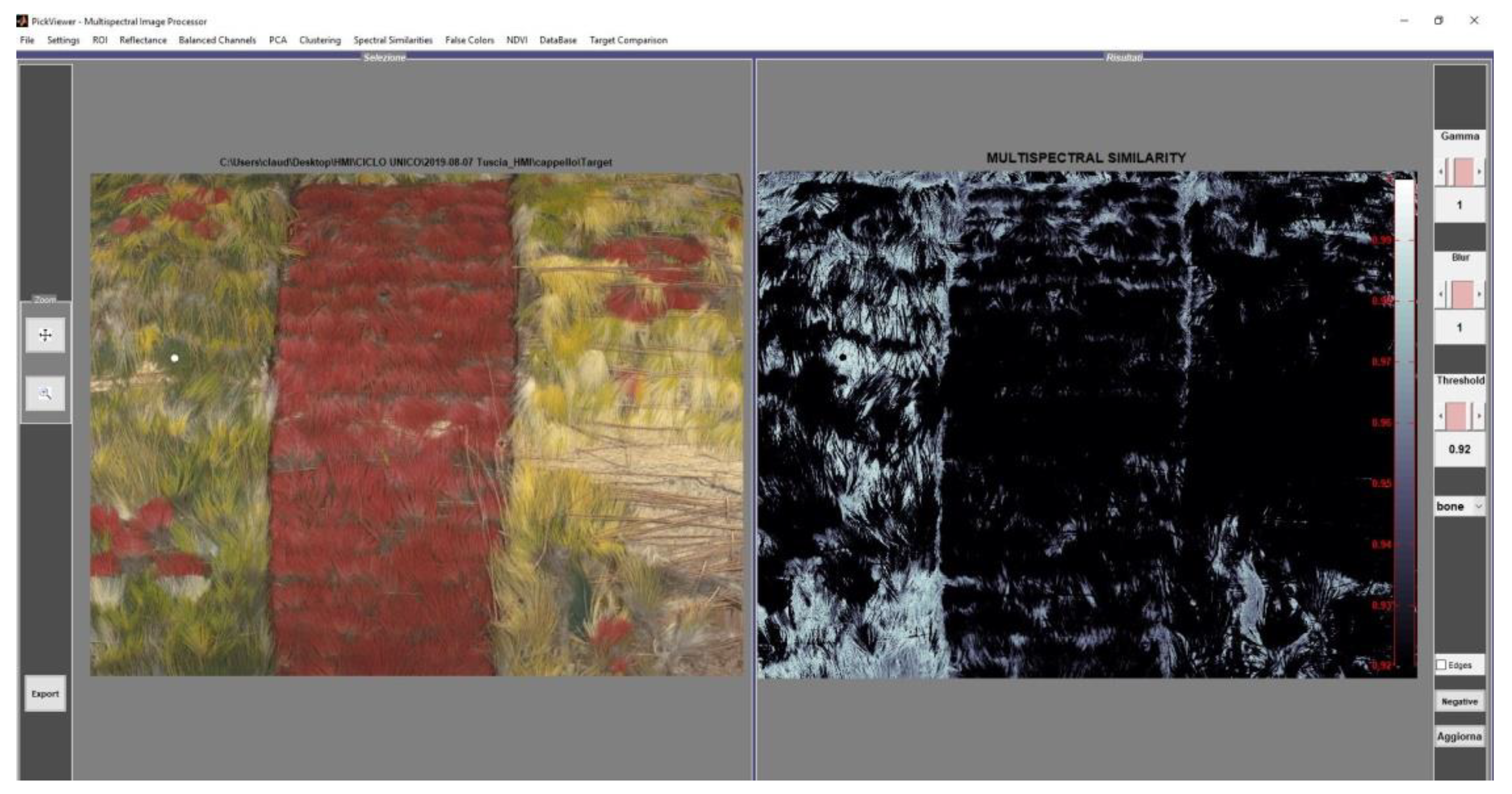
In order to expand on this aspect, a multispectral similarity tool was applied to the green feathers (
Figure 7). The obtained result confirms the similarities, in terms of the spectral response, of the green feathers with that of the zone of the red feathers, close to the quill.
The color in the feathers is due to different mechanisms based on both chemical composition (presence of bio-pigments) and to light scattering phenomena [
31]. In the case of the headdress from Picpus Museum, the homogeneity of the IRFC responses, associated with the careful inspection of the feathers during restoration activities and supported by an ornithologist, allows for assessing the provenance of the feathers as being from a single kind of bird (
Vini kuhlii bird) [
32].
3.3. Scanning Electron Microscopy (SEM)
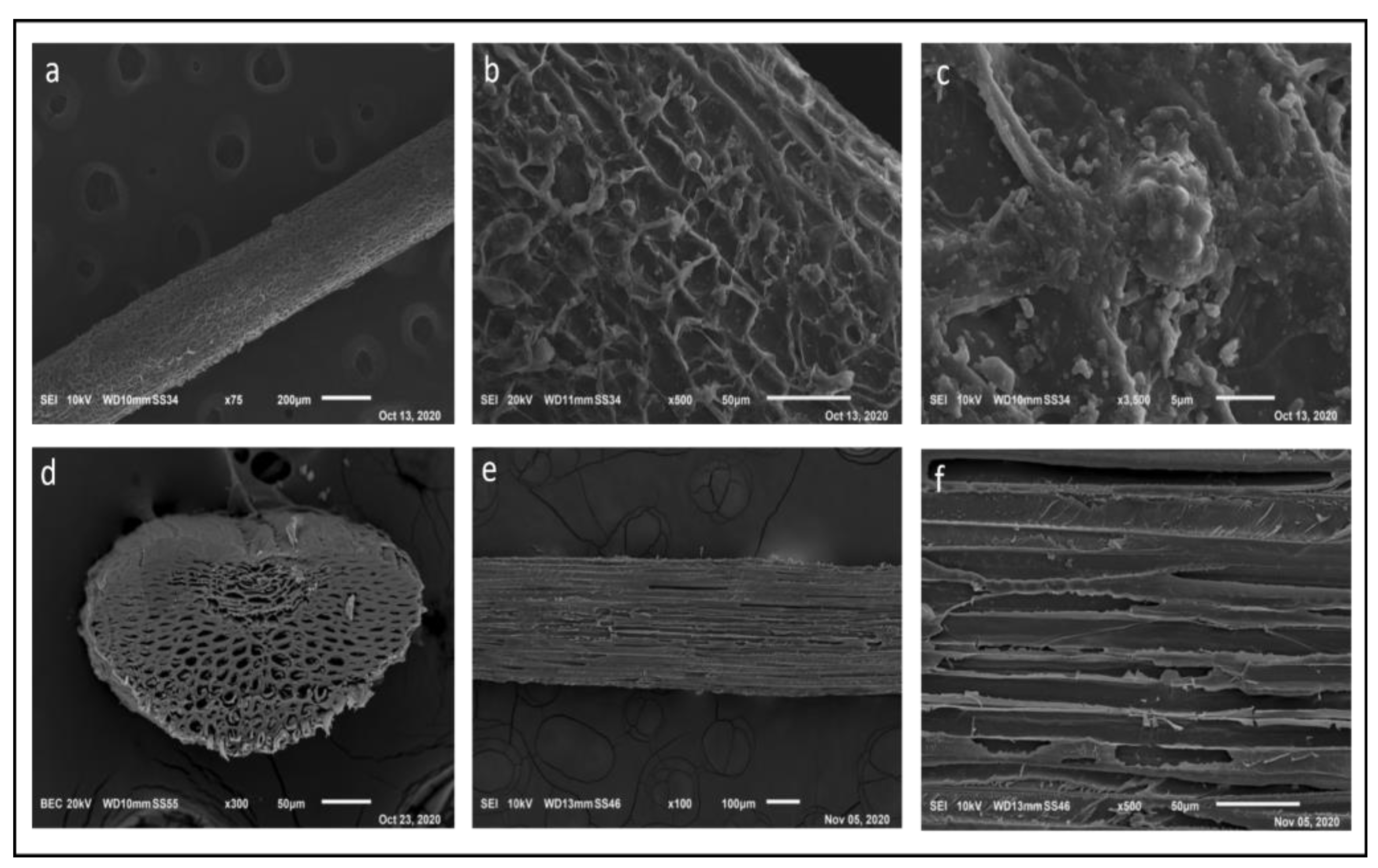
For the confident identification of the botanical species used for the realization of the two Polynesian art objects, molecular investigations certainly are the diagnostic approaches with the highest rate of success, but to provide precise information, they require a sample of at least 1 cm
2, which is too large for most artifacts [
33,
34]. Therefore, micro sampling for SEM microscopy observation was preferred as a diagnostic method aimed at studying the morphological aspects of the fibers that characterize the support of the tapa Ta262 and of the tiara M098 in order to identify the botanical species used for their realization. The images obtained at different magnifications, enhancing details from 200 µm down to 5 µm, were compared with the data from the available literature [
35].
The SEM images acquired on single fibers of the tapa (
Figure 8) highlight some morphological characteristics of the fibers of the tapa, which appear rather long and are protected by an external membrane (
Figure 8a,b). It is possible to perceive the presence of fragmented intracellular material (
Figure 8c,d). The outer membrane does not appear to be fully adherent to the fibers in several spots. Furthermore, the sample seems to be covered with a globular-looking material that can be assumed to be a starch-based adhesive (
Figure 8e,f).
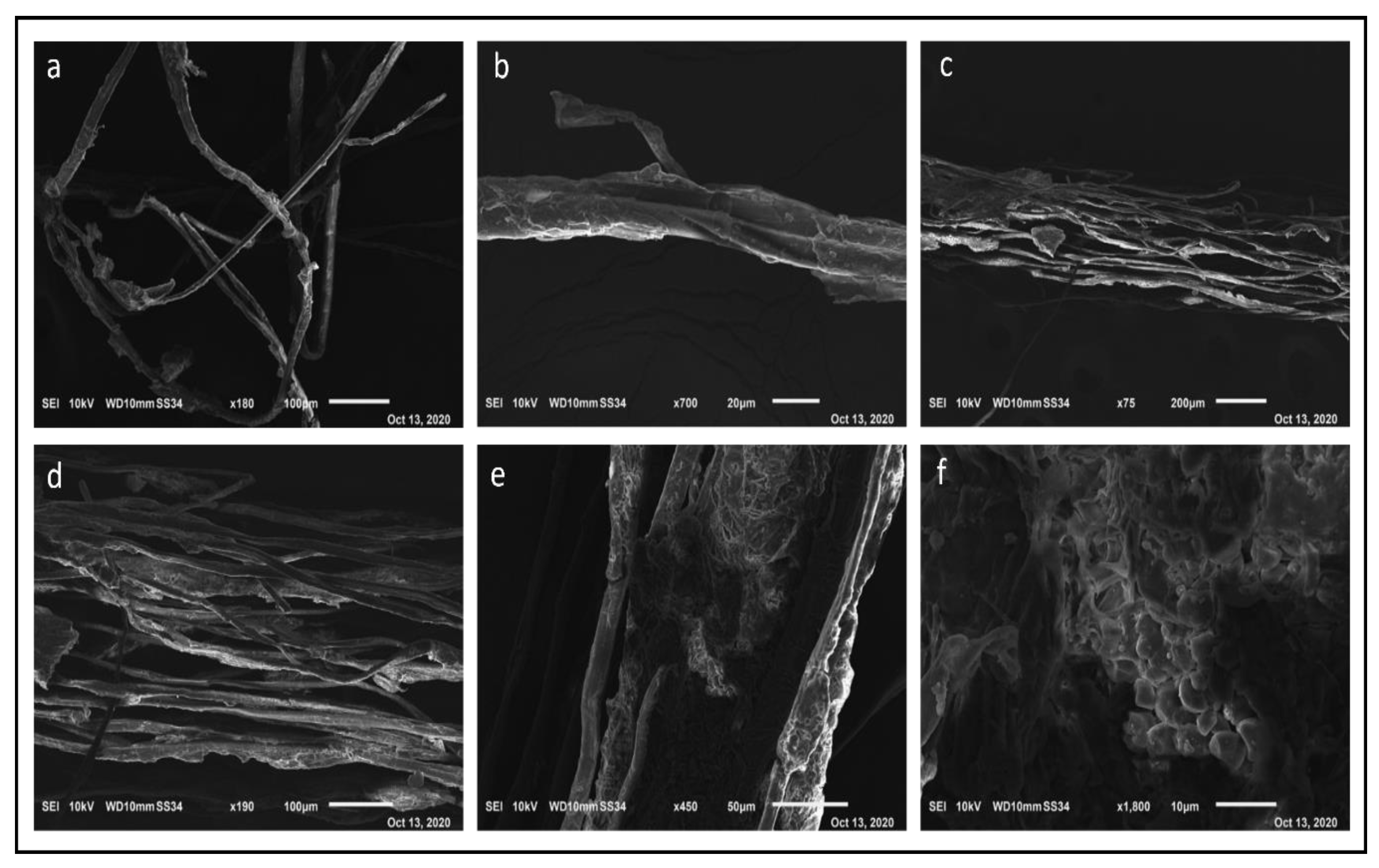
SEM analyses were also performed on single vegetable fibers taken from the primary structure of the tiara (
Figure 9).
The main morphological traits of the fiber were investigated from magnifications of 75× to 3500×. A comparison with several studies published in the scientific literature, especially those focusing on the transverse section of the coconut fiber with its multi-cellular pattern (micro fibrils of different sizes), polygonal shape and a thick wall, as well as a central lumen (
Figure 9d), was performed. Another characterizing aspect is the natural presence of silica dots, as observed in the SEM image at 3500× (
Figure 9c) [
36]. EDS analysis performed on these dots revealed the following chemical elements: Si, C and O, confirming that
Cocos nucifera L. is the species that was used to build the main body of the Polynesian headdress.
3.4. X-ray Fluorescence Spectroscopy (XRF)
As the two objects are essentially made of organic materials, XRF spectroscopy was applied only to detect the possible presence of elements that can be associated with minerals, and their eventual restoration treatments.
The constant presence of Ca and K in the examined points of the tapa may be associated with residues of the instruments (shells and/or rocks fragments) used for scratching the fibrous material in the phases preceding and following the beating [
26]. The presence of iron can be ascribed to the use of red ocher (named
’ele in the local language of Polynesia), probably mixed with organic dyes, but also to the tannins of some of the barks of the most common species that contain Fe.
Lastly, the presence of Cl may be explained by the contact of the tapa with sea water, which contains NaCl. The high counts of chlorine correspond with evidence of water streams, where the tapa probably came into contact with sea water, but we cannot say if this was during its use or as it traveled from Polynesia to Europe [
37].
XRF analysis of the headdress revealed the presence of Ca, Fe, K and As. The first three elements may be associated with the original materials and/or the process used for producing the artifact. Arsenic has been attributed to the use of pesticides in a previous intervention. The XRF investigation shows that the arsenic residues are more concentrated in the feathers, which are known to be easily infested with dermestids. The presence of arsenic, combined with the discovery of three exuviae of Anthrenus verbasci and the extensive structural damage caused to the plumage, would justify the pesticide hypothesis as a treatment against a previous infestation. Arsenic is present in significantly lower concentrations on the backside of the artwork.
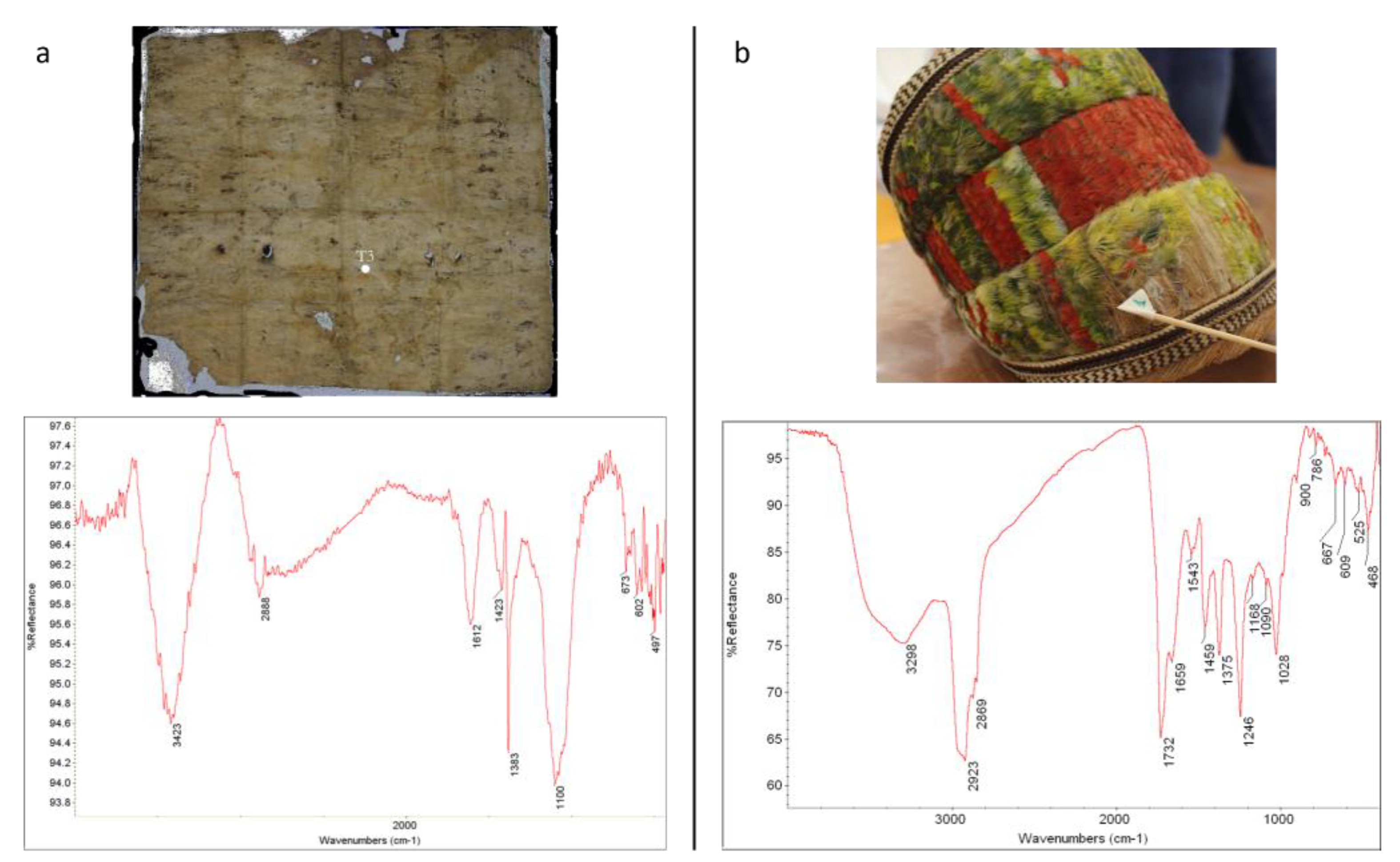
3.5. Fourier Transform Infrared Spectroscopy (FTIR)
IR spectroscopy was used in the study of the Ta262 tapa to verify the substances that emerged under ultraviolet fluorescence with a blue color. The fact that the fluorescence response was more concentrated in the overlapping points of the layers that make up the support suggests that an organic adhesive was employed in the production. Furthermore, the substance showed a certain sensitivity towards aqueous solutions in the preliminary verification phase assessing the sensitivity of the decorated areas to different solvent systems for cleaning.
The signals at 3423 cm
−1 (stretching OH), 1612 cm
−1 (intramolecular H
2O), 1423–1383 cm
−1 (deformation of the C–OH bond) and 1100 cm
−1 (deformation of the C–O bond) suggest the presence of some polysaccharide substance and the absence of modern restoration adhesives (
Figure 10a) [
38]. The signatures under 700 cm
−1 (specifically 673 and 602 cm
−1) are attributable to a combination of the ν(CO) rings of polysaccharides and various ring deformation modes [
38]. Lastly, the peak at 497 cm
−1 is due to the vibrations of the C1–O–C4 glucoside bond found in amylopectin and amylose, the two polysaccharides contained in the plant starch granules [
3]. A detailed band assignment regarding the spectrum in
Figure 10a is reported in the
Supplementary Material (Table S2).
FTIR analyses were also run on small areas on the front and back sides of the Polynesian tiara (
Figure 10b) in order to identify the original adhesive used to glue the feathers to the coconut fibers’ body, and the eventual restoration products. The spectrum obtained, compared with our laboratory databases, shows IR absorption bands characteristic of an organic substance of the ester type, with a steroid structure (specifically, we found a 75% match with cholesteryl acetate (Nr. 842 of Nicolet Standard Collection, CAS 604-35-3)). In the spectrum of
Figure 10b, some of the minor signatures may be associated with proteins (cm
−1: 3298, 1659, 1543, 1459) that are natural components of the feathers (see also
Table S3 for the detailed IR band assignment).
A deepen search of the scientific literature identified only an old paper reporting that natural extracts were commonly used as adhesives to fix feathers, and more specifically, extracts of
Cerbera manghas L. [
39], whose seeds contain various types of steroids [
40]. Furthermore, there is no evidence of restoration interventions on the artifact (with the addition of modern adhesives).
These considerations, together with comparisons with characteristic components of plant species reported in the literature, suggest that the adhesive could be one kind of natural rubber derived from
Cerbera manghas L. The species is known as “sea mango”, and is a small evergreen coastal tree distributed from the Indian ocean up to French Polynesia, whose leaves and fruits contain cerberine, an extremely poisonous compound if ingested [
41]. This tree was known as
eva or
reva on Marquesas island, and in the local culture, its fruits were used for religious punishments [
42] and to commit suicide [
43]. Because of its durability and close grains, it was the wood most commonly used, together with
Fragraea berteriana (called
Pu‘a), to produce musical instruments, such as drums (
pahu), which were made of a sharkskin head fastened over the hollowed-out end of a section of tree trunk [
44].
3.6. Surface-Enhanced Raman Spectroscopy (SERS)
The SERS analysis on the tapa was focused on three samples of the front painted side: one (T7) on the upper part and two (T8 and T5) located on the lower portion (
Figure 11a). For all three samples, the collected spectra were not significantly masked by the Raman signal of the colloid. The spectra acquired from different points of the same sample, however, showed reduced reproducibility, especially in the intensity of their signals: although similar signals were observable in each sample, from point to point, the absolute and relative intensities of the different spectral signatures did, in some cases, significantly change. This variability can be attributed to the presence of different molecules in the mixture, deriving from the same matrix or from the use of different dyes. Furthermore, from a comparison of the averaged spectra of the three samples, common peaks not attributable to the colloid can be observed (
Figure 11b): at low wavelengths, there are two signals around 650 and 733 cm
−1, while a weak signal at about 955 cm
−1 and a very intense band between 1329 and 1335 cm
−1 are observable in all spectra, together with broad bands around 1592 and 1620 cm
−1. This set of signals has a remarkable correspondence with the cochineal spectrum [
21,
22,
23,
24,
45]. In the case of sample T8 (
Figure 11c), with an orange color, the correspondence is also particularly accentuated by the presence of signals at 1384 and 1467 cm
−1. However, it should be emphasized that, for this sample, similarities are also observed with the spectrum identified in the literature for the extract of
Myrica rubra, which presents polyhydroxy flavonoids as dyes [
24]; the similarities, however, are less significant than those with the cochineal spectrum.
For the brown-black sample T7 (
Figure 11d), the average spectrum obtained is remarkably complex. The general trend of the spectrum is characterized by broadened bands, while the number of observable signals is higher: in addition to the bands already identified, peaks are observed at 1122, 1153, 1172, 1222, 1262 and 1287 cm
−1, with one shoulder at 1373 cm
−1, three medium intensity peaks at 1426, 1454 and 1496 cm
−1, and a series of nearby signals at 1535, 1569 and 1647 cm
−1. The spectral pattern has notable similarities with the spectrum of cochineal, but also with those of dyes—and anthraquinones—of the morinda and morindone type, as can be seen from the comparison with the spectra of
Morinda citrifolia (
Figure 11d) and
Morinda officinalis reported in the literature [
23]. It can probably be hypothesized that the spectrum of T7 is a combination of the signals of several dyes, such as those of cochineal and morinda.
Finally, for the T5 sample, no particular correspondence is observed, except with the spectrum of the cochineal, although it is important to underline that some characteristic peaks of carminic acid seem to be absent.
Based on the results obtained, it is not easy to accurately identify the coloring species used, even with reference to the scientific literature. The similarity with the cochineal spectra suggests that the coloring agents, for the T5 and T8 samples, belong to the anthraquinone class. It should be emphasized that cochineal, specifically, is not present among the attribution suggestions provided, but the use of coccidia (also of local origin) to obtain red-brown and orange dyes cannot be ruled out. For sample T7, with a brown-black color, it is possible that the same dye was used in a mixture with another matrix, such as the dye extracted from morinda. The use of dyes from different matrices would explain the obtaining of a dark color.
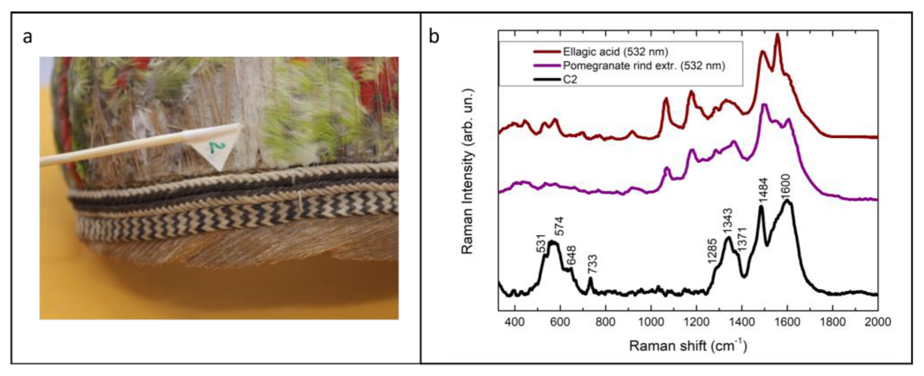
The same analytic procedure was repeated for the C2 sample from the Polynesian tiara (
Figure 12a). Unlike the tapa samples, the spectra obtained at different points of the C2 sample showed good reproducibility. At low Raman shift values, a series of signals at 531, 574, 648 and 667 cm
−1 was observed, together with a signal at 733 cm
−1. At higher wavenumbers, an intense signal was identified at 1345 cm
−1, with shoulders around 1285 and 1371 cm
−1. The strongest signals could be observed at 1484 and 1600 cm
−1, with shoulders at approximately 1560 and 1615 cm
−1, respectively.
The spectrum obtained showed significant correspondence with that characteristic of ellagic acid and dyes rich in ellagitannins [
22,
24], such as pomegranate peel extract (
Figure 12b). The major differences were the absence of the characteristic peaks of ellagic acid at 1065 and 1178 cm
−1. It must be pointed out, however, that the absence of a peak at 1065 cm
−1 has been identified in the literature for archeological samples, and it is attributed to the opening of the lactone rings with aging and the formation of ellagitannin oligomers, while the peak at 1178 cm
−1 is considerably less intense in the spectra reported in the literature of pomegranate dye extracts taken from dyed yarns, with respect to the spectrum of the natural matrix extraction. Consequently, for sample C2, attribution to a dye with a tannin nature is likely, as supported by historical sources.
4. Diagnostics and Main Restoration Decisions
The two ethnographic artworks from the museum collection of the congregation of missionary fathers of the Sacred Hearts of Jesus and Mary (PICPUS) have never been investigated before. The comparison with the literature has returned a complex and heterogeneous picture of the results obtained with different survey methodologies. This is probably due to the large variability in the resources used in the production of the artifacts, and to the evolution of traditional knowledge, handed down through the generations in the different populations dispersed in the Pacific area.
The investigations carried out on the tapa Ta262 were fundamental to understanding the materials used and the production technique of the artwork. Furthermore, the results obtained make it possible to direct the conservation approach and verify the effectiveness of strategies used. The presence of different original organic substances, used to combine the layers of beaten barks and also as coloring materials for the decorations, was reflected in all the analyses. This has led to the use in the conservation approach of different dry-cleaning methods and an extremely limited and controlled use of water (as a dispersing medium for the adhesive) in order to address the problems related to the lack of cohesion and adhesion of the fibers and painted layers. The aim was to avoid any types of solutions or solvents capable of solubilizing or altering the organic elements, such as fibers, coloring materials and even adhesives that, according to FTIR analysis, are composed of polysaccharidic compounds. In order to verify the appropriateness of the conservative approach, and to avoid the removal or alteration of the original substances, the use of UVF and colorimetric measurements was repeated during the restoration activities, using the specific tools of the HMI PickViewer software.
The preliminary investigations were fundamental to identifying the vegetable fibers, feathers and dyes used in the tiara, and to forming a better understanding of their intrinsic characteristics. In particular, the brown dye was identified by SERS as tannin, due to the presence of ellagic acid. The original adhesive was characterized as an ester with a steroid structure, allowing us to assess the use of extracts from Cerbera manghas L., as reported in the literature. Knowledge of the original materials helped in the selection of the adhesive to be used for the affected areas of the tiara. The selection was based on the long-term stability of the chosen product and its chemical compatibility with the original materials, in order to preserve their inherent properties of flexibility, shape and color.
The discovery of the presence of psittacofulvins in the feathered artifact, as a result of UVF analysis, determined the choice of the most suitable intervention treatment for the surface cleaning of the feathers. In this case, the use of organic solvents is not recommended, since they can cause the dissolution of the bio-pigments within the keratin structure. Based on specific data from the literature about the cleaning methods used for psittaciformes feathers, laser cleaning tests were carried out on commercial feather samples with very satisfactory results. According to the preliminary test results, the optimal operating parameters for the laser were established for the cleaning operation, which allowed for the effective removal of undesired materials without causing colorfastness and physical damage to the keratin.
5. Conclusions
Ethnographic art objects are mostly made up of a mixture of different materials (feathers, vegetable fibers, organic dyes, shells, beaten barks, etc.). Multi-material artifacts often have different needs in terms of restoration, turning their preservation into a complex matter, because what could benefit one material may be disadvantageous for another. The production techniques of ethnographic art objects are often scarcely documented or unknown in western culture, and research among native populations may not be successful due to their progressive disappearance, and to the impoverishment and loss of their local heritage as caused by globalization and massive international tourism. A further issue to consider is the deterioration connected with their use, and changes in their usage and locations in collections and museums over time. Once placed in a profoundly different context from their origin, artifacts have to adapt to the new environment, in terms of microclimatic conditions (relative humidity, temperature, lighting), gas and dust pollution (indoor and outdoor sources) and biological degradation (fungi, bacteria, insects, etc.).
In this complex context, a well-consolidated diagnostic approach was used to characterize the constituent materials, the execution techniques, and the states of conservation of the two ethnographic artefacts. Specifically, UVF photography and multispectral imaging were used as non-invasive techniques to detect and map original and possible restoration materials—these include the organic dyes in the tapa, such as the turmeric used to create the yellow color that traces the geometrical pattern, and the bio-pigments (psittacofulvins) responsible of the feathers’ color and the homogeneity of their response to infrared false-color processing, which allowed us to confirm the provenance of the feathers from a single kind of bird (Vini kuhlii).
The SEM-EDS analysis gave information about the morphological characteristics of the fibers used for the tapa and the headdress. In the case of the tapa, it was not possible to define the species due to the lack of morphological characteristics; instead, in the case of the headdress, coconut fibers were clearly identified via their typical silica dots.
The XRF spectroscopy performed on the tapa showed the presence of Ca, K, Fe and Cl; the first two elements are associated with the residues of the shells and/or rock fragments used to scratch the fibers. Fe could be present due to the use of natural ochres and tannins containing this element, and finally Cl, concentrated in correspondence with water streams, is present due to the contact of the tapa with seawater. XRF analysis of the tiara revealed the presence of Ca, Fe, K and As; this last element, concentrated in the feathers, was attributed to the use of pesticides in previous infestation interventions.
FTIR spectroscopy was particularly useful for characterizing the organic materials used as adhesives in the two artefacts. In the tapa, a polysaccharidic compound, probably starch, was revealed, whereas in the headdress a natural rubber extracted from Cernera manghas L. was detected.
SERS analysis was used for studying organic dyes, which, due to their fluorescence, cannot be observed under Raman microscopy. This technique allowed for detecting the presence of different dyes in the tapa with an anthraquinone structure, such as those derived from extracts of Morinda species and cochineal, also revealing complex mixtures of natural dyes. In the case of the headdress, SERS supplied a more defined result, showing the presence of ellagic acid, which suggests the use of tannins as coloring materials.
In conclusion, the careful investigation of the two Polynesian artefacts, made possible thanks to the restorations recently performed, offered an occasion to characterize their materials and execution techniques. Tapa Ta262 and tiara M098 have never been investigated before the present study: the combination of both non-invasive and micro-destructive techniques allowed us to define their material and constructive characteristics, supporting the restoration and expanding the knowledge of these interesting but little-studied objects.
Supplementary Materials
The following are available online at
https://0-www-mdpi-com.brum.beds.ac.uk/article/10.3390/heritage5010012/s1, Table S1: Results of XRF analysis on selected points of tapa and headdress. The values in the table are the counts per second of each detected element, referred to the main signature of the spectrum; Table S2: FTIR band assignment of sample T3 from Tapa whose spectrum is shown in the
Figure 10a; Table S3: FTIR band assignment of sample P3 from headdress whose spectrum is shown in the
Figure 10b; Table S4: Raman signature assignments for samples T5, T7 and T8 of tapa, and sample C2 of tiara
Author Contributions
Conceptualization, C.P., C.C., E.C., D.R. and L.L.; methodology, C.P., E.C. and D.R.; software, L.L. and C.C.; validation, C.P., E.C., D.R., A.C. and I.S.; formal analysis, L.L., C.C., C.P., A.C. and I.S.; investigation, L.L., C.C., C.P., A.C. and I.S.; resources, C.P. and P.P.; data curation, all authors; writing—original draft preparation, all authors; writing—review and editing, all authors; supervision, C.P., P.P. and C.C.; project administration, C.P. and L.L.; funding acquisition, C.P. and P.P. All authors have read and agreed to the published version of the manuscript.
Funding
This research received no external funding.
Institutional Review Board Statement
Not applicable.
Acknowledgments
The authors would like to thank Father Alberto Cataldo Toutin, Father Eric Menout and Luana Tarsi of the Congregation of the Sacred Hearts of Jesus and Mary (Rome) for their availability and the opportunity to study and restore the two objects. The authors thank Andrew Mills, Pacific Art Historian and Curator of The Hunterian Museum & Art Gallery of University of Glasgow, who gave important information about the provenance of the tapa, its aesthetical characteristics, the significance of the inscription, and about the execution technique in general. The authors also thank Fulvio Fraticelli, ornithologist, member of the CITES Scientific Commission of the Ministry of the Environment and Scientific Director at the Bioparco Fondatione in Rome, for support in the identification of the bird species, and Daniele Ciofini, researcher at the “Nello Carrara” Institute of Applied Physics (IFAC) of the National Research Council (Italian CNR) for the laser tests on the feathers. The restoration theses were coordinated by Federica Moretti as the restorer, Fabiana Dimpflmeier as the anthropologist and Paola Pogliani as the historian of artistic techniques. SEM-EDS analysis was performed by Anna Rita Taddei of the University of Tuscia—Special Equipment Center (CGA). The fiber identification was supported by Laura Zucconi of the University of Tuscia’s Department of Ecological and Biological Sciences. The HMI data study was performed with the aid of Eng. Marcello Melis of the society Profilocolore. Laboratory investigations at Sapienza University were coordinated by Roberta Curini. Lastly, spectroscopic analysis through XRF and FTIR were supported by Giorgia Agresti, technician of the Laboratory of Diagnostics and Materials Science.
Conflicts of Interest
The authors declare no conflict of interest.
References
- Greene, V. Using Case Studies to Examine the Decision-Making Process for Cleaning Ethnographic Objects. J. Am. Inst. Conserv. 2006, 45, 183–199. [Google Scholar] [CrossRef]
- Kooijman, S. Tapa in Polynesia; Bishop Museum Press: Honolulu, HI, USA, 1972. [Google Scholar]
- Peña, B.; Saldarriaga, M.; Kardailsky, O.; Moncada, X.; Moraga, M.; Matisoo-Smith, L.; Seelenfreund, D.; Seelenfreund, A. A tale of textiles: Genetic characterization of historical paper mulberry barkcloth from Oceania. PLoS ONE 2020, 15, 1–20. [Google Scholar] [CrossRef]
- Kamen-Kaye, D. Studies in barkcloth: I. Polynesia. Botanical Museum Leaflets; Harvard University, Harvard University Herbaria: Cambridge, MA, USA, 1984; Volume 30, no. 2; pp. 53–84. [Google Scholar]
- Keppler, A.L. Animal Designs on Samoan Siapo and Other Thoughts on West Polynesian Barkcloth Design. J. Polyn. Soc. 2005, 114, 197–225. [Google Scholar]
- Keppler, A.L. Culture, Conservation and Creativity: Two Centuries of Polynesian Barkcloth. In Made in Oceania: Proceedings of the International Symposium on Social and Cultural Meanings and Presentation of Oceanic Tapa, Cologne, Germany, 16–17 January 2014; Mesenholler, P., Stauffer, A., Eds.; Cambridge Scholars Publishing: Newcastle upon Tyne, UK, 2015; pp. 2–14. [Google Scholar]
- Kjellgren, E.; Ivory, C.S. Adorning the World: Art of the Marquesas; The Metropolitan Museum of Art: New York, NY, USA, 2005. [Google Scholar]
- Handy, E.S.C. The Native Culture in the Marquesas Islands; Museum Bulletin n. 9.; Bernice P. Bishop Museum Press: Honolulu, HI, USA, 1923. [Google Scholar]
- Lanteri, L.; Agresti, G.; Pelosi, C. A New Practical Approach for 3D Documentation in Ultraviolet Fluorescence and Infrared Reflectography of Polychromatic Sculptures as Fundamental Step in Restoration. Heritage 2019, 2, 207–215. [Google Scholar] [CrossRef] [Green Version]
- Lanteri, L.; Pelosi, C. 2D and 3D ultraviolet fluorescence applications on cultural heritage paintings and objects through a low cost approach for diagnostics and documentation. In Proceedings of the Optics for Arts, Architecture, and Archaeology VIII, Munich, Germany, 8 July 2021. [Google Scholar] [CrossRef]
- Laureti, S.; Colantonio, C.; Burrascano, P.; Melis, M.; Calabrò, G.; Malekmohammadi, H.; Sfarra, S.; Ricci, M.; Pelosi, C. Development of integrated innovative techniques for the examination of paintings: The case studies of The Resurrection of Christ attributed to Andrea Mantegna and the Crucifixion of Viterbo attributed to Michelangelo’s workshop. J. Cult. Herit. 2019, 40, 1–16. [Google Scholar] [CrossRef]
- Ricci, M.; Laureti, S.; Malekmohammadi, H.; Sfarra, S.; Lanteri, L.; Colantonio, C.; Calabrò, G.; Pelosi, C. Surface and interface investigation of a 15th century wall painting using Multispectral Imaging and Pulse-Compression Infrared Thermography. Coatings 2021, 11, 546. [Google Scholar] [CrossRef]
- Colantonio, C.; Clivet, L.; Laval, E.; Coquinot, Y.; Maury, C.; Melis, M.; Boust, C. Integration of multispectral imaging, XRF mapping and Raman analysis for noninvasive study of illustrated manuscripts: The case study of fifteenth century Humay meets the Princess Humayun Persian masterpiece from Louvre Museum. Eur. Phys. J. Plus 2021, 136, 958. [Google Scholar] [CrossRef]
- Derrick, M.R.; Stulik, D.C.; Landry, J.M. Infrared Spectroscopy in Conservation Science; Getty Conservation Institute: Los Angeles, CA, USA, 1999. [Google Scholar]
- Adrover Gracia, I. Applicazioni della Spettrofotometria IR allo Studio dei Beni Culturali; Il Prato: Padova, Italy, 2001. [Google Scholar]
- Platania, E.; Lombardi, J.R.; Leona, M.; Shibayama, N.; Lofrumento, C.; Ricci, M.; Becucci, M.; Castellucci, E. Suitability of Ag-agar gel for the microextraction of organic dyes on different substrates: The case study of wool, silk, printed cotton and a panel painting mock-up. J. Raman Spectrosc. 2014, 45, 1133–1139. [Google Scholar] [CrossRef]
- Serafini, I.; Lombardi, L.; Fasolato, C.; Sergi, M.; Di Ottavio, F.; Sciubba, F.; Montesano, C.; Guiso, M.; Costanza, R.; Nucci, L.; et al. A new multi analytical approach for the identification of synthetic and natural dyes mixtures. The case of orcein-mauveine mixture in a historical dress of a Sicilian noblewoman of the nineteenth century. Nat. Prod. Res. 2017, 6419, 1–12. [Google Scholar] [CrossRef] [PubMed]
- Serafini, I.; Ciccola, A. Nanotechnologies and Nanomaterials: An Overview for Cultural Heritage. In Nanotechnologies and Nanomaterials for Diagnostic, Conservation and Restoration of Cultural Heritage; Lazzara, G., Fakhrulli, R., Eds.; Elsevier: Amsterdam, The Netherlands, 2018; pp. 325–380. [Google Scholar]
- Leopold, N.; Lendl, B. A new method for fast preparation of highly surface-enhanced Raman scattering (SERS) active silver colloids at room temperature by reduction of silver nitrate with hydroxylamine hydrochloride. J. Phys. Chem. B. 2003, 107, 5723–5727. [Google Scholar] [CrossRef]
- Lee, M.D. PC, Adsorption and surface-enhanced Raman of dyes on silver and gold sols. J Phys Chem. 1982, 86, 3391–3395. [Google Scholar] [CrossRef]
- Bruni, S.; Guglielmi, V.; Pozzi, F. Historical organic dyes: A surface-enhanced Raman scattering (SERS) spectral database on Ag Lee-Meisel colloids aggregated by NaClO4. J. Raman Spectrosc. 2011, 42, 1267–1281. [Google Scholar] [CrossRef]
- Bruni, S.; Guglielmi, V.; Pozzi, F.; Mercuri, A.M. Surface-enhanced Raman spectroscopy (SERS) on silver colloids for the identification of ancient textile dyes. Part II: Pomegranate and sumac. J. Raman Spectrosc. 2011, 42, 465–473. [Google Scholar] [CrossRef]
- De Luca, E.; Redaelli, M.; Zaffino, C.; Bruni, S. A SERS and HPLC study of traditional dyes from native Chinese plants. Vib. Spectrosc. 2018, 95, 62–67. [Google Scholar] [CrossRef]
- Kato, K.; Doherty, B.; Degano, I.; Sabatini, F.; Miliani, C.; Romani, A.; Ito, K.; Brunetti, B.G. An SERS analytical protocol for characterizing native Japanese plant extract. J. Raman Spectrosc. 2020, 51, 892–902. [Google Scholar] [CrossRef]
- Keppler, A.L. West Polynesian Dyes and Decorations as Cultural Signatures. In Material Approaches to Polynesian Barkcloth. Cloth, Collections, Communities; Lennard, F., Mills, A., Eds.; Sidestone Press: Leiden, The Netherlands, 2020; pp. 145–155. [Google Scholar]
- Flowers, T.H.; Smith, M.J.; Brunton, J. Coloring of the Pacific barkcloths: Identification of the brown, red and yellow colorants used in the decoration of historic Pacific barkcloths. Herit. Sci. 2019, 7, 1–15. [Google Scholar] [CrossRef] [PubMed]
- Tamburini, D.; Cartwright, C.; Di Crescenzo, C.; Di Crescenzo, M.; Rayner, G. Scientific characterization of the dyes, pigments, fibres and wood used in the production of barkcloth from Pacific islands. Archaeol. Anthropol. Sci. 2019, 11, 3121–3141. [Google Scholar] [CrossRef]
- Pearlstein, E. Ultraviolet-Induced Visible Fluorescence and Chemical Analysis as Tools for Examining Featherwork. J. Am. Inst. Conserv. 2015, 54, 143–179. [Google Scholar] [CrossRef]
- Available online: https://webimgc.inrim.it/Hyperspectral_imaging/Database.aspx (accessed on 22 November 2021).
- Seelenfreund, A.; Sepúlveda, M.; Petchey, F.; Peña, B.; Payacan, C.; Gutierrez, S.; Cárcamo-Vega, J.; Kardailsky, O.; Moncada, X.; Rojas, A.; et al. Characterization of an archaeological decorated barkcloth from Agakauitai Island, Gambier archipelago, French Polynesia. J. Archaeol. Sci. 2016, 76, 56–69. [Google Scholar] [CrossRef]
- Riedler, R.; Pesme, C.; Druzik, J.; Gleeson, M.; Pearlstein, E. A review of color-producing mechanisms in feathers and their influence on preventive conservation strategies. J. Am. Inst. Conserv. 2014, 53, 44–65. [Google Scholar] [CrossRef]
- Rotari, D. Il Restauro del Diadema Polinesiano Pa’e ku’a della Congregazione dei Sacri Cuori di Gesù e Maria di Roma. Master’s Thesis, University of Tuscia, Viterbo, Italy, 2021. [Google Scholar]
- Di Tullio, V.; Doherty, B.; Capitani, D.; Miliani, C.; Greco, E.; Ciliberto, E.; Rossi, L.; Proietti, N. NMR spectroscopy and micro-analytical techniques for studying the constitutive materials and the state of conservation of an ancient Tapa barkcloth from Polynesia, is. Wallis. J. Cult. Herit. 2020, 45, 379–388. [Google Scholar] [CrossRef]
- Cartwright, C. Scanning Electron Microscope Identification of Fibres and Hairs from the Cook Voyage Collections at The Pitt Rivers Museum, 2013, British Museum, London. Available online: http://web.prm.ox.ac.uk/cookvoyages/images/objects/Plant_Fibre_Identification_report.pdf (accessed on 22 March 2021).
- Lomeli Ramirez, M.G.; Rodriguez Anda, R.; Satyanarayana, K.G.; Muniz, G.; Iwakiri, S. Comparative Study of the Characteristics of Green and Brown Coconut Fibers for the Development of Green Composites. Bioresources 2018, 13, 1637–1660. [Google Scholar] [CrossRef] [Green Version]
- Angel-Monroy, M.; Escobar-Barrios, V.; Peña-Juárez, M.; Camarena-Rangel, R.; Montes-Zavala, I.; Gonzalez-Calderon, J.A.; Pérez, E. Effect of silicon dots into coconut fibers on the nucleating capacity of β-crystals of polypropylene. Rev. Mex. Ing. Química 2020, 20, 479–492. [Google Scholar] [CrossRef]
- Censorii, E. Il Restauro di una Tapa Polinesiana della Congregazione dei Sacri Cuori di Gesù e Maria di Roma. Master’s Thesis, University of Tuscia, Viterbo, Italy, 2021. [Google Scholar]
- Wiercigroch, E.; Szafraniec, E.; Czamara, K.; Pacia, M.Z.; Majzner, K.; Kochan, K.; Kaczor, A.; Baranska, M.; Malek, K. Raman and infrared spectroscopy of carbohydrates: A review. Spectroch. Acta A 2017, 185, 317–335. [Google Scholar] [CrossRef] [PubMed]
- Rollin, L. Les Iles Marquises: Géographie-Ethnographie-Historie, Colonisation et Mise en Valuer; Societé d’Editions, Géographiques, Maritimes et Coloniales: Paris, France, 1929; p. 130. [Google Scholar]
- Carliera, J.; Guitton, J.; Bévalot, F.; Fanton, L.; Gaillard, Y. The principal toxic glycosidic steroids in Cerbera manghas L. seeds: Identification of cerberin, neriifolin, tanghinin and deacetyltanghininby UHPLC–HRMS/MS, quantification by UHPLC–PDA-MS. J. Chromatogr. B 2014, 962, 1–8. [Google Scholar] [CrossRef] [PubMed]
- Maharana, P.K. Ethnobotanical, phytochemical, and pharmacological properties of Cerbera manghas L. J. Biosci. 2021, 46, 25. [Google Scholar] [CrossRef]
- Bambridge, T. The Rahui: Legal Pluralism in Polynesian Traditional Management of Resources and Territories; Anu Press: Canberra, Australia, 2016. [Google Scholar]
- Dunn, L.M. New Plants, New Diseases, New Practices: The Changing Face of Ethnomedicine in Hiva Oa, Marquesas Islands. Master’s Thesis, University of Hawaii at Manoa, Manoa, HI, USA, 2005. [Google Scholar]
- Oliver, D.L. Ancient Tahitian Society; University Press of Hawaii: Honolulu, HI, USA, 1974; p. 328. [Google Scholar]
- Cañamares, M.V.; Garcia-Ramos, J.V.; Domingo, C.; Sanchez-Cortes, S. Surface-enhanced Raman scattering study of the anthraquinone red pigment carminic acid. Vib. Spectrosc. 2006, 40, 161–167. [Google Scholar] [CrossRef]
Figure 1.
Polynesian barkcloth (tapa, Ta262) from the museum of the Congregation of the Sacred Hearts of Jesus and Mary in Rome, front and back side, before the restoration.
Figure 1.
Polynesian barkcloth (tapa, Ta262) from the museum of the Congregation of the Sacred Hearts of Jesus and Mary in Rome, front and back side, before the restoration.
Figure 2.
Polynesian feathered headdress (pa’e ku’a) M098 from the museum of the Congregation of the Sacred Hearts of Jesus and Mary in Rome, front, back and lateral views, before the restoration.
Figure 2.
Polynesian feathered headdress (pa’e ku’a) M098 from the museum of the Congregation of the Sacred Hearts of Jesus and Mary in Rome, front, back and lateral views, before the restoration.
Figure 3.
(a) UVF photography of the tapa with a detail enhancing the yellow fluorescence of the color used to design the hourglasses. (b) A frame of the UVF 3D model of the tiara.
Figure 3.
(a) UVF photography of the tapa with a detail enhancing the yellow fluorescence of the color used to design the hourglasses. (b) A frame of the UVF 3D model of the tiara.
Figure 4.
(a) UV-false color image of the tapa created in HMI processing software. The enlarged detail (bottom right) enhances the pink UV-false color of the yellow pigment used to design the hourglasses. (b) Visible image of the same investigated surface.
Figure 4.
(a) UV-false color image of the tapa created in HMI processing software. The enlarged detail (bottom right) enhances the pink UV-false color of the yellow pigment used to design the hourglasses. (b) Visible image of the same investigated surface.
Figure 5.
Visible image of the front of the tapa (a) and map of distribution of yellow color based on chromatic similarity (b) obtained through the HMI software.
Figure 5.
Visible image of the front of the tapa (a) and map of distribution of yellow color based on chromatic similarity (b) obtained through the HMI software.
Figure 6.
IRFC image of the front of the tiara compared with the visible one in the graphic user interface of PickViewer software.
Figure 6.
IRFC image of the front of the tiara compared with the visible one in the graphic user interface of PickViewer software.
Figure 7.
Multispectral similarity tool of the PickViewer software applied to the dark green-blue feathers.
Figure 7.
Multispectral similarity tool of the PickViewer software applied to the dark green-blue feathers.
Figure 8.
SEM images of the fibers of the Polynesian tapa. (a) Fibers with the external membrane, 180×; (b) the external membrane at higher magnification (700×); (c) presence of intracellular material, 75×; (d) the same of (c) at higher magnification (190×); (e) presence of a globular-looking material, probably starch grains, 450×; (f) the same of (e) at higher magnification (1800×).
Figure 9.
SEM images on the fibers of the tiara at different magnifications in the longitudinal direction (a–c) and transverse section (d–f). In the longitudinal direction, the smooth appearance of the coconut fibers is observable. Silica dots, confirmed by EDS analysis, are visible in (b), and they are clearly appreciable at 3500× (c). In the transverse section (d), the typical multi-cellular pattern of Cocos nucifera is visible.
Figure 10.
FTIR spectra of the T3 sample taken from the tapa’s back side (a) and of the P1 sample taken from the front side of the tiara (b).
Figure 10.
FTIR spectra of the T3 sample taken from the tapa’s back side (a) and of the P1 sample taken from the front side of the tiara (b).
Figure 11.
(
a) Location of the 3 samples used for SERS analysis; (
b) SERS spectra of the three samples and of the colloid; (
c) SERS spectrum of sample T8 compared with the cochineal reference spectrum; (
d) SERS spectrum of sample T7 compared with a reference spectrum of
Morinda citrifolia found in the literature [
22] and with that of the cochineal on wool of the internal database of Sapienza University. The wave numbers reported for the T7 spectrum coincide with those of the cochineal spectrum.
Figure 12.
Sample C2 picked from the fibers of the tiara’s main body (
a) and (
b) its relative SERS spectrum (black line) compared with reference the SERS spectra of ellagic acid and pomegranate rind extract, as reported in the literature [
21].
Figure 12.
Sample C2 picked from the fibers of the tiara’s main body (
a) and (
b) its relative SERS spectrum (black line) compared with reference the SERS spectra of ellagic acid and pomegranate rind extract, as reported in the literature [
21].
| Publisher’s Note: MDPI stays neutral with regard to jurisdictional claims in published maps and institutional affiliations. |
© 2022 by the authors. Licensee MDPI, Basel, Switzerland. This article is an open access article distributed under the terms and conditions of the Creative Commons Attribution (CC BY) license (https://creativecommons.org/licenses/by/4.0/).


 ,
, 


