Analysis of the Interfacial Adhesion between a Stainless-Steel Fiber and an Epoxy Resin by the Single Fiber Microdroplet Test


1
Human Convergence Research Group, Korea Institute of Industrial Technology, 143 Hanggaul-ro, Sangrok-gu, Ansan-si, Gyeonggi-do 15588, Korea
2
Department of Organic Materials Engineering, Chungnam National University, 99 Daehak-ro, Yuseong-gu, Daejeon 34134, Korea
*
Author to whom correspondence should be addressed.
Surfaces 2020, 3(4), 594-604; https://0-doi-org.brum.beds.ac.uk/10.3390/surfaces3040040
Submission received: 16 September 2020
/
Revised: 16 October 2020
/
Accepted: 18 October 2020
/
Published: 24 October 2020
Abstract
:In this study, the surfaces of the stainless-steel fibers of the kind primarily utilized in fiber-reinforced composite materials were modified by an acid treatment to increase the interfacial adhesion between the fibers and epoxy resins in composite materials. The interfacial shear strength between the resins and acid-treated fibers was determined by a single fiber microdroplet test, where the resin droplet was located at the center of the fiber. The etching effect at the surface of the fibers increased with the increase in the acid-treatment time. The interfacial shear strength between the stainless-steel fiber and epoxy resin increased with the increase in the specific surface area of contact between the fiber and resin. Furthermore, there was no significant deterioration in the mechanical properties of the stainless-steel fibers with the increase in the surface etching effect. The modification of the surfaces of the stainless-steel fibers by the acid treatment resulted in an increase in the interfacial shear strength between the fibers and resins. Thus, this study demonstrated the possibility of widening the scope of the applications of stainless-steel fiber/epoxy resin composites.
1. Introduction
Stainless-steel fibers are widely used in various industries for the manufacture of machines, industrial composite materials, and sporting equipment, due to their excellent mechanical and chemical properties. In particular, they are primarily used in fiber-reinforced composite materials, non-woven fabrics, and antistatic fibers. However, these fibers exhibit significant limitations when they are used in conjunction with other dissimilar fibers in textiles or bonded to resins in composite materials. Such limitations are attributed to the smooth and chemically inert surfaces of the fibers that hinder effective bonding to the composite matrix [1,2,3].
Therefore, the mechanical properties of textiles and composite materials are heavily dependent on not only the properties of the fibers and resins, but also on the interfacial adhesion. The interfacial adhesion between the fibers and resins in composite materials is determined by the strength of the chemical, physical, and electrostatic bonds.
There have been attempts to increase the interfacial adhesion by introducing a functional group at the surface of the stainless-steel fibers, which induces the formation of a chemical or electrostatic bond, and changing the shape of the surface of the fibers, which increases the strength of the physical bonds. The effect of introducing a chemical functional group at the surface of a stainless-steel fiber is similar to that of coating the surface of the fiber. Both of the mechanisms lower the interfacial adhesion between the fiber and resin. The surface modification of the stainless-steel fibers involves the etching of the surface by an acid treatment or oxygen plasma. These methods induce the generation of microcracks that increase the specific surface area of the fiber [4,5].
The interfacial adhesion between the fibers and coating resins is a fundamental mechanical property for the application of composite materials that comprise acid-treated fibers and resins. The influence of the repulsive or attractive forces between the fibers and coating resins must be minimized to determine the interfacial strength. The interfacial adhesion of single-fiber composites can be determined by the single fiber pull-out test, single fiber microdroplet test, and single fiber fragmentation test. The interfacial adhesion of the single-fiber composite used in this study was determined by the Kelly–Tyson model. The length of the fiber coating must be lower than the critical length of the fiber in order to determine the interfacial shear strength by the single fiber pull-out test. This prevents the destruction of the fiber before it is pulled out. The ease of the manufacture of the pull-out specimens from high-diameter fibers such as reinforcing bars and wires is attributed to the high critical fiber lengths. However, it is difficult to manufacture pull-out specimens from fine carbon, glass, and Kevlar fibers (diameter ~10 µm), due to the low critical fiber lengths [6,7,8,9,10].
The single fiber microdroplet test to measure the interfacial adhesion can be used to overcome the drawbacks of the single fiber pull-out test. It involves the formation of a microdroplet of the coating resin at the center of the single fiber. The microdroplet specimen is fixed by a micro vise. Subsequently, the fiber is pulled in the direction of the fiber axis to generate a shear stress at the interface between the fiber and resin matrix. The single fiber microdroplet test is an extension of the microbond method for measuring the breaking shear strength at the interface between the fiber and the resin [11,12,13]. The advantages of the method include the ease of fabricating the test specimens and the precise control of the length of the fiber coating. Furthermore, the test can be conveniently performed with liquid epoxy resin at room temperature. However, the microdroplet test is slower than the pull-out test, because the interfacial adhesion can be directly measured by the pull-out test. Therefore, the microdroplet test is slower than the pull-out test. Moreover, it can only be performed for long fibers and the specimens can be fabricated with a limited variety of coating resins [14,15].
The single fiber fragmentation test can overcome the aforementioned drawbacks of the microdroplet test. The dog-bone type test specimen that is used in the fragmentation test can be easily manufactured; furthermore, abundant data can be obtained from a single specimen. The tensile test is performed in order to measure the interfacial shear strength by determining the interfacial adhesion between the fiber and resin. However, the single fiber fragmentation test is time-consuming and requires a transparent coating resin. Furthermore, it is an indirect method of measuring the interfacial adhesion between the fiber and resin, while the pull-out and microdroplet tests are direct methods [16,17,18].
In this study, the interfacial adhesion between stainless-steel fibers and resins was determined by the single fiber microdroplet test. The surfaces of the fibers were modified by acid treatment in order to measure the interfacial shear strength between the fibers and resins.
2. Materials and Methods
2.1. Materials
A round stainless-steel fiber (SUS304, Nippon Seisen Co., Ltd., Osaka, Japan) with an average diameter of 30 µm was used in this experiment. Table 1 shows the chemical composition of SUS304. The surface of the fiber was treated with a solution of 0.2 M hydrochloric acid. YD 115 (Kukdo Chemical Co., Ltd., Seoul, South Korea), a bisphenol-A type liquid resin that is diluted with diglycidyl ether, was used as the epoxy resin in the study. Table 2 shows the physical properties of the epoxy resin.
2.2. Surface Treatment
The surface of the stainless-steel fiber was treated with a solution of 0.2 M hydrochloric acid at 60 °C for 5, 10, 20, 30, 40, 50, 60, 70, 80, 90 and 100 min. The fiber was washed five or more times with distilled water and thoroughly dried before it was used in the experiment to obtain a neutral pH. The etching effect of the acid treatment increased the specific surface area of the fibers.
2.3. Characterization
The dry weight of the untreated stainless-steel fiber was measured. The acid-treated fiber was dried in an oven at 100 °C for 2 h and its weight was measured in order to determine the weight loss that was calculated by the following formula:
where, W1: Dry weight of the SUS304 fiber before the acid treatment, W2: Dry weight of SUS304 fiber after the acid treatment.
The surface morphology of a single fiber was observed with video microscope system equipment (Sometech Co., Ltd., Seoul, South Korea). Furthermore, the diameter of the fiber was measured with video microscope system. The etching effect of the acid treatment and the microcracking at the surface of the stainless-steel fiber were observed by scanning electron microscopy (SEM; JSM-5410, Jeol Ltd., Tokyo, Japan). The elemental composition at the surface of the stainless-steel fiber was analyzed by energy dispersive X-ray spectrometry (EDS) in conjunction with SEM.
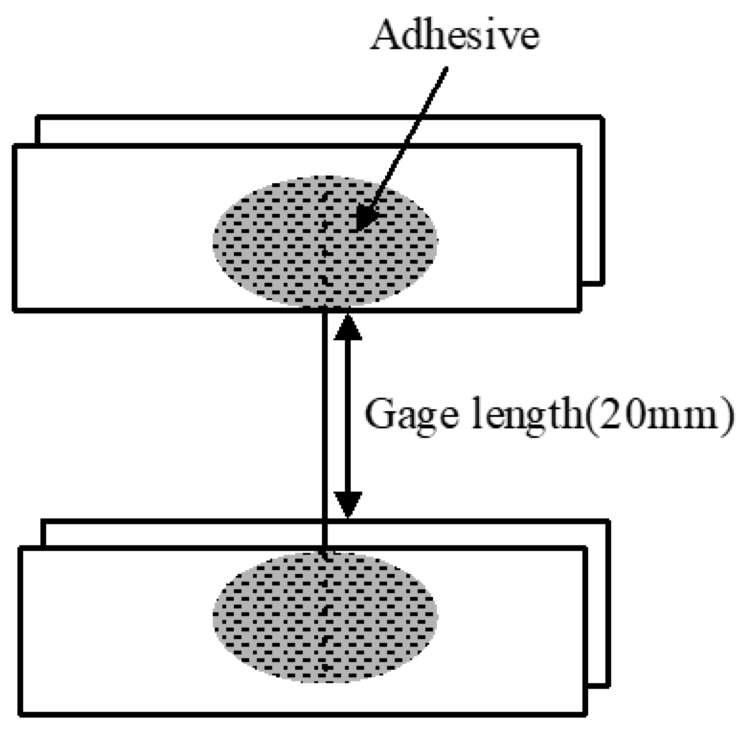
The acid-treated stainless-steel fiber was subjected to tensile testing while using a universal material tester (Instron 4467, Instron, Norwood, MA, USA). The specimens were tested at a crosshead speed of 5 mm/min, with a grip distance of 20 mm by attaching the fibers to the polypropylene (PP) film using an adhesive. Figure 1 shows the schematic of the specimen.
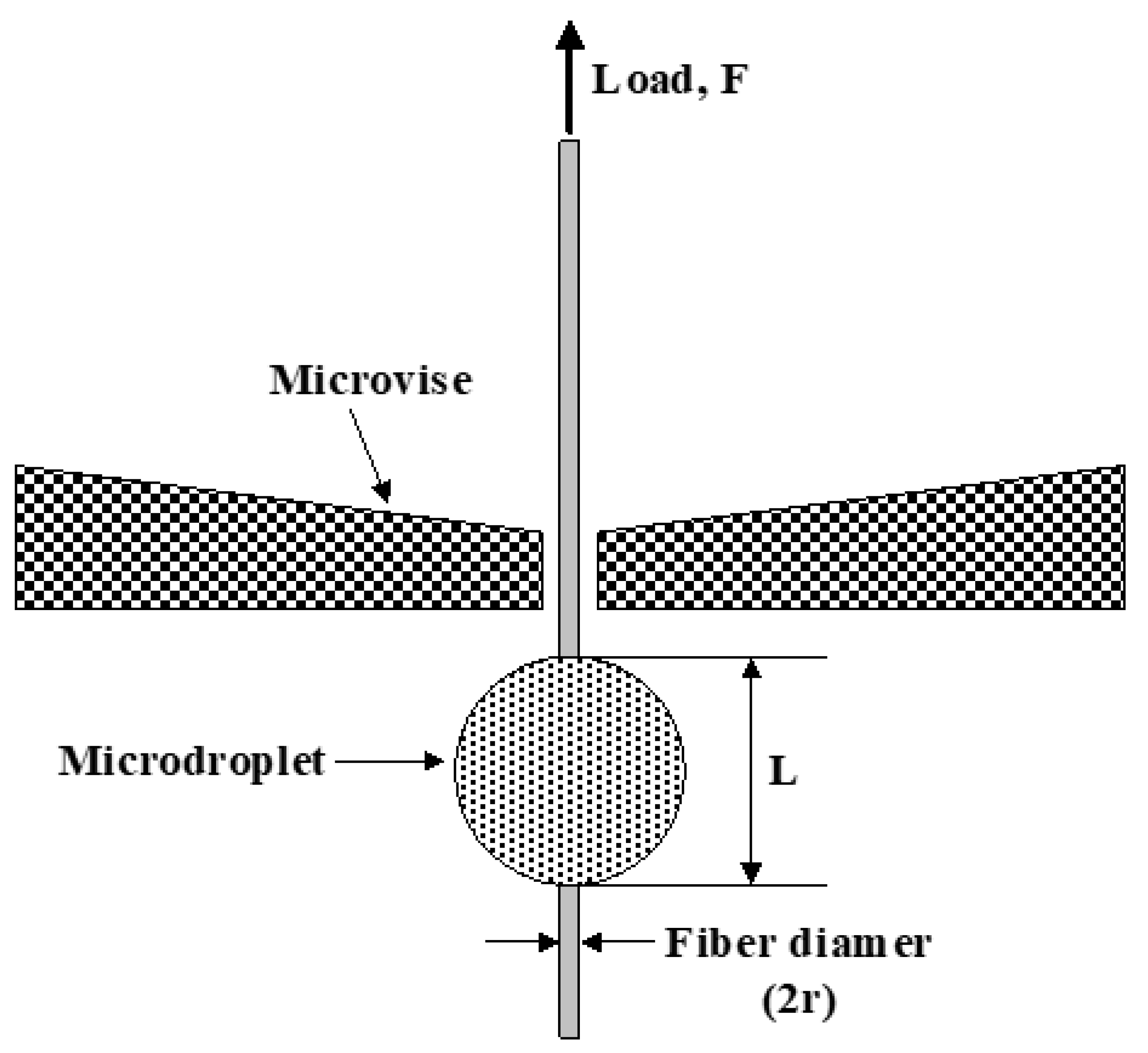
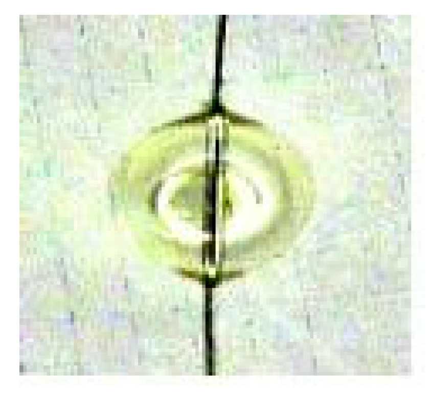
The microdroplet test measured the interfacial shear strength (IFSS) between the epoxy resin and stainless-steel fiber before and after the acid treatment. The epoxy resin was cured in an oven for 2 h at 120 °C. Subsequently, an epoxy resin droplet was attached to the center of the fiber and the microdroplet specimen was fixed by the micro vise (Figure 2). The specimen was pulled in the axial direction of the fiber and the microbond method was used in order to measure the interfacial shear stress between the fiber and the resin matrix. The adhesive strength of the stainless-steel fiber before and after the acid treatment was measured using a universal material tester (Instron 4467, Instron, USA). The specimens were tested at a crosshead speed of 5 mm/min. with a grip distance of 20 mm. The diameter of the fiber and the length of the embedded fiber in the matrix were measured by video microscope system and the interfacial shear strength was calculated while using the following formula:
where τc: Interfacial shear strength, F: Force, L: Embedded length, and 2r: Fiber diameter.
The critical fiber length is the minimum length per given fiber diameter that is essential for a high tensile fracture stress. When the length of the fiber is below the critical fiber length, the maximum fiber stress may never reach the ultimate fiber strength loss. This critical length was calculated from the interfacial shear strength while using the following formula:
where Lc: Critical length, τc: Interfacial shear strength, d: Fiber diameter; and, Fiber stress.
3. Results and Discussion
3.1. Surface Morphology
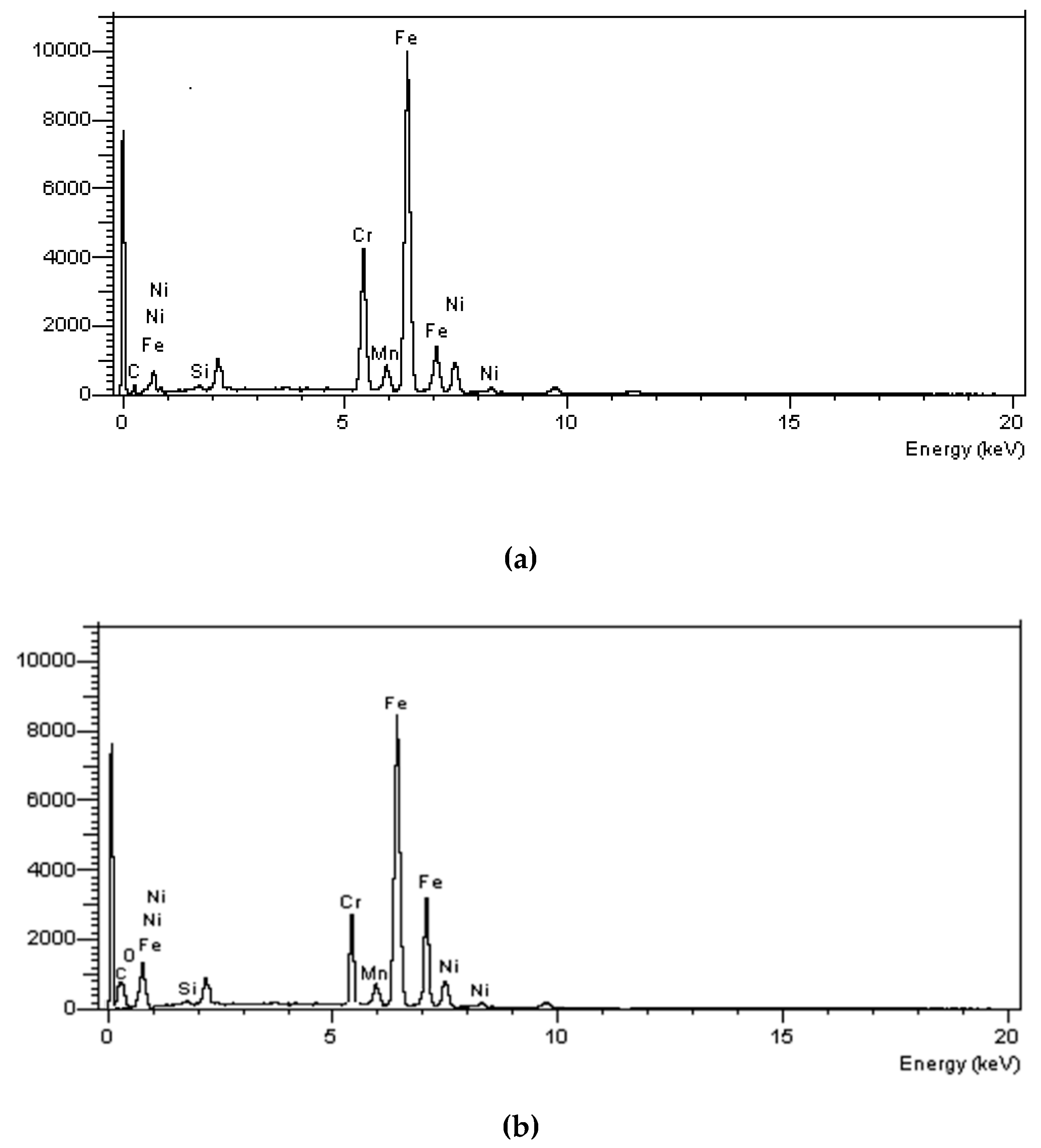
The elemental composition (atomic weight %) at the surface of the stainless-steel fiber was determined by SEM-EDS analysis. Figure 3a showed that the fiber mainly comprised iron along with Cr, Ni, Mn, and Si. Figure 3b shows that the Cr and Ni contents in the surface decreased after the acid treatment, and the C, O, and F contents increased.
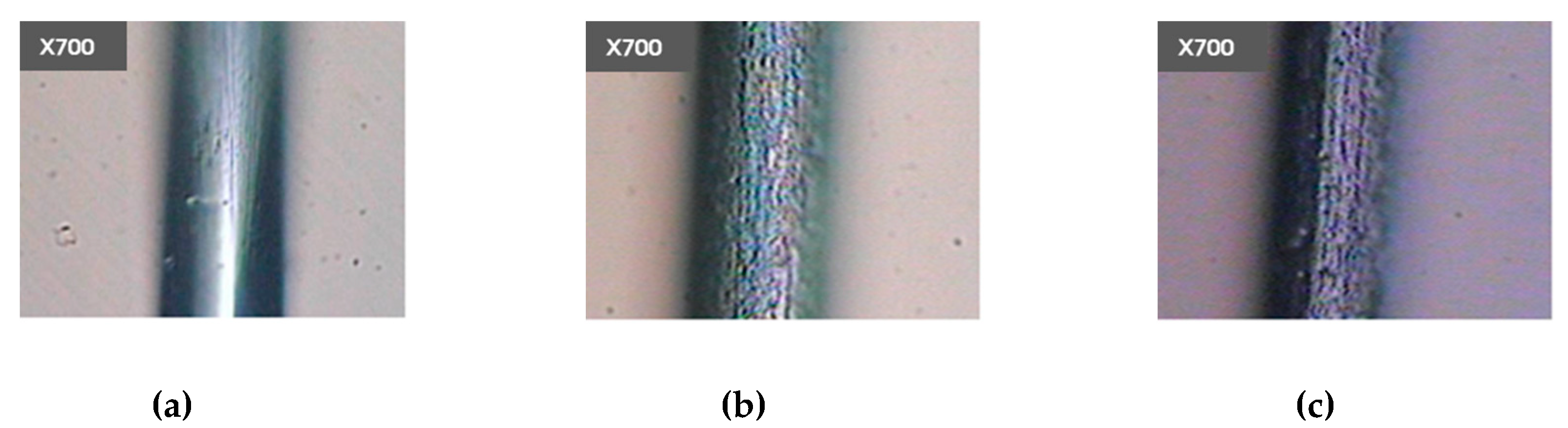
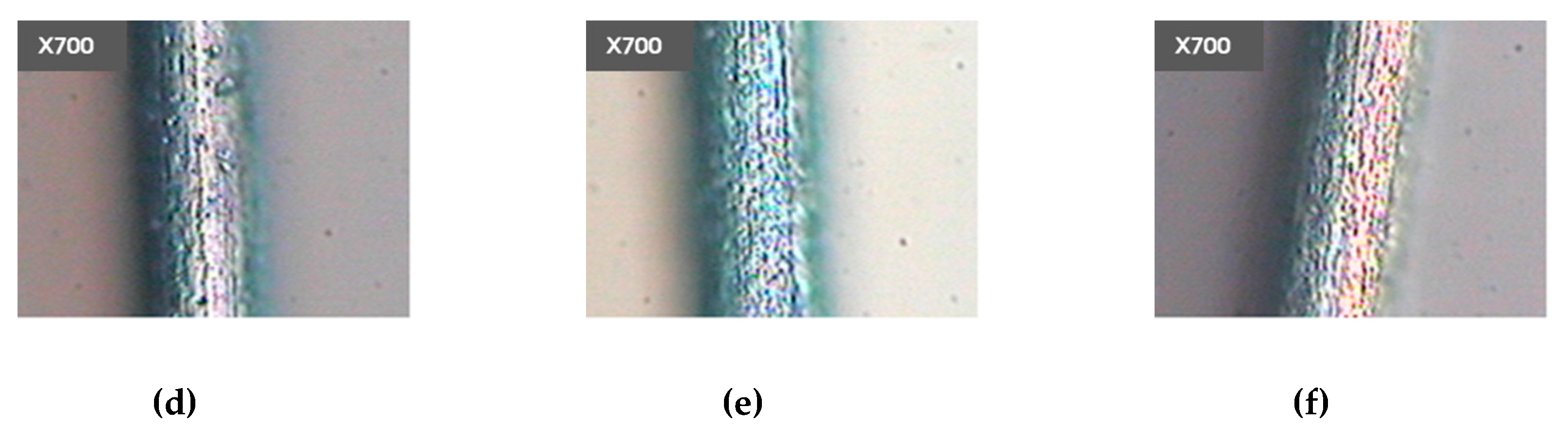
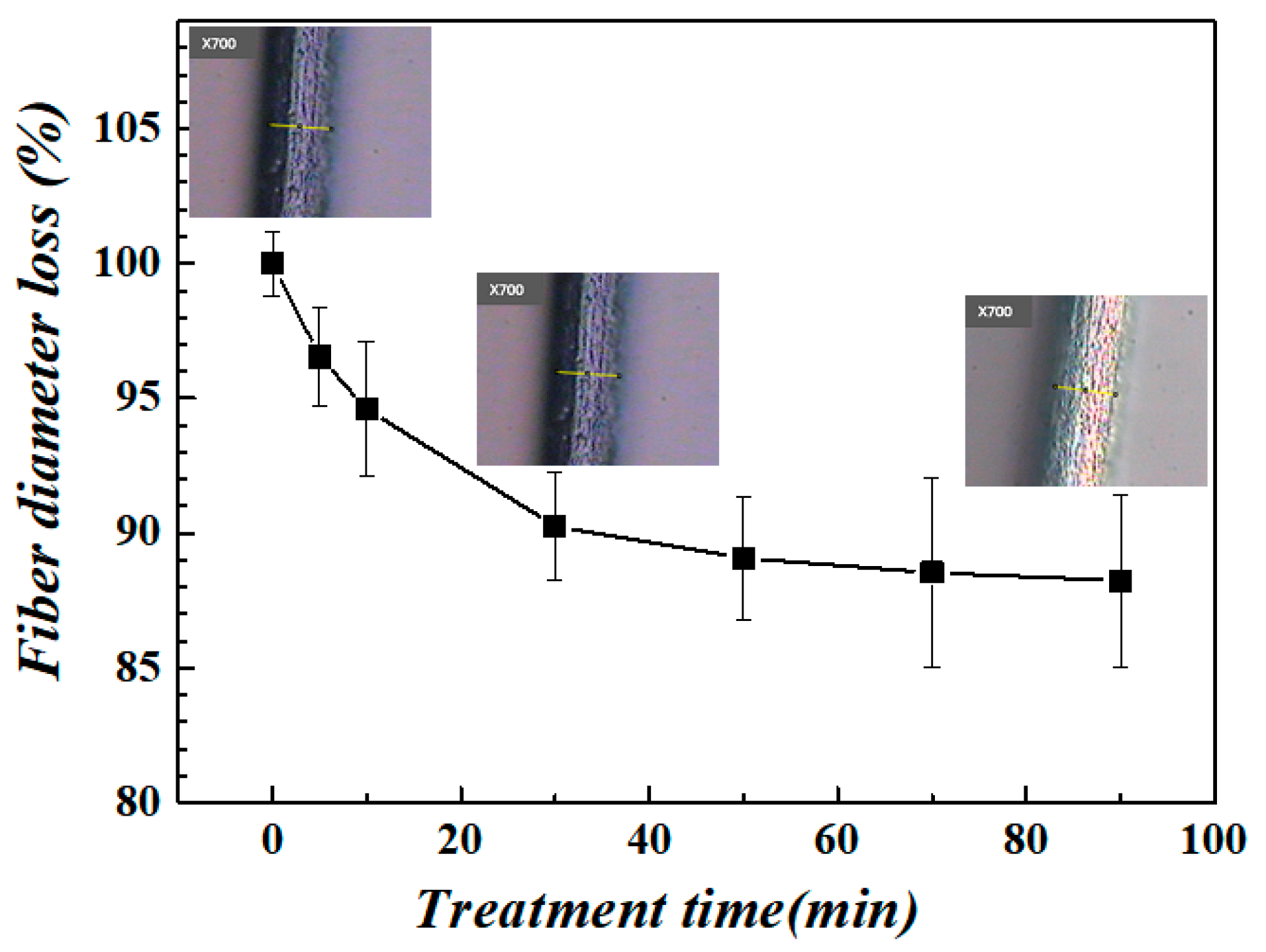
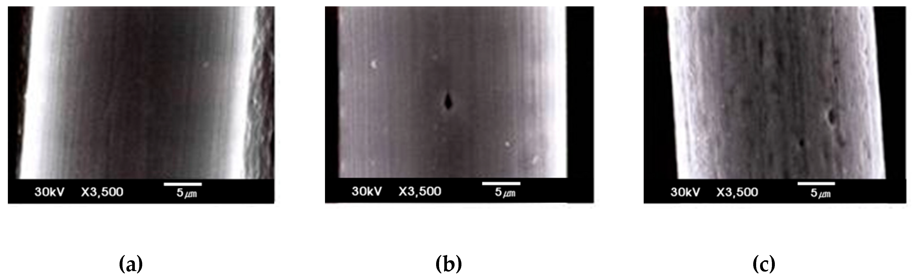
The surface of the fiber was observed before and after the acid treatment with video microscope system (Figure 4). The smooth surface of the untreated stainless-steel fiber showed a few cracks along the fiber axis. However, the rough surface of the acid-treated stainless-steel fiber showed etch pits and microcracks. The number and size of the etch pits and microcracks increased with the increase in the treatment time. However, there was no significant change in the etching effect after 70 min. of the acid treatment. The surface coatings were not peeled off; furthermore, there was an absence of large etch pits or cracks that could decrease the strength of the fiber. Figure 5 shows the fiber diameter remained around 88%, even during 90 min of treatment time. These results were observed in more detail in the scanning electron micrograph. Figure 6 shows the surface morphology of the stainless-steel fiber before and after the acid treatment. The number and size of the etch pits and microcracks at the surface of the stainless-steel fiber were low at low treatment times. There was a significant increase in the number and size of the etch pits and microcracks at the surface of the stainless-steel fibers with the increase in the treatment time.
3.2. Mechanical Properties of a Single Fiber
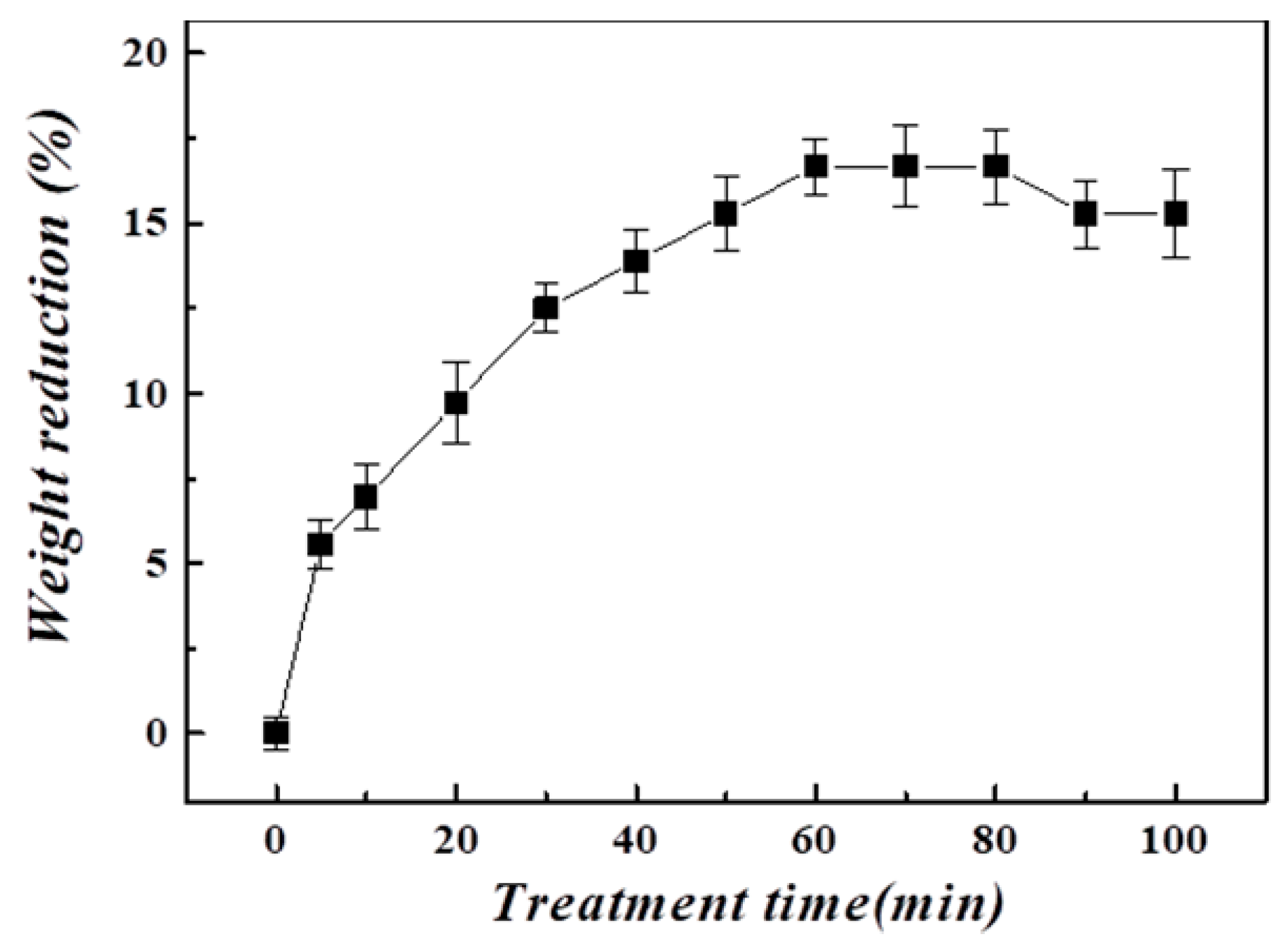
The rate of weight loss of the acid-treated stainless-steel fiber increased with the increase in the treatment time (Figure 7). The images that were observed using an electron injection microscope and video microscope system showed that the number and size of the etch pits and microcracks increased with the increase in the treatment time. The etching effect of the acid treatment resulted in the decrease in the weight of the fiber. There was a continuous increase in the rate of weight loss of the acid-treated fiber for 60 min. of the treatment; subsequently, there was a slight increase in the rate of weight loss. There was a slight decrease in the rate of weight loss after 80 min. of the treatment. The acid treatment corroded the surface of the stainless-steel fiber. This hindered the penetration of the acid to the center of the fiber in order to eliminate the corrosive residues that persisted between the microcracks. Therefore, a significant decrease in the rate of weight loss was not observed.
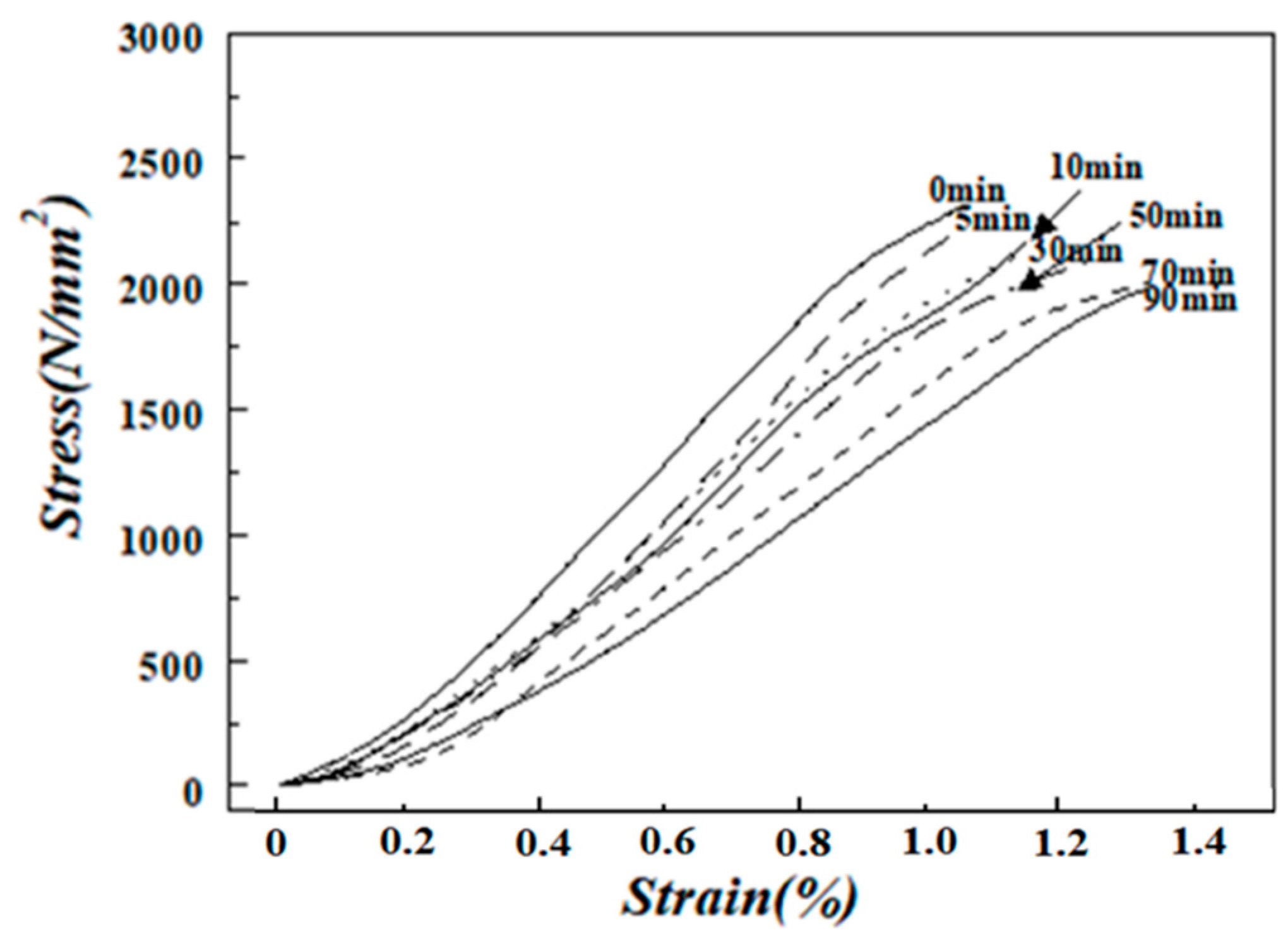
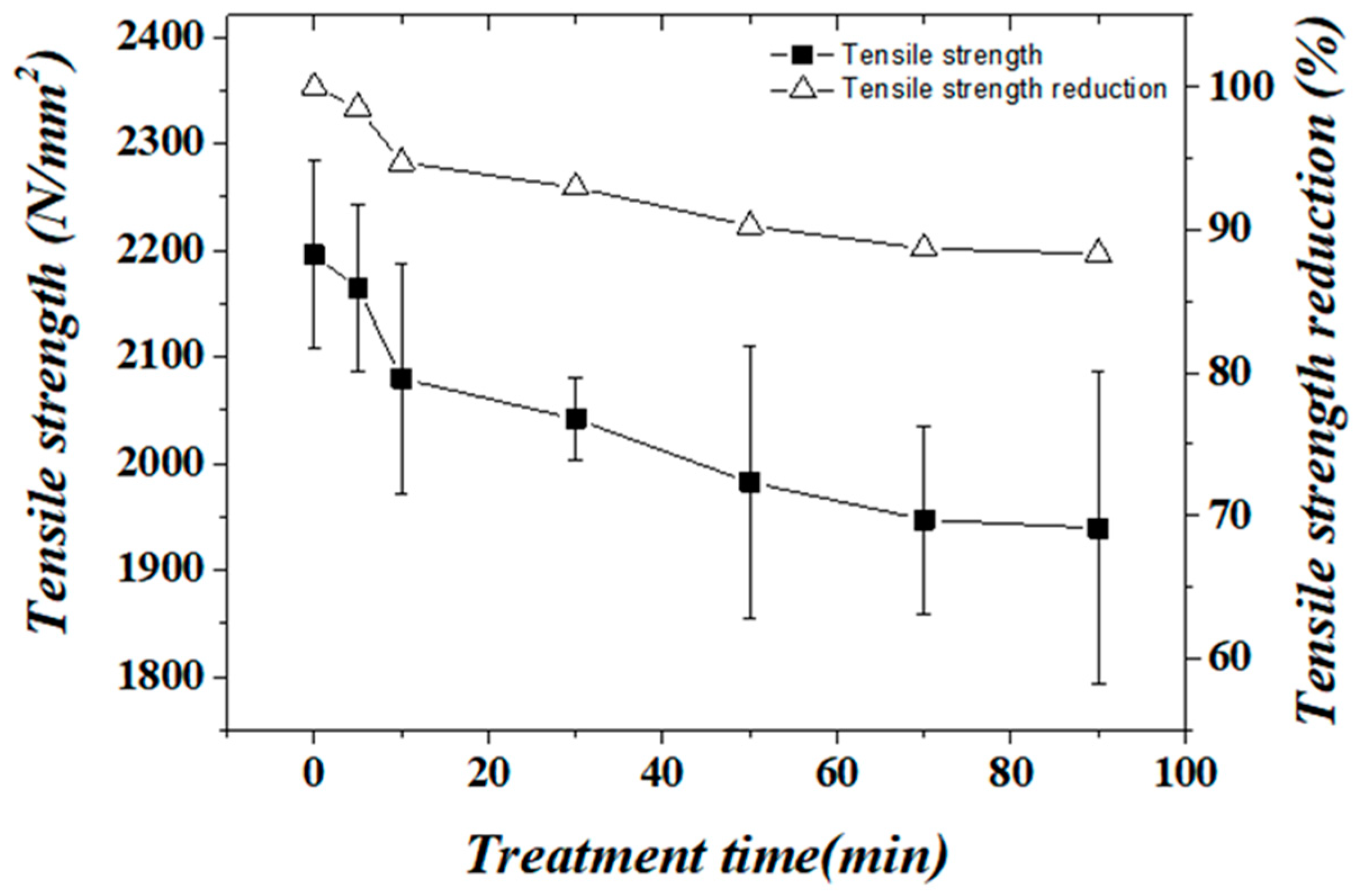
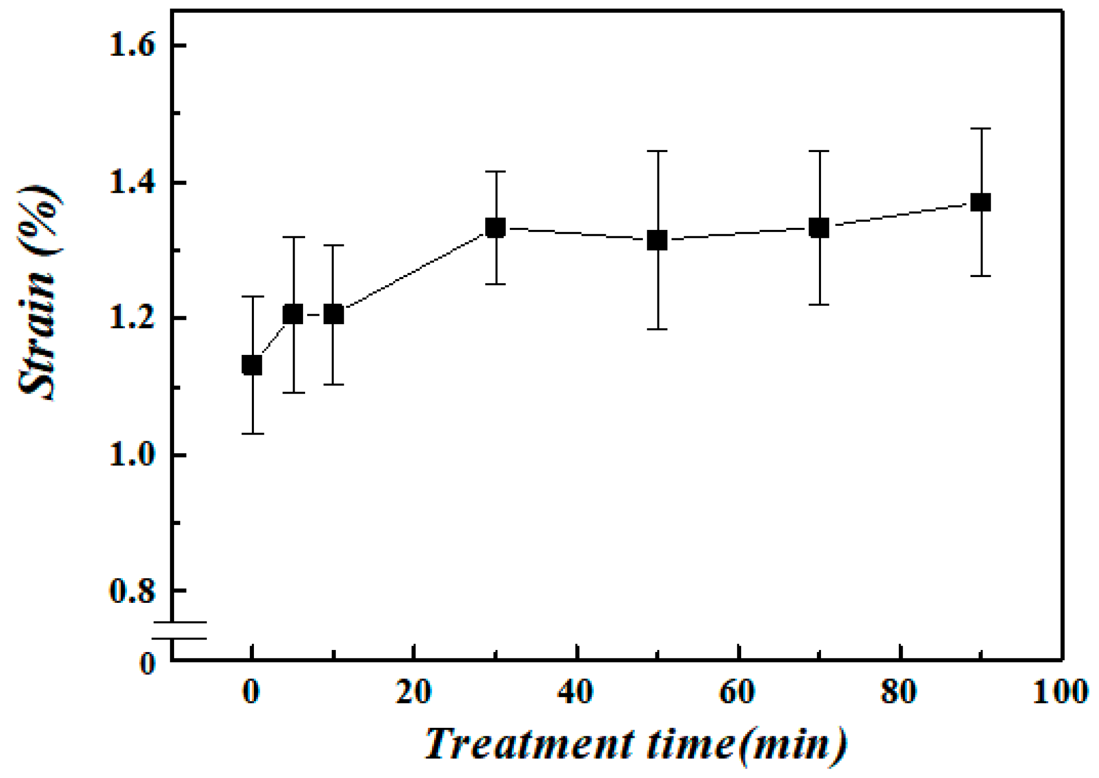
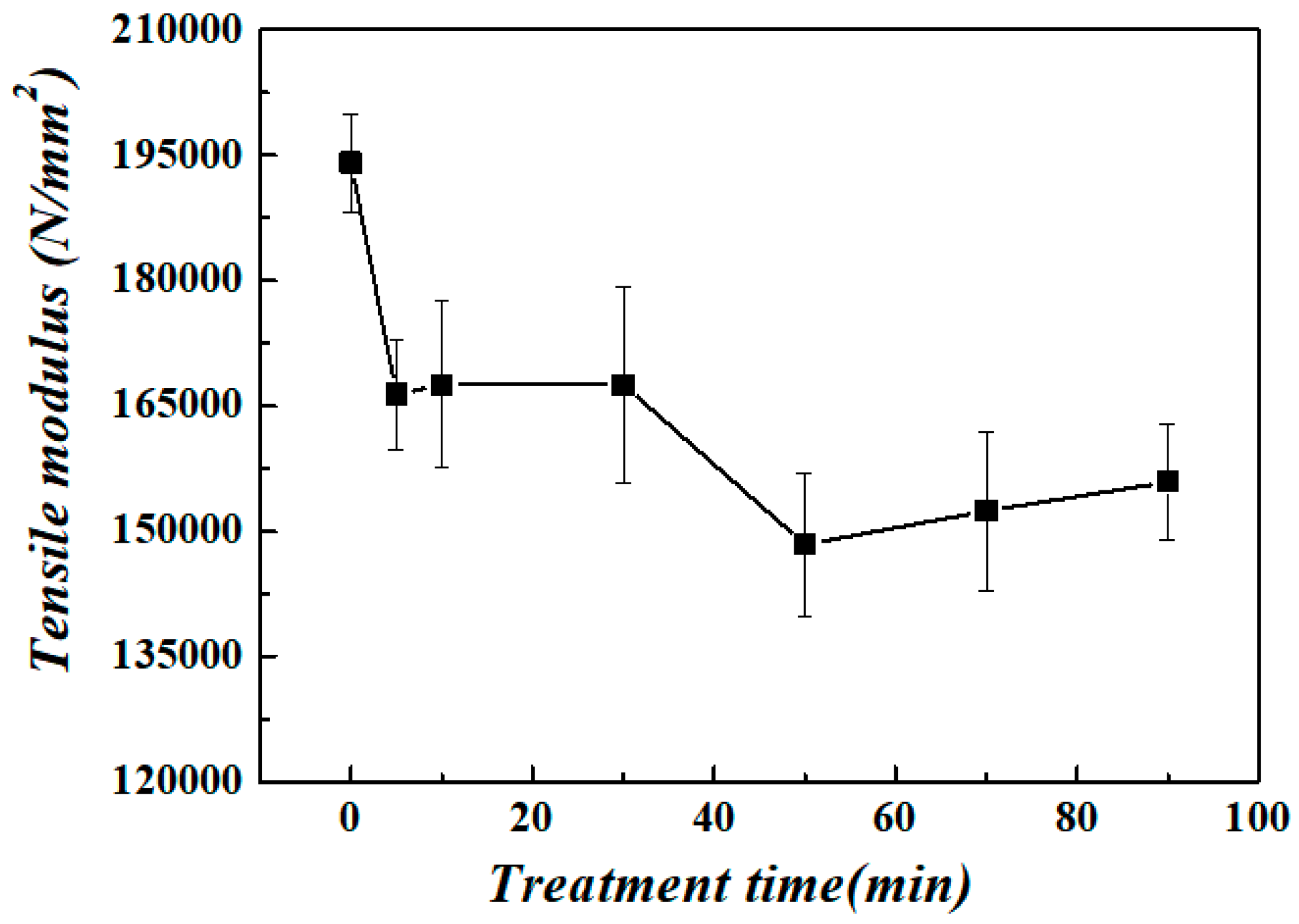
Figure 8, Figure 9, Figure 10 and Figure 11 show the variation in the tensile properties of the stainless-steel fiber with the acid-treatment time. The acid treatment primarily affected the surface of the stainless-steel fiber. It did not have a significant effect on the bulk properties of the fiber. Therefore, there was no significant change in the slope of the stress-strain curve of the fiber due to the acid treatment; however, there was a gradual decrease in the slope of the curve with the increase in the treatment time (Figure 8). Figure 9 shows the gradual decrease in the tensile strength of the fiber with the increase in the acid-treatment time. The rate of weight loss of the fiber remained approximately constant after 70 min. of the acid-treatment; however, the etching effect decreased the tensile strength of the fiber, but it maintained the strength of about 88%, even over 70 min of treatment time. Initially, there was no gradual increase in the elongation of the fiber with the increase in the treatment time (Figure 10). However, the elongation of the fiber increased significantly at a treatment time of 30 min. The variation of the tensile modulus of the stainless-steel fiber with the acid-treatment time was similar to that of the elongation of the fiber with the acid-treatment time. Figure 11 showed a two-step decrease in the tensile modulus of the stainless-steel fiber. The tensile modulus decreased by approximately 11% and 22% after 5 min. and 30 min., respectively, of the acid treatment. The etching effect of the acid treatment induced the formation of microcracks at the surface of the stainless-steel fiber. Thus, a part of the fiber surface was peeled off. This resulted in the slight decrease in the tensile strength, stiffness, and elastic rate of the fibers due to the acid treatment. The skin of the stainless-steel fibers was etched, because its elasticity was higher than that of the core during the manufacturing of the fibers.
3.3. Interfacial Properties of a Single Fiber
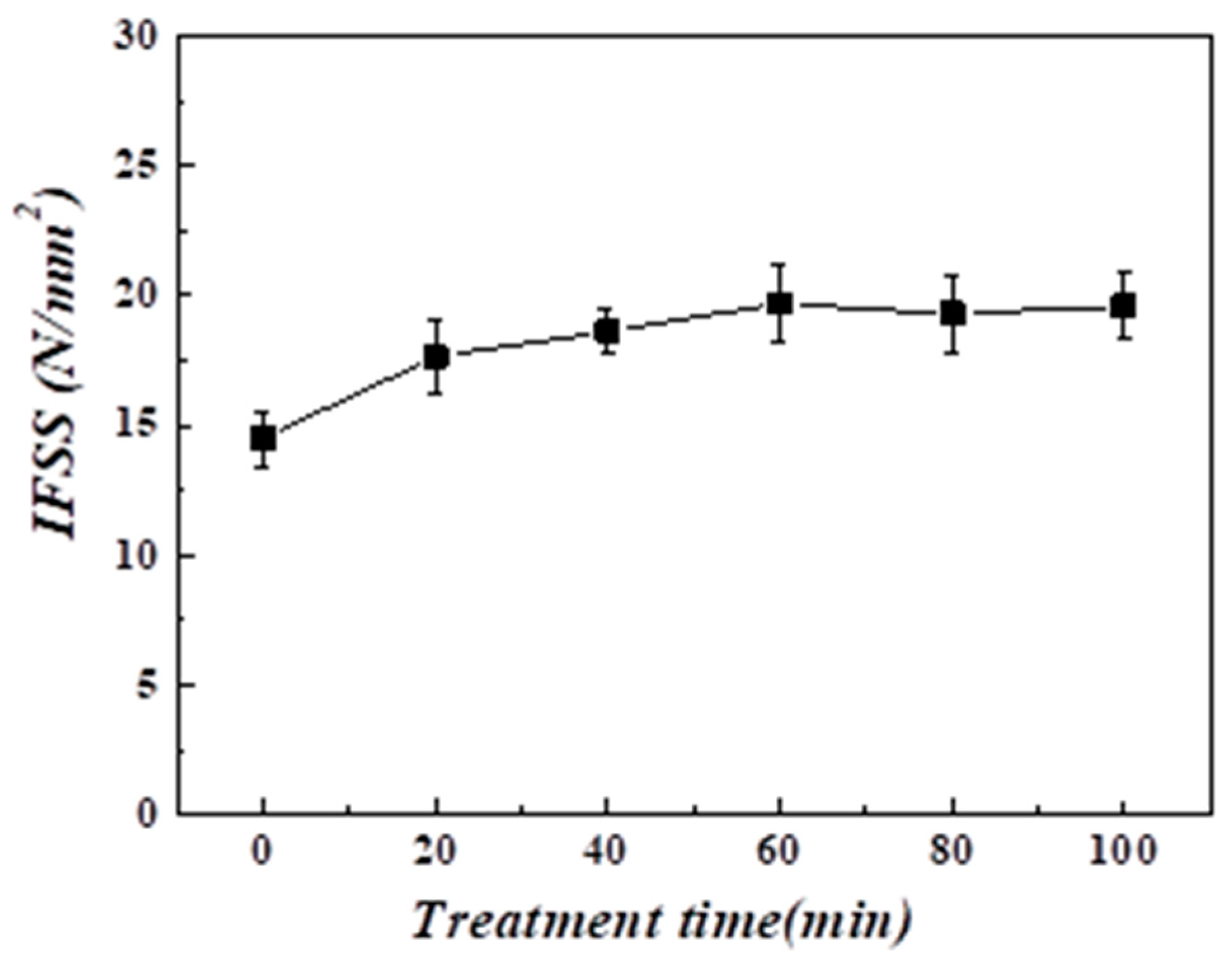
The specimen should be prepared in such a way that the microdroplet is at the center of the fiber in order to measure the interfacial shear strength of the acid-treated stainless-steel fibers (Figure 12). The results of the experiment (Figure 13) showed that the interfacial shear strength between the untreated stainless-steel fiber and epoxy resin was the lowest (14.5 N/mm2). This was attributed to the smooth surface of the untreated fiber. The interfacial shear strength between the acid-treated stainless-steel fiber and the epoxy resin increased by at least 20% as compared to that between the untreated stainless-steel fiber and epoxy resin. Furthermore, the interfacial adhesion increased with the increase in treatment time. The surface adhesion strength increased with the increase in the treatment time and reached a maximum of 19.7 N/mm2 after 60 min. of the treatment. The surface of the acid-treated stainless-steel fiber was rougher than that of the untreated stainless-steel fiber. Therefore, the surface adhesion of the fiber increased with the increase in the surface area per unit volume of the fiber.
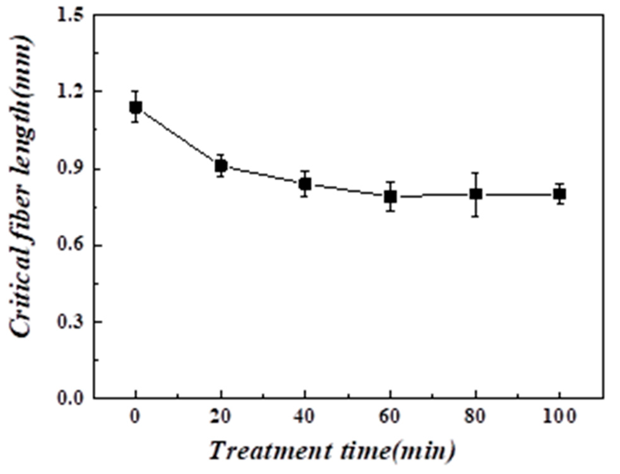
Figure 14 shows the effect of the acid-treatment time on the critical fiber length between the stainless-steel fiber and the epoxy resin matrix. There was an overall decrease in the critical fiber length of the acid-treated stainless-steel fiber from 1.1 mm to 0.88 mm with the increase in the treatment time. Neither the interfacial shear strength nor the critical fiber length underwent significant changes after 40 min. or more of the acid treatment. This confirmed the inverse relationship between the interfacial shear strength of the acid-treated stainless-steel fiber and the critical fiber length.
4. Conclusions
Fine etchings and microcracks were observed at the surface of the 0.2 M hydrochloric acid-treated stainless-steel fiber. The acid treatment increased the specific surface area of the stainless-steel fiber; furthermore, it increased the contact area of the fiber with the epoxy resin. It was confirmed that the mechanical properties did not significantly decrease, because the tensile strength decreased by only about 7% compared to the weight loss of about 13%. When the fiber diameter was maintained at about 88% after 90 min. of acid treatment, the tensile strength was also maintained at approximately 88%, thus confirming the direct correlation between the fiber diameter and tensile strength. Therefore, there was no significant deterioration in the mechanical properties of the fiber. The interfacial shear strength between the stainless-steel fiber and epoxy resin increased from 14.5 N/mm2 to 19.7 N/mm2 with the increase in the acid-treatment time. This was accompanied by a corresponding increase in the surface area of the fiber. There was also a slight decrease in the critical fiber length from 1.1 mm to 0.88 mm with the increase in the acid-treatment time. This was also accompanied by a corresponding increase in the surface area of the fiber. The formation of microcracks at the surface of the acid-treated stainless-steel fiber increased the specific surface area of contact between the fiber with the epoxy resins. This resulted in the increase in adhesion strength between the stainless-steel fiber and epoxy resin without any significant deterioration in the mechanical properties of the fibers.
Author Contributions
Writing—original draft, M.K.; Data curation, M.K.; Formal analysis, M.K. and S.G.L.; Writing—review & editing, M.K. and S.G.L. All authors have read and agreed to the published version of the manuscript.
Funding
This research was funded by the Chungnam National University Academic Research Support Program, grant no. 2019-0654-01.
Acknowledgments
This research was supported by the Chungnam National University Academic Research Support Program, grant no. 2019-0654-01.
Conflicts of Interest
The authors declare no conflict of interest.
References
- Miyahara, Y.; Kako, K. Initiation behavior of stress corrosion cracking for type 316L stainless steel with controlled distribution of surface work hardened layer in high temperature water. Denryoku Chu-ou Kenkyu-jo Houkoku 2010, 42, 1–4. [Google Scholar]
- Chen, L.J.; Chen, M.; Zhou, H.-D.; Chen, J.M. Preparation of super-hydrophobic surface on stainless steel. Appl. Surf. Sci. 2008, 255, 3459–3462. [Google Scholar] [CrossRef]
- Niemi, R.; Mahiout, A.; Siivinen, J.; Mahlberg, R.; Likonen, J.; Nikkola, J.; Mannila, J.; Vuorio, T.; Johansson, L.-S.; Söderberg, O.; et al. Surface pretreatment of austenitic stainless steel and copper by chemical, plasma electrolytic or CO2 cryoblasting techniques for sol–gel coating. Surf. Coat. Technol. 2010, 204, 2424–2431. [Google Scholar] [CrossRef]
- Johnson, A.C.; Hayes, S.A.; Jones, F. The role of matrix cracks and fibre/matrix debonding on the stress transfer between fibre and matrix in a single fibre fragmentation test. Compos. Part A Appl. Sci. Manuf. 2012, 43, 65–72. [Google Scholar] [CrossRef] [Green Version]
- Kim, B.W.; Nairn, J.A. Observations of Fiber Fracture and Interfacial Debonding Phenomena Using the Fragmentation Test in Single Fiber Composites. J. Compos. Mater. 2002, 36, 1825–1858. [Google Scholar] [CrossRef]
- McCarthy, E.D.; Soutis, C. Determination of interfacial shear strength in continuous fibre composites by multi-fibre fragmentation: A review. Compos. Part A Appl. Sci. Manuf. 2019, 118, 281–292. [Google Scholar] [CrossRef] [Green Version]
- Sørensen, B.F.; Lilholt, H. Fiber pull-out test and single fiber fragmentation test-analysis and modelling. IOP Conf. Series Mater. Sci. Eng. 2016, 139, 012009. [Google Scholar] [CrossRef] [Green Version]
- Seghini, M.C.; Touchard, F.; Sarasini, F.; Chocinski-Arnault, L.; Mellier, D.; Tirillò, J. Interfacial adhesion assessment in flax/epoxy and in flax/vinylester composites by single yarn fragmentation test: Correlation with micro-CT analysis. Compos. Part A Appl. Sci. Manuf. 2018, 113, 66–75. [Google Scholar] [CrossRef]
- Zarges, J.; Kaufhold, C.; Feldmann, M.; Heim, H.-P. Single fiber pull-out test of regenerated cellulose fibers in polypropylene: An energetic evaluation. Compos. Part A Appl. Sci. Manuf. 2018, 105, 19–27. [Google Scholar] [CrossRef]
- Kelly, A.; Tyson, W. Tensile properties of fibre-reinforced metals: Copper/tungsten and copper/molybdenum. J. Mech. Phys. Solids 1965, 13, 329–350. [Google Scholar] [CrossRef]
- Wang, D.; Bai, T.; Cheng, W.; Xu, C.; Wang, G.; Cheng, H.; Han, G. Surface Modification of Bamboo Fibers to Enhance the Interfacial Adhesion of Epoxy Resin-Based Composites Prepared by Resin Transfer Molding. Polymers 2019, 11, 2107. [Google Scholar] [CrossRef] [PubMed] [Green Version]
- Hernandez, D.A.; Soufen, C.A.; Orlandi, M.O. Carbon Fiber Reinforced Polymer and Epoxy Adhesive Tensile Test Failure Analysis Using Scanning Electron Microscopy. Mater. Res. 2017, 20, 951–961. [Google Scholar] [CrossRef] [Green Version]
- Herrera-Franco, P.J.; Drzal, L. Comparison of methods for the measurement of fibre/matrix adhesion in composites. Composites 1992, 23, 2–27. [Google Scholar] [CrossRef]
- Graupner, N.; Rößler, J.; Ziegmann, G.; Müssig, J. Fibre/matrix adhesion of cellulose fibres in PLA, PP and MAPP: A critical review of pull-out test, microbond test and single fibre fragmentation test results. Compos. Part A Appl. Sci. Manuf. 2014, 63, 133–148. [Google Scholar] [CrossRef]
- Awal, A.; Cescutti, G.; Ghosh, S.B.; Müssig, J. Interfacial studies of natural fibre/polypropylene composites using single fibre fragmentation test (SFFT). Compos. Part A Appl. Sci. Manuf. 2011, 42, 50–56. [Google Scholar] [CrossRef]
- Dilsiz, N.; Wightman, J. Effect of acid–base properties of unsized and sized carbon fibers on fiber/epoxy matrix adhesion. Colloids Surf. A Physicochem. Eng. Asp. 2000, 164, 325–336. [Google Scholar] [CrossRef]
- Craven, J.; Cripps, R.; Viney, C. Evaluating the silk/epoxy interface by means of the Microbond Test. Compos. Part A Appl. Sci. Manuf. 2000, 31, 653–660. [Google Scholar] [CrossRef]
- Rashkovan, I.; Korabel’Nikov, Y. The effect of fiber surface treatment on its strength and adhesion to the matrix. Compos. Sci. Technol. 1997, 57, 1017–1022. [Google Scholar] [CrossRef]
Figure 1.
Schematic of the specimen for the tensile test.

Figure 2.
Microdroplet test.

Figure 3.
Scanning electron microscopy-energy dispersive X-ray spectrometry (SEM-EDS) analysis of the stainless-steel fiber (a) without the acid treatment and (b) after acid treatemnt.
Figure 3.
Scanning electron microscopy-energy dispersive X-ray spectrometry (SEM-EDS) analysis of the stainless-steel fiber (a) without the acid treatment and (b) after acid treatemnt.

Figure 4.
Photographs (700 X) of the stainless-steel fiber (a) without the acid treatment and after (b) 10 min, (c) 30 min, (d) 50 min, (e) 70 min and (f) 90 min of the acid treatment.
Figure 4.
Photographs (700 X) of the stainless-steel fiber (a) without the acid treatment and after (b) 10 min, (c) 30 min, (d) 50 min, (e) 70 min and (f) 90 min of the acid treatment.


Figure 5.
The effect of the acid-treatment time on the rate of fiber diameter loss of the stainless-steel fiber.
Figure 5.
The effect of the acid-treatment time on the rate of fiber diameter loss of the stainless-steel fiber.

Figure 6.
SEM micrographs (3500 X) of the stainless-steel fibers (a) without the acid treatment and after (b) 20 min and (c) 60 min of the acid treatment.
Figure 6.
SEM micrographs (3500 X) of the stainless-steel fibers (a) without the acid treatment and after (b) 20 min and (c) 60 min of the acid treatment.

Figure 7.
Effect of the acid-treatment time on the rate of weight loss of the stainless-steel fiber.
Figure 7.
Effect of the acid-treatment time on the rate of weight loss of the stainless-steel fiber.

Figure 8.
Effect of the acid-treatment time on the stress–strain curves of the stainless-steel fiber.
Figure 8.
Effect of the acid-treatment time on the stress–strain curves of the stainless-steel fiber.

Figure 9.
Effect of the acid treatment time on the tensile strength of the stainless-steel fiber.

Figure 10.
Effect of the acid-treatment time on the breaking strain (%) of the stainless-steel fiber.
Figure 10.
Effect of the acid-treatment time on the breaking strain (%) of the stainless-steel fiber.

Figure 11.
Effect of the acid treatment time on the tensile modulus of stainless-steel fiber.

Figure 12.
Specimen of the stainless-steel fiber with the epoxy resin microdroplet.

Figure 13.
Effect of the acid-treatment time on the interfacial shear strength of the stainless-steel fiber.
Figure 13.
Effect of the acid-treatment time on the interfacial shear strength of the stainless-steel fiber.

Figure 14.
Effect of the acid treatment time on the critical fiber length of the stainless-steel fibers.
Figure 14.
Effect of the acid treatment time on the critical fiber length of the stainless-steel fibers.


Table 1.
Chemical composition of SUS304.
| C | Si | Mn | P | S | Ni | Cr | |
|---|---|---|---|---|---|---|---|
| SUS 304 | ≤0.08 % | ≤1.00% | ≤2.00% | ≤0.04% | ≤0.03% | 8–10% | 18–20% |
Table 2.
The physical characteristics of the epoxy resin.
| Equivalent Weight | Viscosity (cps at 25 °C) | Specific Gravity (20 °C) | |
|---|---|---|---|
| Epoxy resin | 180–190 g/eq | ~700–1100 | 1.14 |
Publisher’s Note: MDPI stays neutral with regard to jurisdictional claims in published maps and institutional affiliations. |
© 2020 by the authors. Licensee MDPI, Basel, Switzerland. This article is an open access article distributed under the terms and conditions of the Creative Commons Attribution (CC BY) license (http://creativecommons.org/licenses/by/4.0/).
Share and Cite
MDPI and ACS Style
Kwon, M.; Lee, S.G. Analysis of the Interfacial Adhesion between a Stainless-Steel Fiber and an Epoxy Resin by the Single Fiber Microdroplet Test. Surfaces 2020, 3, 594-604. https://0-doi-org.brum.beds.ac.uk/10.3390/surfaces3040040
AMA Style
Kwon M, Lee SG. Analysis of the Interfacial Adhesion between a Stainless-Steel Fiber and an Epoxy Resin by the Single Fiber Microdroplet Test. Surfaces. 2020; 3(4):594-604. https://0-doi-org.brum.beds.ac.uk/10.3390/surfaces3040040
Chicago/Turabian StyleKwon, MiYeon, and Seung Goo Lee. 2020. "Analysis of the Interfacial Adhesion between a Stainless-Steel Fiber and an Epoxy Resin by the Single Fiber Microdroplet Test" Surfaces 3, no. 4: 594-604. https://0-doi-org.brum.beds.ac.uk/10.3390/surfaces3040040