Zein/Bioactive Glass Coatings with Controlled Degradation of Magnesium under Physiological Conditions: Designed for Orthopedic Implants


Department of Materials Science and Engineering, Institute of Space Technology Islamabad, 1, Islamabad Highway, Islamabad 44000, Pakistan
Prosthesis 2020, 2(3), 211-224; https://0-doi-org.brum.beds.ac.uk/10.3390/prosthesis2030018
Submission received: 4 August 2020
/
Revised: 12 August 2020
/
Accepted: 13 August 2020
/
Published: 19 August 2020
Abstract
:Magnesium and its alloys are widely considered as temporary bio-implants owing to their mechanical properties and biocompatibility. However, the high corrosion rates and degradation in the physiological environment restrict the practical application of Mg as a biomedical device. Therefore, in this study, Zein/45S5 bioactive glass (BG) coatings were deposited via electrophoretic deposition (EPD) on pretreated pure magnesium (Mg) substrates, which controls the rapid degradation of magnesium. The set of EPD parameters was first optimized on stainless steel (SS) and then the optimum EPD parameters were applied to obtain zein/BG composite coatings on Mg substrates. The morphology of the obtained coatings was studied by scanning electron microscopy (SEM). SEM results showed that both zein and BG were successfully deposited on the surface of the Mg substrate. Electrochemical measurements consisting of open circuit potential (OCP), electrochemical impedance spectroscopy (EIS), and potentiodynamic polarization confirmed that the corrosion resistance of Mg improved after the deposition of zein/BG coatings. The in-vitro bioactivity study was carried out by immersing the zein/BG coatings in simulated body fluid for 3, 7, and 21 days. SEM, energy dispersive X-ray spectroscopy (EDX), and Fourier transform infrared spectroscopy results elucidated that the hydroxyapatite layer developed after 21 days of immersion in SBF, which confirmed the bone binding ability of the coatings.
1. Introduction
According to the Global Opportunity Analysis and Industry Forecast (2017–2023), the value of the global bio implant market is expected to reach $124,154 million by the year 2023. The major driving factors are the prevalence of rapid aging and chronic diseases in humans [1]. This increase in the market has consequently increased research in the area. To overcome the shortcomings of conventional metallic bioimplants, a new class of degradable Mg implants is studied nowadays [2]. Mg implants degrade via corrosion in the physiological environment which makes them potential candidates for temporary implants [3]. Furthermore, the elastic modulus of Mg and its alloys is close to that of the human bone—thus minimizing the chances of stress-shielding [4]. The major shortcoming is its fast degradation which can result in premature degradation of the implant [5]. The degradation of Mg can be governed by different mechanisms such as galvanic corrosion, pitting corrosion, and stress corrosion [4]. To overcome this drawback, many biomaterials can be used as coating materials that significantly enhance bioactivity and biocompatibility [6]. A polymer such as zein has attracted great attraction owing to its unique properties. Zein is obtained from the endosperm of maize that is composed of prolamins. It is a biocompatible polymer having applications in tissue engineering and drug delivery. Zein is a biodegradable protein with the ability to produce self-assembled colloidal particles for drug delivery [7]. Another challenge for the Mg-based biomedical devices is their low mechanical strength compared to the other competitive metallic alloys. The mechanical properties of Mg based biomedical devices can be tailored by designing the coatings with appropriate composition for biomedical applications [4].
Biopolymers such as zein can be combined with the various bio-ceramics such as hydroxyapatite, bioactive glasses (BGs), and calcium phosphate types of cement. These bio-ceramics can enhance the bone binding ability of the composites [8,9]. One of the popular bio-ceramics is bioactive glass (BG). BG is capable of forming a strong bond with the natural bone [10]. However, BG has poor mechanical properties under the loading conditions. Moreover, the elastic modulus of BG is higher than that of the cortical bone, which may lead to stress shielding issues [11]. To overcome these problems associated with BG, BG is often incorporated into the biocompatible polymers [12]. Zein is a natural protein and is known for improving the attachment and proliferation of osteoblast cells [13,14]. Furthermore, it was shown that the zein-films resist the formation of biofilm. Since humans are threatened by the formation of biofilms and colonization of bacteria which is harmful for the human body and may lead to serious illness [15]. Therefore, the combination of zein/BG can provide good bone binding ability, resistance to the formation of biofilm in addition to the improved attachment and proliferation of the osteoblast cells [16,17]. During the last few years, intensive research efforts have been made to design organic/inorganic systems with favorable osteogenic properties and enhanced antibacterial activity. Several nanoscale metallic ions have also been added in the designed systems to develop anti-infectious surfaces [18].
Recently, an Mg-Zn-Ca-Mn alloy was heat treated in order to improve mechanical compatibility and biocompatibility. The heat treated samples were subjected to the biphasic ceramic nanoparticles via plasma electrolytic oxidation and a sol-gel coating. It was shown that the coating process lead to the improved corrosion resistance of Mg alloy. It was suggested that the integrity of Mg-based implants can be maintained in the physiological environment even after 6–9 months [19]. Zein/BG coatings were fabricated via electrophoretic deposition (EPD), which is widely considered as a suitable option owing to the simplicity of the process. Moreover, EPD is a room temperature processing technique; therefore, proteins and biomolecules can successfully be coated without altering their structure [19,20]. EPD has successfully been employed for the co-deposition of bio-ceramics and biopolymers [21]. However, the mechanism behind the co-EPD is not well understood due to the complexities involved in this process. Accordingly, this study will focus on the EPD of zein/BG coatings. Another advantage of EPD is its applicability to the wide range of substrates independent of their shapes and materials. EPD has been employed for coating 3D substrates [20].
EPD was used earlier for the deposition of zein/BG coatings on the 316 L SS substrate [16]. However, EPD of zein/BG coatings has not been investigated on the Mg substrates. Mg being suitable implant material is owing to its mechanical compatibility with bone and due to its biodegradable nature in the physiological environment [3]. However, successful EPD of zein/BG on the Mg substrate is a challenging task due to the fast degradation of Mg. To control the degradation rate of Mg, Zhao et al. [22] and Höhlinger et al. [23] employed different pre-treatment reagents. Accordingly, this research work is comprised of the pre-treatment of Mg with the phosphoric acid (H3PO4) and di-ammonium hydrogen phosphate (DAHP), following the previous studies by Ahmed et al. [24].
In this study, we deposited zein/BG coatings on the Mg substrate via EPD process. Zein/BG coatings developed an apatite-like layer after three days of incubation in simulated body fluid (SBF), which indicated the possibility of achieving a close interaction between the implant and the bone. Furthermore, zein/BG coatings control the degradation rate of Mg, as confirmed by the potentiodynamic polarization scan and electrochemical impedance spectroscopy (EIS) results.
2. Materials and Methods
2.1. Materials
Zein powder (CAS number 9010-66-6) and 45S5 Bioactive glass were obtained from Sigma-Aldrich, China. A magnesium rod in the rolled form with a purity of 99.98% and a diameter of 10 mm was purchased from chemPUR, China (CAS number 7439-95-4) and was used as the substrate.
2.2. Electrophoretic Deposition
Zein solution was prepared by the magnetic stirring of 6 wt.% zein, 20 wt.% distilled water and 74 wt.% ethanol on a hot plate with gentle heating. The pH was adjusted using acetic acid. An amount of 10 mL of acetic acid was used for 100 mL of zein solution. To ensure homogeneity, the solution was magnetically stirred for 30 min. at ambient temperature. Subsequently, 5 g of 45S5 bioactive glass (BG) was added and the suspension was placed in an ultrasonication bath for 90 min. The optimum concentration of zein and BG was chosen based on previous studies, which has shown appropriate results for orthopedic applications [9,17].
Magnesium samples were cut into 10 mm diameter disks of 2–3 mm thickness. The edges were smoothed with 600–800 grit size SiC abrasive papers and a flat side of the samples was grinded with 800 grit size abrasive paper. A grinding solution of ethanol and glycerol (3:1) was repeatedly applied to the sandpaper to prevent overheating. The following step involved polishing each sample three times for 2 min with cotton cloths using three different diamond suspensions with a particle size of 6 μm, 3 μm, and 1 μm. The lubricant used was a mixture of ethanol and neutral soap. Finally, the samples were placed in an ultrasonic bath for 5 min, in ethanol, to remove the polishing residue. Later samples were rinsed with fresh ethanol and dried under hot air.
2.3. Chemical Treatment
The chemical treatment was performed according to Zhao et al. [18] with slight modifications. Firstly, a solution of 200 mL of ultrapure distilled water and 2 drops of phosphoric acid (H3PO4) with a mass fraction of ≤85 wt.% Carl Roth® (Karlsruhe, Germany) (CAS number 7664-38-2) was prepared by magnetic mixing (pH ≈ 2.5). Subsequently, the solution is heated to 40 °C with constant magnetic stirring and samples were placed in this suspension for 30 min later dried under cold air. Next, a solution of 200 mL of ultrapure distilled water and 1.32 g of diammonium hydrogen phosphate (DAHP) ((NH4) 2HPO4) from Merck® (Kenilworth, NJ, USA) (CAS number 7783-28-0) was prepared (0.05 mol/L DAHP). With constant magnetic stirring, the suspension is heated to 80 °C and samples were immersed for 60 min. In the end, the samples were dried under hot air.
2.4. EPD Parameters
The EPD of zein/BG coating was first optimized on 316 L SS and then optimized parameters were translated on to the Mg substrate. The optimum EPD parameters for the deposition of zein/BG coatings on the Mg substrate were an applied electric field of 14.5 V/cm, deposition time of 5 min, 5 g/L of BG in the zein suspension, and a pH value of 3. The design experiment approach was utilized to optimize the zein/BG coatings (data not shown here).
2.5. Microstructural Examinations
To investigate the effectiveness of the pre-treatment of magnesium, the samples were examined before and after with a light microscope of the type Eclipse LV150N from Nikon® (Tokyo, Japan). The data were also evaluated by Nikon® with the software NIS Elements.
SEM (AURIGA 4750 from Carl Zeiss AG®, Oberkochen, Germany) was used to study the surface morphology of the coatings. The samples were fixed to the holders with silver paste (ACHESON 1415 from Plano®, Collin, TX, USA) and sputtered with gold by Q150R Rotary-Pumped Sputter Coater (Quorum Technologies®, Lewes, UK) to prevent the effect of charging.
2.6. Electrochemical Measurements
The electrochemical behavior of the samples was investigated using the Zennium IM6eX workstation from Zahner® (Kansas City, MI, USA). The software Thales by Zahner was used for the evaluation of the data. All electrochemical measurements were carried out in 60 to 80 mL of Dulbecco’s Modified Eagle Medium (DMEM) (Merck) at room temperature.
The resting potential (OCP) was measured for 10 min, with the duration of a period set to one second. Obtained values were then plotted into a diagram (x-axis: time [s]; y-axis: OCP [V]). The current range was set between −2 V (cathodic) and 2 V (anodic).
In electrochemical impedance spectroscopy (EIS), the graphic representation takes the form of various plots. The Nyquist plot represents the imaginary impedance (ZImag [Ohm*cm2]) depending on the real impedance (ZReal [Ohm*cm2]). The resulting curve corresponds to the impedance in its frequency dependency [25]. The alternating voltage used in this work had an amplitude of 10 mV in a frequency range from 100 kHz to 10 MHz.
Finally, potentiodynamic polarization tests (I/E) were performed. In this process, the polarization curves were recorded, which indicated electrochemical reactions taking place on the surface of the body (to be examined) [21]. The examination range from −300 mV to 0 V was defined with a scan rate of 1 mV/s. The current range was between −2 A (cathodic) and 2 A (anodic).
2.7. In Vitro Bioactivity Study
To test coatings for the ability to form a hydroxyapatite (HA) layer, they were immersed in a simulated bodily fluid (SBF). The SBF was prepared according to Kokubo and Takadama [26]. Coated samples were immersed in 50 mL of SBF at 37 °C in an orbital shaking incubator (IKA®-Werke GmbH & Co. KG, Staufen, Germany) and kept for 3, 7, and 21 days. Circulating movements, a temperature of 37 °C, and a pH of 7.4 simulate the conditions of the human body in-vitro. Samples were dried at room temperature and examined employing SEM and Fourier-transform infrared spectroscopy (FTIR), Nicolet 6700 device (Thermo Scientific®, Waltham, MA, USA), in the wavelength range of 4000–400 cm−1. The examinations were carried out under a resolution of 4 cm−1 and 40 spectral scans. After that, the spectrum was smoothed, but at most with a factor of 15. Furthermore, electrochemical measurements were also done in in vitro studies.
3. Results
3.1. Microstructural Examination of Mg after Pre-Treatment
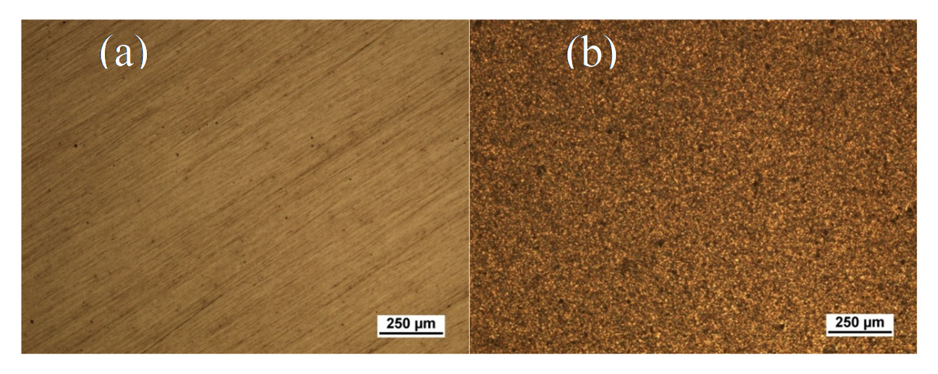
Figure 1a shows the optical microscope images of pure magnesium, which exhibits that the residues of the grinding process are recognizable by small grooves. Figure 1b shows the image of the pre-treatment of magnesium with phosphoric acid (H3PO4) and di-ammonium hydrogen phosphate (DAHP). After pre-treatment, the surface of the substrate is completely coated with small crystals.
3.2. SEM Analysis of Zein/BG Coatings
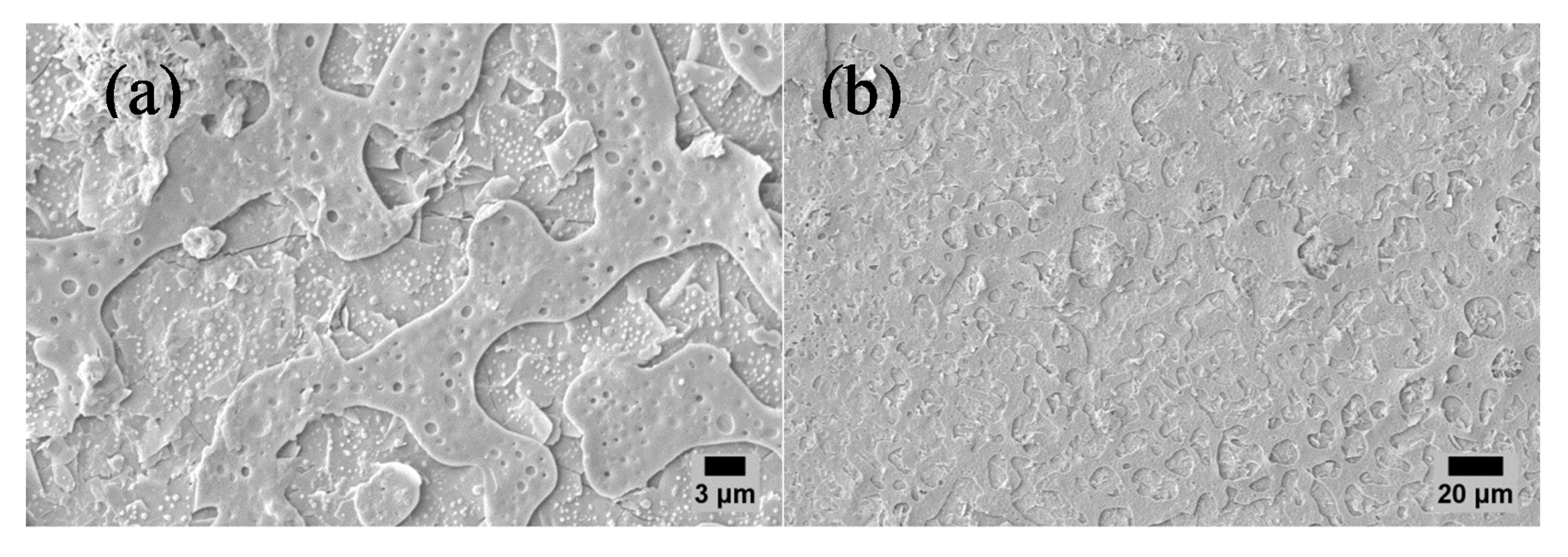
Figure 2 shows the SEM images of the zein/BG coatings on Mg substrates at various magnifications. EPD was used to coat zein/BG coatings on the surface of Mg. EPD parameters were the deposition voltage of 14.5 V, deposition time of 5 min, and the concentration of BG particles in the zein suspension was 5 g/L. Figure 2a (high magnification) shows the typical porous structure of zein. It was observed that the BG particles are distributed unevenly over the surface and in some instances covering the pores in the structure of zein [27]. Figure 2b (low magnification) shows that the zein/BG coatings are fairly uniform and homogenous at the microscopic level.
3.3. Electrochemical Investigations
The raw data obtained from the electrochemical measurements were evaluated with the Thales software from Zahner® and stored in the ASCII format. The plots were then carried out with the software Origin (OriginLab Corporation, Northhampton, MA, USA).
Figure 3 shows that the resting potential of pure magnesium is unstable [28]. Nevertheless, the resting potential settles after 10 min between about −1.73 V and −1.75 V.
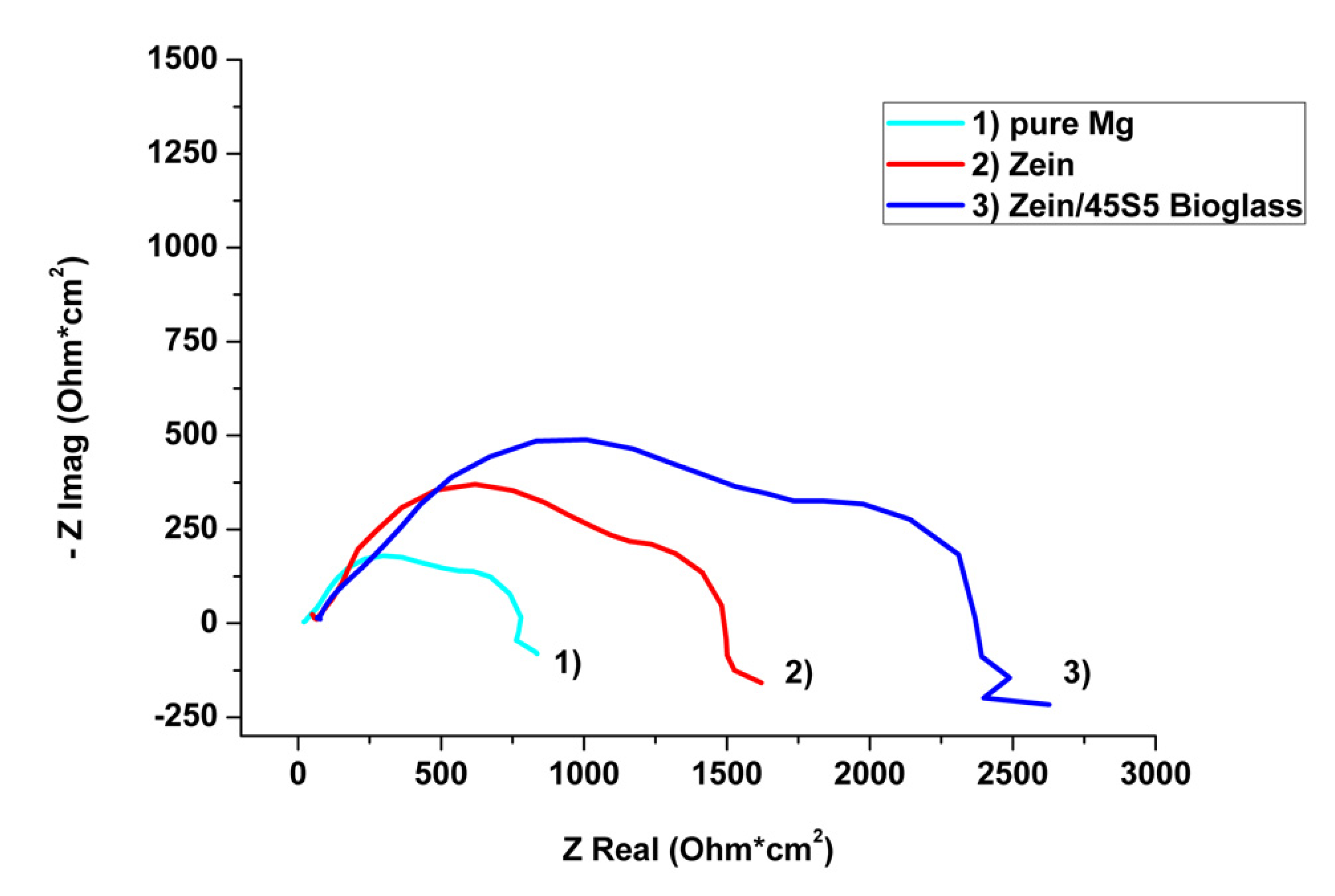
The impedance was measured from an initial time (t = 0), immediately after the stabilization of the potential. The Nyquist plot of all the curves is illustrated in Figure 4, which indicates the formation of a oxide layer on the surface of the pure substrate, wherein the second circle represents the resistance of this layer [29].
Zein/BG coatings show a significantly higher increase in impedance than the coating with zein alone. Figure 5 shows the potentiodynamic polarization curves of pure magnesium compared to coated magnesium. It can be seen that in the anodic area the curves of zein and zein/BG have a clear shoulder at −1.2 V, while the curve of pure magnesium is very steep.
In the case of pure magnesium, an active dissolution could be seen in the anodic area, while the coated samples had a clear protective effect up to a specific breakthrough potential [30]. This is the same for zein and zein/BG and is represented by the kink at −1.2 V. The cathodic current density is reduced for the coated magnesium compared to the pure substrate [31].
3.4. In Vitro Bioactivity
The zein/BG coatings obtained via EPD on Mg substrates were immersed in SBF for 3, 7, and 21 days indicated bioactivity, which is fundamental to the formation of a bond between the implant and bone [32]. Figure 6 shows the SEM images of the zein/BG coatings after treatment in SBF for 21 days.
Figure 6 shows that the change in the morphology of the coatings after 21 day days of immersion in SBF. The coating structure becomes more porous, which is the indication for the degradation of the zein. Moreover, plate-like morphology was also observed in SEM images which is the qualitative indication for the formation of HA crystals on the top of the coatings [33].
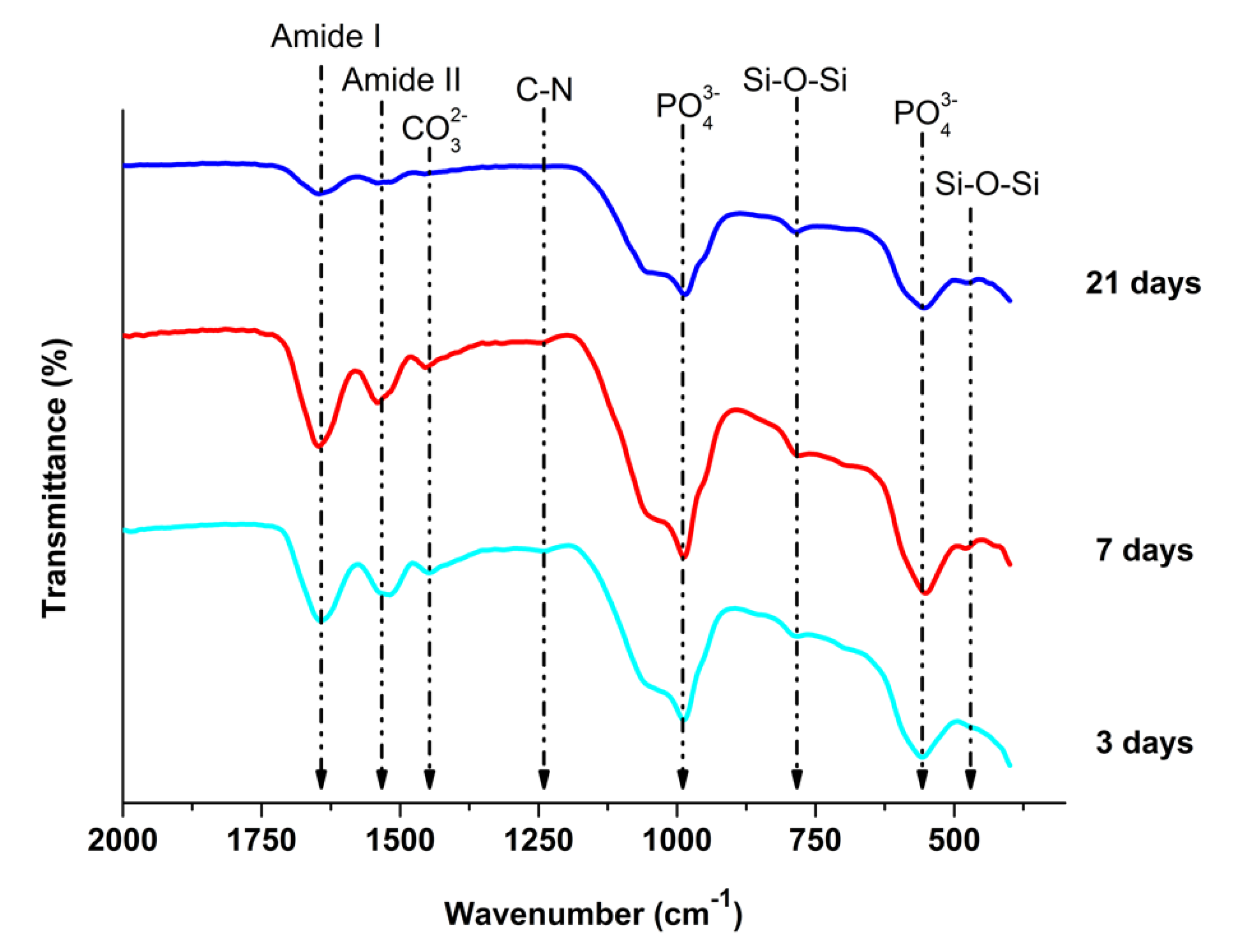
Figure 7 showed the FTIR spectrum of the zein/BG coatings after 3, 7, and 21 days of immersion in SBF. It was observed that the peaks attribute to the phosphate (560 cm−1) and carbonate (CO32−) groups were observed after immersion in SBF.
To further investigate the degradation behavior of the zein/BG coatings after immersion in SBF, electrochemical studies were carried out. Figure 8 exhibited an unstable OCP on the zein/BG coatings after 3 days of immersion in SBF. The curve of the 7th day depicted advanced degradation of the coating in the form of a clear peaks substrate [34].
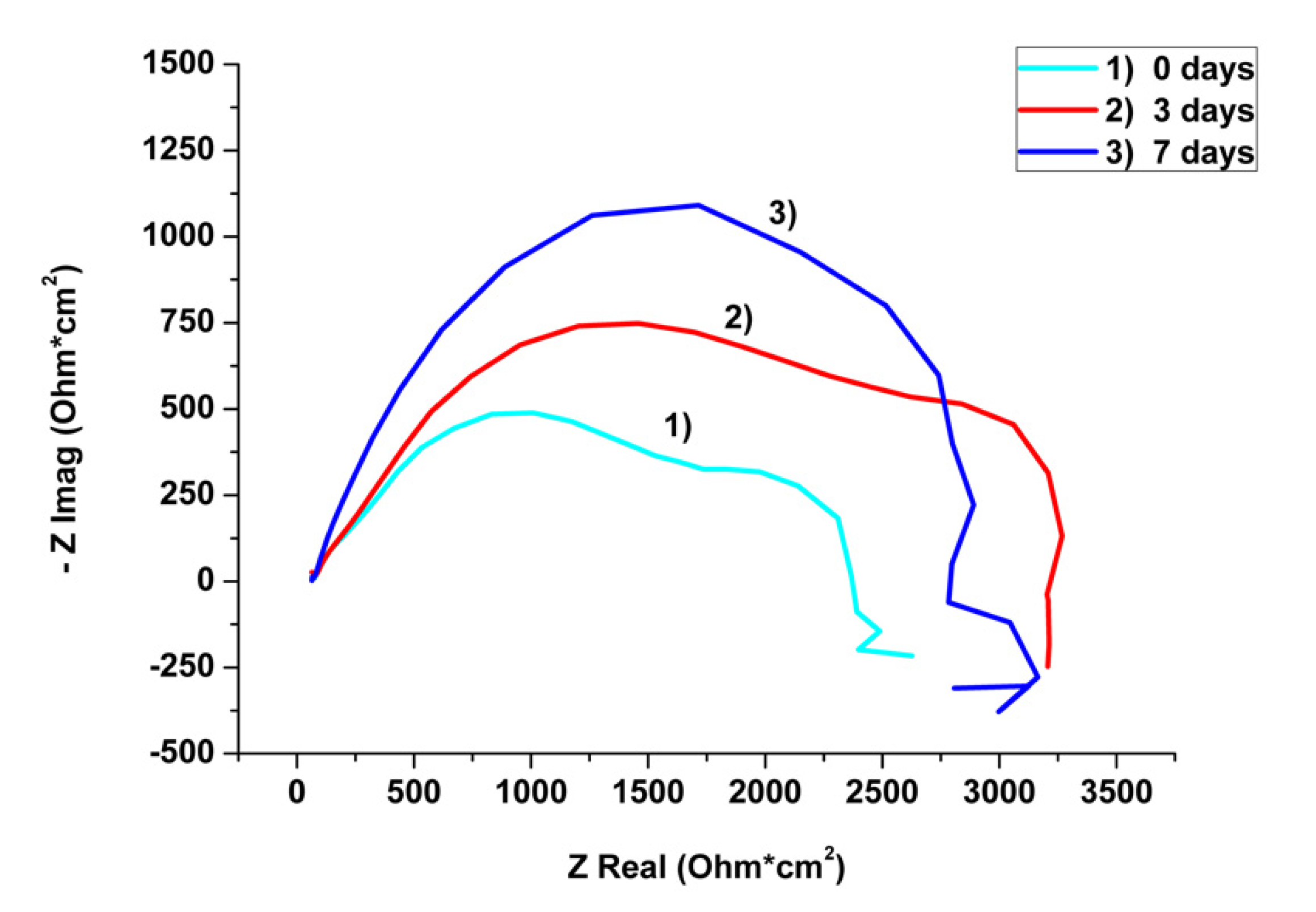
Figure 9 shows the Nyquist plot before contact with the sample with simulated bodily fluid (0 days) and after three and seven days in SBF.
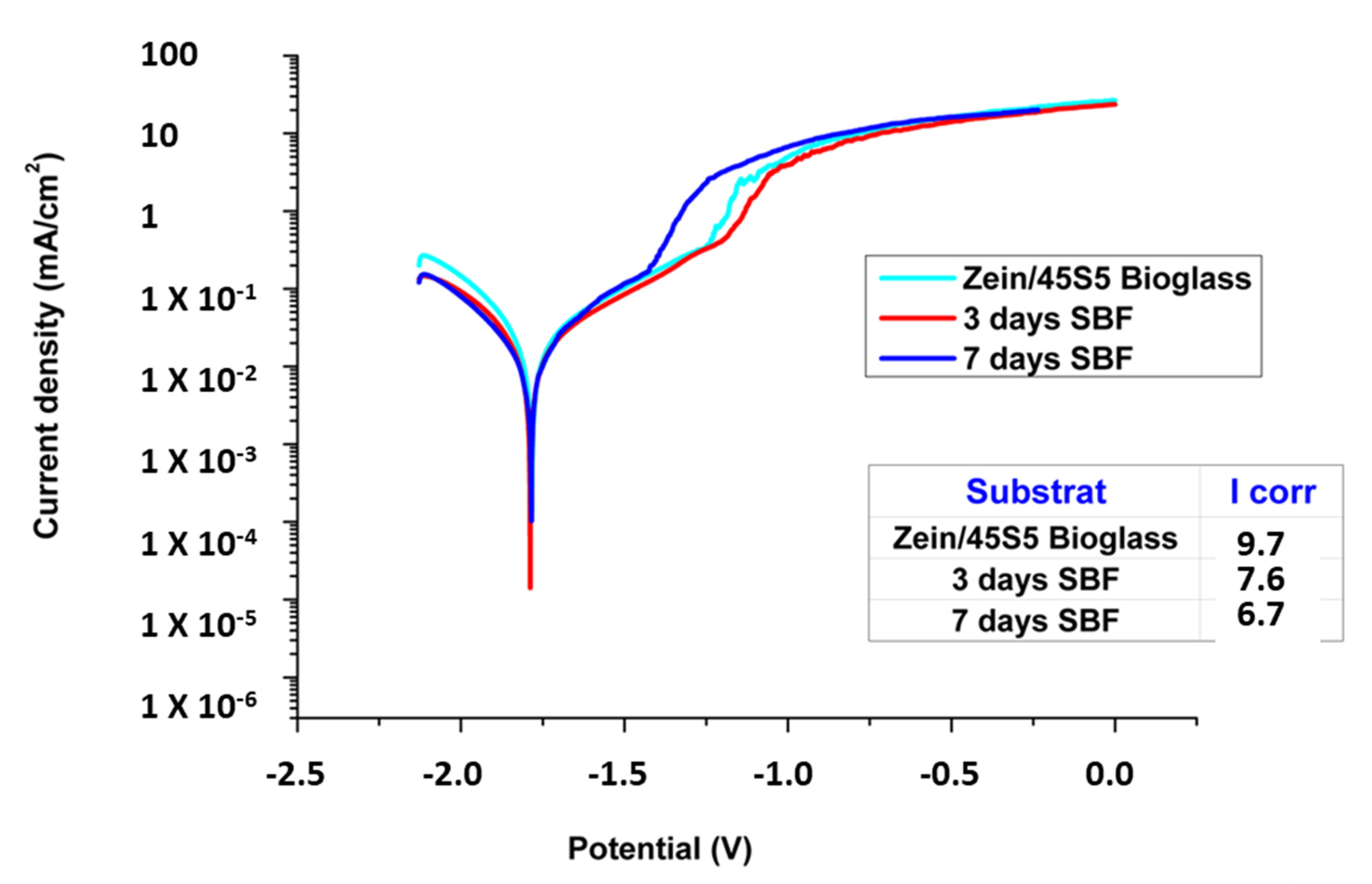
The potentiodynamic polarization curves (Figure 10) showed no clear difference in cathodic areas. The corrosion potential Ecorr was the same for all graphs. The zein/BG coatings before immersion in SBF show an almost identical course of graphs compared to the zein/BG coatings after 3 days of immersion in SBF. However, after 7 days of immersion in the SBF anodic range from the sample that was inserted in SBF for seven days, a slight shoulder at −1.4 V was presented.
It was concluded that, with the increasing duration of the samples in SBF, the protective effect of the coating against corrosion decreases.
4. Discussion
In this work, EPD was successfully used to deposit zein/BG coatings on pretreated pure Mg. Firstly, Mg samples were pretreated with phosphoric acid (H3PO4) and di-ammonium hydrogen phosphate (DAHP). The surface of pure Mg after pretreatment appears to be rough in comparison to that of the pure Mg before pretreatment. It has been reported in the literature that the pretreatment of pure Mg imparts the hydrophilic behavior to the Mg substrate, which led to strong adhesion between the coating and the substrate [20]. Thus, the pretreatment of Mg is an important process in improving the quality of the coatings (zein/BG) to be deposited.
4.1. EPD Kinetics and Suspension Stability
The deposition kinetics, deposition mechanism, and the effect of EPD parameters on the morphology and thickness of zein/BG coatings have been discussed in the literature [17]. To deposit zein/BG coatings, a stable suspension with a zeta potential of +25 mV at pH 3 was prepared. The relatively high value of zeta potential indicates the stability of the suspension, which is important to obtain uniform coatings by EPD. The EPD mechanism of zein/BG deposits has been explained by Rivera et al. [17]. It was suggested that uncharged zein is insoluble in water and organic solvents. However, protonated zein dissolves in water-ethanol acetic acid mixtures at low pH (pH < 4). During the application of an electric field (EPD process), positively charged zein molecules move towards the cathode, lose their charge, and form an insoluble deposit at the cathode. Moreover, BG is positively charged under acidic pH, hence they are expected to move towards the cathode and deposit by coagulation. The electrophoretic mobility of BG particles is much higher than that of zein. Therefore, at a low concentration of BG, co-deposition of zein/BG is expected. However, higher concentrations of BG will lead to a substantial decrease in the electrophoretic mobility of zein (due to the increase in pH and conductivity). These findings formed the basis for choosing the concentration of chitosan BG (5 g/L) in this study.
4.2. EPD of Zein/BG on Mg
The EPD process of zein/BG was first optimized on 316 L SS by applying a Taguchi Design of Experiment approach (data not shown here). The optimized parameters were then applied to obtain coatings on the Mg substrate. The value of current density at the optimum EPD parameters (deposition voltage of 12 V, deposition time of 5 min, and concentration of BG = 5 g/L) for deposition of zein/BG on 316 L SS was noted. The applied voltage was adjusted (14.5 V) to obtain the same value of the current density for the deposition of zein/BG on the Mg substrate.
SEM images of the top surface of the zein/BG coating on the Mg substrate showed a uniform distribution of BG particles in the zein matrix (Figure 2). Moreover, zein dissolved completely during the suspension preparation, which may have contributed to the formation of films (Figure 2b). However, slight variations in the coating homogeneity have been observed among different researchers, as well as in the current study, which is possibly due to the difference in particle size and concentration of BG along with the variations in EPD parameters [17,18,24]. The typical porous structure of zein was observed in this study, which is consistent with the research by Zhao et al. [18]. It was suggested that a dense film of flower-like clusters formed. Although there are some cavities between the clusters, but the protection of the coating was sufficient [22].
4.3. Corrosion Study
Figure 3 shows that the curve of the resting potential of zein and zein/BG coatings was seen to be more stable than that of pure magnesium. Both graphs were in a range of 0.015 and showed no outliers. This affirms the intended purpose of coating, improving the corrosion resistance of magnesium.
Figure 4 confirmed that the zein/BG coatings increased the impedance more than the zein coating alone. Therefore, impedance increased to ~750 ohms*cm2 for the pure zein coating and 1–600 ohms*cm2 for zein/BG coating. This is in line with the results of Heise et al. [29], who has investigated the corrosion behavior of chitosan/BG on pure magnesium. Unfortunately, it was not possible to compare with values of the literature, since corrosion tests on magnesium, especially with zein as a coating, are not standardized.
Figure 5 also confirmed that the zein/BG coatings showed the corrosion protection behavior as determined from the polarization curve. It is known from the literature that the lower the current density, the higher the resistance to corrosion [30]. A Tafel extrapolation was carried out leading to corrosion current densities (icorr) of 15.3 μA cm−2, 13.7 μA cm−2, and 9.7 μA cm−2 for bare Mg, zein, and zein/BG coatings, respectively The value of Icorr decreased for zein and zein/BG coating on the pretreated Mg sample. The strongest decrease in the value of current density was observed in the case of zein/BG coatings on the Mg sample. According to Zhao et al. [22], the decrease in the value of Icorr indicates that the coatings provide effective protection against corrosion. Mehdipour et al. [31] also describe that the addition of BG to the coating improves the corrosion resistance of the substrate. The coated samples show a continuous increase of the current densities upon anodic polarization, indicating coating dissolution. The strong decrease of the cathodic current densities for the coated samples is noteworthy. The zein/BG coatings showed a good corrosion protection effect, as the EPD process leads to the formation of robust zein film reinforced with BG particles that effectively acts as a barrier on the Mg substrate.
4.4. In Vitro Bioactivity
SEM images show that plate-like hydroxyapatite (HA) completely covered the surface of zein/BG coating after 21 days of immersion in SBF (Figure 6a). The plate-like structure indicates the formation of a calcium-enriched apatite layer. Considering that the iso-electric point (IEP) of the zein/BG layer occurs at a pH lower than of the SBF (7.40), the zein/BG coatings likely exhibit a negative charge in SBF and attract more positive Ca2+ ions. This fact may be responsible for a calcium dominated “plate-like” form of apatite. Figure 6b,c revealed a porosity between the HA crystals, which could be associated to the densification of the apatite-like layer. Figure 6d shows the characteristic cauliflower-like structure on the top of the coatings, which indicates the formation of HA on the coatings [35], which is due to the ion exchange between BG particles and SBF.
Figure 7 showed that the typical amide peaks for the zein (between 1750–1600 cm−1 (Amide I) and 1550–1400 cm−1 (Amide II)). FTIR spectra of zein/BG coatings after immersion in SBF (Figure 7) exhibited a reduction in the intensity of the peaks related to BG (Si-O-Si at 459 cm−1) and amide peaks of zein after 3 days of immersion in SBF, which indicates the degradation of zein and BG or the formation of thick layer of HA crystals [30]. Peaks that indicated HA formation were phosphate peaks and carbonate-peaks (1450 cm−1 (CO32−), 1000 cm−1, and 560 cm−1 (PO43−)) [27]. After immersion in SBF, the P-O band from the crystalline phase of BG becomes wider at 560 cm−1, as is typically found in the amorphous phase of calcium phosphate. This indicates the formation of HA [36]. The peak at 800 cm−1 (Si-O-Si) appears after 3 days of immersion in SBF [31] (Changing the Si-O-Si Peak at 440 cm−1 according to Hoppe et al. [35]) attributed to the formation of a phosphate phase. The zein/BG coating showed bioactivity after 3 days of immersion in SBF. The results of the current study are in agreement with the literature, where it was shown that the Mg, Ca, P, and O are the degradation products of the Mg upon immersion in SBF. Furthermore, it was suggested that the degradation products indicate the formation calcium phosphate hydroxide. The formation of calcium phosphate on the surface of Mg is an indication for the good bone binding ability of the coatings [37,38].
Figure 8 showed a steeper OCP curve for 3 days of immersion in SBF, which indicates that the coating starts to degrade. This concluded that the stability of coating decreased with increasing duration in SBF.
Figure 9 showed that the impedance increased significantly after 3 days of immersion in SBF. In addition, 0 and 3-day curves showed the same shape, whereas the 7th-day curve indicated a different course which concluded that the layer applied to the substrate has started to degrade, which is in agreement with the OCP results. Moreover, Figure 10 also affirmed that the protective effect of the coatings is decreasing with an increase in the immersion time in SBF. The effect of the coating was observed in the electrochemical studies after immersion in SBF. It was concluded that the coatings remain in contact even after 21 days of immersion in SBF. In the future, it will be interesting to monitor the change in the mass of the samples after immersion in SBF at different time points. However, the effect of the coatings on corrosion resistance decreased with an increase in immersion time. Thus, future studies will include further improvements in the stability of the coatings upon immersion in SBF.
5. Conclusions
- The zein/BG composite coatings were deposited on pre-treated pure magnesium substrate via EPD.
- The optimum deposition parameters were deduced from a series of experiments on SS. The initial experiments inferred that the deposition voltage of 14.5 V, BG concentration of 5 g/L, pH of 3, deposition time of 5 min, and an electrode distance of 10 mm are optimum for the deposition of zein/BG composite coatings on the Mg substrate.
- SEM images showed that a homogeneous film of zein was embedded with BG particles.
- Electrochemical tests confirmed that the coating provides effective protection against corrosion.
- Zein/BG coatings developed hydroxyapatite crystals on the surface of the coatings after immersion in SBF.
- FTIR analysis showed the dissolution of zein and BG upon immersion in SBF.
- The electrochemical tests after immersion in SBF revealed that the zein/BG slows down the degradation rate of Mg compared to the available literature, which was the main aim of the present study.
Funding
MAUR would like to thank Higher Education Commission Pakistan for the award of SRGP grant#2307. APC was waived by the MDPI being the reviewer board’s member.
Conflicts of Interest
The author declares no conflict of interest.
References
- Prateeksha Kaul, P.J. Bioimplants Market by Type (Cardiovascular Bioimplants, Dental Bioimplants, Orthopedic Bioimplants, Spinal Bioimplants, and Ophthalmology Bioimplants), Material (Metallic Biomaterials, Ceramic Biomaterials, Polymers Biomaterials, and Natural Biomaterials)—Global Opportunity Analysis and Industry Forecast, 2017–2023. Available online: https://www.alliedmarketresearch.com/bioimplants-market (accessed on 1 August 2020).
- Manivasagam, G.; Dhinasekaran, D.; Rajamanickam, A. Biomedical implants: Corrosion and its prevention—A review. Recent Pat. Corros. Sci. 2010, 2, 40–54. [Google Scholar] [CrossRef] [Green Version]
- Staiger, M.P.; Pietak, A.M.; Huadmai, J.; Dias, G. Magnesium and its alloys as orthopedic biomaterials: A review. Biomaterials 2006, 27, 1728–1734. [Google Scholar] [CrossRef] [PubMed]
- Dehghanghadikolaei, A.; Ibrahim, H.; Amerinatanzi, A.; Elahinia, M. 9—Biodegradable magnesium alloys. In Metals for Biomedical Devices; Woodhead Publ. Ser., Biomater.; Niinomi, S.E., Ed.; Woodhead Publishing: Cambridge, UK, 2019; pp. 265–289. [Google Scholar] [CrossRef]
- Zhang, L.-N.; Hou, Z.-T.; Ye, X.; Xu, Z.-B.; Bai, X.-L.; Shang, P. The effect of selected alloying element additions on properties of Mg-based alloy as bioimplants: A literature review. Front. Mater. Sci. 2013, 7, 227–236. [Google Scholar] [CrossRef]
- Ahmed, Y.; Rehman, M.A.U. Improvement in the surface properties of stainless steel via zein/hydroxyapatite composite coatings for biomedical applications. Surf. Interfaces 2020, 100589. [Google Scholar] [CrossRef]
- Makvandi, P.; Ghomi, M.; Padil, V.V.T.; Shalchy, F.; Ashrafizadeh, M.; Askarinejad, S.; Mokhtari, B. Biofabricated Nanostructures and Their Composites in Regenerative Medicine. ACS Appl. Nano Mater. 2020, 3, 6210–6238. [Google Scholar] [CrossRef]
- Makvandi, P.; Ali, G.W.; Della Sala, F.; Abdel-Fattah, W.I.; Borzacchiello, A. Hyaluronic acid/corn silk extract based injectable nanocomposite: A biomimetic antibacterial scaffold for bone tissue regeneration. Mater. Sci. Eng. C 2020, 107, 110195. [Google Scholar] [CrossRef]
- Meyer, N.; Rivera, L.R.; Ellis, T.; Qi, J.; Ryan, M.P.; Boccaccini, A.R. Bioactive and antibacterial coatings based on zein/bioactive glass composites by electrophoretic deposition. Coatings 2018, 8, 27. [Google Scholar] [CrossRef] [Green Version]
- Filho, O.P.; La Torre, G.P.; Hench, L.L. Effect of crystallization on apatite-layer formation of bioactive glass 45S5. J. Biomed. Mater. Res. Off. J. Soc. Biomater. Jpn. Soc. Biomater. 1996, 30, 509–514. [Google Scholar] [CrossRef]
- Hench, L.L. The story of Bioglass®. J. Mater. Sci. Mater. Med. 2006, 17, 967–978. [Google Scholar] [CrossRef]
- Lu, H.H.; El-Amin, S.F.; Scott, K.D.; Laurencin, C.T. Three-dimensional, bioactive, biodegradable, polymer–bioactive glass composite scaffolds with improved mechanical properties support collagen synthesis and mineralization of human osteoblast-like cells in vitro. J. Biomed. Mater. Res. Part. A An. Off. J. Soc. Biomater. Jpn. Soc. Biomater. Aust. Soc. Biomater. Korean Soc. Biomater. 2003, 64, 465–474. [Google Scholar] [CrossRef]
- Ahmed, Y.; Yasir, M.; Ur Rehman, M.A. Fabrication and Characterization of Zein/Hydroxyapatite Composite Coatings for Biomedical Applications. Surfaces 2020, 3, 237–250. [Google Scholar] [CrossRef]
- Corradini, E.; Curti, P.S.; Meniqueti, A.B.; Martins, A.F.; Rubira, A.F.; Muniz, E.C. Recent advances in food-packing, pharmaceutical and biomedical applications of zein and zein-based materials. Int. J. Mol. Sci. 2014, 15, 22438–22470. [Google Scholar] [CrossRef] [PubMed] [Green Version]
- Makvandi, P.; Wang, C.Y.; Zare, E.N.; Borzacchiello, A.; Niu, L.N.; Tay, F.R. Metal-Based Nanomaterials in Biomedical Applications: Antimicrobial Activity and Cytotoxicity Aspects. Adv. Funct. Mater. 2020, 30, 1910021. [Google Scholar] [CrossRef]
- Ramos Rivera, L.; Dippel, J.; Boccaccini, A.R. Formation of Zein/Bioactive Glass Layers Using Electrophoretic Deposition Technique. ECS Trans. 2018, 82, 73–80. [Google Scholar] [CrossRef]
- Kaya, S.; Boccaccini, A.R. Electrophoretic deposition of zein coatings. J. Coat. Technol. Res. 2017, 14, 683–689. [Google Scholar] [CrossRef]
- Wang, C.Y.; Makvandi, P.; Zare, E.N.; Tay, F.R.; Niu, L.N. Advances in Antimicrobial Organic and Inorganic Nanocompounds in Biomedicine. Adv. Ther. 2020, 3, 2000024. [Google Scholar] [CrossRef]
- Fotovvati, B.; Namdari, N.; Dehghanghadikolaei, A. On coating techniques for surface protection: A review. J. Manuf. Mater. Process. 2019, 3, 28. [Google Scholar] [CrossRef] [Green Version]
- Avcu, E.; Baştan, F.E.; Abdullah, H.Z.; Rehman, M.A.U.; Avcu, Y.Y.; Boccaccini, A.R. Electrophoretic deposition of Chitosan-based Composite Coatings for Biomedical Applications: A Review. Prog. Mater. Sci. 2019, 103, 69–108. [Google Scholar] [CrossRef]
- Boccaccini, A.R.; Dickerson, J.H. Electrophoretic Deposition: Fundamentals and Applications; ACS Publications: Washington, DC, USA, 2013. [Google Scholar]
- Zhao, H.; Cai, S.; Ding, Z.; Zhang, M.; Li, Y.; Xu, G. A simple method for the preparation of magnesium phosphate conversion coatings on a AZ31 magnesium alloy with improved corrosion resistance. RSC Adv. 2015, 5, 24586–24590. [Google Scholar] [CrossRef]
- Höhlinger, M.; Heise, S.; Wagener, V.; Boccaccini, A.R.; Virtanen, S. Developing surface pre-treatments for electrophoretic deposition of biofunctional chitosan-bioactive glass coatings on a WE43 magnesium alloy. Appl. Surf. Sci. 2017, 405, 441–448. [Google Scholar] [CrossRef]
- Ahmed, Y.; Nawaz, A.; Singh Virk, R.; Wadood, A.; Rehman, M.A.U. Fabrication and characterization of zein/bioactive glass (BG) deposited on pre-treated magnesium via electrophoretic deposition. Int. J. Ceram. Eng. Sci. 2020. [Google Scholar] [CrossRef]
- Hertel, M. Elektrochemische Charakterisierung von Stents mit Hilfe des Adaptierten Mini-Cell-Systems (MCS). Ph.D. Thesis, Freie Universität, Berlin, Germany, 2012. [Google Scholar]
- Kokubo, T.; Takadama, H. How useful is SBF in predicting in vivo bone bioactivity? Biomaterials 2006, 27, 2907–2915. [Google Scholar] [CrossRef] [PubMed]
- Naseri, S.; Hum, J.; Lepry, W.C.; Miri, A.K.; Nazhat, S.N.; Boccaccini, A.R. Fabrication and characterization of zein–bioactive glass scaffolds. BioinspiredBiomim. Nanobiomater. 2015, 4, 73–78. [Google Scholar] [CrossRef]
- Hornberger, H.; Virtanen, S.; Boccaccini, A.R. Biomedical coatings on magnesium alloys—A review. Acta Biomater. 2012, 8, 2442–2455. [Google Scholar] [CrossRef] [PubMed]
- Heise, S.; Höhlinger, M.; Hernández, Y.T.; Palacio, J.J.P.; Ortiz, J.A.R.; Wagener, V.; Virtanen, S.; Boccaccini, A.R. Electrophoretic deposition and characterization of chitosan/bioactive glass composite coatings on Mg alloy substrates. Electrochim. Acta 2017, 232, 456–464. [Google Scholar] [CrossRef]
- Amiri, H.; Mohammadi, I.; Afshar, A. Electrophoretic deposition of nano-zirconia coating on AZ91D magnesium alloy for bio-corrosion control purposes. Surf. Coat. Technol. 2017, 311, 182–190. [Google Scholar] [CrossRef]
- Mehdipour, M.; Afshar, A.; Mohebali, M. Electrophoretic deposition of bioactive glass coating on 316L stainless steel and electrochemical behavior study. Appl. Surf. Sci. 2012, 258, 9832–9839. [Google Scholar] [CrossRef]
- Ur Rehman, M.A.; Bastan, F.E.; Nawaz, A.; Nawaz, Q.; Wadood, A. Electrophoretic deposition of PEEK/bioactive glass composite coatings on stainless steel for orthopedic applications: An optimization for in vitro bioactivity and adhesion strength. Int. J. Adv. Manuf. Technol. 2020, 108, 1849–1862. [Google Scholar] [CrossRef]
- Zhao, X.-Y.; Zhu, Y.-J.; Chen, F.; Lu, B.-Q.; Wu, J. Nanosheet-assembled hierarchical nanostructures of hydroxyapatite: Surfactant-free microwave-hydrothermal rapid synthesis, protein/DNA adsorption and pH-controlled release. CrystEngComm 2013, 15, 206–212. [Google Scholar] [CrossRef]
- Demir, M.; Ramos-Rivera, L.; Silva, R.; Nazhat, S.N.; Boccaccini, A.R. Zein-based composites in biomedical applications. J. Biomed. Mater. Res. Part. A 2017, 105, 1656–1665. [Google Scholar] [CrossRef]
- Hoppe, A.; Meszaros, R.; Stähli, C.; Romeis, S.; Schmidt, J.; Peukert, W.; Marelli, B.; Nazhat, S.N.; Wondraczek, L.; Lao, J. In vitro reactivity of Cu doped 45S5 Bioglass® derived scaffolds for bone tissue engineering. J. Mater. Chem. B 2013, 1, 5659–5674. [Google Scholar] [CrossRef] [PubMed]
- Wang, Y.; Cui, L.Y.; Zeng, R.C.; Li, S.Q.; Zou, Y.H.; Han, E.H. In Vitro Degradation of Pure Magnesium―The Effects of Glucose and/or Amino Acid. Materials 2017, 10, 725. [Google Scholar] [CrossRef] [PubMed] [Green Version]
- Ibrahim, H.; Dehghanghadikolaei, A.; Advincula, R.; Dean, D.; Luo, A.; Elahinia, M. Ceramic coating for delayed degradation of Mg-1.2Zn-0.5Ca-0.5Mn bone fixation and instrumentation. Thin Solid Films 2019, 687, 137456. [Google Scholar] [CrossRef]
- Steiner Petrovič, D.; Mandrino, D.; Šarler, B.; Horky, J.; Ojdanic, A.; Zehetbauer, M.J.; Orlov, D. Surface Analysis of Biodegradable Mg-Alloys after Immersion in Simulated Body Fluid. Materials 2020, 13, 1740. [Google Scholar] [CrossRef] [PubMed] [Green Version]
Figure 1.
Optical microscope images (a) pure magnesium; (b) pre-treated magnesium, and the pretreatment was done with the phosphoric acid (H3PO4) and di-ammonium hydrogen phosphate (DAHP).
Figure 1.
Optical microscope images (a) pure magnesium; (b) pre-treated magnesium, and the pretreatment was done with the phosphoric acid (H3PO4) and di-ammonium hydrogen phosphate (DAHP).

Figure 2.
SEM image of zein/BG on magnesium substrates obtained via EPD at pH = 3, deposition voltage = 14.5 V, deposition time = 5 min, and concentration of BG = 5 g/L (a) low resolution; (b) high resolution.
Figure 2.
SEM image of zein/BG on magnesium substrates obtained via EPD at pH = 3, deposition voltage = 14.5 V, deposition time = 5 min, and concentration of BG = 5 g/L (a) low resolution; (b) high resolution.

Figure 3.
Open circuit potential of (a) pure Mg before the deposition, and (b) zein and zein/BG coatings deposited via EPD on the Mg substrate.
Figure 3.
Open circuit potential of (a) pure Mg before the deposition, and (b) zein and zein/BG coatings deposited via EPD on the Mg substrate.

Figure 4.
Nyquist Plot of pure Mg and coated and zein, zein/BG coatings deposited on the Mg substrate via EPD.
Figure 4.
Nyquist Plot of pure Mg and coated and zein, zein/BG coatings deposited on the Mg substrate via EPD.

Figure 5.
Comparison of the potentiodynamic polarization curves of pure and coated magnesium and an indication of Icorr.
Figure 5.
Comparison of the potentiodynamic polarization curves of pure and coated magnesium and an indication of Icorr.

Figure 6.
SEM images showing the change in the morphology of the zein/BG coatings after 21 days of immersion in SBF at different magnifications; (a) 50,000×, (b) 25,000×, (c) 5000×, and (d) 1000×.
Figure 6.
SEM images showing the change in the morphology of the zein/BG coatings after 21 days of immersion in SBF at different magnifications; (a) 50,000×, (b) 25,000×, (c) 5000×, and (d) 1000×.

Figure 7.
FTIR spectrum of zein/BG coating after treatment with SBF for 3, 7, and 21 days.

Figure 8.
OCP of Zein/BG coatings before and after immersion in SBF for 3 days and 7 days.

Figure 9.
Nyquist Plot showing the degradation of zein/BG coatings before and after immersion in SBF for 3 and 7 days.
Figure 9.
Nyquist Plot showing the degradation of zein/BG coatings before and after immersion in SBF for 3 and 7 days.

Figure 10.
Potentiodynamic polarization curve of coated magnesium before and after removal in SBF for 3 and 7 days.
Figure 10.
Potentiodynamic polarization curve of coated magnesium before and after removal in SBF for 3 and 7 days.

© 2020 by the author. Licensee MDPI, Basel, Switzerland. This article is an open access article distributed under the terms and conditions of the Creative Commons Attribution (CC BY) license (http://creativecommons.org/licenses/by/4.0/).
Share and Cite
MDPI and ACS Style
Ur Rehman, M.A. Zein/Bioactive Glass Coatings with Controlled Degradation of Magnesium under Physiological Conditions: Designed for Orthopedic Implants. Prosthesis 2020, 2, 211-224. https://0-doi-org.brum.beds.ac.uk/10.3390/prosthesis2030018
AMA Style
Ur Rehman MA. Zein/Bioactive Glass Coatings with Controlled Degradation of Magnesium under Physiological Conditions: Designed for Orthopedic Implants. Prosthesis. 2020; 2(3):211-224. https://0-doi-org.brum.beds.ac.uk/10.3390/prosthesis2030018
Chicago/Turabian StyleUr Rehman, Muhammad Atiq. 2020. "Zein/Bioactive Glass Coatings with Controlled Degradation of Magnesium under Physiological Conditions: Designed for Orthopedic Implants" Prosthesis 2, no. 3: 211-224. https://0-doi-org.brum.beds.ac.uk/10.3390/prosthesis2030018